Journal Description
Diagnostics
Diagnostics
is an international, peer-reviewed, open access journal on medical diagnosis published semimonthly online by MDPI. The British Neuro-Oncology Society (BNOS), the International Society for Infectious Diseases in Obstetrics and Gynaecology (ISIDOG) and the Swiss Union of Laboratory Medicine (SULM) are affiliated with Diagnostics and their members receive a discount on the article processing charges.
- Open Access— free for readers, with article processing charges (APC) paid by authors or their institutions.
- High Visibility: indexed within Scopus, SCIE (Web of Science), PubMed, PMC, Embase, Inspec, CAPlus / SciFinder, and other databases.
- Journal Rank: JCR - Q2 (Medicine, General & Internal)
- Rapid Publication: manuscripts are peer-reviewed and a first decision is provided to authors approximately 20.7 days after submission; acceptance to publication is undertaken in 2.8 days (median values for papers published in this journal in the second half of 2023).
- Recognition of Reviewers: reviewers who provide timely, thorough peer-review reports receive vouchers entitling them to a discount on the APC of their next publication in any MDPI journal, in appreciation of the work done.
- Companion journal: LabMed.
Impact Factor:
3.6 (2022);
5-Year Impact Factor:
3.7 (2022)
Latest Articles
Pharmacologically Induced Accommodation Palsy and the Bioelectrical Activity of the Muscular System: A Preliminary Investigation
Diagnostics 2024, 14(9), 961; https://doi.org/10.3390/diagnostics14090961 (registering DOI) - 04 May 2024
Abstract
The aim of this study was to pharmacologically induce accommodative paralysis and evaluate its effects on the bioelectrical activity of the muscular system. The study included two participant groups: those with myopia and those with normal vision (emmetropes). Electromyographic assessments were performed using
[...] Read more.
The aim of this study was to pharmacologically induce accommodative paralysis and evaluate its effects on the bioelectrical activity of the muscular system. The study included two participant groups: those with myopia and those with normal vision (emmetropes). Electromyographic assessments were performed using the Noraxon Ultium DTS 8-K MR 3 myo Muscle Master Edition system. The muscles analyzed in this study were the temporalis, masseter, sternocleidomastoid, trapezius, abdominal muscles, biceps brachii, and the external oblique muscles of the abdomen. It is important to acknowledge that, based on the current findings, it cannot be definitively stated that the observed effects have clinical significance, and additional studies are encouraged.
Full article
(This article belongs to the Section Pathology and Molecular Diagnostics)
►
Show Figures
Open AccessReview
Intelligent Liver Function Testing (iLFT): An Intelligent Laboratory Approach to Identifying Chronic Liver Disease
by
Jennifer Nobes, Damien Leith, Sava Handjiev, John F. Dillon and Ellie Dow
Diagnostics 2024, 14(9), 960; https://doi.org/10.3390/diagnostics14090960 (registering DOI) - 04 May 2024
Abstract
The intelligent Liver Function Testing (iLFT) pathway is a novel, algorithm-based system which provides automated laboratory investigations and clinical feedback on abnormal liver function test (LFT) results from primary care. iLFT was introduced to NHS Tayside, Scotland, in August 2018 in response to
[...] Read more.
The intelligent Liver Function Testing (iLFT) pathway is a novel, algorithm-based system which provides automated laboratory investigations and clinical feedback on abnormal liver function test (LFT) results from primary care. iLFT was introduced to NHS Tayside, Scotland, in August 2018 in response to vast numbers of abnormal LFTs, many of which were not appropriately investigated, coupled with rising mortality from chronic liver disease. Here, we outline the development and implementation of the iLFT pathway, considering the implications for the diagnostic laboratories, primary care services and specialist hepatology clinics. Additionally, we describe the utility, outcomes and evolution of iLFT, which was used over 11,000 times in its first three years alone. Finally, we will consider the future of iLFT and propose areas where similar ‘intelligent’ approaches could be used to add value to laboratory investigations.
Full article
(This article belongs to the Special Issue Laboratory Medicine: Extended Roles in Healthcare Delivery)
►▼
Show Figures
Figure 1
Open AccessArticle
CD3 and CD20 Expressions and Infiltrating Patterns in Salivary Gland Tumors
by
Rukhsar R. Hussein and Balkees T. Garib
Diagnostics 2024, 14(9), 959; https://doi.org/10.3390/diagnostics14090959 - 03 May 2024
Abstract
Tumor-infiltrating lymphocytes (TILs) represent a subset of immunological constituents within the tumor microenvironment that can influence cancer growth. We retrospectively evaluate the density and pattern of CD3 and CD20 expression in salivary gland tumors and their relation to clinical pathologic parameters. A total
[...] Read more.
Tumor-infiltrating lymphocytes (TILs) represent a subset of immunological constituents within the tumor microenvironment that can influence cancer growth. We retrospectively evaluate the density and pattern of CD3 and CD20 expression in salivary gland tumors and their relation to clinical pathologic parameters. A total of 44 formalin-fixed paraffin-embedded blocks of salivary gland tumors were included. These tumors were stained immunohistochemically with CD3 and CD20. The chi-square test was used to relate immune scoring, intensity, and clinical pathological parameters to different salivary tumors. p-value < 0.05 was considered statistically significant. The intra-tumoral CD3 infiltrating count was high and diffused in (71.4%) of pleomorphic adenomas (PAs) followed by mucoepidermoid carcinomas (MECs) (66.7%). At the same time, adenoid cystic carcinomas (AdCCs) exhibited significantly low infiltration (71.4%) (p = 0.046). The three types of tumors exhibited high tumor-infiltrating counts diffused in peripheral areas with significant differences between malignant tumors (p = 0.047). The intra-tumoral CD20 infiltrating count significantly differed among the tumors (p = 0.002); it was low in all PAs and AdCCs, while MECs showed an equal percentage of expression. However, in the peripheral area, PAs and MECs exhibited significantly (p = 0.007) high infiltrating counts (69.2% and 84.6), and the lowest infiltrating count was predominantly found for AdCCs. The two markers had a significant positive correlation between the mean of CD3 in the intra-tumoral and peripheral regions and CD20 in the peripheral zone across the total samples. In conclusion, the density of CD3 expression is notably higher than CD20 across tumor types. PAs and MECs showed high-density scores, while AdCCs were characterized by low scores. TIL expression was found to be significantly associated with patients’ outcomes in the intra-tumoral area.
Full article
(This article belongs to the Special Issue Advances in the Diagnosis and Management of Head and Neck Disease)
►▼
Show Figures
Figure 1
Open AccessReview
Pathological and Molecular Diagnosis of Uveal Melanoma
by
Mihai Adrian Păsărică, Paul Filip Curcă, Christiana Diana Maria Dragosloveanu, Alexandru Călin Grigorescu and Cosmin Ionuț Nisipașu
Diagnostics 2024, 14(9), 958; https://doi.org/10.3390/diagnostics14090958 - 02 May 2024
Abstract
(1) Background: Uveal melanoma (UM) is a common malignant intraocular tumor that presents with significant genetic differences to cutaneous melanoma and has a high genetic burden in terms of prognosis. (2) Methods: A systematic literature search of several repositories on uveal melanoma diagnosis,
[...] Read more.
(1) Background: Uveal melanoma (UM) is a common malignant intraocular tumor that presents with significant genetic differences to cutaneous melanoma and has a high genetic burden in terms of prognosis. (2) Methods: A systematic literature search of several repositories on uveal melanoma diagnosis, prognosis, molecular analysis, and treatment was conducted. (3) Results: Recent genetic understanding of oncogene-initiation mutations in GNAQ, GNA11, PLCB4, and CYSLTR2 and secondary progression drivers of BAP1 inactivation and SF3B1 and EIF1AX mutations offers an appealing explanation to the high prognostic impact of adding genetic profiling to clinical UM classification. Genetic information could help better explain peculiarities in uveal melanoma, such as the low long-term survival despite effective primary tumor treatment, the overwhelming propensity to metastasize to the liver, and possibly therapeutic behaviors. (4) Conclusions: Understanding of uveal melanoma has improved step-by-step from histopathology to clinical classification to more recent genetic understanding of oncogenic initiation and progression.
Full article
(This article belongs to the Special Issue Pathological and Molecular Diagnosis of Melanoma)
Open AccessArticle
Abnormal Static Sagittal Cervical Curvatures following Motor Vehicle Collisions: A Retrospective Case Series of 41 Patients before and after a Crash Exposure
by
Jason W. Haas, Paul A. Oakley, Joseph R. Ferrantelli, Evan A. Katz, Ibrahim M. Moustafa and Deed E. Harrison
Diagnostics 2024, 14(9), 957; https://doi.org/10.3390/diagnostics14090957 - 02 May 2024
Abstract
Previous investigations have found a correlation between abnormal curvatures and a variety of patient complaints such as cervical pain and disability. However, no study has shown that loss of the cervical curve is a direct result of exposure to a motor vehicle collision
[...] Read more.
Previous investigations have found a correlation between abnormal curvatures and a variety of patient complaints such as cervical pain and disability. However, no study has shown that loss of the cervical curve is a direct result of exposure to a motor vehicle collision (MVC). This investigation presents a retrospective consecutive case series of patients with both a pre-injury cervical lateral radiograph (CLR) and a post-injury CLR after exposure to an MVC. Computer analysis of digitized vertebral body corners on CLRs was performed to investigate the possible alterations in the geometric alignment of the sagittal cervical curve. Methods: Three spine clinic records were reviewed over a 2-year period, looking for patients where both an initial lateral cervical X-ray and an examination were performed prior to the patient being exposed to a MVC; afterwards, an additional exam and radiographic analysis were obtained. A total of 41 patients met the inclusion criteria. Examination records of pain intensity on numerical pain rating scores (NPRS) and neck disability index (NDI), if available, were analyzed. The CLRs were digitized and modeled in the sagittal plane using curve fitting and the least squares error approach. Radiographic variables included total cervical curve (ARA C2–C7), Chamberlain’s line to horizontal (skull flexion), horizontal translation of C2 relative to C7, segmental translations (retrolisthesis and anterolisthesis), and circular modelling radii. Results: There were 15 males and 26 females with an age range of 8–65 years. Most participants were drivers (28) involved in rear-end impacts (30). The pre-injury NPRS was 2.7 while the post injury was 5.0; p < 0.001. The NDI was available on 24/41 (58.5%) patients and increased after the MVC from 15.7% to 32.8%, p < 0.001. An altered cervical curvature was identified following exposure to MVC, characterized by an increase in the mean radius of curvature (265.5 vs. 555.5, p < 0.001) and an approximate 8° reduction of lordosis from C2–C7; p < 0.001. The mid-cervical spine (C3–C5) showed the greatest curve reduction with an averaged localized mild kyphosis at these levels. Four participants (10%) developed segmental translations that were just below the threshold of instability, segmental translations < 3.5 mm. Conclusions: The post-exposure MVC cervical curvature was characterized by an increase in radius of curvature, an approximate 8° reduction in C2–C7 lordosis, a mild kyphosis of the mid-cervical spine, and a slight increase in anterior translation of C2–C7 sagittal balance. The modelling result indicates that the post-MVC cervical sagittal alignment approximates a second-order buckling alignment, indicating a significant alteration in curve geometry. Future biomechanics experiments and clinical investigations are needed to confirm these findings.
Full article
(This article belongs to the Special Issue Imaging Diagnosis in Musculoskeletal Medicine)
►▼
Show Figures
Figure 1
Open AccessArticle
Integrating Routine Hematological and Extended Inflammatory Parameters as a Novel Approach for Timely Diagnosis and Prognosis in Sepsis Management
by
Sianny Herawati, I Ketut Agus Somia, Sully Kosasih, I Nyoman Wande, Jethro Felim and I Made Dwi Payana
Diagnostics 2024, 14(9), 956; https://doi.org/10.3390/diagnostics14090956 - 02 May 2024
Abstract
Sepsis is one of the major causes of morbidity and mortality in hospitals, especially in low- and middle-income countries, and represents a challenge to health care providers to carry out early detection, and accurate diagnosis and prognosis with cost-effective diagnostic tools. An observational
[...] Read more.
Sepsis is one of the major causes of morbidity and mortality in hospitals, especially in low- and middle-income countries, and represents a challenge to health care providers to carry out early detection, and accurate diagnosis and prognosis with cost-effective diagnostic tools. An observational prospective study was conducted from December 2021 to December 2022 to investigate the extended inflammatory parameters (EIPs) for sepsis management and analyze the survival of septic patients in the emergency unit, intensive care unit (ICU) and inpatient ward. Patients suspected of having sepsis underwent a sequential organ failure assessment (SOFA) evaluation and had blood drawn for complete blood counts (CBCs). Significant changes were observed in various CBC parameters and EIPs, and the sepsis group was followed up with for 30-day mortality. The study highlighted a significant difference yet strong discriminatory power to differentiate sepsis with an AUC of 0.924 against the non-sepsis group and an AUC of 0.991 against the healthy control group using combination of white blood cells and EIPs. Furthermore, the study showed good predictive ability for 30-day mortality with a hazard ratio of 2.311. In summary, this study provides evidence that the utilization of EIPs may be valuable in diagnosing and predicting patient outcomes, and thus will be beneficial for sepsis management in the hospital.
Full article
(This article belongs to the Special Issue Advances in Cell-Based Technologies for Precision Diagnostics)
►▼
Show Figures
Figure 1
Open AccessArticle
Pancreatic Iodine Density and Fat Fraction on Dual-Energy Computed Tomography in Acute Pancreatitis
by
Zrinka Matana Kaštelan, Ivan Brumini, Goran Poropat, Lovro Tkalčić, Tiana Grubešić and Damir Miletić
Diagnostics 2024, 14(9), 955; https://doi.org/10.3390/diagnostics14090955 - 02 May 2024
Abstract
The aim of our study was to investigate iodine density (ID) and fat fraction (FF) on dual-energy computed tomography (DECT) in patients with acute pancreatitis (AP). This retrospective study included 72 patients with clinically confirmed AP and 62 control subjects with DECT of
[...] Read more.
The aim of our study was to investigate iodine density (ID) and fat fraction (FF) on dual-energy computed tomography (DECT) in patients with acute pancreatitis (AP). This retrospective study included 72 patients with clinically confirmed AP and 62 control subjects with DECT of the abdomen. Two radiologists assessed necrosis and measured attenuation values, ID, and FF in three pancreatic segments. We used receiver operating characteristic (ROC) analysis to determine the optimal threshold for ID for the differentiation between AP groups. The ID was significantly higher in interstitial edematous AP compared to necrotizing AP and the control group (both p < 0.05). The ROC curve analysis revealed the thresholds of ID for detecting pancreatic necrosis ≤ 2.2, ≤2.3, and ≤2.4 mg/mL (AUC between 0.880 and 0.893, p > 0.05) for the head, body, and tail, respectively. The FF was significantly higher for pancreatitis groups when compared with the control group in the head and body segments (both p < 0.001). In the tail, the difference was significant in necrotizing AP (p = 0.028). The ID values were independent of attenuation values correlated with the FF values in pancreatic tissue. Iodine density values allow for differentiation between morphologic types of AP.
Full article
(This article belongs to the Special Issue Advances in Computed Tomography Imaging for Clinical Diagnosis)
►▼
Show Figures
Figure 1
Open AccessArticle
A Neoteric Feature Extraction Technique to Predict the Survival of Gastric Cancer Patients
by
Warid Islam, Neman Abdoli, Tasfiq E. Alam, Meredith Jones, Bornface M. Mutembei, Feng Yan and Qinggong Tang
Diagnostics 2024, 14(9), 954; https://doi.org/10.3390/diagnostics14090954 - 01 May 2024
Abstract
Background: At the time of cancer diagnosis, it is crucial to accurately classify malignant gastric tumors and the possibility that patients will survive. Objective: This study aims to investigate the feasibility of identifying and applying a new feature extraction technique to predict the
[...] Read more.
Background: At the time of cancer diagnosis, it is crucial to accurately classify malignant gastric tumors and the possibility that patients will survive. Objective: This study aims to investigate the feasibility of identifying and applying a new feature extraction technique to predict the survival of gastric cancer patients. Methods: A retrospective dataset including the computed tomography (CT) images of 135 patients was assembled. Among them, 68 patients survived longer than three years. Several sets of radiomics features were extracted and were incorporated into a machine learning model, and their classification performance was characterized. To improve the classification performance, we further extracted another 27 texture and roughness parameters with 2484 superficial and spatial features to propose a new feature pool. This new feature set was added into the machine learning model and its performance was analyzed. To determine the best model for our experiment, Random Forest (RF) classifier, Support Vector Machine (SVM), K-Nearest Neighbors (KNN), and Naïve Bayes (NB) (four of the most popular machine learning models) were utilized. The models were trained and tested using the five-fold cross-validation method. Results: Using the area under ROC curve (AUC) as an evaluation index, the model that was generated using the new feature pool yields AUC = 0.98 ± 0.01, which was significantly higher than the models created using the traditional radiomics feature set (p < 0.04). RF classifier performed better than the other machine learning models. Conclusions: This study demonstrated that although radiomics features produced good classification performance, creating new feature sets significantly improved the model performance.
Full article
(This article belongs to the Section Machine Learning and Artificial Intelligence in Diagnostics)
►▼
Show Figures
Figure 1
Open AccessCase Report
Mucin-Producing Lobular Breast Carcinoma Metastasis to an Ovarian Fibroma: Histopathological and Immunohistochemical Analysis of a Rare Case and Literature Review
by
Alexandra Corina Faur, Camelia Vidiţa Gurban, Ecaterina Dăescu, Răzvan Vlad Tîrziu, Daniela Cornelia Lazăr and Laura Andreea Ghenciu
Diagnostics 2024, 14(9), 953; https://doi.org/10.3390/diagnostics14090953 - 30 Apr 2024
Abstract
Breast cancer stands as the primary cause of cancer-related mortality among women worldwide, often presenting with distant metastases upon diagnosis. Ovarian metastases originating from breast cancer represent a range of 3–30% of all ovarian neoplasms. Case Report: Herein, we present the histopathological, histochemical,
[...] Read more.
Breast cancer stands as the primary cause of cancer-related mortality among women worldwide, often presenting with distant metastases upon diagnosis. Ovarian metastases originating from breast cancer represent a range of 3–30% of all ovarian neoplasms. Case Report: Herein, we present the histopathological, histochemical, and immunohistochemical findings of a rare case involving mucin-producing lobular breast carcinoma metastasizing to an ovarian fibroma in an 82-year-old female previously diagnosed with lobular breast carcinoma. Histopathological examination of the excised tissues revealed a biphasic neoplasm characterized by tumor cells expressing AE-1/AE-3 cytokeratin, mammaglobin, GCDFP-15, inhibin, and calretinin. Positive mucin staining was observed using histochemical techniques, and reticulin fibers were demonstrated using the Gordon–Sweets technique. A final diagnosis of mucin-producing lobular breast carcinoma metastatic to a benign ovarian fibroma was rendered. Conclusion: The occurrence of metastatic breast carcinoma overlaid on an ovarian tumor represents a rare and diagnostically challenging scenario.
Full article
(This article belongs to the Special Issue Advances in the Diagnosis and Management of Gynecological Oncology)
Open AccessArticle
Deep Learning-Based Classification and Semantic Segmentation of Lung Tuberculosis Lesions in Chest X-ray Images
by
Chih-Ying Ou, I-Yen Chen, Hsuan-Ting Chang, Chuan-Yi Wei, Dian-Yu Li, Yen-Kai Chen and Chuan-Yu Chang
Diagnostics 2024, 14(9), 952; https://doi.org/10.3390/diagnostics14090952 - 30 Apr 2024
Abstract
We present a deep learning (DL) network-based approach for detecting and semantically segmenting two specific types of tuberculosis (TB) lesions in chest X-ray (CXR) images. In the proposed method, we use a basic U-Net model and its enhanced versions to detect, classify, and
[...] Read more.
We present a deep learning (DL) network-based approach for detecting and semantically segmenting two specific types of tuberculosis (TB) lesions in chest X-ray (CXR) images. In the proposed method, we use a basic U-Net model and its enhanced versions to detect, classify, and segment TB lesions in CXR images. The model architectures used in this study are U-Net, Attention U-Net, U-Net++, Attention U-Net++, and pyramid spatial pooling (PSP) Attention U-Net++, which are optimized and compared based on the test results of each model to find the best parameters. Finally, we use four ensemble approaches which combine the top five models to further improve lesion classification and segmentation results. In the training stage, we use data augmentation and preprocessing methods to increase the number and strength of lesion features in CXR images, respectively. Our dataset consists of 110 training, 14 validation, and 98 test images. The experimental results show that the proposed ensemble model achieves a maximum mean intersection-over-union (MIoU) of 0.70, a mean precision rate of 0.88, a mean recall rate of 0.75, a mean F1-score of 0.81, and an accuracy of 1.0, which are all better than those of only using a single-network model. The proposed method can be used by clinicians as a diagnostic tool assisting in the examination of TB lesions in CXR images.
Full article
(This article belongs to the Special Issue Recent Advances in Artificial Intelligence-Based Medical Image Analysis)
Open AccessArticle
Novel Tools for Single Comparative and Unified Evaluation of Qualitative and Quantitative Bioassays: SS/PV–ROC and SS-J/PV-PSI Index–ROC Curves with Integrated Concentration Distributions, and SS-J/PV-PSI Index Cut-Off Diagrams
by
Peter Oehr
Diagnostics 2024, 14(9), 951; https://doi.org/10.3390/diagnostics14090951 - 30 Apr 2024
Abstract
Background: This investigation is both a study of potential non-invasive diagnostic approaches for the bladder cancer biomarker UBC® Rapid test and a study including novel comparative methods for bioassay evaluation and comparison that uses bladder cancer as a useful example. The objective
[...] Read more.
Background: This investigation is both a study of potential non-invasive diagnostic approaches for the bladder cancer biomarker UBC® Rapid test and a study including novel comparative methods for bioassay evaluation and comparison that uses bladder cancer as a useful example. The objective of the paper is not to investigate specific data. It is used only for demonstration, partially to compare ROC methodologies and also to show how both sensitivity/specificity and predictive values can be used in clinical diagnostics and decision making. This study includes ROC curves with integrated cut-off distribution curves for a comparison of sensitivity/specificity (SS) and positive/negative predictive values (PPV/NPV or PV), as well as SS-J index/PV-PSI index–ROC curves and SS-J/PV-PSI index cut-off diagrams (J = Youden, PSI = Predictive Summary Index) for the unified direct comparison of SS-J/PV results achieved via quantitative and/or qualitative bioassays and an identification of optimal separate or unified index cut-off points. Patients and Methods: According to the routine diagnostics, there were 91 patients with confirmed bladder cancer and 1152 patients with no evidence of bladder cancer, leading to a prevalence value of 0.073. This study performed a quantitative investigation of used-up test cassettes from the visual UBC® Rapid qualitative point-of-care assay, which had already been applied in routine diagnostics. Using a photometric reader, quantitative data could also be obtained from the test line of the used cassettes. Interrelations between SS and PV values were evaluated using cumulative distribution analysis (CAD), SS/PV–ROC curves, SS-J/PV-PSI index–ROC curves, and the SS-J/PV-PSI index cut-off diagram. The maximum unified SS-J/PV-PSI index value and its corresponding cut-off value were determined and calculated with the SS-J/PV-PSI index cut-off diagram. 
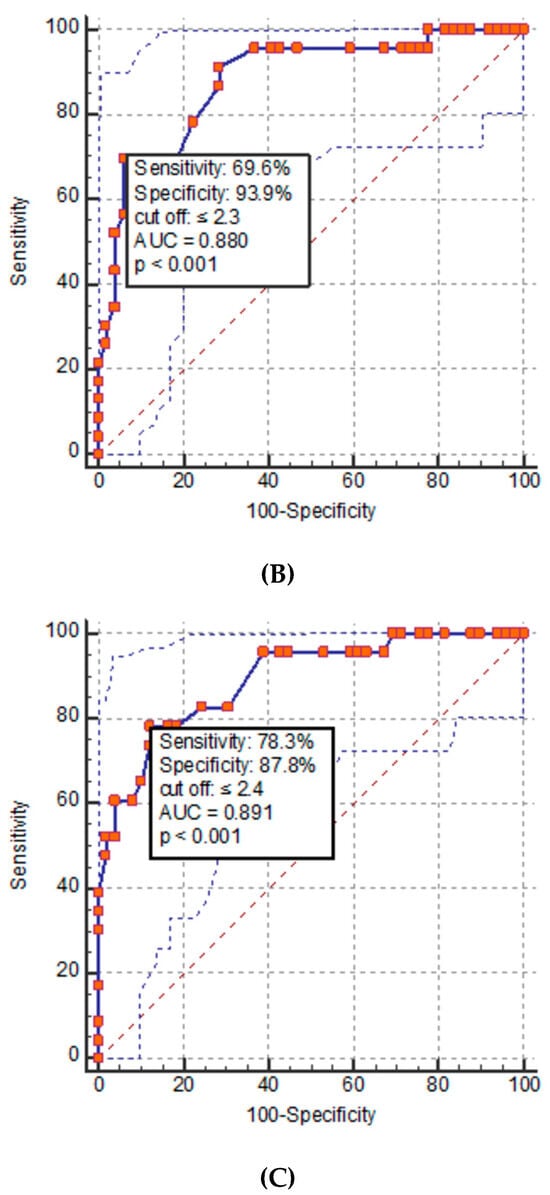
Results: The use of SS/PV–ROC curves with integrated cut-off concentration distribution curves provides improved diagnostic information compared to “traditional” ROC curves. The threshold distributions integrated as curves into SS/PV–ROC curves and SS-J/PV-PSI index–ROC curves run in opposite directions. In contrast to the SS–ROC curves, the PV–ROC and the novel PV-PSI index–ROC curves had neither an area under the curve (AUC) nor a range from 0% to 100%. The cut-off level of the qualitative assay was 7.5 µg/L, with a sensitivity of 65.9% and a specificity of 63.3%, and the PPV was 12.4% and the NPV was 95.9%, at a threshold value of 12.5 µg/L. Based on these set concentrations, the reader-based evaluation revealed a graphically estimated 5% increase in sensitivity and a 13% increase in specificity, as compared to the visual qualitative POC test. In the case of predictive values, there was a gain of 8% for PPV and 10% for NPV. The index values and cut-offs were as follows: visual SS-J index, 0.328 and 35 µg/L; visual PV-PSI index, 0.083 and 5.4 µg/L; maximal reader Youden index, 0.0558 and 250 µg/L; and maximal PV-PSI index, 0.459 and 250 µg/L, respectively. The maximum unified SS-J/PV-PSI index value was 0.32, and the cut-off was 43 µg/L. The reciprocal SS-J index correctly detected one out of three patients, while the reciprocal PV-PSI index gave one out of twelve patients a correct diagnosis. Conclusions: ROC curves including cut-off distribution curves supplement the information lost in “traditionally plotted” ROC curves. The novel sets of ROC and index–ROC curves and the new SS/PV index cut-off diagrams enable the simultaneous comparison of sensitivity/specificity and predictive value profiles of diagnostic tools and the identification of optimal cut-off values at maximal index values, even in a unifying SS/PV approach. Because the curves within an SS-J/PV-PSI index cut-off diagram are distributed over the complete cut-off range of a quantitative assay, this field is open for special clinical considerations, with the need to vary the mentioned clinical diagnostic parameters. Complete or partial areas over the x-axis (AOX) can be calculated for summarized quantitative or qualitative effectivity evaluations with respect to single and/or unified SS-J and PV-PSI indices and with respect to single, several, or several unified assays. The SS-J/PV-PSI index-AOX approach is a new tool providing additional joint clinical information, and the reciprocal SS-J indices can predict the number of patients with a correct diagnosis and the number of persons who need to be examined in order to correctly predict a diagnosis of the disease. These methods could be used in applications like medical or plant epidemiology, machine learning algorithms, and neural networks.
Full article
(This article belongs to the Section Clinical Laboratory Medicine)
Open AccessReview
Vascular Endothelial Damage in COPD: Where Are We Now, Where Will We Go?
by
Gianluca Screm, Lucrezia Mondini, Francesco Salton, Paola Confalonieri, Liliana Trotta, Mariangela Barbieri, Antonio Romallo, Alessandra Galantino, Michael Hughes, Selene Lerda, Marco Confalonieri and Barbara Ruaro
Diagnostics 2024, 14(9), 950; https://doi.org/10.3390/diagnostics14090950 - 30 Apr 2024
Abstract
Background: Chronic obstructive pulmonary disease (COPD) has higher rates among the general population, so early identification and prevention is the goal. The mechanisms of COPD development have not been completely established, although it has been demonstrated that endothelial dysfunction plays an important role.
[...] Read more.
Background: Chronic obstructive pulmonary disease (COPD) has higher rates among the general population, so early identification and prevention is the goal. The mechanisms of COPD development have not been completely established, although it has been demonstrated that endothelial dysfunction plays an important role. However, to date, the measurement of endothelial dysfunction is still invasive or not fully established. Nailfold video capillaroscopy (NVC) is a safe, non-invasive diagnostic tool that can be used to easily evaluate the microcirculation and can show any possible endothelial dysfunctions early on. The aim of this review is to evaluate if nailfold microcirculation abnormalities can reflect altered pulmonary vasculature and can predict the risk of cardiovascular comorbidities in COPD patients. Methods: A systematic literature search concerning COPD was performed in electronic databases (PUBMED, UpToDate, Google Scholar, ResearchGate), supplemented with manual research. We searched in these databases for articles published until March 2024. The following search words were searched in the databases in all possible combinations: chronic obstructive pulmonary disease (COPD), endothelial damage, vascular impairment, functional evaluation, capillaroscopy, video capillaroscopy, nailfold video capillaroscopy. Only manuscripts written in English were considered for this review. Papers were included only if they were able to define a relationship between COPD and endothelium dysfunction. Results: The search selected 10 articles, and among these, only three previous reviews were available. Retinal vessel imaging, flow-mediated dilation (FMD), and skin autofluorescence (AF) are reported as the most valuable methods for assessing endothelial dysfunction in COPD patients. Conclusions: It has been assumed that decreased nitric oxide (NO) levels leads to microvascular damage in COPD patients. This finding allows us to assume NVC’s potential effectiveness in COPD patients. However, this potential link is based on assumption; further investigations are needed to confirm this hypothesis.
Full article
(This article belongs to the Special Issue Diagnosis, Classification, and Monitoring of Pulmonary Diseases)
►▼
Show Figures
Graphical abstract
Open AccessReview
Sensitivity and Specificity of Selected Biomarkers and Their Combinations in the Diagnosis of Ovarian Cancer (Review)
by
Aleksandra Englisz, Marta Smycz-Kubańska and Aleksandra Mielczarek-Palacz
Diagnostics 2024, 14(9), 949; https://doi.org/10.3390/diagnostics14090949 - 30 Apr 2024
Abstract
One of the greatest challenges in modern gynecological oncology is ovarian cancer. Despite the numerous studies currently being conducted, it is still sometimes detected at late clinical stages, where the prognosis is unfavorable. One significant contributing factor is the absence of sensitive and
[...] Read more.
One of the greatest challenges in modern gynecological oncology is ovarian cancer. Despite the numerous studies currently being conducted, it is still sometimes detected at late clinical stages, where the prognosis is unfavorable. One significant contributing factor is the absence of sensitive and specific parameters that could aid in early diagnosis. An ideal screening test, in view of the low incidence of ovarian cancer, should have a sensitivity of greater than 75% and a specificity of at least 99.6%. To enhance sensitivity and specificity, diagnostic panels are being created by combining individual markers. The drive to develop better screening tests for ovarian cancer focuses on modern diagnostic methods based on molecular testing, which in turn aims to find increasingly effective biomarkers. Currently, researchers’ efforts are focused on the search for a complementary parameter to those most commonly used that would satisfactorily enhance the sensitivity and specificity of assays. Several biomarkers, including microRNA molecules, autoantibodies, cDNA, adipocytokines, and galectins, are currently being investigated by researchers. This article reviews recent studies comparing the sensitivity and specificity of selected parameters used alone and in combination to increase detection of ovarian cancer at an early stage.
Full article
(This article belongs to the Special Issue Diagnosis and Management of Gynecological Cancers: Volume 3)
Open AccessReview
Emerging Trends in Left Ventricular Thrombus: A Comprehensive Review of Non-Ischemic and Ischemic Cardiopathies, Including Eosinophilic Myocarditis, Chagas Cardiomyopathy, Amyloidosis, and Innovative Anticoagulant Approaches
by
Benjamin Colle, Fabian Demeure, Julien Higny, Martin Benoit, Jean-Philippe Henry, Isabelle Michaux, Robaye Benoit, Olivier Xhaët, Laurence Gabriel, Antoine Guedes, Dominique Blommaert, Nathalie Dulieu, Yannick Berners, Fabian Wery, Steven Droogmans, Bernard Cosyns and Maria-Luiza Luchian
Diagnostics 2024, 14(9), 948; https://doi.org/10.3390/diagnostics14090948 - 30 Apr 2024
Abstract
This comprehensive review explores the intricate aspects of left ventricular thrombus (LVT), a potential complication in both ischemic and non-ischemic cardiomyopathies. It provides a thorough understanding of left ventricular thrombus, revealing its uncommon incidence in the general population (7 cases per 10,000 patients),
[...] Read more.
This comprehensive review explores the intricate aspects of left ventricular thrombus (LVT), a potential complication in both ischemic and non-ischemic cardiomyopathies. It provides a thorough understanding of left ventricular thrombus, revealing its uncommon incidence in the general population (7 cases per 10,000 patients), predominantly linked to ischemic heart diseases (ICMs) at an 80% prevalence rate. Diagnostic tools, notably transthoracic echocardiography (TTE) and cardiac magnetic resonance imaging (CMR), demonstrate varying sensitivity but remain indispensable in specific clinical contexts related to LVT as non-invasive diagnostic modalities. A detailed comparison between ICM patients and those with non-ischemic cardiomyopathy (NICM) who have left ventricular thrombus reveals subtle distinctions with significant clinical implications. This analysis underscores the importance of these imaging techniques in distinguishing between the two conditions. Additionally, we explored the occurrence of LVT in specific non-ischemic cardiomyopathies, including Takotsubo syndrome, hypertrophic cardiomyopathy, eosinophilic myocarditis, Chagas disease, cardiac amyloidosis, and several other conditions. The article further delves into anticoagulation strategies, thoroughly examining their impact on LVT regression and patient outcomes. Pharmacological interventions, with a focus on direct oral anticoagulants, emerge as promising alternatives; however, there is insufficient information on their efficiency and safety, especially in NICM population. In conclusion, this review highlights the complex nature of LVT, incorporating a range of etiopathogenic factors, diagnostic complexities, and evolving therapeutic approaches. It emphasizes the pressing need for ongoing research in this field.
Full article
(This article belongs to the Special Issue New Progress in Diagnosis and Management of Cardiovascular Diseases)
►▼
Show Figures
Figure 1
Open AccessArticle
Urinary Epidermal Growth Factor Level as a Noninvasive Indicator of Tubular Repair in Patients with Acute Kidney Injury
by
Kazutoshi Ono, Akito Maeshima, Izumi Nagayama, Taro Kubo, Takashi Yagisawa and Daisuke Nagata
Diagnostics 2024, 14(9), 947; https://doi.org/10.3390/diagnostics14090947 - 30 Apr 2024
Abstract
Epidermal growth factor (EGF), an essential factor for the proliferation and survival of renal tubular cells, is expressed by distal tubules and normally excreted via urine. Previous studies in rats demonstrated that acute tubular injury reduces urinary EGF levels. However, it is unclear
[...] Read more.
Epidermal growth factor (EGF), an essential factor for the proliferation and survival of renal tubular cells, is expressed by distal tubules and normally excreted via urine. Previous studies in rats demonstrated that acute tubular injury reduces urinary EGF levels. However, it is unclear whether urinary EGF is a suitable monitoring marker of tubular repair status after acute kidney injury (AKI) in humans. To address this question, we measured serum and urinary EGF in patients with AKI (n = 99) using ELISA and investigated whether urinary EGF levels were associated with the severity of tubular injury and renal prognosis. Urinary EGF was abundant in healthy controls but showed a significant decrease in AKI patients (14,522 ± 2190 pg/mL vs. 3201 ± 459.7 pg/mL, p < 0.05). The urinary EGF level in patients with renal AKI was notably lower than that in patients with pre-renal AKI. Furthermore, the urinary EGF level in patients with AKI stage 3 was significantly lower than that in patients with AKI stage 1. Urinary EGF levels were negatively correlated with urinary β-2MG and serum creatinine levels but positively correlated with hemoglobin levels and eGFR. Urinary EGF was not significantly correlated with urinary NAG, α-1MG, L-FABP, NGAL, KIM-1, or urinary protein concentrations. No significant correlation was observed between serum and urinary EGF levels, suggesting that urinary EGF is derived from the renal tubules rather than the blood. In living renal transplantation donors, the urinary EGF/Cr ratio was approximately half the preoperative urinary EGF/Cr ratio after unilateral nephrectomy. Collectively, these data suggest that urinary EGF is a suitable noninvasive indicator of not only the volume of functional normal renal tubules but also the status of tubular repair after AKI.
Full article
(This article belongs to the Special Issue Kidney Disease: Biomarkers, Diagnosis, and Prognosis: 2nd Edition)
Open AccessArticle
Long-Term Effects of 0.1 mg Recombinant-Human-Thyrotropin-Stimulated Fixed-Dose Radioiodine Therapy in Patients with Recurrent Multinodular Goiter after Surgery
by
Nicholas Angelopoulos, Ioannis Iakovou, Grigoris Effraimidis and Sarantis Livadas
Diagnostics 2024, 14(9), 946; https://doi.org/10.3390/diagnostics14090946 - 30 Apr 2024
Abstract
(1) Background: After thyroid malignancy is ruled out, treatment options for multinodular goiter patients include surgery, levothyroxine suppressive therapy, and 131-I therapy. Surgery effectively reduces goiter size but carries risks of surgical and anesthetic complications. 131-I therapy is the only nonsurgical alternative, but
[...] Read more.
(1) Background: After thyroid malignancy is ruled out, treatment options for multinodular goiter patients include surgery, levothyroxine suppressive therapy, and 131-I therapy. Surgery effectively reduces goiter size but carries risks of surgical and anesthetic complications. 131-I therapy is the only nonsurgical alternative, but its effectiveness diminishes with goiter size and depends on iodine sufficiency. This study aimed to assess the efficacy and safety of 0.1 mg rhTSH as an adjuvant to a fixed dose of 131-I therapy in patients with a recurrence of large multinodular goiter, several years after the initial thyroidectomy. (2) Methods: 14 patients (13 females and 1 male), aged 59.14 ± 15.44 (range, 35–78 years) received 11mciu of 131-I, 24 h after the administration of 0.1 mg rhTSH. The primary endpoint was the change in thyroid volume (by ultrasound measurements) as well as in the diameter of the predominant nodule during a follow-up period of 10 years. Secondary endpoints were the alterations in thyroid function and potential adverse effects. (3) Results: A significant decrease in the volume of initial thyroid remnants (32.16 ± 16.66 mL) was observed from the first reevaluation (at 4 months, 23.12 ± 11.59 mL) as well as at the end of the follow-up period (10 years, 12.62 ± 8.76 mL), p < 0.01. A significant reduction in the dominant nodule was also observed (from 31.71 ± 10.46 mm in the beginning to 26.67 ± 11.05 mm). (4) Conclusions: Further investigation is needed since this approach could be attractive in terms of minimizing the potential risks of reoperation in these patients.
Full article
(This article belongs to the Section Clinical Laboratory Medicine)
►▼
Show Figures
Figure 1
Open AccessArticle
Hair Evaluation in Orthodontic Patients with Oligodontia
by
Małgorzata Zadurska, Adriana Rakowska, Ewa Czochrowska, Małgorzata Laskowska, Konrad Perkowski, Izabela Strużycka, Lidia Rudnicka and Agnieszka Jurek
Diagnostics 2024, 14(9), 945; https://doi.org/10.3390/diagnostics14090945 - 30 Apr 2024
Abstract
Oligodontia can be isolated or syndromic, associated with other ectodermal abnormalities. The aim of the study was to perform hair examination in orthodontic patients diagnosed with oligodontia with a low clinical expression of symptoms of ectodermal origin. All available orthodontic patients diagnosed with
[...] Read more.
Oligodontia can be isolated or syndromic, associated with other ectodermal abnormalities. The aim of the study was to perform hair examination in orthodontic patients diagnosed with oligodontia with a low clinical expression of symptoms of ectodermal origin. All available orthodontic patients diagnosed with oligodontia in the permanent dentition were enrolled. Hair examination included clinical evaluation of the patients’ hair, trichoscopy, trichogram and evaluation of the hair shafts under a polarized light microscope. In total, 25 patients, 18 males and 7 females, aged 6 to 24 years were evaluated for the presence of dental and hair abnormalities. The number of congenitally absent teeth ranged from 6 to 24 teeth and diastemas, microdontia, taurodontism and altered tooth shape were found in 23 patients. Hair disorders were found in 68% of the subjects. Hypotrichosis, the heterogeneity of shaft color and loss of pigment, androgenetic alopecia, telogen effluvium, trichoschisis, pili canaliculi, trichorrhexis nodosa and pseudomoniletrix were observed. Trichoscopy and trichogram are valid non-invasive diagnostic tests which could be used to differentiate between isolated and syndromic oligodontia in patients with a low clinical expression of ectodermal symptoms.
Full article
(This article belongs to the Special Issue New Insights into Diagnosis of Orthodontics)
Open AccessArticle
Radiomic Features of Acute Cerebral Hemorrhage on Non-Contrast CT Associated with Patient Survival
by
Saif Zaman, Fiona Dierksen, Avery Knapp, Stefan P. Haider, Gaby Abou Karam, Adnan I. Qureshi, Guido J. Falcone, Kevin N. Sheth and Seyedmehdi Payabvash
Diagnostics 2024, 14(9), 944; https://doi.org/10.3390/diagnostics14090944 - 30 Apr 2024
Abstract
The mortality rate of acute intracerebral hemorrhage (ICH) can reach up to 40%. Although the radiomics of ICH have been linked to hematoma expansion and outcomes, no research to date has explored their correlation with mortality. In this study, we determined the admission
[...] Read more.
The mortality rate of acute intracerebral hemorrhage (ICH) can reach up to 40%. Although the radiomics of ICH have been linked to hematoma expansion and outcomes, no research to date has explored their correlation with mortality. In this study, we determined the admission non-contrast head CT radiomic correlates of survival in supratentorial ICH, using the Antihypertensive Treatment of Acute Cerebral Hemorrhage II (ATACH-II) trial dataset. We extracted 107 original radiomic features from n = 871 admission non-contrast head CT scans. The Cox Proportional Hazards model, Kaplan–Meier Analysis, and logistic regression were used to analyze survival. In our analysis, the “first-order energy” radiomics feature, a metric that quantifies the sum of squared voxel intensities within a region of interest in medical images, emerged as an independent predictor of higher mortality risk (Hazard Ratio of 1.64, p < 0.0001), alongside age, National Institutes of Health Stroke Scale (NIHSS), and baseline International Normalized Ratio (INR). Using a Receiver Operating Characteristic (ROC) analysis, “the first-order energy” was a predictor of mortality at 1-week, 1-month, and 3-month post-ICH (all p < 0.0001), with Area Under the Curves (AUC) of >0.67. Our findings highlight the potential role of admission CT radiomics in predicting ICH survival, specifically, a higher “first-order energy” or very bright hematomas are associated with worse survival outcomes.
Full article
(This article belongs to the Special Issue Advances in Cerebrovascular Imaging and Interventions)
►▼
Show Figures
Figure 1
Open AccessArticle
Case Ascertainment of Measles during a Large Outbreak—Laboratory Compared to Epidemiological Confirmation
by
Chen Stein-Zamir, Nitza Abramson, Irina Sokolov, Lia Mor-Shimshi and Hanna Shoob
Diagnostics 2024, 14(9), 943; https://doi.org/10.3390/diagnostics14090943 - 30 Apr 2024
Abstract
Measles is a highly contagious viral disease, and hence, sufficient herd immunity is obligatory to prevent infection transmission. Measles is still a cause of considerable disease burden globally, mainly in children. During a national measles outbreak in Israel in 2018–2019, the peak incidence
[...] Read more.
Measles is a highly contagious viral disease, and hence, sufficient herd immunity is obligatory to prevent infection transmission. Measles is still a cause of considerable disease burden globally, mainly in children. During a national measles outbreak in Israel in 2018–2019, the peak incidence rates occurred in the Jerusalem district. Most measles cases in the Jerusalem district (75.5%, 1702) were observed in children younger than 15 years of age, 49.2% (1109) were in children under 5 years of age, and 18.9% (425) were in infants under 1 year of age. The routine measles vaccination schedule includes two doses at 1 and 6 years of age. Most cases (1828, 81.1%) were unvaccinated (zero measles vaccine doses). These cases comprised the 425 affected infants under 1 year of age, who were ineligible for vaccination, along with the 1403 children over 1 year of age, who were otherwise unvaccinated. This study aimed to describe the epidemiologic and laboratory features of this measles outbreak, and to investigate case ascertainment (laboratory confirmed compared to epidemiologically confirmed cases). The study population included 2254 measles cases notified during the period spanning June 2018 to May 2019 in the Jerusalem district (incidence rate 176 per 10,000 population). Of the 2254 cases, 716 (31.8%) were laboratory confirmed, and 1538 (68.2%) were confirmed as epidemiologically linked. Most laboratory confirmed cases (420, 58.7%) underwent real-time PCR tests. Serological tests (measles IgM and IgG) were used in 189 (26.4%) cases, and a combination of RT-PCR and serology was used in 107 (14.9%) cases. In a multivariate model analysis, the variables significantly associated (after adjustment) with higher odds for laboratory confirmation included month of disease onset (late), additional measles cases in the household (single case), place of medical treatment (hospital; either emergency department, or hospitalization) and vaccination status (at least one prior vaccine dose). The measles outbreak described demonstrates the urgency of addressing vaccination gaps with appropriate outbreak prevention programs. The road to measles elimination needs to be paved with robust public health infrastructure, excellent field epidemiology for outbreak surveillance, investigation, and control, and laboratory proficiency.
Full article
(This article belongs to the Special Issue Laboratory Diagnosis of Infectious Diseases)
►▼
Show Figures
Figure 1
Open AccessArticle
Intraoperative Ultrasound: Bridging the Gap between Laparoscopy and Surgical Precision during 3D Laparoscopic Partial Nephrectomies
by
Ionela Mihai, Horatiu Dura, Cosmin Adrian Teodoru, Samuel Bogdan Todor, Cristian Ichim, Nicolae Grigore, Cosmin Ioan Mohor, Alin Mihetiu, George Oprinca, Nicolae Bacalbasa, Denisa Tanasescu, Dan Georgian Bratu, Adrian Boicean, Bogdan Oros and Adrian Hasegan
Diagnostics 2024, 14(9), 942; https://doi.org/10.3390/diagnostics14090942 - 30 Apr 2024
Abstract
The use of 3D laparoscopic partial nephrectomy has emerged as a cornerstone in the surgical arsenal for addressing renal tumors, particularly in managing challenging cases characterized by deeply seated tumors embedded within the renal parenchyma. In these intricate scenarios, the utilization of intraoperative
[...] Read more.
The use of 3D laparoscopic partial nephrectomy has emerged as a cornerstone in the surgical arsenal for addressing renal tumors, particularly in managing challenging cases characterized by deeply seated tumors embedded within the renal parenchyma. In these intricate scenarios, the utilization of intraoperative ultrasound (IOUS) acquires paramount importance, serving as an indispensable tool for guiding and meticulously monitoring the surgical process in real time. To further explore the efficacy of IOUS-guided techniques, we conducted a retrospective study comparing outcomes in patients who underwent partial nephrectomy with IOUS guidance (n = 60) between 2020 and 2022 with a cohort from 2018 to 2019 without IOUS guidance (n = 25). Our comprehensive analysis encompassed various post-operative parameters, including the duration until food resumption, analgesia requirements, and length of the hospital stay. While these parameters exhibited comparable outcomes between the two groups, notable distinctions emerged in the intraoperative metrics. The IOUS-guided cohort demonstrated significantly reduced blood loss, a shorter median operative duration, and diminished ischemia time (p = 0.001). These compelling findings underscore the undeniable benefits of IOUS-guided techniques in not only facilitating the attainment of negative surgical margins but also in enhancing procedural safety and precision, thereby contributing to improved patient outcomes in the management of renal tumors.
Full article
(This article belongs to the Section Medical Imaging and Theranostics)
►▼
Show Figures
Figure 1
Journal Menu
► ▼ Journal Menu-
- Diagnostics Home
- Aims & Scope
- Editorial Board
- Reviewer Board
- Topical Advisory Panel
- Instructions for Authors
- Special Issues
- Topics
- Sections & Collections
- Article Processing Charge
- Indexing & Archiving
- Editor’s Choice Articles
- Most Cited & Viewed
- Journal Statistics
- Journal History
- Journal Awards
- Society Collaborations
- Conferences
- Editorial Office
Journal Browser
► ▼ Journal BrowserHighly Accessed Articles
Latest Books
E-Mail Alert
News
Topics
Topic in
Algorithms, Diagnostics, Entropy, Information, J. Imaging
Application of Machine Learning in Molecular Imaging
Topic Editors: Allegra Conti, Nicola Toschi, Marianna Inglese, Andrea Duggento, Matthew Grech-Sollars, Serena Monti, Giancarlo Sportelli, Pietro CarraDeadline: 31 May 2024
Topic in
Biomedicines, Current Oncology, Diagnostics, Gastrointestinal Disorders, JCM
Advances in Gastrointestinal and Liver Disease: From Physiological Mechanisms to Clinical Practice
Topic Editors: Davide Giuseppe Ribaldone, Gian Paolo CavigliaDeadline: 20 June 2024
Topic in
Biomolecules, Diagnostics, JCM, Metabolites, JPM, Nutrients
Metabolic Syndrome, Biomarkers and Lifestyles
Topic Editors: Sang Yeoup Lee, Young Hye ChoDeadline: 30 June 2024
Topic in
Cancers, Diagnostics, JCM, Current Oncology, Gastrointestinal Disorders, Biomedicines
Hepatobiliary and Pancreatic Diseases: Novel Strategies of Diagnosis and Treatments
Topic Editors: Alessandro Coppola, Damiano Caputo, Roberta Angelico, Domenech Asbun, Chiara MazzarelliDeadline: 20 July 2024
Conferences
Special Issues
Special Issue in
Diagnostics
Fifth Anniversary of "Machine Learning and Artificial Intelligence in Diagnostics" Section
Guest Editors: Dania Cioni, Salvatore Claudio FanniDeadline: 10 May 2024
Special Issue in
Diagnostics
Monitoring and Diagnosis of Patients during Rehabilitation Training
Guest Editor: Redha TaiarDeadline: 31 May 2024
Special Issue in
Diagnostics
Improvement of Diagnostic Sensitivity for Tumor Markers
Guest Editors: Jong-Han Lee, Jooyoung ChoDeadline: 16 June 2024
Special Issue in
Diagnostics
Endoscopy and Diagnostic Tools in Hepatobiliary and Pancreatic Diseases
Guest Editor: Stefano Francesco CrinòDeadline: 30 June 2024
Topical Collections
Topical Collection in
Diagnostics
Editorial Board Members' Collection Series: Diagnostic Approaches to Gastrointestinal and Pancreatic Diseases
Collection Editors: Paolo Aseni, Ervin Toth