Article

Influence of Additive Firing on the Surface Characteristics, Streptococcus mutans Viability and Optical Properties of Zirconia

Department of Dental Biomaterials Science, Dental Research Institute, School of Dentistry, Seoul National University, Seoul 03080, Korea
*
Authors to whom correspondence should be addressed.
Equal contribution.
Materials 2021, 14(5), 1286; https://doi.org/10.3390/ma14051286
Submission received: 24 January 2021 / Revised: 5 March 2021 / Accepted: 5 March 2021 / Published: 8 March 2021

Abstract

:
The purpose of this study was to observe whether the repetitive firing of dental zirconia caused changes in surface characteristics, S. mutans viability, and optical properties of zirconia. Dental zirconia blocks were sintered and randomly distributed into seven experimental groups: F0–F6. Except for F0, which only went through sintering, the additive firing was performed in order for F1–F6. Surface roughness, contact angle, S. mutans viability by fluorescence, and translucency parameter were measured. They were all highest after sintering (F0) and decreased after additive firings (F1–F6). The additive firing of zirconia after sintering decreased surface roughness, contact angle, S. mutans viability, and translucency. The number of firings after the first firing was not found to be critical in surface characteristics, S. mutans viability, and optical property. Changes in surface characteristics might have led to a decrease in S. mutans viability, while the change of translucency was not clinically significant. This implies that additive firing may prevent secondary caries under zirconia restorations, not compromising esthetic appearance.

1. Introduction

Dental zirconia has been widely used for esthetic prostheses that required high physical properties due to its superior mechanical properties and chemical stability compared to conventional ceramic materials [1,2,3]. It has been used not only as all-ceramic restorations, core materials, and orthodontic brackets, but also as an implant material because of its excellent biocompatibility and bone fusion with the alveolar bone [4].
The method of using zirconia for tooth restoration can be divided into two categories. One is its use as a core material; ceramic material is then fabricated on the top of zirconia in a bilayer form (porcelain fused zirconia; PFZ). Due to the optical impermeability of zirconia, it is covered with veneering porcelain. Feldspar is mainly used to reproduce the esthetic characteristics of natural teeth. The veneering process of placing ceramics on the zirconia surface generally requires five stages of firings (750–900 °C) Another method is a pre-colored dental zirconia block that overcomes the porcelain chipping, which is the biggest drawback of PFZ. Zirconia is manufactured and used in a crown shape without ceramic material (full-contour zirconia; FCZ). However, the colored zirconia blocks are also insufficient to reproduce the colors of various teeth, so additional coloring or firing in the dental laboratory is required. Therefore, additive firing is one of the essential parts in the fabrication of dental zirconia prostheses.
Firing process and its effect on properties of zirconia are not fully understood, but there have been several reports on the negative effects of firing. Although the additive firing temperature was lower than the sintering temperature of the core material, it decreased the flexural strength and microhardness of zirconia [5]. In addition, even at lower temperatures, low-temperature degradation was found to be accelerated in yttria-stabilized zirconia implants used in orthopedics [6]. In contrast, one of the clinical advantages of firing was that it improved marginal fit [7]. With regard to translucency, previous studies reported its decrease after repeated firing [8,9], but the clinical significance of such decrease was not fully investigated.
Surface characteristics such as surface roughness and contact angle are closely related [10,11]. Their relationship that an increase in one increases another and vice versa has been well known and shown in the researches regarding dental zirconia [12,13]. To understand the effect of surface roughness and contact angle on zirconia, various processes such as laser scanning, sandblasting, polishing, machining, and heat treatment were performed to produce diverse topography [14,15,16]. However, the effect of repeated firing on the surface roughness and contact angle of zirconia has not been studied yet.
Analysis of surface roughness and contact angle in terms of biofilm accumulation is also important since they affect microbial adhesion to induce biofilm formation [17,18,19]. In particular, increased surface roughness [20,21] and specific surface topography [22] were reported to be major contributing factors of microbial adhesion. Biofilm on dental restorations has been known to have harmful effects since it causes secondary caries and peri-implantitis [23,24,25]. Among various microorganisms participating in biofilm formation and caries, S. mutans plays a main role [26]. For successful dental restorations, reducing S. mutans adhesion might be important, so there has been research on adhesion of S. mutans on zirconia [27,28]. To provide a more favorable surface to reduce S. mutans adhesion, reduction in surface roughness and hydrophobicity has been found to be successful [29,30]. However, the previous studies were limited to zirconia without firing, and there have been a lack of studies in relation to adhesion after firing.
Despite the importance and advantage of firing in zirconia, its effect has not been clearly identified in relation to surface roughness, contact angle, S. mutans viability, and translucency. In addition, those characteristics have not been integrated to suggest clinical relevance. Therefore, the purpose of this study was to observe whether the repetitive firing of dental zirconia caused changes in surface characteristics, S. mutans viability, and optical property of zirconia and to discuss the importance of them in clinical perspective.

2. Materials and Methods

2.1. Preparation of Specimen

Dental zirconia blocks (Lava Plus, 3M ESPE, St. Paul, MN, USA) were cut and finished by a low-speed diamond disc (MD-Piano, Struers, Ballerup, Denmark). They were then sintered, up to 1450 °C, according to the manufacturer’s instructions. The specimens were randomly distributed into seven experimental groups: F0 (control; sintering only), F1 (first additive firing (ZirLiner; zirconia lining material)), F2 (second additive firing (Margin)), F3 (third additive firing (Wash)), F4 (fourth additive firing (Dentin and Enamel)), F5 (fifth additive firing (Stain)), and F6 (sixth additive firing (Glazing)); n = 7 each. Except for F0, which only went through sintering, the additive firing was performed for F1–F6 according to the manufacturer’s instructions (Figure 1). They were embedded in an epoxy resin (Cold Mounting Systems Epoxy Systems, Metallurgical Supplies, Buffalo, NY, USA) and went through final finishing and polishing up to 0.06 μm abrasive (LaboPol-5, Struers, Ballerup, Denmark).

2.2. Surface Characteristics

For all the groups (F0–F6), surface roughness was measured by confocal laser scanning microscopy (CLSM) (LMS 5-Pascal, Carl Zeiss, Oberhausen, Germany) at 20 × objective to obtain the image field of 500 × 500 μm. High pass filter cut-off ( λ c ) of 800 μm and low pass filter cut-off ( λ s ) of 2.5 μm were used to determine roughness from noise and waviness. Ra (arithmetic mean roughness of profile), Sa (mean height of the surface), Sz (maximum height of surface), and Sv (maximum pit height of surface) values were obtained for the roughness parameters. Representative surface profiles were also obtained and also contact angles were measured (Phoenix 150, SEO, Suwon, Korea). At room temperature, one drop of distilled water was placed at the center of the zirconia surface and the camera acquired the profile image. A computer software determined the contact angle between the surface and the tangent of the water drop on the zirconia. Roughness and contact angles were measured with three specimens from each group.

2.3. Streptococcus Mutans Viability

Streptococcus mutans (UA159) was cultured in Brain Heart Infusion Broth (Sigma-Aldrich, St. Louis, MO, USA) at 37 °C of 100% relative humidity. Zirconia specimens were prepared by ultrasonication with ethanol and distilled water. Then S. mutans (OD600 = 0.5) was cultured over the prepared specimen for 24 h. S. mutans was treated by bacterial viability kit (LIVE/DEAD® BacLightTM, Invitrogen by Thermo Fisher Scientific, San Jose, CA, USA) for 15 min to be observed under CLSM (LSM700, Carl Zeiss Meditec, Jena, Germany). Based on the images, the area fraction of the fluorescent cells was calculated with computer software (ImageJ, NIH, Bethesda, MD, USA).

2.4. Calculation of Translucency

The translucency was measured using the translucency parameter (TP) [31] with three specimens of each group. The CIE L*a*b* values (L* referred to brightness, a* to redness-greenness, and b* to yellowness–blueness) were measured at the center of each specimen over a black (B) and white (W) background using a spectrophotometer (Ci7600, X-rite, Grand Rapids, IL, USA). Then the TP values were calculated by the following equation:
T P = L * W L * B 2 + a * W a * B 2 + b * W b * B 2 1 / 2

2.5. Statistical Analysis

Surface roughness, contact angle, and translucency parameters were analyzed by a nonparametric Kruskal–Wallis test and Mann–Whitney test with Bonferroni correction as they failed the normality test. All statistical analyses were performed with a significance level of 0.05 using SPSS statistics for Windows (version 26.0, IBM, Armonk, NY, USA).

3. Results

3.1. Surface Characteristics

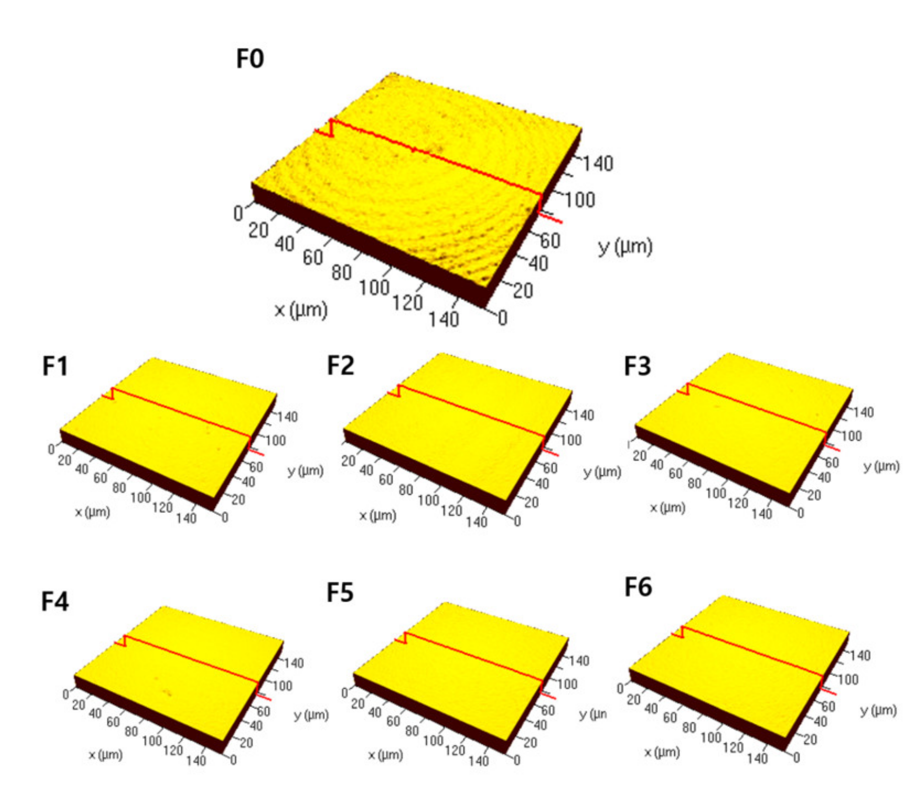
The surface roughness measured as mean height (Sa) was the highest (0.17 (0.15, 0.18) μm) immediately after sintering (F0; control), and the roughness decreased to 0.07 (0.06, 0.07) μm after the first firing (F1). Second firing (F2) (0.06 (0.06, 0.06) μm), third firing (F3) (0.06 (0.06, 0.06) μm), fourth firing (F4) (0.06 (0.06, 0.07) μm), fifth firing (F5) (0.06 (0.06, 0.06) μm), and sixth firing (F6) (0.06 (0.06, 0.07) μm) also decreased the surface roughness compared to F0. For the arithmetic mean roughness of profile (Ra), maximum height of surface (Sz), and maximum pit height of surface (Sv), the same trend as in mean height (Sa) was observed (Table 1). Moreover, representative surface profiles were obtained (Figure 2). It was consistent with the roughness values in that F1–F6, in contrast to F0, had decreased roughness and showed no identifiable surface structures.
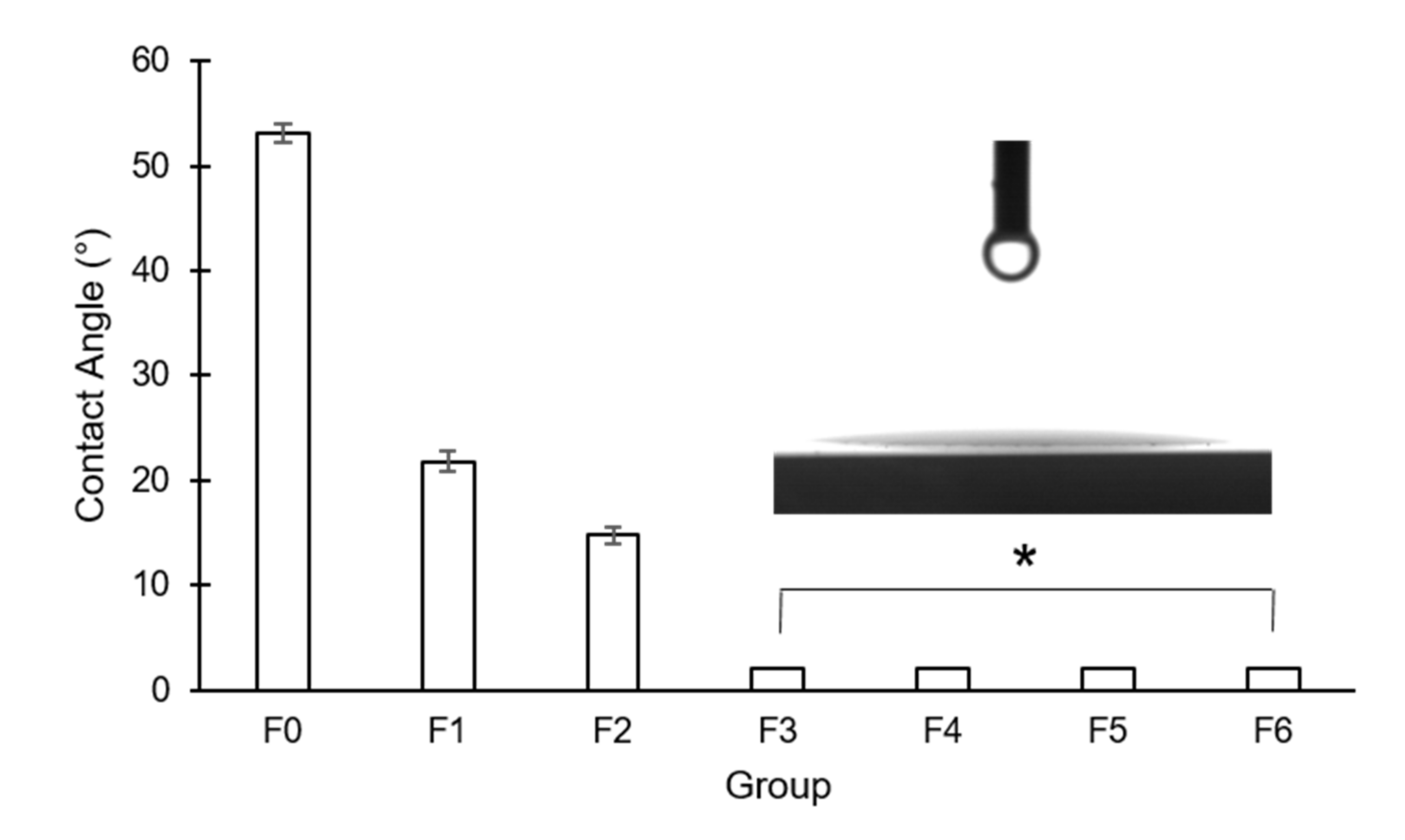
The contact angle was 53.13 ± 0.82° in F0, and it decreased to 21.83 ± 0.97° in F1 and to 14.75 ± 0.84° in F2. In F3–F6, the contact angle was less than 10°, indicating their hydrophilic properties (Figure 3). There was a significant difference between every pair of the groups (p < 0.05).

3.2. Streptococcus Mutans Viability

The area fraction of fluorescence, which represented the viability of S. mutans, observed with CLSM was the highest in F0 (7.66%), and it decreased to 1.47% (F1), 1.02% (F2), 0.88% (F3), 0.66% (F4), 0.92% (F5), and 0.85% (F6). Its decrease was visualized as a series of fluorescence images (Figure 4).

3.3. Optical Property

Median translucency parameters decreased after firing (F1–F6) compared to F0. It was 5.05 in F0 and decreased to 4.39 (F1), 4.21 (F2), 4.43 (F3), 4.66 (F4), 4.53 (F5), and 4.79 (F6) (Table 2).

4. Discussion

Dental zirconia undergoes additive firing during the veneering process for esthetic purposes, and their physical properties may change during the process. It was reported that the veneering process reduced mechanical properties such as flexural strength, microhardness [5], and bond strength of ceramic [32]. In this study, surface characteristics, microbial viability, and translucency were newly tested, and firing conditions were more specifically divided (F0–F6). The surface roughness was the highest in F0 and decreased rapidly after the first firing (F1). After that, the roughness did not show radical change according to the number of firing times from the second to sixth firing. To the best of the authors’ knowledge, there have been no previous reports on the decrease of roughness after firing. The possible explanation for the phenomenon was that the microstructure or debris on the surface could have been destroyed and removed due to high energy transmitted by firing processes which were conducted at a maximum of 750–900 °C. This might also explain that there was only a small change in surface topography after the first firing. Decreased roughness in dental zirconia has been known to be more favorable to initial fibroblast adhesion [33]. Fibroblast adhesion has been typically used for testing cytotoxicity of dental materials [34]. Considering its high possibility of contact with surrounding soft tissue, dental zirconia’s lowered roughness might provide higher biocompatibility.
For the bacteria, on the other hand, increased roughness induces their adhesion due to irregularities and increased surface area [35]. This phenomenon was well presented in various biomaterials [36,37]. In this study, higher roughness in F0 led to higher S. mutans viability, so it was consistent with the previous studies. The significance of this study was that such phenomenon was reproduced by repeated firings. Increased roughness and the following hydrophobicity are also well-known [38]. Typically, roughness-based hydrophilicity was obtained after polishing by reducing roughness [39], but the firing, which was a part of zirconia veneering, showed that it could reduce roughness to induce hydrophilicity more efficiently.
To analyze the surface roughness both on a profile and surface, profile roughness parameter (Ra) and areal roughness parameters (Sa, Sz, and Sv) were taken. Although Ra could provide the arithmetic mean of the roughness on a profile, it only measures 2D roughness. To include 3D information about the roughness, the areal parameters were additionally used in the study [40]. First, Sa was measured to give an overall picture of the surface. As a result, the specimens in the study were found to be full of peaks and valleys, so the parameters which were sensitive to peaks and valleys were measured to capture them. Sz values which represented the sum of maximum peak heights and maximum pit heights and Sv values which represented the maximum pit heights, were used to supplement Sa values. If additional feature parameters such as Spd, Spc, etc. can be provided in future studies, it would suggest more information about the surfaces.
The contact angle was significantly reduced after the first and second firing (F1 and F2), and it was consistently smaller than 10° in the following firing. This might be explained in relation to the physicochemistry alteration of the surface. Although there have been no explicit results about the effect of firing, numerous approaches, including oxygen plasma and ultraviolet treatment, were known to induce hydrophilicity by removal of hydrocarbons and insertion of polar groups on the surface [41]. As the firing also conveys heat energy, hydrophilicity might have been obtained by similar mechanisms. Since hydrophilicity of dental zirconia as restorative material may improve its surface adaptation of adhesives, primers, or resin cement [42], firing-induced hydrophilicity may have advantages in the bonding process. However, to utilize the increased hydrophilicity after firing, further studies should be performed to test whether such property is maintained for the intended period of time.
Among the various types of bacteria residing in the oral cavity, S. mutans forms a biofilm on the tooth surface and demineralizes it, causing dental caries [43]. Reducing S. mutans viability to inhibit biofilm formation is important in preventing dental caries. After firing zirconia in this study, the viability of S. mutans was reduced in all stages (F1–F6), which can be said to be a clinical advantage. However, although S. mutans is one of the major players in biofilm formation, it is not the only one. Other microorganisms such as F. nucleatum, P. gingivalis, and S. sanguinis are also involved in biofilm formation [44]. Thus, more studies on various microorganisms should be conducted to further discuss biofilm.
Multiple firings bring changes in color and translucency due to alterations in crystalline structure and surface specifications [45,46]. The decrease in translucency after firing could be an obstacle in obtaining the desired appearance since additive firing is basically to suffice esthetic purposes by veneering. In this study, the decrease in translucency parameter after firing was between 0.5 and 0.92. According to the previous study, a decrease in translucency parameter as much as 1.24 was translated to an increase of 0.02 in contrast ratio [47]. If the contrast ratio, which is another parameter to determine translucency, is smaller than 0.07, it is known that the difference in translucency is not detectable by human eyes [48]. The decrease in translucency parameter in this study was smaller than 1.24, so the increase in contrast ratio would be even smaller, leading to an undetectable change of translucency. Therefore, despite the decrease in translucency parameter after firing, it was not large enough to be detected. This can be an important advantage since potentially beneficial changes in roughness, contact angle, and S. mutans viability, as previously discussed, are achieved not affecting translucency.
The physical changes by multiple firing of zirconia and their potential advantages were discussed in this study. However, it may be necessary to investigate whether multiple firing could affect other factors such as longevity, structural stability, color, and marginal integrity that might influence the function of dental restorations. If there is a negative effect on those factors, the clinical importance of the presented results might be questioned. Thus, additional research that confirms the original function as dental restoration is needed. In addition, the effect of multiple firings without the presence of additives (liner, stain, etc.) might also be investigated to observe possible interfacial interaction. By doing so, it would allow more sophisticated control of the experimental conditions and improve the clinical significance of the current study.
In a dental prosthesis, the veneering material covers most of the outer surface and core material, so two different materials can affect each other. Veneering ceramic increases the bulk thickness and may cause a change in the mechanical strength. According to one study, the cause of failure of the zirconia-based all-ceramic restorations occurred between the two interfaces or inside the veneer ceramic [49]. Thus, the result of firing after applying veneering porcelain may appear different from this study. Also, thermal behavior in the veneered zirconia is also a significant factor since many failures occur due to thermal incompatibility [50,51]. To evaluate thermal behavior in porcelain-zirconia restorations, variations on firing and cooling rate were studied [52]. Rather than constant firing and cooling rate presented in this study, different conditions may be applied to adjust thermal compatibility between zirconia and veneered porcelain. Optimization of the sintering and firing conditions is also important for the other properties discussed in this study. The effect of various sintering and firing conditions on roughness, contact angle, microbial viability, and translucency is still unknown. Though the current conditions are based on the manufacturer’s instructions, further studies may discover better conditions to achieve superior properties.
This study focused on the phenomenon that took place on the surface. However, zirconia goes through phase transformation from tetragonal to monoclinic under stress [53], so structural changes can also be related to surface characteristics and optical properties. Previous studies of zirconia phase transformation were mostly about changes in mechanical properties such as flexural strength [54,55]. Other than phase transformation, but still in the context of structural analysis, the grain size was also known to affect zirconia properties [56]. Therefore, establishing correlation between structural changes and surface characteristics, more specifically in the additive firing process, may add insights to this study.
The reported characteristics of zirconia in the present study, if reinforced by further studies, might be applied to various clinical situations. For example, zirconia implants can go through firings for better hydrophilicity and less S. mutans viability to be used with grafting biomaterials to induce bone growth more efficiently [57,58,59] or for immunodeficient patients [60,61,62]. Also, zirconia after firings might have the potential to be used as the tissue engineering scaffold for stem cells in the field of regenerative dentistry [63].

5. Conclusions

The additive firing of zirconia after sintering decreased surface roughness, contact angle, S. mutans viability, and translucency. The number of firings after the first firing was not found to be critical in surface characteristics, S. mutans viability, and optical property. Changes in surface characteristics might have led to decrease in S. mutans viability, while the change of translucency was not clinically significant. This study, if reinforced by further research on microbial viability, implies that additive firing may prevent secondary caries under zirconia restorations, not compromising esthetic appearance.

Author Contributions

Conceptualization, methodology, software, writing—original draft, W.M. and J.H.P.; data curation, B.-S.L. and H.-A.L.; supervision, project administration, B.-S.L.; writing—review and editing, W.M. and J.H.P.; investigation, validation, H.-A.L. and S.H.C.; project administration, visualization, funding acquisition, S.H.C. All authors have read and agreed to the published version of the manuscript.

Funding

This research was funded by the National Research Foundation (NRF) of Korea, grant number 2020R1A2C1102316.

Institutional Review Board Statement

Not applicable.

Informed Consent Statement

Not applicable.

Data Availability Statement

Data sharing is not applicable for this article.

Conflicts of Interest

The authors declare no conflict of interest. The funders had no role in the design of the study; in the collection, analyses, or interpretation of data; in the writing of the manuscript, or in the decision to publish the results.

References

  1. Pittayachawan, P.; McDonald, A.; Young, A.; Knowles, J.C. Flexural strength, fatigue life, and stress-induced phase trans-formation study of Y-TZP dental ceramic. J. Biomed. Mater. Res. B 2009, 88, 366–377. [Google Scholar] [CrossRef]
  2. Guazzato, M.; Albakry, M.; Ringer, S.P.; Swain, M.V. Strength, fracture toughness and microstructure of a selection of all-ceramic materials. Part II. Zirconia-based dental ceramics. Dent. Mater. 2004, 20, 449–456. [Google Scholar] [CrossRef]
  3. Ardlin, B.I. Transformation-toughened zirconia for dental inlays, crowns and bridges: Chemical stability and effect of low-temperature aging on flexural strength and surface structure. Dent. Mater. 2002, 18, 590–595. [Google Scholar] [CrossRef]
  4. Kosmač, T.; Oblak, C.; Jevnikar, P.; Funduk, N.; Marion, L. The effect of surface grinding and sandblasting on flexural strength and reliability of Y-TZP zirconia ceramic. Dent. Mater. 1999, 15, 426–433. [Google Scholar] [CrossRef]
  5. Øilo, M.; Gjerdet, N.R.; Tvinnereim, H.M. The firing procedure influences properties of a zirconia core ceramic. Dent. Mater. 2008, 24, 471–475. [Google Scholar] [CrossRef] [PubMed]
  6. Chevalier, J. What future for zirconia as a biomaterial? Biomaterials 2006, 27, 535–543. [Google Scholar] [CrossRef]
  7. Kohorst, P.; Brinkmann, H.; Dittmer, M.P.; Borchers, L.; Stiesch, M. Influence of the veneering process on the marginal fit of zirconia fixed dental prostheses. J. Oral Rehabil. 2010, 37, 283–291. [Google Scholar] [CrossRef] [PubMed]
  8. Li, S.; Pang, L.; Yao, J. The effects of firing numbers on the opening total pore volume, translucency parameter and color of dental all-ceramic systems. Hua Xi Kou Qiang Yi Xue Za Zhi 2012, 30, 417–419. [Google Scholar]
  9. Nejatidanesh, F.; Azadbakht, K.; Savabi, O.; Sharifi, M.; Shirani, M. Effect of repeated firing on the translucency of CAD-CAM monolithic glass-ceramics. J. Prosthet. Dent. 2020, 123, 530.e1–530.e6. [Google Scholar] [CrossRef]
  10. Ryan, B.J.; Poduska, K.M. Roughness effects on contact angle measurements. Am. J. Phys. 2008, 76, 1074–1077. [Google Scholar] [CrossRef] [Green Version]
  11. Yang, C.; Tartaglino, U.; Persson, B.N.J. Influence of Surface Roughness on Superhydrophobicity. Phys. Rev. Lett. 2006, 97, 116103. [Google Scholar] [CrossRef] [PubMed] [Green Version]
  12. Yang, Y.; Zhou, J.; Liu, X.; Zheng, M.; Yang, J.; Tan, J. Ultraviolet light-treated zirconia with different roughness affects function of human gingival fibroblasts in vitro: The potential surface modification developed from implant to abutment. J. Biomed. Mater. Res. B 2015, 103, 116–124. [Google Scholar] [CrossRef]
  13. Pantea, M.; Antoniac, I.; Trante, O.; Ciocoiu, R.; Fischer, C.A.; Traistaru, T. Correlations between connector geometry and strength of zirconia-based fixed partial dentures. Mater. Chem. Phys. 2019, 222, 96–109. [Google Scholar] [CrossRef]
  14. Abdullah, A.O.; Muhammed, F.K.; Yu, H.; Pollington, S.; Xudong, S.; Liu, Y. The impact of laser scanning on zirconia coating and shear bond strength using veneer ceramic material. Dent. Mater. J. 2019, 38, 452–463. [Google Scholar] [CrossRef] [Green Version]
  15. Khan, A.A.; Mohamed, B.A.; Mirza, E.H.; Syed, J.; Divakar, D.D.; Vallittu, P.K. Surface wettability and nano roughness at different grit blasting operational pressures and their effects on resin cement to zirconia adhesion. Dent. Mater. J. 2019, 38, 388–395. [Google Scholar] [CrossRef] [Green Version]
  16. Rohr, N.; Zeller, B.; Matthisson, L.; Fischer, J. Surface structuring of zirconia to increase fibroblast viability. Dent. Mater. 2020, 36, 779–786. [Google Scholar] [CrossRef]
  17. Han, A.; Tsoi, J.K.; Matinlinna, J.P.; Zhang, Y.; Chen, Z. Effects of different sterilization methods on surface characteristics and biofilm formation on zirconia in vitro. Dent. Mater. 2018, 34, 272–281. [Google Scholar] [CrossRef]
  18. Chen, L.; Yang, S.; Yu, P.; Wu, J.; Guan, H.; Wu, Z. Comparison of bacterial adhesion and biofilm formation on zirconia fabricated by two different approaches: An in vitro and in vivo study. Adv. Appl. Ceram. 2020, 119, 323–331. [Google Scholar] [CrossRef]
  19. Kunrath, M.F.; Monteiro, M.S.; Gupta, S.; Hubler, R.; De Oliveira, S.D. Influence of titanium and zirconia modified surfaces for rapid healing on adhesion and biofilm formation of Staphylococcus epidermidis. Arch. Oral Biol. 2020, 117, 104824. [Google Scholar] [CrossRef] [PubMed]
  20. Kang, D.-H.; Choi, H.; Yoo, Y.-J.; Kim, J.-H.; Park, Y.-B.; Moon, H.-S. Effect of polishing method on surface roughness and bacterial adhesion of zirconia-porcelain veneer. Ceram. Int. 2017, 43, 5382–5387. [Google Scholar] [CrossRef]
  21. Go, H.; Park, H.; Lee, J.; Seo, H.; Lee, S. Effect of various polishing burs on surface roughness and bacterial adhesion in pediatric zirconia crowns. Dent. Mater. J. 2019, 38, 311–316. [Google Scholar] [CrossRef] [PubMed] [Green Version]
  22. Hsu, L.C.; Fang, J.; Borca-Tasciuc, D.A.; Worobo, R.W.; Moraru, C.I.J.A. Effect of micro-and nanoscale to-pography on the adhesion of bacterial cells to solid surfaces. Appl. Environ. Microbiol. 2013, 79, 2703–2712. [Google Scholar] [CrossRef] [PubMed] [Green Version]
  23. Li, Y.; Carrera, C.; Chen, R.; Li, J.; Lenton, P.; Rudney, J.D.; Jones, R.S.; Aparicio, C.; Fok, A. Degradation in the dentin–composite interface subjected to multi-species biofilm challenges. Acta Biomater. 2014, 10, 375–383. [Google Scholar] [CrossRef] [PubMed] [Green Version]
  24. Park, J.; Song, C.; Jung, J.; Ahn, S.; Ferracane, J.; Ferracane, J. The Effects of Surface Roughness of Composite Resin on Biofilm Formation of Streptococcus mutans in the Presence of Saliva. Oper. Dent. 2012, 37, 532–539. [Google Scholar] [CrossRef] [Green Version]
  25. Cheng, L.; Zhang, K.; Zhang, N.; Melo, M.; Weir, M.; Zhou, X.; Bai, Y.; Reynolds, M.; Xu, H.H.K. Developing a new gen-eration of antimicrobial and bioactive dental resins. J. Dent. Res. 2017, 96, 855–863. [Google Scholar] [CrossRef]
  26. Forssten, S.D.; Björklund, M.; Ouwehand, A.C. Streptococcus mutans, Caries and Simulation Models. Nutrients 2010, 2, 290–298. [Google Scholar] [CrossRef] [Green Version]
  27. Lee, B.-C.; Jung, G.-Y.; Kim, D.-J.; Han, J.-S. Initial bacterial adhesion on resin, titanium and zirconiain vitro. J. Adv. Prosthodont. 2011, 3, 81–84. [Google Scholar] [CrossRef] [PubMed] [Green Version]
  28. Souza, J.C.M.; Mota, R.R.C.; Sordi, M.B.; Passoni, B.B.; Benfatti, C.A.M.; Magini, R.S. Biofilm Formation on Different Materials Used in Oral Rehabilitation. Braz. Dent. J. 2016, 27, 141–147. [Google Scholar] [CrossRef]
  29. Lee, D.-H.; Mai, H.-N.; Thant, P.P.; Hong, S.-H.; Kim, J.; Jeong, S.-M.; Lee, K.-W. Effects of different surface finishing protocols for zirconia on surface roughness and bacterial biofilm formation. J. Adv. Prosthodont. 2019, 11, 41–47. [Google Scholar] [CrossRef] [PubMed]
  30. Yu, P.; Wang, C.; Zhou, J.; Jiang, L.; Xue, J.; Li, W.J.B.R.I. Influence of surface properties on adhesion forces and attachment of Streptococcus mutans to zirconia in vitro. Biomed. Res. Int. 2016, 2016, 1–10. [Google Scholar]
  31. Kaizer, M.D.R.; Diesel, P.G.; Mallmann, A.; Jacques, L.B. Ageing of silorane-based and methacrylate-based composite resins: Effects on translucency. J. Dent. 2012, 40, e64–e71. [Google Scholar] [CrossRef]
  32. Queiroz, J.R.C.; Benetti, P.; Massi, M.; Junior, L.N.; Della Bona, A. Effect of multiple firing and silica deposition on the zirconia–porcelain interfacial bond strength. Dent. Mater. 2012, 28, 763–768. [Google Scholar] [CrossRef] [PubMed]
  33. Takamori, E.R.; Cruz, R.; Gonçalvez, F.; Zanetti, R.V.; Zanetti, A.; Granjeiro, J.M. Effect of Roughness of Zirconia and Titanium on Fibroblast Adhesion. Artif. Organs 2008, 32, 305–309. [Google Scholar] [CrossRef] [PubMed]
  34. Geurtsen, W.; Lehmann, F.; Spahl, W.; Leyhausen, G. Cytotoxicity of 35 dental resin composite monomers/additives in permanent 3T3 and three human primary fibroblast cultures. J. Biomed. Mater. Res. 1998, 41, 474–480. [Google Scholar] [CrossRef]
  35. Bohinc, K.; Dražić, G.; Abram, A.; Jevšnik, M.; Jeršek, B.; Nipič, D.; Kurinčič, M.; Raspor, P. Metal surface characteristics dictate bacterial adhesion capacity. Int. J. Adhes. Adhes. 2016, 68, 39–46. [Google Scholar] [CrossRef]
  36. Wu, Y.; Zitelli, J.P.; TenHuisen, K.S.; Yu, X.; Libera, M.R. Differential response of Staphylococci and osteoblasts to varying titanium surface roughness. Biomaterials 2011, 32, 951–960. [Google Scholar] [CrossRef]
  37. Wu, S.; Zhang, B.; Liu, Y.; Suo, X.; Li, H. Influence of surface topography on bacterial adhesion: A review (Review). Biointerphases 2018, 13, 060801. [Google Scholar] [CrossRef] [Green Version]
  38. Andrukhov, O.; Behm, C.; Blufstein, A.; Wehner, C.; Gahn, J.; Pippenger, B.; Wagner, R.; Rausch-Fan, X. Effect of implant surface material and roughness to the susceptibility of primary gingival fibroblasts to inflammatory stimuli. Dent. Mater. 2020, 36, e194–e205. [Google Scholar] [CrossRef] [PubMed]
  39. Schünemann, F.H.; Galárraga-Vinueza, M.E.; Magini, R.; Fredel, M.; Silva, F.; Souza, J.C.M.; Zhang, Y.; Henriques, B. Zirconia surface modifications for implant dentistry. Mater. Sci. Eng. C 2019, 98, 1294–1305. [Google Scholar] [CrossRef]
  40. Wennerberg, A.; Albrektsson, T. Suggested guidelines for the topographic evaluation of implant sur-faces. Int. J. Oral Maxillofac. Surg. 2000, 15, 331–344. [Google Scholar]
  41. Noro, A.; Kaneko, M.; Murata, I.; Yoshinari, M. Influence of surface topography and surface physicochemistry on wettability of zirconia (tetragonal zirconia polycrystal). J. Biomed. Mater. Res. Part B Appl. Biomater. 2012, 101, 355–363. [Google Scholar] [CrossRef]
  42. Lopes, B.B.; Ayres, A.A.P.; Lopes, L.B.; Negreiros, W.M.; Giannini, M. The effect of atmospheric plasma treatment of dental zirconia ceramics on the contact angle of water. Appl. Adhes. Sci. 2014, 2, 17. [Google Scholar] [CrossRef]
  43. Yoshida, A.; Kuramitsu, H.K. Multiple Streptococcus mutans Genes Are Involved in Biofilm Formation. Appl. Environ. Microbiol. 2002, 68, 6283–6291. [Google Scholar] [CrossRef] [Green Version]
  44. Rigolin, M.S.M.; Barbugli, P.A.; Jorge, J.H.; Reis, M.R.D.; Adabo, G.L.; Casemiro, L.A.; Martins, C.H.G.; de Lima, O.J.; Junior, F.D.A.M. Effect of the aging of titanium and zirconia abutment surfaces on the viability, adhesion, and proliferation of cells and the adhesion of microorganisms. J. Prosthet. Dent. 2019, 122, 564.e1–564.e10. [Google Scholar] [CrossRef]
  45. Kurtulmus-Yilmaz, S.; Ulusoy, M. Comparison of the translucency of shaded zirconia all-ceramic systems. J. Adv. Prosthodont. 2014, 6, 415–422. [Google Scholar] [CrossRef] [PubMed] [Green Version]
  46. Dongdong, Q.; Lei, Z.; Xiaoping, L.; Wenli, C. Effect of repeated sintering on the color and translucency of dental lithium disilicate-based glass ceramic. West China J. Stomatol. 2015, 33, 50–53. [Google Scholar]
  47. Walczak, K.; Meißner, H.; Range, U.; Sakkas, A.; Boening, K.; Wieckiewicz, M.; Konstantinidis, I. Translucency of Zirconia Ceramics before and after Artificial Aging. J. Prosthodont. 2018, 28, e319–e324. [Google Scholar] [CrossRef] [PubMed] [Green Version]
  48. Liu, M.-C.; Aquilino, S.A.; Lund, P.S.; Vargas, M.A.; Gratton, D.G.; Qian, F.; Diaz-Arnold, A.M. Human Perception of Dental Porcelain Translucency Correlated to Spectrophotometric Measurements. J. Prosthodont. 2010, 19, 187–193. [Google Scholar] [CrossRef]
  49. Zeighami, S.; Mahgoli, H.; Farid, F.; Azari, A. The Effect of Multiple Firings on Microtensile Bond Strength of Core-Veneer Zirconia-Based All-Ceramic Restorations. J. Prosthodont. 2012, 22, 49–53. [Google Scholar] [CrossRef] [PubMed]
  50. Dehoff, P.H.; Barrett, A.A.; Lee, R.B.; Anusavice, K.J. Thermal compatibility of dental ceramic systems using cylindrical and spherical geometries. Dent. Mater. 2008, 24, 744–752. [Google Scholar] [CrossRef] [PubMed] [Green Version]
  51. Benetti, P.; Della Bona, A.; Kelly, J.R. Evaluation of thermal compatibility between core and veneer dental ceramics using shear bond strength test and contact angle measurement. Dent. Mater. 2010, 26, 743–750. [Google Scholar] [CrossRef] [PubMed]
  52. Benetti, P.; Kelly, J.R.; Della Bona, A. Analysis of thermal distributions in veneered zirconia and metal restorations during firing. Dent. Mater. 2013, 29, 1166–1172. [Google Scholar] [CrossRef] [PubMed]
  53. Witek, S.R.; Butler, E.P. Zirconia Particle Coarsening and the Effects of Zirconia Additions on the Mechanical Properties of Certain Commercial Aluminas. J. Am. Ceram. Soc. 1986, 69, 523–529. [Google Scholar] [CrossRef]
  54. Karakoca, S.; Yılmaz, H. Influence of surface treatments on surface roughness, phase transformation, and biaxial flexural strength of Y-TZP ceramics. J. Biomed. Mater. Res. Part B Appl. Biomater. 2009, 91, 930–937. [Google Scholar] [CrossRef]
  55. Guazzato, M.; Quach, L.; Albakry, M.; Swain, M.V. Influence of surface and heat treatments on the flexural strength of Y-TZP dental ceramic. J. Dent. 2005, 33, 9–18. [Google Scholar] [CrossRef]
  56. Nakamura, K.; Adolfsson, E.; Milleding, P.; Kanno, T.; Örtengren, U. Influence of grain size and veneer firing process on the flexural strength of zirconia ceramics. Eur. J. Oral Sci. 2012, 120, 249–254. [Google Scholar] [CrossRef]
  57. Crespi, R.; Cappare, P.; Gherlone, E. Dental implants placed in extraction sites grafted with different bone substitutes: Radi-ographic evaluation at 24 months. J. Periodontol. 2009, 80, 1616–1621. [Google Scholar] [CrossRef] [PubMed]
  58. Crespi, R.; Cappare, P.; Gherlone, E. Comparison of magnesium-enriched hydroxyapatite and porcine bone in human ex-traction socket healing: A histologic and histomorphometric evaluation. Int. J. Oral Maxillofac. Implants 2011, 26, 1057–1062. [Google Scholar]
  59. Crespi, R.; Capparé, P.; Romanos, G.E.; Mariani, E.; Benasciutti, E.; Gherlone, E. Corticocancellous porcine bone in the healing of human extraction sockets: Combining histomorphometry with osteoblast gene expression profiles in vivo. Int. J. Oral Maxillofac. Implant. 2011, 26, 866–872. [Google Scholar]
  60. Gherlone, E.F.; Capparé, P.; Tecco, S.; Polizzi, E.; Pantaleo, G.; Gastaldi, G.; Grusovin, M.G. A Prospective Longitudinal Study on Implant Prosthetic Rehabilitation in Controlled HIV-Positive Patients with 1-Year Follow-Up: The Role of CD4+ Level, Smoking Habits, and Oral Hygiene. Clin. Implant. Dent. Relat. Res. 2015, 18, 955–964. [Google Scholar] [CrossRef] [PubMed]
  61. Gherlone, E.F.; Capparé, P.; Tecco, S.; Polizzi, E.; Pantaleo, G.; Gastaldi, G.; Grusovin, M.G. Implant Prosthetic Rehabilitation in Controlled HIV-Positive Patients: A Prospective Longitudinal Study with 1-Year Follow-Up. Clin. Implant. Dent. Relat. Res. 2016, 18, 725–734. [Google Scholar] [CrossRef] [PubMed]
  62. Capparé, P.; Teté, G.; Romanos, G.E.; Nagni, M.; Sannino, G.; Gherlone, E.F. The ‘All-on-four’ protocol in HIV-positive patients: A prospective, longitudinal 7-year clinical study. Int. J. Oral Implantol. 2019, 12, 501–510. [Google Scholar]
  63. Capparè, P.; Tetè, G.; Sberna, M.T.; Panina-Bordignon, P. The Emerging Role of Stem Cells in Regenerative Dentistry. Curr. Gene Ther. 2020, 20, 259–268. [Google Scholar] [CrossRef] [PubMed]
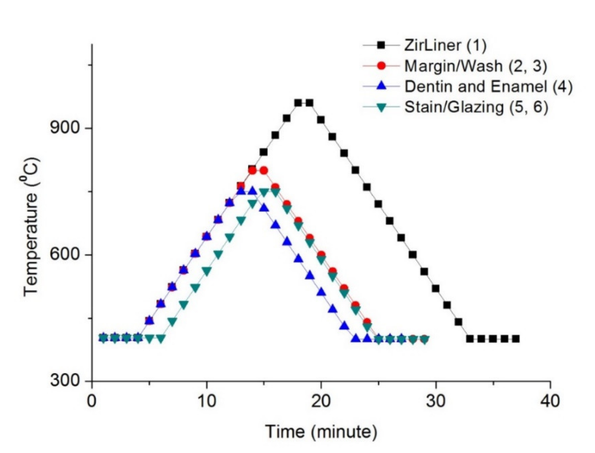
Figure 1. Firing schedule of sintered zirconia (ZirLiner: zirconia lining material).
Materials 14 01286 g001
Figure 2. Representative surface profiles with enlarged pictures for the experimental groups (F0–F6); red lines indicate surface profile.
Materials 14 01286 g002
Figure 3. Measurement of contact angles of the experimental groups (F0–F6) (*: The values were less than 10°, indicating their hydrophilic properties).
Materials 14 01286 g003
Figure 4. S. mutans cultured over zirconia specimens (F0–F6) for 24 h; fluorescence images and the area fraction of fluorescence in F0–F6.
Materials 14 01286 g004
Table 1. Surface roughness parameters of the experimental groups (F0–F6).
Groups
F0F1F2F3F4F5F6
Ra
(μm)
0.16 A
(0.15, 0.16)
0.06 B
(0.06, 0.06)
0.04 C
(0.04, 0.04)
0.05 CD
(0.05, 0.05)
0.06 BE
(0.06, 0.06)
0.05 BDF
(0.05, 0.06)
0.05 BDG
(0.05, 0.06)
Sa
(μm)
0.17 A
(0.15, 0.18)
0.07 B
(0.06, 0.07)
0.06 C
(0.06, 0.06)
0.06 BCD
(0.06, 0.06)
0.06 BCE
(0.06, 0.07)
0.06 BCF
(0.06, 0.06)
0.06 BCG
(0.06, 0.07)
Sz
(μm)
2.25 A
(1.95, 2.32)
0.48 B
(0.45, 0.49)
0.45 BC
(0.43, 0.46)
0.47 BD
(0.45, 0.49)
0.48 BE
(0.44, 0.48)
0.55 F
(0.55, 0.57)
0.54 BFG
(0.50, 0.59)
Sv
(μm)
2.09 A
(2.05, 2.40)
0.75 B
(0.59, 0.75)
0.98 BC
(0.63, 1.30)
0.51 BD
(0.43, 0.63)
0.28 BE
(0.28, 0.63)
0.83 CEF
(0.83, 1.02)
0.83 BEG
(0.63, 0.87)
1. Values with the different uppercase superscripts within the rows are significantly different (p < 0.05). 2. Interquartile ranges (first quartile, third quartile) are in parentheses.
Table 2. Median translucency parameters of the experimental groups (F0–F6).
Groups
F0F1F2F3F4F5F6
TP5.05 A
(5.05, 5.20)
4.39 BCDEFG
(4.34, 4.50)
4.21 C
(4.20, 4,25)
4.43 D
(4.38, 4.44)
4.66 E
(4.58, 4.68)
4.53 EF
(4.51, 4.57)
4.79 DEG
(4.57, 4.80)
1. TP: Translucency parameter. 2. Values with the different uppercase superscripts are significantly different (p < 0.05). 3. Interquartile ranges (first quartile, third quartile) are in parentheses.
Publisher’s Note: MDPI stays neutral with regard to jurisdictional claims in published maps and institutional affiliations.