Article

Dietary Aspergillus oryzae Modulates Serum Biochemical Indices, Immune Responses, Oxidative Stress, and Transcription of HSP70 and Cytokine Genes in Nile Tilapia Exposed to Salinity Stress

by
Mustafa Shukry
1,
Marwa F. Abd El-Kader
2,
Basma M. Hendam
3,
Mahmoud A. O. Dawood
4,
Foad A. Farrag
5,
Salama Mostafa Aboelenin
6,
Mohamed Mohamed Soliman
7 and
Hany M. R. Abdel-Latif
8,*
1
Department of Physiology, Faculty of Veterinary Medicine, Kafrelsheikh University, Kafrelsheikh 33516, Egypt
2
Fish Diseases and Management, Sakha Aquaculture Research Unit, Central Laboratory for Aquaculture Research (A.R.C.), Cairo 11511, Egypt
3
Genetics and Genetic Engineering, Department of Husbandry and Development of Animal Wealth, Faculty of Veterinary Medicine, Mansoura University, Mansoura 35516, Egypt
4
Department of Animal Production, Faculty of Agriculture, Kafrelsheikh University, Kafrelsheikh 33516, Egypt
5
Department of Anatomy and Embryology, Faculty of Veterinary Medicine, Kafrelsheikh University, Kafrelsheikh 33516, Egypt
6
Biology Department, Turabah University College, Taif University, P.O. Box 11099, Taif 21944, Saudi Arabia
7
Clinical Laboratory Sciences Department, Turabah University College, Taif University, P.O. Box 11099, Taif 21944, Saudi Arabia
8
Department of Poultry and Fish Diseases, Faculty of Veterinary Medicine, Alexandria University, Alexandria 22758, Egypt
*
Author to whom correspondence should be addressed.
Animals 2021, 11(6), 1621; https://doi.org/10.3390/ani11061621
Submission received: 17 March 2021 / Revised: 19 May 2021 / Accepted: 28 May 2021 / Published: 31 May 2021
(This article belongs to the Section Aquatic Animals)

Abstract

:

Simple Summary

Probiotics are live microbial adjuncts with numerous beneficial effects on fish. This study aims to evaluate the roles of Aspergillus oryzae (ASP) in the modulation of serum haemato-biochemical measurements, immunity, antioxidative capacity, and transcriptomic responses of Nile tilapia juveniles exposed to salinity stress. Findings revealed that dietary supplementation with A. oryzae mitigated the harmful influences of salinity stress on the exposed Nile tilapia.

Abstract

Nile tilapia Juveniles (19.50 ± 0.5 g) were fed on a basal diet (control group (CTR)) and a diet supplemented with 1 g Aspergillus oryzae (ASP) per kg diet for 12 weeks. Fish were then subjected to different salinity levels (0, 10, 15, and 20 practical salinity units (psu)) for another 15 days. Two-way ANOVA analysis revealed that the individual effects of ASP in Nile tilapia exposed to salinity levels presented a significant decrease (p < 0.05) in values of haemato-biochemical indices (such as glucose, cortisol, alanine transaminase, aspartate transaminase, and malondialdehyde) compared to those in the CTR group exposed to the same salinity levels. Moreover, significant increases (p < 0.05) of blood protein profile (albumin, globulin, and total protein), non-specific immune responses (lysozyme activity, phagocytic activity, and phagocytic index), and antioxidant enzymes activities (glutathione peroxidase, catalase, and superoxide dismutase) were observed in ASP-supplemented groups. Interestingly, there was significant (p < 0.05) downregulation of the mRNA expression values of heat shock protein 70 and interferon-gamma genes, alongside upregulation of the mRNA expression values of interleukin 1 beta and interleukin 8 genes, in the hepatic tissues of Nile tilapia in ASP-supplemented groups exposed to different salinities compared to those in the CTR group exposed to the same salinity levels. Taken together, these findings supported the potential efficacy of dietary supplementation with ASP in alleviating salinity stress-induced haemato-biochemical alterations, immune suppression, and oxidative stress in the exposed Nile tilapia.

1. Introduction

Over the past decade, the production rates of Nile tilapia (Oreochromis niloticus) have been greatly increased because of increased consumer preferences, relatively reasonable prices, and to some extent, its resistance to diseases [1]. During intensive culture systems, Nile tilapia are exposed to several stressors, which will weaken the fish immune responses and become susceptible to diseases [2]. The overuse of antibiotics for controlling disease occurrence has several drawbacks (for instance, the existence of multiple resistant bacteria harmfully affects the fish health and the persistence of drug residues in the tissues of treated fish), which will negatively affect human health [3]. Therefore, finding safe and environmentally friendly alternatives to improve fish health and productivity is of great interest to several researchers [4,5].
Probiotics are live microbial adjuncts that present several beneficial effects to a host, such as improving feed efficiency, enhancing growth performance, and fortifying the host immunity of fish against challenging environmental conditions [6]. The proficiency of probiotics to promote the health status is closely linked to their capability to augment the fish immunity and prevent the growth of pathogenic bacteria [7]. Aspergillus oryzae (ASP), as a probiotic, has been recognized as a significant source of various enzymes such as protease, glucoamylase, and alpha-amylase [8], and could be used as a probiotic feed supplement for poultry and livestock [9]. In Nile tilapia, dietary supplementation with A. oryzae could potentially enhance the immune responses and disease resistance [10], growth performance indices, intestinal histomorphometry [11], and antioxidative capacity [12], and improve defensive mechanisms against hypoxia stress [13]. Moreover, it enhances the growth and hematological parameters of common carp [14].
Salinity is one of the environmental factors that negatively affect the growth and reproduction performance of fish [15]. Salinity stress negatively influences the metabolic responses of shortnose sturgeon (Acipenser brevirostrum) juveniles [16] and the hematological indices of great sturgeon (Huso huso) [17]. Recently, Tian et al. [18] illuminated that exposure of the yellow drum (Nibea albiflora) to high salinities induced growth suppression, alterations of the intestinal microbiome, and changes in the physiological and immune-related parameters. It has been found that exposure of tilapia spp. to long-term hypersaline stress deleteriously induced several side effects such as impairment of reproductive performance [19], low survival rates [20], poor growth performance and impaired antioxidant capacity [21], histopathological alterations of spleen and kidneys [22], and disruption of the gut microbiome [23].
Cytokines are signaling molecules generated by immune cells to promote inflammation at the infection site and subsequently enhance the arrival of phagocytic cells to engulf and kill the invading pathogenic agents [24]. Moreover, Heat shock protein 70 (HSP70) is an indicator of stress in aquatic animals [25]. Some previously published studies illustrated that exposure of animals to chemicals and stress factors will induce liver injury [26,27]. In a similar sense, the mRNA expression levels of cytokines and HSP70 were increased in the livers of Nile tilapia exposed to stressors and toxicants [28,29].
Several attempts have been made using dietary supplements to alleviate the effects of salinity stress, such as galacto-oligosaccharide [30], L-Tryptophan [30,31,32], Lactobacillus rhamnosus [33], taurine [34], bovine lactoferrin [35], mannan oligosaccharides [36], inulin [37], a host-derived Bacillus subtilis [38], and myo-inositol [39]. Hence, the current study aimed to evaluate the mitigative effect of A. oryzae on salinity stress through an exploration of the non-specific immunity, haemato-biochemical indices, antioxidative capacity, as well as the transcriptomic profile of HSP70 and cytokine genes in Nile tilapia.

2. Materials and Methods

2.1. Ethical Approval

The guidelines of the Local Experimental Animal Care Committee were closely followed during the present study. Moreover, this study was also approved by the Institutional Ethics Committee of the Faculty of Veterinary Medicine, Kafrelsheikh University (Approval No. 70 20-03-2018).

2.2. Probiotic Feed Supplement

Aspergillus oryzae (ASP) (Bio’c Co., Ltd., Uchida, Japan) contained 1 × 10⁸ CFU (colony forming units) per 1 g and was used as a feed supplement in the current study.

2.3. Fish Rearing and Management

Nile tilapia (Oreochromis niloticus) (19.50 ± 0.5 g) were procured from a local fish farm (Kafrelsheikh, Egypt) and then kept for 14 days in 500 L tanks, in the wet laboratory that belonged to Sakha Aquaculture Research Unit (Central Laboratory for Aquaculture Research, Kafrelsheikh, Egypt), to be acclimated to the laboratory conditions. During acclimation, fish were fed on a commercially purchased basal diet that was used as the control (CTR) diet (30% protein) (Aller Aqua Co., Giza, 3rd. Giza Governorate, Egypt). Ration ingredients and their chemical compositions were adapted to assemble the nutritional requirements for optimum growth of Nile tilapia (Table S1—please see Supplementary Materials). During the experimental periods, fish were reared in glass aquaria contained 100 L water and sized 1 × 1 × 1 m and supplemented with compressed air via air stones using aquarium air pumps. One third of the water per each aquarium was siphoned every 2 or 3 days to get rid of faecal matter and then substituted with new water from the storage tanks.

2.4. Water Quality Parameters

The physical and chemical parameters of the rearing water samples were examined weekly and maintained throughout the whole experimental period at 27.0–28.5 °C, 7.50 ± 0.50 mg/L, 7.5–8.5, and 0.01 ± 0.03 mg/L for temperature, dissolved oxygen, pH, and un-ionized ammonia, respectively [40].

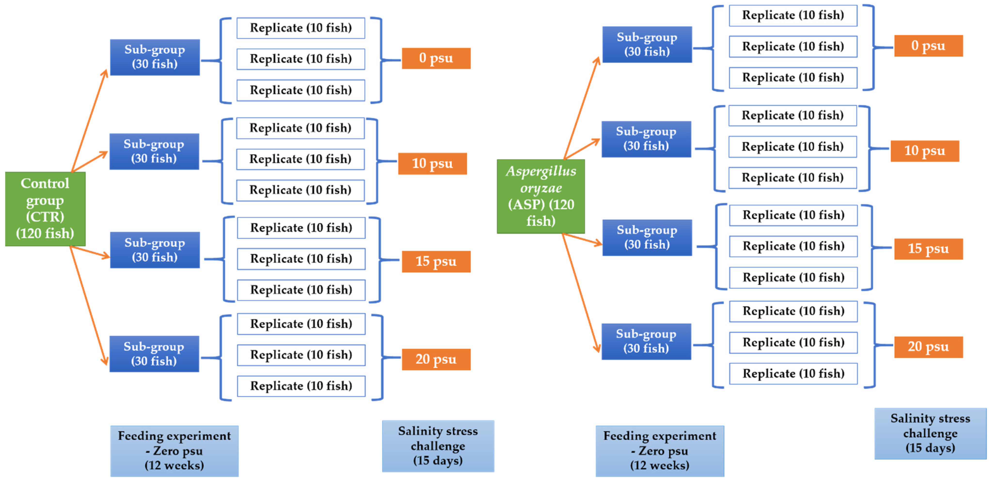
2.5. Experimental Setup

2.5.1. Experiment I: Feeding Trial

A total number of 240 fish were allocated into two main groups (each group was divided into 4 sub-groups) (30 fish per each sub-group) (Figure 1). Fish were fed on two experimental diets that were prepared to contain two levels of A. oryzae (ASP) (0 g per kg diet) and served as a control group (CTR) (the commercially purchased diet), and the other group was fed the CTR diet supplemented with 1 g A. oryzae (ASP) per kg. ASP level used in the current study was selected according to our previously published studies on Nile tilapia [11,12,13]. Sodium carboxymethyl cellulose was used for pelleting the experimental diets (to make the diets as pellets to fit the fish feeding). Pellets were produced by an extruder pelleting machine and then kept in polyethylene bags at 4 °C until use. The experiment lasted for 12 weeks, and fish were fed daily until apparent satiety (3 times daily). During experiment I, fish were reared on freshwater with zero practical salinity units (psu).

2.5.2. Experiment II: Challenge with Salinity Stress

After the end of the first experiment, the fish in each sub-group (30 fish) were allocated into 3 other replicates (10 fish per each) (Figure 1). Moreover, the fish continued to be fed on the same previously prepared and corresponding experimental diets (ASP and CTR). Afterward, the fish in each sub-group were exposed to a different salinity level (0, 10, 15, or 20 psu) for an additional 15 days (Figure 1).
During the salinity stress test, the fish in each glass aquarium were provided with continuous aeration. For the salinity challenge, the tap water was dechlorinated by strong aeration for 24 h and then mixed with seawater to raise the salinity to 10, 15, and 20 psu. Salinity concentrations were determined by a salinometer (Thermo Electron corporation model Orion 150 A+). After exposure to salinity levels, mortalities were recorded, and blood and tissue specimens were sampled.

2.6. Sampling

2.6.1. Blood Sampling

Fish in all experimental groups were starved for 24 h before collection of blood samples. Three fish were sampled from each replicate (9 fish per sub-group). Fish were anesthetized by tricaine methane sulphonate, then blood was sampled from the caudal veins. Blood was allotted into two sets: one was collected with heparin for hematological parameters; the second set was left at room temperature (for serum collection into centrifuge tubes). Centrifugation was conducted at 3000× g for 15 min to separate serum, which were then stored at −20 °C until use.

2.6.2. Liver Samples

Three liver samples from each replicate (9 samples per sub-group) were sampled and then immediately stored in liquid nitrogen and stored at −80 °C in TRIzol reagent (iNtRON Biotechnology, Inc., Gyeonggi, Korea) to be employed for PCR assays.

2.7. Serum Biochemical Measurements

Serum samples were investigated to obtain their protein profiles, which include total protein (TP) [41] and albumin (ALB) contents [42]. Globulin (GLO) levels were also determined by subtraction of ALB from TP values. Blood glucose (GLU), cortisol (CORT) levels, serum alanine transaminase (ALT) and aspartate transaminase (AST) activities were measured colorimetrically using commercially purchased specific kits (Bio diagnostic Co., Giza, Egypt) (all laboratory procedures were accomplished according to the protocol provided from the manufacturer).

2.8. Immunity and Antioxidant Parameters

2.8.1. Immune Parameters

Serum lysozyme (LYZ) activity was evaluated by the turbidimetric assay using lyophilized Micrococcus lysodeikticus cells (Sigma-Aldrich, St. Louis, MN, USA) according to the methodology described by Ellis [43] and Mörsky [44].
Phagocytic activity (PA) and phagocytic index (PI) were calculated in the whole blood samples following the methodology of Kawahara et al. [45] according to the following equations:
  • PA = Total No. of macrophages encompassing yeast cells/total No. of macrophages × 100
  • PI = No. of phagocytized yeast cells/No. of phagocytic cells

2.8.2. Oxidative Stress Biomarkers

Serum catalase (CAT), superoxide dismutase (SOD), and glutathione peroxidase (GPX) enzyme activities, as well as malondialdehyde (MDA) levels (as a marker of lipid peroxidation), were evaluated colorimetrically using commercially purchased specific diagnostic kits (Bio diagnostic Company, Giza, Egypt).

2.9. Gene Transcription

The total RNA was extracted from 100 mg of the hepatic tissues using Trizol reagent (in accordance with the protocol obtained from the manufacturer). The quality and quantity of the RNA were then confirmed by Nanodrop (Uv–Vis spectrophotometer Q5000/Quawell, USA). The complementary DNA (cDNA) was synthesized using the SensiFAST™ cDNA synthesis kit (Bioline, London, UK) (according to the protocol of the manufacturer). The cDNA samples were then stored at −20 °C until use.
Specific primer sequences were used for evaluation of the mRNA expression levels of some immune-related genes such as interleukin 1beta (IL-1β), IL-8, and interferon-gamma (INF-γ), and stress-related genes such as heat shock protein 70 (HSP70). Moreover, beta-actin (β-actin) was used as a reference gene (Table S2—please see Supplementary Materials).
The SYBR green method was used to quantify the gene transcription folds using an RT-PCR machine (SensiFast SYBR Lo-Rox kit, Bioline, London, UK). The thermal cycle conditions for the reaction were adjusted as follows: 10 min at 95 °C, 40 cycles at 95 °C for 15 s, 30 min at 60 °C, and finally 5 min at 85 °C (61 °C for IFN-γ) for 1 min. The transcription folds were standardized to β-actin through using the 2−ΔΔCT method [46].

2.10. Statistical Analysis

Data were checked for normality and homogeneity using the Levene test. The non-percentage data were directly represented, but the percentage of data was arcsine transformed before analysis. A Two Way-ANOVA in a 2 × 4 factorial design which represents two main factors—two levels of ASP and four levels of salinities—was used to obtain the main effects of ASP, salinity, and the interaction between them. The significant differences were determined at the level of p < 0.05. The differences between the calculated means were determined using Tukey’s range test. The statistics were calculated using the SPSS program version 22 (SPSS, v 22.0; SPSS Inc., Chicago, IL, USA).

3. Results

The fish efficiently ingested all tested diets, and no mortalities were found in all experimental groups (CTR and ASP) after exposure to different salinity levels for 15 days.

3.1. Serum Biochemical Measurements

Two-way ANOVA analysis showed that the effects of salinity levels on the CTR group showed significant increases (p < 0.05; Table 1) in GLU, CORT, ALT, and AST levels. These results suggested the occurrence of stress in the exposed fish. Moreover, there were significant decreases in TP, ALB, and GLO values of salinity-exposed fish in the CTR group. The opposite trend was, however, noticed in the ASP-supplemented group, which was exposed to the same salinities, whereas the individual effects of ASP in Nile tilapia exposed to different salinity levels showed a significant decrease in blood GLU, CORT, ALT, and AST values (p < 0.05; Table 1), alongside a significant increase in ALB, GLO, and TP values (p < 0.05; Table 1) compared to the CTR group exposed to the same salinity levels.

3.2. Non-Specific Immune Parameters

Through pairwise comparisons with the CTR group exposed to different salinity levels, it was found that the individual effects of ASP-supplemented diets in Nile tilapia exposed to the same salinities included a significant increase in the serum LYZ activity and phagocytic capacity of blood macrophages (PA % and PI) (p < 0.05; Table 2).

3.3. Antioxidative Capacity Markers

Compared with the CTR group exposed to different salinities, ASP-supplementation induced significant effects in the antioxidative biomarkers of treated fish (p < 0.05). Nile tilapia fed on ASP-supplemented diets and concurrently exposed to different salinities had significantly lowered serum MDA levels alongside elevated serum CAT, SOD, and GPX activities compared with the CTR group exposed to the same salinities (p < 0.05; Table 3).

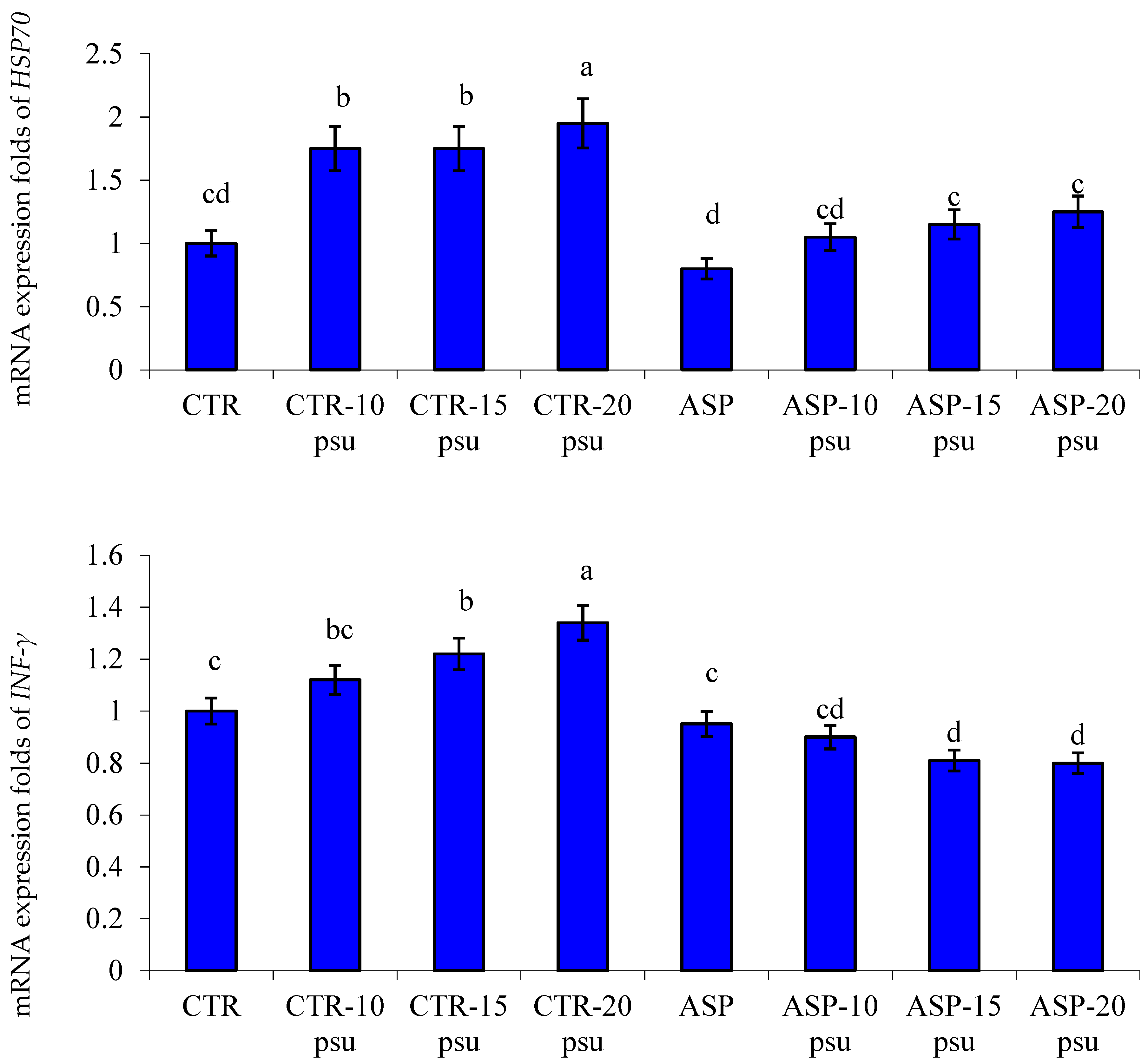
3.4. Transcriptomic Profile

There was significant downregulation of the expression profile of HSP70 and INF-γ genes in hepatic tissues of Nile tilapia fed on ASP-supplemented diets and exposed to different salinity levels compared to those in the CTR group and exposed to the same salinities (p < 0.05; Figure 2). Besides, there was significant upregulation of the expression profile of IL-8 and IL-1β genes in the hepatic tissues of Nile tilapia fed an ASP diet and exposed to different salinities than the CTR group exposed to the same salinities (p < 0.05; Figure 3).
From the aforementioned findings, the potential capacity of ASP in the improvement of fish immunity, via the upregulation of immune-related genes and the down regulation of stress-related genes, in salinity stress-exposed Nile tilapia can be suggested.

4. Discussion

For a long period, the use of probiotics in aquaculture had gained great interest because of their growth-promoting and immune-stimulating characteristics [47,48], and recently, they have been increasingly used to alleviate the health problems induced after the exposure of fish to certain adverse environmental stressors and toxic contaminants [49]. Among the probiotics, A. oryzae has attracted the interest of researchers for its beneficial roles in maintaining fish health [11,14].
This study evaluated the roles of dietary supplementation with ASP to mitigate the undesirable influences of salinity stress on exposed Nile tilapia. The tested dose was 1 g ASP per kg diet. This dose was selected based on the previous studies conducted in our laboratory. The results suggest that supplementation with 1 g ASP per kg of diet considerably improved growth, intestinal histomorphometry, and hematological profiles [11], enhanced immunity and disease resistance [12], and fortified antioxidative capacities against hypoxia [13].

4.1. Serum Biochemical Indices

High CORT levels indicate exposure of fish to stress [50]. Blood GLU is normally secreted for energy production to diminish the adverse impacts of stress on fish [51]. Serum transaminases (AST and ALT) are declarative of the fish liver functions [52]. Blood protein profiles (TP, ALB, and GLO) are considered as effective indicators of the humoral immunity of fish [53]. There was a significant decrease in blood GLU, CORT, ALT, and AST values, alongside a considerable increase in ALB, GLO, and TP values in the ASP groups exposed to different salinity levels compared to those in the CTR group exposed to the same salinities. The elevation of blood GLU and CORT levels in the CTR group exposed to higher salinities are indicative of the effects of salinity stress [54]. The decrease in CORT and GLU levels in ASP-supplemented groups, and concurrently, exposure to different salinities suggest the beneficial effects of ASP in the relief of the side effects of salinity stress. Our findings were consistent with those obtained by Dawood et al. [13], who demonstrated that dietary ASP noticeably decreased the CORT and GLU levels in Nile tilapia exposed to hypoxia stress. Moreover, dietary supplementation with Pediococcus acidilactici considerably lowered the blood GLU and CORT in green terror during hypoxia [55]. Similarly, CORT and GLU levels were significantly decreased in red sea bream fed diets supplemented with Lactobacillus rhamnosus and exposed to low salinity stress [33].
The increases in ALT and AST enzyme activities are also indicative of damage, necrosis, and degeneration of hepatic tissues [56]. There were significant decreases in the ALT and AST levels in the serum of Nile tilapia fed on ASP-supplemented diets and exposed to salinity stress as compared with the CTR group exposed to salinity stress. These results were in line with those obtained by Dawood et al. [11]. Furthermore, dietary supplementation with the prebiotic mannan oligosaccharide considerably decreased the ALT and AST levels in red sea bream exposed to low salinity stress [36].

4.2. Immune and Antioxidant Measurements

Phagocytic cells are an important component of the cellular immune system in fish, with protective mechanisms against invading pathogens [57]. Moreover, lysozyme is important for lysis of the cell walls of the pathogenic G+ve and G-ve bacteria [43,58]. It has been reported that exposure of Nile tilapia to physical stressors such as heat stress and high stocking densities has led to immune suppression of the exposed fish [2,59]. In the present study, compared to the CTR group exposed to different salinity levels, the effects of ASP-supplemented diets in Nile tilapia exposed to the same salinities showed a significant increase in the serum LYZ activity and phagocytic capacity of the macrophages (PA % and PI). These findings suggest the immune-modulating roles of dietary ASP against the salinity stress-induced decline in the non-specific immunity of exposed Nile tilapia. Several probiotics significantly increased the LYZ activity and phagocytic capacity of fish [7,49,60,61].
Oxidative stress is defined as the state induced through the overproduction of reactive oxygen species combined with the inability of the host body to counteract their side effects, leading to lysis of the cell membrane, disruption of the cellular components, and lipid peroxidation (LPO) [62,63]. It has been reported that oxidative stress occurs after exposure of the living organisms to harmful substances [64,65,66,67]. On the other hand, the CAT, SOD, and GPX enzymes comprise part of the enzymatic antioxidant mechanism to alleviate oxidative stress [68], and MDA is a bioindicator of LPO [69]. In the existing study, Nile tilapia fed on ASP-supplemented diets and exposed to different salinities had significantly lowered serum MDA levels alongside elevated serum CAT, SOD, and GPX activities compared with the CTR group exposed to the same salinities. These findings suggest the potential role of dietary ASP in potentiating the antioxidative capacity in salinity stress-exposed fish.
In line with our findings, Esteban et al. [70] showed that dietary probiotics potentiate the enzymatic antioxidant mechanisms in gilthead seabream (Sparus aurata). Dawood et al. [12] illustrated that the synbiotic effects of ASP and beta-glucan significantly potentiate the antioxidant capacity of Nile tilapia. Moreover, Dawood et al. [13] reported a similar substantial increase in SOD, CAT, and GPX activities alongside a decrease in MDA levels in Nile tilapia fed ASP-supplemented diets and exposed to hypoxia stress.

4.3. Gene Transcription

Cytokines are signaling molecules generated by immune cells to promote inflammation at the infection site, and consequently, accelerate the arrival of phagocytic cells to engulf and kill the invading pathogenic agents [24,71]. IL-1β is a pro-inflammatory cytokine that performs a pivotal role in regulation of the inflammatory and immune processes [72]. INF-γ is one of the interferons that play an important role in fish immunity [73,74]. HSP70 is a stress marker in aquatic animals [25,75,76]. In the present study, it was noted that before and after exposure to the salinity stress, there was a significant upregulation of the mRNA expression values of IL-8 and IL-1β genes alongside downregulation of the mRNA expression values of HSP70 and INF-γ genes in the liver of Nile tilapia fed on ASP-supplemented diets. Xia et al. [77] showed that dietary probiotics considerably increased the expression of IL-1β in Nile tilapia. Moreover, the expression of IL-8 and IL-1β was significantly increased in grass carp fed on diets supplemented with probiotics [78].

5. Conclusions

The findings of the existing study showed that the exposure to higher salinities induced haemato-biochemical alterations, a decline in the immune parameters, and oxidative stress in the exposed Nile tilapia. Moreover, the dietary probiotic effect of A. oryzae was clearly noticed in the improvement of the growth performance and alleviation of the side effects of salinity stress. Taken together, the improvement of serum biochemistry, such as cortisol and glucose values, the enhancement of fish immunity, the modulation of antioxidative capacity, and the transcriptomic profile of the exposed fish were indicative of the beneficial roles of A. oryzae-supplemented diets in the alleviation of the side effects of salinity stress in Nile tilapia.

Supplementary Materials

The following are available online at https://www.mdpi.com/article/10.3390/ani11061621/s1.

Author Contributions

H.M.R.A.-L. and B.M.H., Methodology; M.A.O.D., S.M.A. and M.M.S., Software; H.M.R.A.-L. and M.S., Validation; H.M.R.A.-L., M.S. and M.F.A.E.-K., Formal analysis; H.M.R.A.-L., Writing—original draft preparation; M.A.O.D., S.M.A. and M.M.S., Visualization; M.S., B.M.H., F.A.F. and M.F.A.E.-K., Supervision. All authors have read and agreed to the published version of the manuscript.

Funding

The current work was funded by Taif University, Saudi Arabia, through its Researchers Supporting Project (TURSP-2020-105).

Institutional Review Board Statement

The study was conducted according to the guidelines of the Declaration of the Local Experimental Animal Care Committee and approved by the Institutional Ethics Committee of the Faculty of Veterinary Medicine, Kafrelsheikh University (Approval No. 70 20-03-2018).

Informed Consent Statement

Not applicable.

Data Availability Statement

All datasets collected and analyzed during the current study are available from the corresponding author on fair request.

Acknowledgments

All authors appreciate and thank Taif University, Saudi Arabia, for financial support through its Researchers Supporting Project (TURSP-2020-105).

Conflicts of Interest

The authors declare no conflict of interest.

References

  1. Abdel-Latif, H.M.R.; Dawood, M.A.O.; Menanteau-Ledouble, S.; El-Matbouli, M. The nature and consequences of co-infections in tilapia: A review. J. Fish Dis. 2020, 43, 651–664. [Google Scholar] [CrossRef]
  2. Dawood, M.A.; Metwally, A.E.-S.; El-Sharawy, M.E.; Atta, A.M.; Elbialy, Z.I.; Abdel-Latif, H.M.; Paray, B.A. The role of β-glucan in the growth, intestinal morphometry, and immune-related gene and heat shock protein expressions of Nile tilapia (Oreochromis niloticus) under different stocking densities. Aquaculture 2020, 523, 735205. [Google Scholar] [CrossRef]
  3. Vincent, A.T.; Gauthier, J.; Derome, N.; Charette, S.J. The Rise and Fall of Antibiotics in Aquaculture. In Microbial Communities in Aquaculture Ecosystems; Springer: Cham, Switzerland, 2019; pp. 1–19. [Google Scholar]
  4. Ahmadifar, E.; Yousefi, M.; Karimi, M.; Raieni, R.F.; Dadar, M.; Yilmaz, S.; Dawood, M.A.O.; Abdel-Latif, H.M.R. Benefits of Dietary Polyphenols and Polyphenol-Rich Additives to Aquatic Animal Health: An Overview. Rev. Fish. Sci. Aquac. 2020, 10, 1–34. [Google Scholar] [CrossRef]
  5. Dawood, M.A.O.; El Basuini, M.F.; Zaineldin, A.I.; Yilmaz, S.; Hasan, M.T.; Ahmadifar, E.; El Asely, A.M.; Abdel-Latif, H.M.R.; Alagawany, M.; Abu-Elala, N.M.; et al. Antiparasitic and Antibacterial Functionality of Essential Oils: An Alternative Approach for Sustainable Aquaculture. Pathogens 2021, 10, 185. [Google Scholar] [CrossRef] [PubMed]
  6. Gatesoupe, F.J. Chapter 32—Probiotics and Other Microbial Manipulations in Fish Feeds: Prospective Health Benefits. In Bio-active Foods in Promoting Health; Watson, R.R., Preedy, V.R., Eds.; Academic Press: Boston, MA, USA, 2010; pp. 541–552. [Google Scholar]
  7. Gobi, N.; Vaseeharan, B.; Chen, J.-C.; Rekha, R.; Vijayakumar, S.; Anjugam, M.; Iswarya, A. Dietary supplementation of probiotic Bacillus licheniformis Dahb1 improves growth performance, mucus and serum immune parameters, antioxidant enzyme activity as well as resistance against Aeromonas hydrophila in tilapia Oreochromis mossambicus. Fish Shellfish Immunol. 2018, 74, 501–508. [Google Scholar] [CrossRef]
  8. Machida, M.; Asai, K.; Sano, M.; Tanaka, T.; Kumagai, T.; Terai, G.; Kusumoto, K.-I.; Arima, T.; Akita, O.; Kashiwagi, Y.; et al. Genome sequencing and analysis of Aspergillus oryzae. Nat. Cell Biol. 2005, 438, 1157–1161. [Google Scholar] [CrossRef] [Green Version]
  9. Lee, K.; Lee, S.K.; Lee, B.D. Aspergillus oryzae as probiotic in poultry—A review. Int. J. Poult. Sci. 2006, 5, 1–3. [Google Scholar]
  10. Iwashita, M.K.P.; Nakandakare, I.B.; Terhune, J.S.; Wood, T.; Ranzani-Paiva, M.J.T. Dietary supplementation with Bacillus subtilis, Saccharomyces cerevisiae and Aspergillus oryzae enhance immunity and disease resistance against Aeromonas hydrophila and Streptococcus iniae infection in juvenile tilapia Oreochromis niloticus. Fish Shellfish Immunol. 2015, 43, 60–66. [Google Scholar] [CrossRef] [PubMed]
  11. Dawood, M.A.O.; Eweedah, N.M.; Moustafa, E.M.; Shahin, M.G. Effects of feeding regimen of dietary Aspergillus oryzae on the growth performance, intestinal morphometry and blood profile of Nile tilapia (Oreochromis niloticus). Aquac. Nutr. 2019, 25, 1063–1072. [Google Scholar] [CrossRef]
  12. Dawood, M.A.O.; Eweedah, N.M.; Moustafa, E.M.; Shahin, M.G. Synbiotic Effects of Aspergillus oryzae and β-Glucan on Growth and Oxidative and Immune Responses of Nile Tilapia, Oreochromis niloticus. Probiotics Antimicrob. Proteins 2019, 12, 172–183. [Google Scholar] [CrossRef] [PubMed]
  13. Dawood, M.A.; Eweedah, N.M.; Moustafa, E.M.; Farahat, E.M. Probiotic effects of Aspergillus oryzae on the oxidative status, heat shock protein, and immune related gene expression of Nile tilapia (Oreochromis niloticus) under hypoxia challenge. Aquaculture 2020, 520, 734669. [Google Scholar] [CrossRef]
  14. Hedayati, S.A.; Bagheri, T.; Hoseinifar, S.H.; Van Doan, H. Growth performances and hemato-immunological responses of common carp (Cyprinus carpio Linnaeus, 1758) to fermented Aspergillus oryzae. Iran. J. Fish. Sci. 2020, 19, 1749–1756. [Google Scholar]
  15. Kültz, D. Physiological mechanisms used by fish to cope with salinity stress. J. Exp. Biol. 2015, 218, 1907–1914. [Google Scholar] [CrossRef] [PubMed] [Green Version]
  16. Jarvis, P.L.; Ballantyne, J.S. Metabolic responses to salinity acclimation in juvenile shortnose sturgeon Acipenser brevirostrum. Aquaculture 2003, 219, 891–909. [Google Scholar] [CrossRef]
  17. Zarejabad, A.M.; Jalali, M.A.; Sudagar, M.; Pouralimotlagh, S. Hematology of great sturgeon (Huso huso Linnaeus, 1758) juvenile exposed to brackish water environment. Fish Physiol. Biochem. 2010, 36, 655–659. [Google Scholar] [CrossRef]
  18. Tian, L.; Tan, P.; Yang, L.; Zhu, W.; Xu, D. Effects of salinity on the growth, plasma ion concentrations, osmoregulation, non-specific immunity, and intestinal microbiota of the yellow drum (Nibea albiflora). Aquaculture 2020, 528, 735470. [Google Scholar] [CrossRef]
  19. Watanabe, W.O.; Kuo, C.-M. Observations on the reproductive performance of Nile tilapia (Oreochromis niloticus) in laboratory aquaria at various salinities. Aquaculture 1985, 49, 315–323. [Google Scholar] [CrossRef]
  20. Kang’Ombe, J.; Brown, J.A. Effect of Salinity on Growth, Feed Utilization, and Survival of Tilapia rendalli Under Laboratory Conditions. J. Appl. Aquac. 2008, 20, 256–271. [Google Scholar] [CrossRef]
  21. Gan, L.; Xu, Z.X.; Ma, J.J.; Xu, C.; Wang, X.D.; Chen, K.; Chen, L.Q.; Li, E.C. Effects of salinity on growth, body composition, muscle fatty acid composition, and antioxidant status of juvenile Nile tilapia Oreochromis niloticus (Linnaeus, 1758). J. Appl. Ichthyol. 2016, 32, 372–374. [Google Scholar] [CrossRef]
  22. Xu, C.; Li, E.; Suo, Y.; Su, Y.; Lu, M.; Zhao, Q.; Qin, J.G.; Chen, L. Histological and transcriptomic responses of two immune organs, the spleen and head kidney, in Nile tilapia (Oreochromis niloticus) to long-term hypersaline stress. Fish Shellfish Immunol. 2018, 76, 48–57. [Google Scholar] [CrossRef] [Green Version]
  23. Zhang, M.; Sun, Y.; Liu, Y.; Qiao, F.; Chen, L.; Liu, W.-T.; Du, Z.; Li, E. Response of gut microbiota to salinity change in two euryhaline aquatic animals with reverse salinity preference. Aquaculture 2016, 454, 72–80. [Google Scholar] [CrossRef]
  24. Secombes, C.; Wang, T.; Hong, S.; Peddie, S.; Crampe, M.; Laing, K.; Cunningham, C.; Zou, J. Cytokines and innate immunity of fish. Dev. Comp. Immunol. 2001, 25, 713–723. [Google Scholar] [CrossRef]
  25. Lindquist, S.; Craig, E.A. The Heat-Shock Proteins. Annu. Rev. Genet. 1988, 22, 631–677. [Google Scholar] [CrossRef] [PubMed]
  26. Li, Z.; Miao, Z.; Ding, L.; Teng, X.; Bao, J. Energy metabolism disorder mediated ammonia gas-induced autophagy via AMPK/mTOR/ULK1-Beclin1 pathway in chicken livers. Ecotoxicol. Environ. Saf. 2021, 217, 112219. [Google Scholar] [CrossRef] [PubMed]
  27. Zheng, S.; Song, H.; Gao, H.; Liu, C.; Zhang, Z.; Fu, J. The Antagonistic Effect of Selenium on Lead-Induced Inflammatory Factors and Heat Shock Protein mRNA Level in Chicken Cartilage Tissue. Biol. Trace Element Res. 2016, 173, 177–184. [Google Scholar] [CrossRef] [PubMed]
  28. Abdel-Latif, H.; Dawood, M.; Mahmoud, S.; Shukry, M.; Noreldin, A.; Ghetas, H.; Khallaf, M. Copper Oxide Nanoparticles Alter Serum Biochemical Indices, Induce Histopathological Alterations, and Modulate Transcription of Cytokines, HSP70, and Oxidative Stress Genes in Oreochromis niloticus. Animals 2021, 11, 652. [Google Scholar] [CrossRef]
  29. Abdel-Latif, H.; Shukry, M.; El Euony, O.; Soliman, M.M.; Noreldin, A.; Ghetas, H.; Dawood, M.; Khallaf, M. Hazardous Effects of SiO2 Nanoparticles on Liver and Kidney Functions, Histopathology Characteristics, and Transcriptomic Responses in Nile Tilapia (Oreochromis niloticus) Juveniles. Biology 2021, 10, 183. [Google Scholar] [CrossRef]
  30. Hoseinifar, S.H.; Khalili, M.; Rostami, H.K.; Esteban, M.Á. Dietary galactooligosaccharide affects intestinal microbiota, stress resistance, and performance of Caspian roach (Rutilus rutilus) fry. Fish Shellfish Immunol. 2013, 35, 1416–1420. [Google Scholar] [CrossRef]
  31. Akhtar, M.S.; Pal, A.K.; Sahu, N.P.; Ciji, A. Hemato-immunological responses of Labeo rohita juveniles to temperature and salinity stress: Effect of dietary L-tryptophan. Isr. J. Aquac. 2013, 65, 1–8. [Google Scholar]
  32. Akhtar, M.S.; Pal, A.K.; Sahu, N.P.; Ciji, A.; Gupta, S.K.; Dasgupta, S. Serum Electrolytes, Osmolarity and Selected Enzyme Activities of Labeo rohita Juveniles Exposed to Temperature and Salinity Stress: Effect of Dietary l-Tryptophan; Springer: Berlin/Heidelberg, Germany, 2014; pp. 973–980. [Google Scholar]
  33. Dawood, M.A.O.; Koshio, S.; Ishikawa, M.; El-Sabagh, M.; Yokoyama, S.; Wang, W.-L.; Yukun, Z.; Olivier, A. Physiological response, blood chemistry profile and mucus secretion of red sea bream (Pagrus major) fed diets supplemented with Lactobacillus rhamnosus under low salinity stress. Fish Physiol. Biochem. 2017, 43, 179–192. [Google Scholar] [CrossRef]
  34. Abdel-Tawwab, M.; Monier, M.N. Stimulatory effect of dietary taurine on growth performance, digestive enzymes activity, antioxidant capacity, and tolerance of common carp, Cyprinus carpio L., fry to salinity stress. Fish Physiol. Biochem. 2018, 44, 639–649. [Google Scholar] [CrossRef]
  35. Yokoyama, S.; Ishikawa, M.; Koshio, S. Dietary bovine lactoferrin enhances defense factors on body surface and anti-parasitic effects against Neobenedenia girellae infection, and mitigates low-salinity stress in amberjack (Seriola dumerili) juveniles. Aquaculture 2019, 504, 52–58. [Google Scholar] [CrossRef]
  36. Dawood, M.A.; Koshio, S.; Fadl, S.; Ahmed, H.A.; El Asely, A.; Abdel-Daim, M.M.; Alkahtani, S. The modulatory effect of mannanoligosaccharide on oxidative status, selected immune parameters and tolerance against low salinity stress in red sea bream (Pagrus major). Aquac. Rep. 2020, 16, 100278. [Google Scholar] [CrossRef]
  37. Zhou, L.; Zhang, J.; Yan, M.; Tang, S.; Wang, X.; Qin, J.G.; Chen, L.; Li, E. Inulin alleviates hypersaline-stress induced oxidative stress and dysbiosis of gut microbiota in Nile tilapia (Oreochromis niloticus). Aquaculture 2020, 529, 735681. [Google Scholar] [CrossRef]
  38. Tang, S.; Liu, S.; Zhang, J.; Zhou, L.; Wang, X.; Zhao, Q.; Weng, W.; Qin, J.G.; Chen, L.; Li, E. Relief of hypersaline stress in Nile tilapia Oreochromis niloticus by dietary supplementation of a host-derived Bacillus subtilis strain. Aquaculture 2020, 528, 735542. [Google Scholar] [CrossRef]
  39. Cui, W.; Ma, A.; Huang, Z.; Liu, Z.; Yang, K.; Zhang, W. myo-inositol facilitates salinity tolerance by modulating multiple physiological functions in the turbot Scophthalmus maximus. Aquaculture 2020, 527, 735451. [Google Scholar] [CrossRef]
  40. Boyd, C.E.; Tucker, C.S. Pond Aquaculture Water Quality Management; Kluwer Academic Publishers: Boston, MA, USA, 1998. [Google Scholar]
  41. Henry, R.J. Colorimetric determination of total protein. In Clinical Chemistry; Harper and Row Publishers: New York, NY, USA, 1964. [Google Scholar]
  42. Doumas, B.T.; Watson, W.A.; Biggs, H.G. Albumin standards and the measurement of serum albumin with bromcresol green. Clin. Chim. Acta 1971, 31, 87–96. [Google Scholar] [CrossRef]
  43. Ellis, A.E. Lysozyme assays. In Techniques in Fish Immunology; SOS Publications: Cambridge, UK, 1990; pp. 101–103. [Google Scholar]
  44. Mörsky, P. Turbidimetric determination of lysozyme with Micrococcus lysodeikticus cells: Reexamination of reaction conditions. Anal. Biochem. 1983, 128, 77–85. [Google Scholar] [CrossRef]
  45. Kawahara, E.; Ueda, T.; Nomura, S. In Vitro Phagocytic Activity of White-Spotted Char Blood Cells after Injection with Aeromonas salmonicida Extracellular Products. Fish Pathol. 1991, 26, 213–214. [Google Scholar] [CrossRef]
  46. Schmittgen, T.D.; Livak, K.J. Analyzing real-time PCR data by the comparative CT method. Nat. Protoc. 2008, 3, 1101–1108. [Google Scholar] [CrossRef]
  47. Nayak, S. Probiotics and immunity: A fish perspective. Fish Shellfish Immunol. 2010, 29, 2–14. [Google Scholar] [CrossRef]
  48. Sakai, M. Current research status of fish immunostimulants. Aquaculture 1999, 172, 63–92. [Google Scholar] [CrossRef]
  49. Euony, O.I.E.; Elblehi, S.S.; Abdel-Latif, H.M.; Abdel-Daim, M.M.; El-Sayed, Y.S. Modulatory role of dietary Thymus vulgaris essential oil and Bacillus subtilis against thiamethoxam-induced hepatorenal damage, oxidative stress, and immunotoxicity in African catfish (Clarias garipenus). Environ. Sci. Pollut. Res. 2020, 27, 23108–23128. [Google Scholar] [CrossRef] [PubMed]
  50. Barton, B.A. Stress in Fishes: A Diversity of Responses with Particular Reference to Changes in Circulating Corticosteroids. Integr. Comp. Biol. 2002, 42, 517–525. [Google Scholar] [CrossRef] [PubMed]
  51. Bonga, S.E.W. The stress response in fish. Physiol. Rev. 1997, 77, 591–625. [Google Scholar] [CrossRef] [PubMed]
  52. Abdel-Latif, H.M.; Abdel-Tawwab, M.; Khafaga, A.F.; Dawood, M.A. Dietary oregano essential oil improved the growth performance via enhancing the intestinal morphometry and hepato-renal functions of common carp (Cyprinus carpio L.) fingerlings. Aquaculture 2020, 526, 735432. [Google Scholar] [CrossRef]
  53. Patriche, T.; Patriche, N.; Tenciu, M. Cyprinids total blood proteins determination. J. Anim. Sci. Biotechnol. 2009, 42, 95–101. [Google Scholar]
  54. Laiz-Carrión, R.; Sangiao-Alvarellos, S.; Guzmán, J.M.; Del Río, M.P.M.; Míguez, J.M.; Soengas, J.L.; Mancera, J.M. Energy Metabolism in Fish Tissues Related to Osmoregulation and Cortisol Action. Fish Physiol. Biochem. 2002, 27, 179–188. [Google Scholar] [CrossRef]
  55. Neissi, A.; Rafiee, G.; Nematollahi, M.; Razavi, S.H.; Maniei, F. Influence of supplemented diet with Pediococcus acidilactici on non-specific immunity and stress indicators in green terror (Aequidens rivulatus) during hypoxia. Fish Shellfish Immunol. 2015, 45, 13–18. [Google Scholar] [CrossRef]
  56. Bruslé, J.; Anadon, G.G. The structure and function of fish liver. Fish Morphol. 1996, 76, 545–551. [Google Scholar]
  57. Secombes, C.; Fletcher, T. The role of phagocytes in the protective mechanisms of fish. Annu. Rev. Fish Dis. 1992, 2, 53–71. [Google Scholar] [CrossRef]
  58. Saurabh, S.; Sahoo, P.K. Lysozyme: An important defence molecule of fish innate immune system. Aquac. Res. 2008, 39, 223–239. [Google Scholar] [CrossRef]
  59. Dawood, M.A.; Metwally, A.E.-S.; El-Sharawy, M.E.; Ghozlan, A.M.; Abdel-Latif, H.M.; Van Doan, H.; Ali, M.A. The influences of ferulic acid on the growth performance, haemato-immunological responses, and immune-related genes of Nile tilapia (Oreochromis niloticus) exposed to heat stress. Aquaculture 2020, 525, 735320. [Google Scholar] [CrossRef]
  60. Ferguson, R.; Merrifield, D.; Harper, G.; Rawling, M.; Mustafa, S.; Picchietti, S.; Balcázar, J.L.; Davies, S. The effect of Pediococcus acidilactici on the gut microbiota and immune status of on-growing red tilapia (Oreochromis niloticus). J. Appl. Microbiol. 2010, 109, 851–862. [Google Scholar] [CrossRef] [PubMed]
  61. Das, A.; Nakhro, K.; Chowdhury, S.; Kamilya, D. Effects of potential probiotic Bacillus amyloliquifaciens FPTB16 on systemic and cutaneous mucosal immune responses and disease resistance of catla (Catla catla). Fish Shellfish Immunol. 2013, 35, 1547–1553. [Google Scholar] [CrossRef]
  62. Le Bras, M.; Clément, M.-V.; Pervaiz, S.; Brenner, C. Reactive oxygen species and the mitochondrial signaling pathway of cell death. Histol. Histopathol. 2005, 20, 205–219. [Google Scholar] [CrossRef] [PubMed]
  63. Nordberg, J.; Arnér, E.S. Reactive oxygen species, antioxidants, and the mammalian thioredoxin system. Free. Radic. Biol. Med. 2001, 31, 1287–1312. [Google Scholar] [CrossRef]
  64. Chen, J.; Chen, D.; Li, J.; Liu, Y.; Gu, X.; Teng, X. Cadmium-induced Oxidative Stress and Immunosuppression Mediated Mitochondrial Apoptosis via JNK-FoxO3a-PUMA pathway in Common Carp (Cyprinus carpio L.) Gills. Aquat. Toxicol. 2021, 233, 105775. [Google Scholar] [CrossRef]
  65. Han, Q.; Zhang, J.; Sun, Q.; Xu, Y.; Teng, X. Oxidative stress and mitochondrial dysfunction involved in ammonia-induced nephrocyte necroptosis in chickens. Ecotoxicol. Environ. Saf. 2020, 203, 110974. [Google Scholar] [CrossRef] [PubMed]
  66. Jiao, X.; Yang, K.; An, Y.; Teng, X.; Teng, X. Alleviation of lead-induced oxidative stress and immune damage by selenium in chicken bursa of Fabricius. Environ. Sci. Pollut. Res. 2017, 24, 7555–7564. [Google Scholar] [CrossRef] [PubMed]
  67. Shah, S.W.A.; Chen, D.; Zhang, J.; Liu, Y.; Ishfaq, M.; Tang, Y.; Teng, X. The effect of ammonia exposure on energy metabolism and mitochondrial dynamic proteins in chicken thymus: Through oxidative stress, apoptosis, and autophagy. Ecotoxicol. Environ. Saf. 2020, 206, 111413. [Google Scholar] [CrossRef]
  68. Halliwell, B. Antioxidant defence mechanisms: From the beginning to the end (of the beginning). Free. Radic. Res. 1999, 31, 261–272. [Google Scholar] [CrossRef]
  69. Abdel-Daim, M.M.; Eissa, I.A.; Abdeen, A.; Abdel-Latif, H.M.; Ismail, M.M.; Dawood, M.A.; Hassan, A.M. Lycopene and resveratrol ameliorate zinc oxide nanoparticles-induced oxidative stress in Nile tilapia, Oreochromis niloticus. Environ. Toxicol. Pharmacol. 2019, 69, 44–50. [Google Scholar] [CrossRef] [PubMed]
  70. Esteban, M.; Cordero, H.; Martínez-Tomé, M.; Jiménez-Monreal, A.; Bakhrouf, A.; Mahdhi, A. Effect of dietary supplementation of probiotics and palm fruits extracts on the antioxidant enzyme gene expression in the mucosae of gilthead seabream (Sparus aurata L.). Fish Shellfish Immunol. 2014, 39, 532–540. [Google Scholar] [CrossRef]
  71. Wang, T.; Secombes, C.J. The cytokine networks of adaptive immunity in fish. Fish Shellfish Immunol. 2013, 35, 1703–1718. [Google Scholar] [CrossRef] [PubMed]
  72. Bird, S.; Zou, J.; Wang, T.; Munday, B.; Cunningham, C.; Secombes, C.J. Evolution of interleukin-1β. Cytokine Growth Factor Rev. 2002, 13, 483–502. [Google Scholar] [CrossRef]
  73. Robertsen, B. The interferon system of teleost fish. Fish Shellfish Immunol. 2006, 20, 172–191. [Google Scholar] [CrossRef]
  74. Zou, J.; Secombes, C.J. Teleost fish interferons and their role in immunity. Dev. Comp. Immunol. 2011, 35, 1376–1387. [Google Scholar] [CrossRef]
  75. Iwama, G.K.; Vijayan, M.M.; Forsyth, R.B.; Ackerman, P.A. Heat Shock Proteins and Physiological Stress in Fish. Am. Zool. 1999, 39, 901–909. [Google Scholar] [CrossRef] [Green Version]
  76. Molina, A.; Biemar, F.; Müller, F.; Iyengar, A.; Prunet, P.; MacLean, N.; Martial, J.A.; Muller, M. Cloning and expression analysis of an inducibleHSP70gene from tilapia fish. FEBS Lett. 2000, 474, 5–10. [Google Scholar] [CrossRef] [Green Version]
  77. Xia, Y.; Lu, M.; Chen, G.; Cao, J.; Gao, F.; Wang, M.; Liu, Z.; Zhang, D.; Zhu, H.; Yi, M. Effects of dietary Lactobacillus rhamnosus JCM1136 and Lactococcus lactis subsp. lactis JCM5805 on the growth, intestinal microbiota, morphology, immune response and disease resistance of juvenile Nile tilapia, Oreochromis niloticus. Fish Shellfish Immunol. 2018, 76, 368–379. [Google Scholar] [CrossRef] [PubMed]
  78. Wu, Z.-Q.; Jiang, C.; Ling, F.; Wang, G.-X. Effects of dietary supplementation of intestinal autochthonous bacteria on the innate immunity and disease resistance of grass carp (Ctenopharyngodon idellus). Aquaculture 2015, 438, 105–114. [Google Scholar] [CrossRef]
Figure 1. Experimental design of the present study showing the two experiments (feeding trial and salinity stress challenge).
Animals 11 01621 g001
Figure 2. Changes in the mRNA expression folds of hepatic HSP70 and INF-γ genes in Nile tilapia fed a basal diet (CTR) in comparison to those fed on diets supplemented with Aspergillus oryzae (ASP) for 12 weeks and then exposed to different salinity levels for 15 days (10, 15, and 20 psu). Bars of each variable assigned by different letters are statistically significant. p values of Two-way ANOVA (ASP = 0.022, salinity = 0.034, and ASP × salinity interaction = ˂0.001).
Animals 11 01621 g002
Figure 3. Changes in the mRNA expression folds of hepatic IL-8 and IL-1β genes in Nile tilapia fed a basal diet (CTR) in comparison to those fed on diets supplemented with Aspergillus oryzae (ASP) for 12 weeks and then exposed to different salinity levels for 15 days (10, 15, and 20 psu). Bars of each variable assigned by different letters are statistically significant. p values of Two-way ANOVA (ASP = 0.022, salinity = 0.034, and ASP × salinity interaction = ˂0.001).
Animals 11 01621 g003
Table 1. Serum biochemical measurements of Nile tilapia fed a basal diet (CTR) compared to those fed on diets supplemented with Aspergillus oryzae (ASP) for 12 weeks and then exposed to different salinity levels for 15 days.
TreatmentSerum Biochemical Parameters
DietsSalinity LevelsGLU
(mg/dL)
CORT
(ng/mL)
GLO
(g/dL)
ALB
(g/dL)
TP
(g/dL)
AST
(U/L)
ALT
(U/L)
CTR0.0 psu10.2554.281.461.262.7224.1130.01
10 psu10.9265.731.421.252.6724.9535.42
15 psu11.1569.941.401.242.6325.6236.69
20 psu11.5070.711.371.202.5726.1538.04
ASP0.0 psu10.1341.461.621.413.0323.9729.11
10 psu10.7652.181.531.382.9124.6630.33
15 psu10.8853.161.581.292.8724.8932.70
20 psu10.9750.901.581.212.7824.9934.22
SEM0.050.650.020.010.010.040.11
Two-way ANOVA (p values)
ASP˂0.001˂0.001˂0.001˂0.001˂0.001˂0.001˂0.001
Salinity˂0.0010.0310.266˂0.001˂0.001˂0.001˂0.001
ASP × Salinity0.0340.0120.001˂0.0010.097˂0.0010.001
Data were presented as means ± standard error. GLU: Glucose, CORT: Cortisol, GLO: Globulin, ALB: Albumin, TP: Total protein, AST: Aspartate transaminase, ALT: Alanine transaminase.
Table 2. Non-specific immunity parameters of Nile tilapia fed a basal diet (CTR) compared to those fed on diets supplemented with Aspergillus oryzae (ASP) for 12 weeks and exposed to different salinity levels for 15 days.
TreatmentsMeasurements
DietsSalinity LevelsLYZ (Units/mL)PIPA (%)
CTR0.0 psu8.800.779.12
10 psu8.840.839.16
15 psu8.840.889.30
20 psu8.640.819.19
ASP0.0 psu9.381.0210.20
10 psu9.701.1010.16
15 psu9.741.1510.21
20 psu9.551.0110.05
SEM0.020.010.03
Two- way ANOVA (p values)
ASP˂0.001˂0.001˂0.001
Salinity˂0.001˂0.0010.001
ASP × Salinity0.3230.0010.081
Data were presented as means ± standard error. LYZ: Lysozyme activity, PI: Phagocytic index, PA: Phagocytic activity.
Table 3. Serum antioxidant biomarkers of Nile tilapia fed a basal diet (CTR) compared to those fed on diets supplemented with Aspergillus oryzae (ASP) for 12 weeks and then exposed to different salinity levels for 15 days.
TreatmentSerum Parameters
DietSalinityMDA (nmol/mL)GPX (IU/L)CAT (IU/L)SOD (IU/L)
CTR0.0 psu10.718.287.576.90
10 psu11.168.787.736.94
15 psu11.178.777.707.03
20 psu11.348.707.676.84
ASP0.0 psu8.639.628.057.14
10 psu8.8210.118.378.20
15 psu8.9710.278.488.13
20 psu9.0110.288.468.29
SEM0.030.010.020.04
Two-way ANOVA (p values)
ASP˂0.001˂0.001˂0.001˂0.001
Salinity˂0.0010.0010.0220.937
ASP × Salinity0.015˂0.001˂0.0010.002
Data were presented as means ± standard error. MDA: Malondialdehyde, GPX: Glutathione peroxidase, CAT: Catalase, SOD: Superoxide dismutase.
Publisher’s Note: MDPI stays neutral with regard to jurisdictional claims in published maps and institutional affiliations.

