Article

Isolation, Characterization and Identification of a New Lysinibacillus fusiformis Strain ZC from Metlaoui Phosphate Laundries Wastewater: Bio-Treatment Assays

1
Laboratory of Analysis, Treatment and Valorization of the Pollutants of the Environment and Products, Faculty of Pharmacy, University of Monastir, Avicenne Street, Monastir 5000, Tunisia
2
Faculté des Sciences de Gafsa, Campus Universitaire Sidi Ahmed Zarroug, Université de Gafsa, Gafsa 2112, Tunisia
3
Unité de Recherches n°17/ES/13, Faculté de Médecine de Tunis, Université de Tunis El Manar, Tunis 1007, Tunisia
4
Laboratory of Molecular and Cellular Screening Processes, Centre of Biotechnology of Sfax, P.O. Box 1177, Sfax 3018, Tunisia
5
Centro de Biologia Molecular “Severo Ochoa”, Campus Cantoblanco, CSIC—Consejo Superior de Investigaciones Científicas, 28049 Madrid, Spain
*
Author to whom correspondence should be addressed.
Sustainability 2021, 13(18), 10072; https://doi.org/10.3390/su131810072
Submission received: 30 May 2021 / Revised: 18 July 2021 / Accepted: 20 July 2021 / Published: 8 September 2021
(This article belongs to the Section Environmental Sustainability and Applications)

Abstract

:
The aim of the present study is to isolate, characterize and identify a novel strain ZC from the Metlaoui phosphate laundries wastewater (MPLW). The chemical characterization of this phosphate rich effluent showed an alkaline pH and is saline, highly turbid and rich in suspended matter and total solids. The MPLW samples were loaded with potentially toxic metals, presented in decreasing order as follows: magnesium (5655 mg L−1), potassium (45 mg L−1), lead (1 mg L−1), iron (0.7 mg L−1), cadmium (0.5 mg L−1), copper (0.3 mg L−1) and zinc (0.1 mg L−1). Due to the high COD/BOD5 ratio, a poorly biodegradable organic load is underlining. The newly isolated strain was identified as Lysinibacillus fusiformis using 16S rDNA sequencing analysis. The viability of this new strain was tested in presence of the zinc, lead, cadmium, manganese and copper at 1, 10 and 100 mM. The L. fusiformis survival, under metallic stress, was inversely proportional to metal ion concentrations, while lead and zinc were the most toxic ones using MTT assay. Then, the newly isolated strain was characterized in terms of enzyme production, proteomic alteration and antibiotic resistance. The strain ZC revealed some modifications in the biochemical and enzymatic profiles by either the appearance or/and the disappearance of some activities. In addition, the increase in metal ions stress and concentrations was proportional to the adherence and to the hydrophobicity. The presence of the metal ions suggested the change of sensitivity to the resistance of this strain towards tobramycin, kanamycin, neomycin, netilmicin and cefoxitin, showing an increase in the MARindex. The strain ZC, used as a biological tool for MPLW treatment, showed a reduction in the metal ion contents. This reduction was due to accumulation and/or adsorption, showing a bioprocessing performance of the newly isolated L. fusiformis.

1. Introduction

The Company of Phosphates of Gafsa (CPG, Gafsa, Tunisia), the world’s fifth largest phosphate producer, was producing up to 8 million metric tons (t) of phosphate a year until 2010. Social unrest in Tunisia, however, has subsequently affected production [1]. The phosphate is “washed” in several phases, including the mechanical separation and treatment to increase the P2O5 content [1], consuming 5 t of water per ton of phosphate. Thus, the production of 8 million tons per year of marketable phosphate consumes approximately 10.5 million tons of water. Moreover, the CPG used water is paleowater from Infill Continental and is not renewable [2]. Although mining provides various social and economic solutions in Tunisia [2], the long-term negative effects on the environment and public health cannot be overlooked. Around 11 million m3 of wastewater per year is discharged into the receiving hydrographic network [3]. In this context, several studies proved that the analysis of phosphate wastewater in CPG showed its richness in various toxic heavy metals [1,4,5,6]. These non-degradable contaminants are able to infiltrate deep into groundwater sources and the surface water [7,8,9]. In addition, the persistent nature and bioaccumulation capacity of heavy metals in the food chain make these metals serious pollutants [10]. Some diseases, such as children’s dental fluorosis, are strongly related to the fluoride in the phosphate rock. In addition, fluoride exposure to the developing brain, which is much more susceptible to injury caused by toxicants than the mature brain, may possibly lead to permanent damage. A meta-analysis of published studies showed that increased fluoride exposure in drinking water is associated with neurodevelopmental delays [11]. In addition, the human respiratory system is negatively affected by fine phosphate particles generated by mining, causing respiratory diseases such as bronchitis and asthma and increasing the incidence of cancer [1,12].
Heavy metals are defined as metals with atomic weights between 63.5 and 200.6 g mol−1 and a specific gravity greater than 5 g cm−3 [13]. Several heavy metals such as copper (Cu), chromium (Cr), magnesium (Mg), zinc (Zn), manganese (Mn), cobalt (Co), nickel (Ni) calcium (Ca), sodium (Na) and molybdenum (Mo), at low concentrations, play a vital role in the life processes of organisms which serve as microelements needed for metabolic and redox functions. These metals are categorized as essential heavy metals. On the other hand, non-essential metals, including cadmium (Cd), lead (Pb), arsenic (As), mercury (Hg) and aluminum (Al), do not have any biological function and are toxic to living organisms [14]. Both categories of metals may exert their toxicity because they are non-degradable and are only transformable through methylation, sorption, complexation and alteration to a valence state, which influences their bioavailability, mobility and accumulation in tissues with the potential to transfer to higher trophic levels, deteriorating human health [9,10]. To solve this serious problem of metal pollution, numerous conventional methods have been practiced in order to eliminate heavy metal ions from industrial wastewater. Among the most commonly used techniques are chemical precipitation, oxido-reduction, filtration, electrochemical techniques and separation processes via membranes, which are far more expensive. These conventional techniques raise various problems such as unpredictable metal ion removal and the generation of toxic sludge [15].
Recently, a bioremediation process using biological agents to remove organic and inorganic toxic wastes from the environment has become an innovative efficient tool that could be the key to resolving this problem [12].
Microorganisms deploy many mechanisms to survive and resist metallic environments. These include metal oxidation, methylation, enzymatic degradation, metal-organic complexion, metal concentration decrease, metal ligand degradation, metal efflux pumps, demethylation, intracellular and extracellular metal sequestration, exclusion by permeability barrier and the production of metal chelators such as metallothioneins and bio-surfactants. Thus, to remove metals contaminants, microorganisms involve valence conversion, volatilization or extracellular chemical precipitation [12,16,17].
In this study, the physicochemical characterization of the Metlaoui phosphate laundries wastewater was investigated. The aims of our work were: (i) to isolate, characterize and identify the bacterial strain ZC, and (ii) to use the ZC strain as a biological tool for phosphate laundries wastewater rich in metal ions.

2. Material and Methods

2.1. Study Site and Sampling

Metlaoui Phosphate laundries wastewaters (MPLW) sampling was carried out at the Metlaoui phosphate laundries, Gafsa Governorate, southwest (north latitude 34°20′, east longitude 8°22′) of Tunisia. Sample was taken in plastic recipient, on March 2015, at the exit of the laundries and stored at +4 °C until analyses.

2.2. Physicochemical Characterization of Phosphate Laundries Wastewaters

The pH, electrical conductivity (EC) and turbidity were measured using STARTER 2100 pH meter (Ohaus, Parsippany NJ, USA), Cond1970i conductivity meter (WTW, London UK) [4]. The chemical oxygen demand (COD), the biochemical oxygen demand (BOD5), the total organic carbon (TOC), the total solids (TS), the total suspended solids (TSS) and the volatile suspended solids (VSS) were determined according to Ben Younes et al., 2013 [18]. Heavy metals and cations concentrations were determined by flame atomic absorption spectrometry (Analytik Jena NOVA 400, Jena, Germany) of samples digested with an acid mixture of HCl and HNO3.

2.3. Isolation of Bacteria from Phosphate Laundries Wastewaters

Nine strains were isolated from the MPLW and were preserved as pure cultures. The bacteria were isolated from MPLW supplemented with agar-agar.

2.4. DNA Extraction Protocol, Bacterial Identification Using 16S rDNA Sequencing and Phylogenetic Analysis

Bacterial isolate ZC was identified by 16S rDNA sequencing using universal bacterial forward primer Bac27F (5′-AGAGTTTGATCCTGGCTCAG-3′) and universal reverse primer Univ1492R (5′CGGTTACCTTGTTACGACTT-3′) as previously described [19]. The DNA was extracted using an extraction kit “Wizard Genomic DNA Purification Kit” (Promega, Madison, WI, USA). PCR was performed using a 50 µL reaction mixture containing the following components (per reaction): 50 ng of extracted DNA; 10 μL Taq buffer (10×, 15mM MgCl2) (Invitrogen, Waltham, MA, USA); 4 μL dNTPs (2 mM) (Invitrogen, Waltham, MA, USA); 5 μL MgCl2 (25 mM) (Invitrogen, Waltham, MA, USA); 2 μL of each primer (25 mM) (Invitrogen, Waltham, MA, USA); 0.5 µL Taq DNA polymerase (Promega, Madison, WI, USA). Amplifications were performed by thermo cycler (Gene Amp 9700, Applied Biosystem, Waltham, MA, USA) as follows: 94 °C for 5 min, which was followed by 30 cycles of denaturation at 94 °C for 1 min, annealing at 50, 52, 55 and 57 °C for 1 min, and extension at 72 °C for 90 s, followed by a 10 min final extension at 72 °C. Ten microliters of the mixture were analyzed by electrophoresis on agarose gels (1.5%) and visualized by ethidium bromide (0.5 mg ml−1). The PCR products were purified with PCR Clean-Up System (PCR) kit (Promega, Madison, WI, USA) according to the manufacturer’s instructions. Sequences (DNA Analyzer ABI3730xl, Applied Biosystems, Waltham, MA, USA) were compared with those in the NCBI database (GenBank, Bethesda, MD, USA) using the Basic Local Alignment Search Tool (BLAST, Newport Beach, CA, USA).

2.5. Nucleotide Sequence Accession Number

The nucleotide sequence of the amplified region of DNA strain ZC was determined in this study and was submitted on 22 April 2016 to GenBank under the accession number ID: KX148607.

2.6. Characterization of the Bacterial ZC Strain

2.6.1. MTT Colorimetric Assay

Viability, a colorimetric assay, based on the reduction of tetrazolium salt (3-[4,5-dimethylthiazol-2-yl]-2,5-diphenyltetrazolium bromide (MTT), initially yellow, to a formazon salt (violet color) soluble by the metabolically active cell was performed. One hundred µL MTT solution (Sigma-Aldrich, Buchs, Switzerland) (1mg mL−1 MTT in PBS (7 mM, pH = 7.2)) was added to 900 µL of each microcosm. After incubation for 2 h in the dark at room temperature, cells were lysed to solubilize the formed formazon crystals with a lyse solution (HCl (5 M) in isopropanol) for 1 h at 37 °C. Finally, the solubilized formazon produced was quantified by photometry at 570 nm. Viability was determined in each microcosm at the time of inoculation, after 24 h, 48 h and 72 h [20].

2.6.2. Enzymatic Profile of Strain ZC

The enzymatic profile of the isolated strain ZC was done according to the method described previously by Ben Kahla-Nakbi et al., 2009 [21]. The presence of several activities was determined following inoculation onto Trypticase soy agar (TSA) (Difco, Franklin Lakes, NJ, USA) by adding the following substrates: 0.2% (w/v) starch for amylase, 1% (w/v) skim milk for caseinase, 1% tween 80 for lipase, 5% (v/v) egg yolk for phospholipase (lecithinase), 1% (w/v) gelatin for gelatinase and 5% (v/v) sheep red blood cells for haemolysin. After incubation of 24–48 h at 37 °C, a positive reaction was seen by a clear halo around of the colony. Only for the amylase activity, the activity was revealed by flooding plates with iodine solution (0.2% (w/v) iodine in 2% (w/v) KI). When amylolytic enzymes were secreted by the bacteria, a clear zone could be seen around the colony. The undigested starch would be stained [22].

2.6.3. Determination of Heavy Metal Content Using Atomic Absorption Spectrometry (AAS) Technique in the Absence and in the Presence of ZC Strain

The metal processing ability of ZC bacterial strain was evaluated by measuring the changes in the metal ion amounts in the culture medium using atomic absorption spectrophotometer. After 24 h, a sample was taken from microcosms incubated at 37 °C and at 120 rpm for the metal ion amount determination [23].

2.6.4. Biofilm Formation Assays by L. fusiformis

Phenotypic (Qualitative) Characterization of Slime-Producing Bacteria

Qualitative detection of biofilm formation by L. fusiformis was studied, in triplicate, by culturing the strains on Congo red agar (CRA) plates [24]. The newly isolated strain ZC, identified as L. fusiformis, was inoculated at the CRA surface plates (0.8 g Congo red, 36 g sucrose (Sigma-Aldrich, Buchs, Switzerland) in 1 L of brain heart infusion agar), and incubated for 24 h at 37 °C and overnight at room temperature. Slime-producing bacteria appeared as black colonies, whereas non-slime producers remained unpigmented [25].

Characterization of Semi-Quantitative Slime-Producing Bacteria

Biofilm production by L. fusiformis was determined using a semi-quantitative adherence assay on 96-well tissue culture plates [26]. Strain was grown in Trypticase soy broth supplemented with various concentrations (1, 10 and 100 mM) of metal ions at 37 °C. The optical density (OD600) of the bacterial culture was measured at 600 nm. Two hundred µL of cell suspensions (OD = 0.3) was transferred in a U-bottomed 96-well microtiter plate (Nunc, Roskilde, Denmark). Each strain was tested in triplicate. Tryptic soy broth (TSB) (Difco, Franklin Lakes, NJ, USA) was used as control. After incubation at 37 °C for 24 h, the cultures were removed and the wells were washed twice with phosphate-buffered saline (PBS) (7 mM Na2HPO4, 3 mM NaH2PO4 and 130 mM NaCl at pH 7.4) to remove non-adherent cells and dried in an inverted position. Adherent bacteria were fixed with 95% ethanol and stained with crystal violet (1%) (Merck, France) for 5 min. The excess stain was rinsed, and the wells were washed three times with sterile distilled water. Then, the water was cleared, and the microplates were air-dried. The optical density of each well was measured at 570 nm (OD570) using an automated Multiskan reader (Gio. de Vita E C, Rome, Italy). Biofilm formation was interpreted as highly positive (OD570 ≥ 1), low-grade positive (0.1 ≤ OD570 < 1) or negative (OD570 < 0.1) [27].

2.6.5. Quantitative Adherence Assays

Quantitative adherence assays were performed with human oral cavity epidermoid carcinoma (KB) cell line [28]. KB cells (2 × 10⁵) were seeded and grown overnight in Minimal Essential Medium (MEM) with Earle’s salts and 10% fetal bovine serum in 96-well microtiter plates at 37 °C with 5% CO2. L. fusiformis strain was grown overnight in TSB supplemented with metal ion solutions (1, 10, 100 mM) at 37 °C and 150 rpm. The human cells monolayers were inoculated with L. fusiformis strain (108 UFC/mL) and incubated at 37 °C in 5% CO2 for 60 min. Then, bacterial suspension was removed to exclude the unattached bacteria. The monolayers of KB cells were washed 3 times with Dulbecco’s Modified Eagle Medium (DMEM), and 1 mL Triton X-100 in PBS was added for 5 min at room temperature to release the bacteria from the cells. The number of bacteria was estimated by plating of the serial dilutions of bacterial suspension on PBS and counted in nutrient agar plate.

2.6.6. Cell Surface Hydrophobicity

Hydrophobicity was measured by the hexane, decane and chloroform partitioning method [29]. Bacterial cells, grown overnight in TSB supplemented with different concentration of metal ions (1, 10 and 100 mM), were washed with PBS and re-suspended in 4 mL of PBS, and the absorbance (OD540) was determined. One ml of hexane, decane and chloroform were added to each cell suspension for 10 min and was re-incubated at 37 °C for 30 min. The aqueous layer was removed and aerated to remove all traces of hexane, decane and chloroform, and absorbance (OD540) was measured using UV spectrophotometer (UV-1800 Shimadzu, Kyoto, Japan) against a hexane-, decane- and chloroform-extracted PBS blank. The hydrophobicity index was expressed as the ratio of absorbance of the hexane-, decane- and chloroform-extracted sample to absorbance of the sample before extraction.

2.6.7. Antibiotic Susceptibility Testing

The Kirby–Bauer disk diffusion assay was used to test resistance of L. fusiformis against seven antibiotics (penicillin (6 µg), kanamicyn (30 µg), neomycin (30 μg), netilimicin (30 μg), tetracyclin (30 μg), tobramycin (10 μg), cefoxitin (30 μg)), in the absence and presence of metallic ions using standardized potencies [30]. The selected antibiotics were based on two criteria, the antibiotics action mechanism (β-lactamas, protein synthesis inhibition, DNA replication inhibition and disruption of the folic acid pathway) as well as the most common antibiotic resistance pattern observed. Multidrug resistance (MDR) is defined as non-susceptibility to at least three or more antimicrobial agents. The multiple antibiotic resistant (MAR) index for each isolate was calculated as a ratio between the number of antibiotics to which the isolate is resistant to the total number of antibiotics against which the isolate was tested [30].

2.7. Bio-Treatment Assays of Phosphate Laundries Wastewaters

The phosphate laundries wastewater bio-treatment assays were carried out in 500 mL Erlenmeyer flasks. First, 250 mL of autoclaved MPLW were inoculated with a pre-culture of L. fusiformis in LB medium. The cultures were incubated in a rotary incubator for one week at 30 °C and 150 rpm. The potential bio-treatment of the newly identified strain was assessed by analyzing the amount of potentially toxic metal ion concentrations in the bio-treated phosphate laundries wastewater. The removal efficiency was measured as follows:
Removal efficiency (Reff (%)) = [(Ci−Cf) × 100]/Ci
where Ci and Cf are the concentration of initial and final metal ions in the solution (mg L−1), respectively.

2.8. Statistical Analysis

Statistical analysis was performed using the S.P.S.S. 13.0 statistics package for Windows (Redmond, WA, USA). The differences in the degree of adhesion assay were examined by the Friedman test, followed by the Wilcoxon signed ranks test. p values of < 0.05 were considered as significant. All experiments were performed in triplicate.

3. Result and Discussion

3.1. Physicochemical Characteristics of Phosphates Laundries Wastewater

The first part of the experimental survey involved the characterization of Metlaoui phosphates laundries wastewater (MPLW) in terms of the conventional parameters. The physicochemical characteristics of the MPLW are summarized in Table 1. A considerable variability in the MPLW characteristics, released by phosphate laundries in Gafsa, Tunisia, and in the world, has been noticed [1]. The studied MPLW presents relatively alkaline pH (pH = 7.5) and slightly saline (EC = 3.5 mS cm−1), which favors the development of microorganism purifiers in aerobic and anaerobic environments [31]. This salinity was associated with the MPLW richness in chlorosity (1.1 g L−1). This effluent contained high amounts of total suspended solids (TSS = 61.8 g L−1), resulting in an elevated value of their turbidity (503 NTU). The values of the total suspended solids obtained were high, which revealed that there were suspended particles in the water samples analysed that invariably decreased the transparency and showed that the samples were highly polluted [31]. In addition, the total organic carbon (TOC), an important parameter in the assessment of organic pollution of water, was high (TOC = 3.7 g L−1) and exceeded the ranges fixed by Tunisian and international standards (Table 1) [32,33].
Biodegradability is the capacity of microorganisms to degrade organic matter. In addition, the COD value reached 1155 mg L−1, and the BOD5 was 280 mg L−1. Hence, the value of the MPLW COD/BOD5 ratio is superior to the border three, underlining an organic load that is poorly biodegradable. The reported DBO5/DCO provides important indications on the origin of the pollution of the employed waters and the proper treatment to be accomplished [31]. It was noticed that the COD, BOD5 and TSS were all exceeding the previously reported ranges for industrial wastewaters [6]. Similar results were reported by many researchers [4,6]. The TSS/DBO5 ratio explains that the material in suspension unsettles the oxygenation and bacterial activity. This report is weak (0.2) at the MPLW effluent.
Moreover, the MPLW effluent seems to be highly loaded with metal ions. In this study, heavy metal ion concentrations were not high, being attributed to the dilution effect when using large amounts of water. In fact, the average amounts of Mg (5655 mg L−1), Pb (1 mg L−1) and Cd (0.5 mg L−1) are considerably higher than the required limits [32,33]. However, the K (45 mg L−1), Fe (0.7 mg L−1), Cu (0.3 mg L−1) and Zn (0.1 mg L−1) contents were relatively comparable to the limit fixed by international standards for wastewater discharges (Table 1). Metal ion concentrations in phosphate laundries wastewaters depend on the water quantity used that generates the waste. In fact, effluents may contain various chemical pollutants that contribute to high-suspended solids contents, high COD and BOD concentrations and intensive color as well as other soluble substances. This process produces multi-component wastewaters, which usually cause difficulties and inhibitory behaviours in treatment processes [18]. In addition, the Metlaoui phosphate laundries release a fine fraction (<70 µm) in effluents directly into the hydrographic network. Such effluents were rich in organic matter, which promotes an additional capacity to trap toxic elements [4,6]. It has been reported that these effluents contain high concentrations of sulfates, fluorine and various metals [36]. In addition, many negative effects of mud on the structure and permeability of soils bordering laundries were reported [6]. On the other hand, it was reported that the MPLW effluents of the laundries of Metlaoui, Gafsa, contain various metal contaminants, such as cadmium, chromium, copper, nickel and strontium [5]. The accumulation of toxic metal ions in the soils receiving phosphate industries effluents as well as their phytotoxicity were equally demonstrated [4,5]. In fact, exposure to many elements present in the MPLW effluents was associated with several health repercussions, such as affecting the human respiratory system and causing dental fluorosis due to fluoride, while mining activities might lead to cancer [4,6].

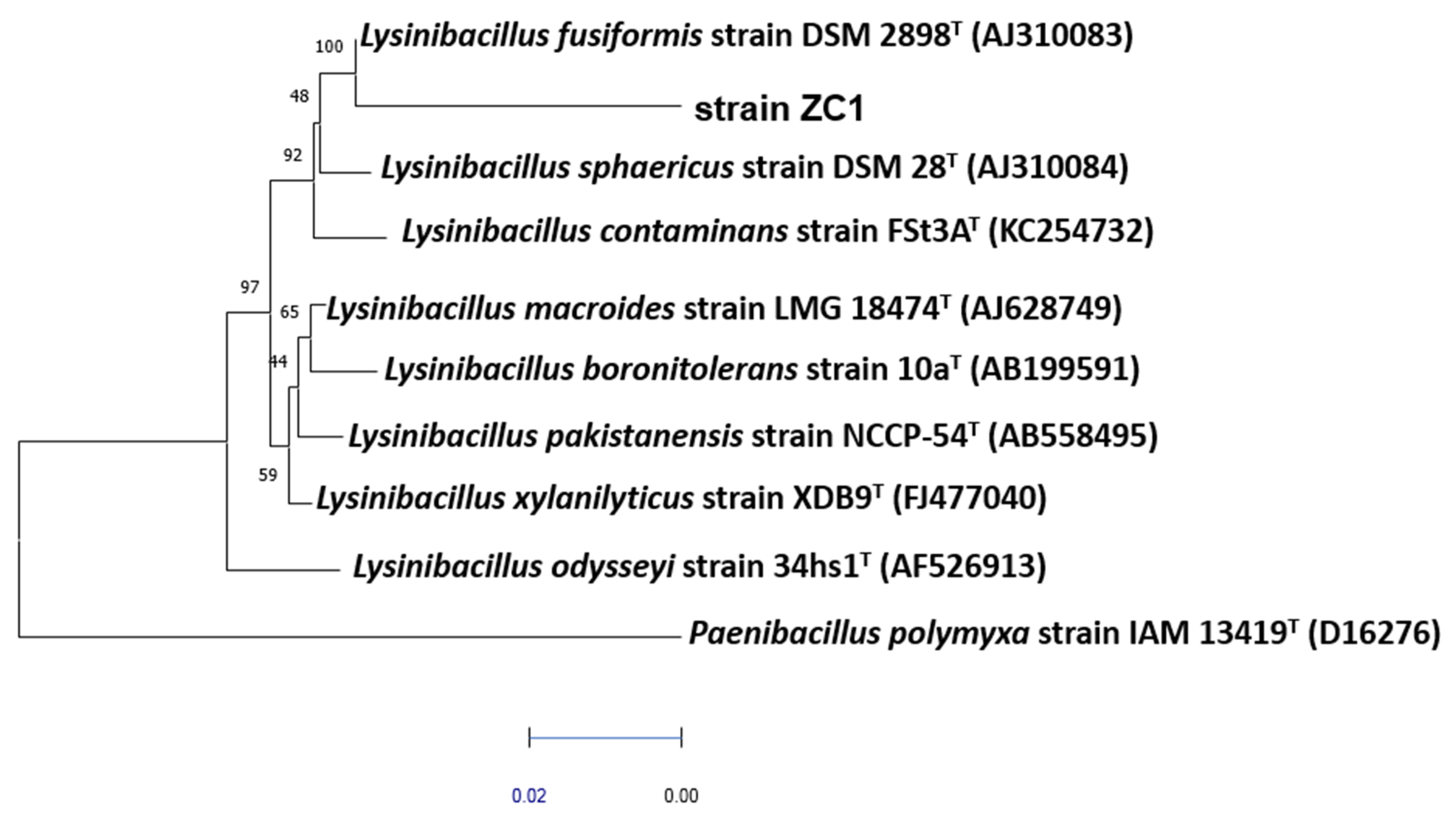
3.2. Phylogenetic Identification of Strain ZC
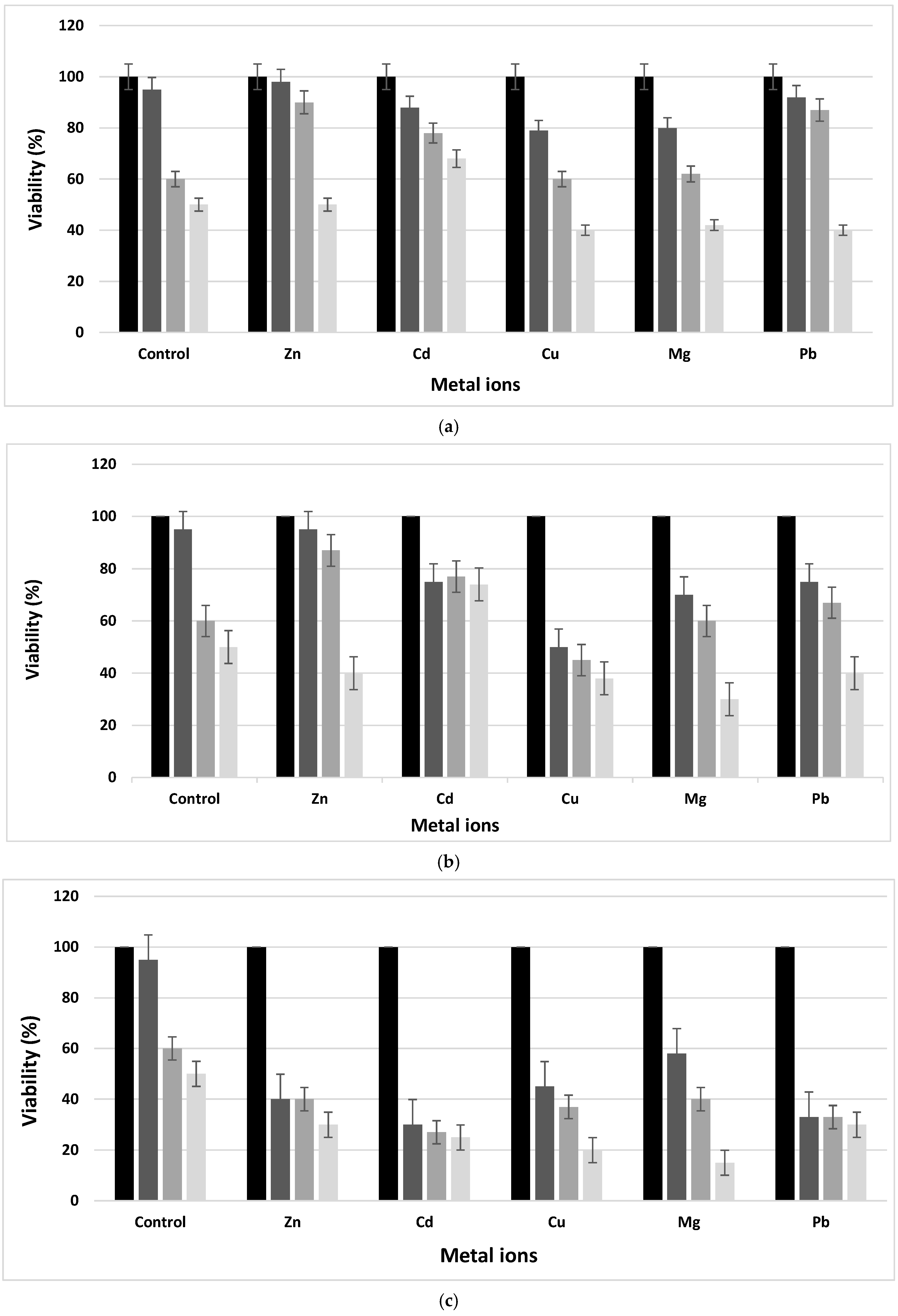
Nine bacterial strains were screened from the MPLW effluent. Among these isolates, strain ZC was selected for its high minimal inhibition concentrations towards Zn2+, Cu2+, Pb2+, Mg2+ and Cd2+ metal ions and its overproduction of exopolysaccharides in stressed conditions (data not shown). In order to identify the ZC isolate, purified DNA was amplified by the polymerase chain reaction using the universal bacterial Bac27F and Univ1492R primers [19]. The amplified DNA nucleotide sequence was about 1223 bp, and it was subjected to BLAST analysis. The DNA was extracted using an extraction kit “Wizard Genomic DNA Purification Kit” (Promega, Madison, WI, USA). The sequences used in the phylogenetic analysis were based on the fragments of sequences of the 16S rDNA of the type strains obtained from the ribosomal data project (RDP) and GenBank databases. Pairwise evolutionary distances were calculated using the method of Jukes and Cantor [37]. A dendrogram was constructed using the neighbor-joining method with MEGA-X software (State College, PA, USA) [38] (Figure 1). Confidence in the topology of the dendrogram was determined using 100-bootstrapped trees.

3.3. MTT Assay

The tolerance of the L. fusiformis strain to metal ions was examined. The results showed a decrease in cell viability with an increase in metal ion concentrations (Figure 2). The newly isolated strain, identified as L. fusiformis, was able to survive in the presence of metal ions containing media at a concentration equal to 1 mM as the control. In fact, increasing the metal ion concentrations, the viability was different from the control and decreased drastically after 24 h of incubation. In fact, the viability of the newly isolated bacteria with different metal ion concentrations within 72 h in TSB broth microcosms showed its viability (not exceeding 30%) in spite of the decrease in cell number after 72 h of incubation. In addition, a drop in the number of bacteria was noticed from the first day of incubation and for all metal ion presences and concentrations. The decrease was significant at 100 mM and reached about 10–30% for all metal ions. This decrease was also proportional to the incubation time. The presence of metal ions was toxic to the bacteria in the medium. The toxicity effect of the metal ions was classified in a decreasing order as follows: Pb, Zn > Cd > Cu > Mg.
Mine wastewater can profoundly influence biological systems. For example, species diversity and the total biomass composition in aquatic and terrestrial ecosystems can be affected by heavy metal contamination [39]. The newly isolated L. fusiformis from MPLW is able to adapt and survive under extremely stressful conditions.

3.4. Determination of Heavy Metal Content Using Atomic Absorption Spectrometry (AAS) Technique

The determination of metal ion contents, using AAS, was assayed to show whether the bacteria, in each microcosm accumulate and/or adsorb these toxic ions. By analyzing the results illustrated in Table 2, it is clear that strain ZC can accumulate and/or adsorb a small content of all the studied metal ions. In fact, the percentage of accumulation and/or adsorption did not exceed 20% for all metal ions at a concentration of 1 mM with the most minor ones for cadmium (4.6%). Increasing the concentration at 10 mM, the accumulation percentage was proportional to the concentration. It ranged between 11 and 35%. Similar results were demonstrated at 100 mM. The stressed strain ZC accumulated a high amount of metal ions exceeding 45%. In fact, it has been found that there is a decrease in the level of trace metals in the culture media, regardless of the concentration. The ZC strain manifested a bioaccumulation performance according to the following increasing order of metal ions: Cu > Zn > Cd > Mg > Pb. This accumulation into ZC strain was confirmed by the decreasing amount of metal ions in the supernatant before sonication.
Biosorption is defined as an interaction between the living and non-living microorganisms and the metallic ions in the system. This process is used for removing metal or metalloid species, compounds and particulates from solutions by biological materials [12,40]. The most important types of biosorbents that have been developed from diverse raw biomass are bacteria due to their ubiquity, size, ability to grow under controlled conditions and resilience to environmental conditions (e.g., Pseudomonas fluorescens; Bacillus safensis; Pseudomonas aeruginosa) [41,42]. L. fusiformis is able to adapt and survive under extremely stressful conditions. In addition, we find that the number of L. fusiformis cells decrease with the increase in metal concentration, but they remain viable. Thus, this resistance could be explained by the metallic pollution induced by the effluents of the Company of Phosphate Gafsa (CPG). Previous studies have noted that the high frequencies of metal-resistant germs encountered at a given site are related to the selective pressures exerted by heavy metals within this site [32,43]. Similarly, contact with other bacterial organisms at such frequency due to the process of transfer of genes enabling resistance to heavy metals, which are mainly located on plasmids between strains [33]. Generally, to survive under metal stress conditions, bacteria have adopted various types of heavy metal resistance mechanisms, such as alteration of their membrane by which bacteria protect their essential cellular components sensitive to metals. Some bacteria have a membrane or envelope that is capable of passively adsorbing high levels of dissolved metals, generally via a charge-mediated attraction [44,45], accumulation and complexation in the metal ion inside the cell, reduction of the toxicity of this metal [16], enzymatic transformation [46], as well as the decrease in the level of metal ions in the culture media, whatever the concentration. This confirms that there is intracellular or extracellular sequestration. In fact, many bacteria have developed a cytosolic sequestration mechanism for protection from metals through converting heavy metals into more inoffensive forms using cytoplasmic proteins, such as metallothioneins, to bind, sequester or store metals [16,47,48]. In the bacterium, a fine regulation for the maintenance (homoeostasis) and the control of their intracellular concentration [46] prevents the expulsion of essential metals present at homeostatic concentrations or the entry of metals in toxic amounts. Furthermore, extracellular polymers and siderophores produced by bacteria to withstand heavy metals can trap or precipitate metal ions in the extracellular environment [49,50]. These compounds bind heavy metals and subsequently detoxify metals simply via complex formation or by forming an effective barrier surrounding the cell [51]. Metals might also attach to, or precipitate at, bacterial cell surfaces by interactions involving proteins or cell-associated polysaccharides [32].

3.5. Enzymatic Changes

The newly isolated ZC strain identified as L. fusiformis was tested to produce exoenzymes such as amylase, lipase, gelatinase, caseinase, lecithinase and haemolysin using specific media on agar plates (Table 3). The control strain could produce gelatinase, caseinase, amylase and lecithinase and was unable to secrete lipase and haemolysin. The production was revealed by the appearance of a halo around the colony in each specific medium. Under the metallic stress, the new strain produced a new enzymatic activity named lipase. On the other hand, the loss of caseinase activity in the presence of 100 mM of copper and magnesium has been noticed, as well as lecithinase and gelatinase at 10 and 100 mM of zinc, respectively. In fact, gelatinase and lecithinase, zinc-metalloproteinases, are inhibited at high concentrations of zinc [52,53]. The mechanism of enzyme inactivation by metals is not completely understood. It is assumed that metal ions bind to specific sites, causing conformational changes that inactivate the catalytic function of enzymes. The non-competitive inhibition by other heavy metal ions is attributed to the binding of the ion to a site distinct from the active site [54]. Consequently, zinc is essential for the activity of these enzymes. It can either be directly active at their catalytic site or intervene in their conformation.
The biochemical modifications, by the appearance of new enzymatic activities, observed in the stressed L. fusiformis were a form of adaptation to the stressful conditions of the environment. This new enzymatic activity is the result of the expression of genes involved in survival under the stressful conditions of the environment. The high concentration of zinc can also be at the origin of the loss of some characters by the repression of the genes, which code for this enzyme. Transcriptional regulation is under the control of many proteins, which bind to the promoter sequences of genes, thus preventing their production of the transcripts. When the genes are exposed to metals, they bind these metals, inducing an allosteric change so that RNA polymerase can transcribe the target genes [45].

3.6. Qualitative Slime Production

The qualitative detection of biofilm formation was studied by culturing L. fusiformis on congo red agar. The newly isolated strain ZC shows colonies with a red center and a lighter outer area (Figure 3). These later properties showed its negative slime character. After the metal ions effect, the ZC strain changes its phenotypic profile and becomes brown. Indeed, these changes are considered to be a variable phenotype [55]. Slime-producing bacteria appeared as black colonies, whereas non-slime producers remained unpigmented. This difference between the strain in the normal and stressed state is a state of transition to biofilm formation. It could be proposed that the bacterium is developing a variety of resistance mechanisms to neutralize the toxic effect of the metal. In fact, biofilm-combining microorganisms have been shown to play a crucial role in the nutrient cycling and biodegradation of environmental pollutants [32,56]. In addition, biofilm bacteria are generally embedded in a polymeric extracellular substance. However, this substance, called slime, seems to play an important role [32,56]. In addition, exopolysaccharide can also act as an ion exchange and is able to positively sequester the charged pollutants [57].

3.7. Quantitative Adhesion and Culture Cells Adherence Assays

Quantitative adherence assays were performed with a human oral cavity epidermoid carcinoma (KB) cell line. The strain is considered weakly biofilm positive (0.1 < OD570 < 1) in the absence of any stress and in the presence of various metal ions such as Cu, Cd, Mg, Pb and Zn. In fact, the unchangeable property was improved by a significant (p < 0.05) increase in adherence in presence and with an increase in metal ions stress and concentrations, respectively (Table 4). In addition, the obtained results showed that the stress applied leads to a significant (p < 0.05) increase in adhesion (Table 4). In fact, the unstressed strain was weakly adherent, and in the presence in metal ions stress, the adherence became strong. In fact, bacteria have a natural tendency to adhere to surfaces as a survival mechanism under adverse conditions. Bacterial colonization of solid surfaces has been described as a basic ploy in a wide variety of environments [57]. Generally, biofilms, composed of extracellular polymeric substances, have also been reported to adsorb heavy metals [32,50].

3.8. Cell Surface Hydrophobicity

Hydrophobicity was measured by the hexane, decane and chloroform partitioning method. The obtained results (Table 5) indicate that strain L. fusiformis is weakly hydrophobic in the absence of metal ions. A significant variation (p < 0.05) in the hydrophobicity of L. fusiformis depends on the type and the concentration of metal ions and also of the used solvent. Indeed, the hydrophobic state was changed from the weakly to the hydrophilic state using chloroform as the solvent. On the other hand, the hydrophobicity increased when decane was used. In addition, an increase in hydrophobicity was noticed using hexane in the presence of Zn and Mg, but there was also a decrease in the presence of Cu. For microorganisms, hydrophobicity is considered as a virulence factor that detracts from the adhesion of microorganisms to biotic and abiotic surfaces. This variation in hydrophobicity is probably due to the metal stress effect as well as the nature of the solvent. Indeed, the surface properties of stressed strains are different from those of logarithmically growing strains. Among these properties, hydrophobicity is the most important one. This change is expected to increase the chances of survival in adverse conditions. Under adverse conditions, the bacterium regulates its protein synthesis by reorganizing the membrane, degrading certain proteins and synthesizing new “stress” proteins [58]. Generally, the modification of properties of the cell surface of bacteria plays a major role in resistance to harsh conditions as well as in removal of contaminants [59,60].

3.9. Antibiotic Sensitivity of Bacteria

The sensitivity or antibiotic resistance of L. fusiformis mentioned in Table 6 varied according to the metal ion as well as its concentration. The ZC strain was tested against seven antibiotics. Two were β-lactams (penicillin and cefoxitin), three inhibited protein synthesis (kanamycin, tetracycline and netilmicin) and two inhibited DNA replication (neomycin and tobramycin). The new ZC strain was sensitive to the majority of the used antibiotics except for penicillin. In fact, the incubation of the strain with metal ions in the different microcosms, revealed the change of sensitivity to the resistance of this strain towards tobramycin, kanamycin, neomycin, netilmicin, and cefoxitin. MARindex is defined as resistance to ≥3 antibiotics; in this study, seven antibiotics were tested. Consequently, a MAR index of 0.7 and 0.8 translate to antibiotic resistance towards five and six antibiotics respectively. The MARindex of the ZC strain fell into this range within incubation with metal ions at different concentrations (Table 6). Only, in presence of 1 mM of manganese, ZC strain was resistant to all the seven tested antibiotics, showing its multi-resistance. This L. fusiformis resistance can be essentially enabled by four mechanisms: the reduction of the membrane permeability, the efflux, the modification of the target and the inactivation of the antibiotic. As a result, metal contamination functions should be taken seriously as a selective agent for the proliferation of antibiotic resistance. Indeed, previous research has shown mechanisms including co-resistance to heavy metal ions and antibiotics [61]. The genetic determinants involved in these mechanisms are present in the bacterial genome [62]. Furthermore, different studies show a positive correlation between the presence of metal ions in an ecosystem and the resistance to both metal ions and antibiotics in native bacteria, regardless of the metal or the antibiotic [30,63].

3.10. Bio-Treatment Assays of Phosphate Laundries Wastewater Using Lysinibacillus fusiformis

The richness of MPLW in cultivable mesophilic microflora (38 104 CFU ml−1), used as a parameter, encourages the bio-treatment processing assay of the effluent using an indigenous strain. After successive sub-cultures on autoclaved MPLW supplemented with agar-agar, the ZC isolated strain was used in order to study their bioprocessing performance. The variations of the used metal ions concentration in phosphate laundries wastewater induced by reacting with new ZC isolated strain are presented in Table 7. The result showed a very high decrease in the concentration of Cu, Cd, Pb, Zn and Mg after adding L. fusiformis. The results showed different performance rates of bioprocessing with the addition of the metal ions. Indeed, results showed that the removal efficiency (Reff) of magnesium (Mg) was the mostly reduced element followed by lead (Pb) and zinc (Zn) with a Reff equal to 86–87%, then cadmium (Cd) and finally copper (Cu) with Reff equal to 82.3 % and 80.6%, respectively.
The bio-treatment assays of phosphate laundries wastewater by L. fusiformis new isolate showed a very important decrease in the concentration of the heavy metal ions. Indeed, biosorption is becoming a potential alternative to the existing technologies for the removal and/or recovery of toxic metals and it has been reported that some bacterial strains naturally have ability to uptake heavy metal ions, such as Cu, Zn and Mg from solutions at a relatively lower concentration. The bioremediation process is an approach for removing these toxic elements from the polluted sites and/or transforming into less toxic form by applying live or dead microbes. Microorganisms are omnipresent in the environment and play a pivotal role in the biogeochemical cycles [64]. The bioremediation technique can be implemented in in situ and ex situ methods. The remediation is carried out in the site of contamination by stimulating the growth of indigenous microbes, supplying adequate nutrition or application of engineered microbes [65]. Therefore, many organisms have been used from different raw biomass, such as bacteria (e.g., Pseudomonas fluorescens; Bacillus safensis; Pseudomonas aeruginosa; Bacillus thuringiensis) [41,42,66], fungi (e.g., Botrytis cinerae) [12,67] and algae (e.g., Anabaena sphaerica) [68]. However, there are several advantages of microbial-based biosorption in the removal of metal ions, such as the high metal removal efficiency thanks to their selectivity towards particular metal [64].

4. Conclusions

In this study, a new strain ZC was isolated from the Metlaoui phosphate laundries wastewater (MPLW) and identified as L. fusiformis. Indeed, the MPLW was especially rich in various heavy metal ions and had a high COD/BOD5 ratio underlining a poorly biodegradable organic load. The newly isolated strain showed a decreasing viability and an increasing adherence in presence of metal ions at 100 mM. In addition, the enzymatic and biochemical profiles as well as resistance or sensibility to antibiotics was changed. The strain ZC, used as a biological tool for MPLW treatment, showed a reduction of the metal ion contents. This reduction was due to accumulation and/or adsorption showing a bioprocessing performance of the newly isolated L. fusiformis. This potential can be used safely for the bio-treatment of effluents.

Author Contributions

Conceptualization, A.E.; software, S.M.; validation, A.E.; formal analysis, I.T., S.B.Y., S.M., A.E.; investigation, I.T., B.M.; resources, R.M., A.B., C.J., J.A.A.S.; data curation, I.T.; writing—original draft preparation, I.T., A.E.; writing—review and editing, S.B.Y.; supervision, R.M., A.B., C.J., J.A.A.S., A.E.; project administration, A.E. All authors have read and agreed to the published version of the manuscript.

Funding

This research received no external funding.

Institutional Review Board Statement

Not applicable.

Informed Consent Statement

Not applicable.

Data Availability Statement

Not applicable.

Acknowledgments

The authors thank Haythem Chiboub from the Faculty of Sciences of Gafsa for his help with English.

Conflicts of Interest

The authors declare no conflict of interest.

References

  1. Mekki, A.; Awali, A.; Aloui, F.; Loukil, S.; Sayadi, S. Characterization and toxicity assessment of wastewater from rock phosphate processing in Tunisia. Mine Water Environ. 2017, 36, 502–507. [Google Scholar] [CrossRef]
  2. Tijani, A.; Fakhfakh, H. Les projets sociaux sont-ils des leviers de création de la valeur? J. Bus. Econ. 2011, 1, 94–105. [Google Scholar]
  3. Galfati, I.; Bilal, E.; Abdallah, H.; Beji Sassi, A. Geochemistry of solid effluents and phosphate ore washed from Métlaoui-Gafsa basin, Tunisia. Rom. J. Miner. Deposits. 2014, 87, 83–86. [Google Scholar]
  4. Mekki, A.; Sayadi, S. Study of heavy metal accumulation and residual toxicity in soil saturated with phosphate processing wastewater. Water Air Soil Pollut. 2017, 228, 215–224. [Google Scholar] [CrossRef] [Green Version]
  5. Srarfi, F.; Rachdi, R.; Bol, R.; Gocke, M.I.; Brahim, N.; SlimShimi, N. Stream sediments geochemistry and the influence of flood phosphate mud in mining area, Metlaoui, Western south of Tunisia. Environ. Earth. Sci. 2019, 78, 211–223. [Google Scholar] [CrossRef]
  6. Moula, A.; Borgi, M.A.; Loukil, S.; Chaieb, M.; Mekki, A. Assessment of phosphate laundries waste water phytotoxicity and biotreatment assays. Clean Soil Air Water 2020, 48, 1–8. [Google Scholar] [CrossRef]
  7. Chen, H.; Teng, Y.; Lu, S.; Wang, Y.; Wang, J. Contamination features and health risk of soil heavy metals in China. Sci. Total Environ. 2015, 512, 143–153. [Google Scholar] [CrossRef]
  8. François, F.; Lombard, C.; Guigner, J.-M.; Soreau, P.; Brian-Jaisson, F.; Martino, G.; Vandervennet, M.; Garcia, D.; Molinier, A.-L.; Pignol, D.; et al. Isolation and characterization of environmental bacteria capable of extracellular biosorption of mercury. Appl. Environ. Microbiol. 2012, 78, 1097–1106. [Google Scholar] [CrossRef] [PubMed] [Green Version]
  9. Anyanwu, C.U.; Nwankwo, S.C.; Moneke, A.N. Soil bacterial response to introduced metal stress. Int. J. Basic Appl. Sci. 2011, 11, 109–115. [Google Scholar]
  10. Gall, J.E.; Boyd, R.S.; Rajakaruna, N. Transfer of heavy metals through terrestrial food webs: A review. Environ. Monit. Assess. 2015, 187, 201–221. [Google Scholar] [CrossRef] [Green Version]
  11. Choi, A.L.; Sun, G.; Zhang, Y.; Grandjean, P. Developmental fluoride neurotoxicity: A systematic review and meta-analysis. Environ. Health Perspect. 2012, 120, 1362–1368. [Google Scholar] [CrossRef] [Green Version]
  12. Siddiquee, S.; Rovina, K.; Al Azad, S.; Naher, L.; Suryani, S.; Chaikaew, P. Heavy metal contaminants removal from wastewater using the potential filamentous fungi biomass: A review. J. Microb. Biochem. Technol. 2015, 7, 384–393. [Google Scholar] [CrossRef]
  13. Srivastava, N.K.; Majumder, C.B. Novel biofiltration methods for the treatment of heavy metals from industrial wastewater. J. Hazard. Mater. 2008, 151, 1–8. [Google Scholar] [CrossRef] [PubMed]
  14. Alloway, B.J. Heavy Metals and Metalloids as Micronutrient for Plants and Animals; Springer: Dordrecht, The Netherlands, 2013; pp. 195–209. [Google Scholar]
  15. Lakherwal, D. Adsorption of heavy metals: A review. Int. J. Environ. Res. Dev. 2014, 4, 41–48. [Google Scholar]
  16. Igiri, B.E.; Okoduwa, S.I.R.; Idoko, G.O.; Akabuogu, E.P.; Adeyi, A.O.; Ejiogu, I.K. Toxicity and bioremediation of heavy metals contaminated ecosystem from tannery wastewater: A review. J. Toxicol. 2018, 2018, 1–16. [Google Scholar] [CrossRef] [PubMed]
  17. Lemire, J.; Mailloux, R.; Auger, C.; Whalen, D.; Appanna, V.D. Pseudomonas fluorescens orchestrates a fine metabolic-balancing act to counter aluminium toxicity. Environ. Microbiol. 2010, 12, 1384–1390. [Google Scholar]
  18. Ben Younes, S.; Ellouze, M.; Sayadi, S. A comparative study of an industrial effluent treatment using enzymatic and alkaline adapted consortium assays. J. Chem. Technol. Biotechnol. 2013, 88, 563–571. [Google Scholar] [CrossRef]
  19. Jiang, H.; Dong, H.; Zhang, G.; Yu, B.; Chapman, L.R.; Fields, M.W. Microbial diversity in water and sediment of Lake Chaka, an athalasso haline lake in northwestern China. Appl. Environ. Microbiol. 2006, 72, 3832–3845. [Google Scholar] [CrossRef] [Green Version]
  20. Kouidhi, B.; Zmantar, T.; Bakhrouf, A. Anti-cariogenic and anti-biofilms activity of Tunisian propolis extract and its potential protective effect against cancer cells proliferation. Anaerobe 2010, 16, 566–571. [Google Scholar] [CrossRef]
  21. Ben Kahla-Nakbi, A.; Besbes, A.; Chaieb, K.; Rouabhia, M.; Bakhrouf, A. Investigation of several virulence properties among Vibrio alginolyticus strains isolated from diseased cultured fish in Tunisia. Dis. Aquat. Org. 2009, 86, 21–28. [Google Scholar] [CrossRef] [Green Version]
  22. Ben Younes, S.; Dallali, C.; Ellafi, A.; Bouslama, L.; Feriani, A.; Sayadi, S. Extracellular enzymatic activities of bacterial strains isolated from Tunisian biotopes: Decolorization and detoxification of indigo carmine. Cata. Lett. 2020, 151, 1248–1261. [Google Scholar] [CrossRef]
  23. Marzan, L.W.; Hossain, M.; Akter Mina, S.; Akter, Y.; Masudul Azad Chowdhury, A.M. Isolation and biochemical characterization of heavy-metal resistant bacteria from tannery effluent in Chittagong city, Bangladesh: Bioremediation viewpoint. Egy. J. Aquat. Res. 2017, 43, 65–74. [Google Scholar] [CrossRef]
  24. Freeman, D.J.; Falkiner, F.R.; Keane, C.T. New method for detecting slime production by coagulase negative staphylococci. J. Clin. Pathol. 1989, 42, 872–874. [Google Scholar] [CrossRef] [Green Version]
  25. Ellafi, A.; Denden, I.; Ben Abdallah, F.; Souissi, I.; Bakhrouf, A. Survival and adhesion ability of Shigella spp. strains after their incubation in seawater microcosms. World J. Microbiol. Biotechnol. 2009, 25, 1161–1168. [Google Scholar] [CrossRef]
  26. Ben Abdallah, F.; Chaieb, K.; Zmantar, T.; Kallel, H.; Bakhrouf, A. Adherence assays and slime production of vibrio alginolyticus and vibrio parahaemolyticus. Braz. J. Microbiol. 2009, 40, 394–398. [Google Scholar] [CrossRef] [PubMed]
  27. Chaieb, K.; Chehab, O.; Zmantar, T.; Rouabhia, M.; Mahdouani, K.; Bakhrouf, A. In vitro effect of pH and ethanol on biofilm formation by clinical ica-positive Staphylococcus epidermidis strains. Ann. Microbiol. 2007, 57, 431–437. [Google Scholar] [CrossRef]
  28. Chatti, A.; Daghfous, D.; Landoulsi, A. Effect of seqA mutation on Salmonella typhimurium virulence. J. Infect. 2007, 54, 241–245. [Google Scholar] [CrossRef] [PubMed]
  29. Van Loosdrecht, M.C.; Lyklema, J.; Norde, W.; Schraa, G.; Zehnder, A.J.B. The role of bacterial cell wall hydrophobicity in adhesion. Appl. Environ. Microbiol. 1987, 531, 1893–1897. [Google Scholar] [CrossRef] [Green Version]
  30. Jardine, J.; Mavumengwana, V.; Ubomba-Jaswa, E. Antibiotic resistance and heavy metal tolerance in cultured bacteria from hot springs as indicators of environmental intrinsic resistance and tolerance levels. Environ. Pollut. 2019, 249, 696–702. [Google Scholar] [CrossRef]
  31. El Rhaouat, O.; El Kherrati, I.; El khayyat, F.; Chiguer, H.; Ezziani, K.; Ibeda, A.; Fareh, M.; Saidi, Y.; El Kharim, K.; Belghyti, D. Physic-chemical evaluation of urban wastewater of the town of Sidi Kacem. Comput. Water Energy Environ. Eng. 2014, 3, 30–35. [Google Scholar] [CrossRef] [Green Version]
  32. Domingues, V.S.; de Souza Monteiro, A.; Júlio, A.D.L.; Queiroz, A.L.L.; dos Santos, V.L. Diversity of metal-resistant and tensoactive-producing culturable heterotrophic bacteria isolated from a copper mine in Brazilian Amazonia. Sci. Rep. 2020, 10, 1–12. [Google Scholar] [CrossRef] [PubMed]
  33. Ünaldi Coral, M.N.; Korkmaz, H.; Arikan, B.; Coral, G. Plasmid mediated heavy metal resistances in Enterobacter spp. isolated from Sofulu landfill, in Adana, Turkey. Ann. Microbiol. 2005, 55, 175–179. [Google Scholar]
  34. Ministère des Affaires Locales et de l’Environnement, Official Printing Office of the Republic of Tunisia. Décret gouvernemental n° 2018-315 du 26 mars 2018, ANNEXE 1, Rejet dans le domaine public maritime, hydraulique et réseau public d’assainissement. J. Off. R. Tunis. 2018, 26, 824–827. (In French) [Google Scholar]
  35. APHA (American Public Health Association). Standard Methods for the Examination of Water and Wastewater, 21st ed.; American Public Health Association: Washington, DC, USA, 2005. [Google Scholar]
  36. Jarvis, I.; Burnett, W.C.; Nathan, Y.; Almbaydin, F.S.M.; Attia, A.K.M.; Castro, L.N.; Flicoteaux, R.; Hilmy, M.E.; Husain, V.; Qutawnah, A.A.; et al. Phosphorite geochemistry: State of-the-art and environmental concerns. Eclogae Geol. Helv. 1994, 87, 643–700. [Google Scholar]
  37. Jukes, T.H.; Cantor, C.R. Evolution of proteins molecules. In Mammalian Protein Metabolism; Munro, H.N., Ed.; Academic Press: New York, NY, USA, 1969; Volume 3, pp. 21–132. [Google Scholar]
  38. Kumar, S.; Stecher, G.; Li, M.; Knyaz, C.; Tamura, K. MEGA X: Molecular evolutionary genetics analysis across computing platforms. Mol. Biol. Evol. 2018, 35, 1547–1549. [Google Scholar] [CrossRef]
  39. Ashraf, M.A.; Maah, M.J.; Yusoff, I. Morphology, geology and water quality assessment of former tin mining catchment. Sci. World J. 2012, 1–15. [Google Scholar] [CrossRef] [Green Version]
  40. Gadd, G.M. Biosorption: Critical review of scientific rationale, environmental importance and significance for pollution treatment. J. Chem. Technol. Biotechnol. 2009, 84, 13–28. [Google Scholar] [CrossRef]
  41. Kalaimurugan, D.; Balamuralikrishnan, B.; Durairaj, K.; Vasudhevan, P.; Shivakumar, M.S.; Kaul, T.; Chang, S.W.; Ravindran, B.; Venkatesan, S. Isolation and characterization of heavy-metal-resistant bacteria and their applications in environmental bioremediation. Int. J. Environ. Sci. Technol. 2020, 17, 1455–1462. [Google Scholar] [CrossRef]
  42. Chellaiah, E.R. Cadmium (heavy metals) bioremediation by Pseudomonasaeruginosa: A minireview. Appl. Water Sci. 2018, 8, 154–163. [Google Scholar] [CrossRef] [Green Version]
  43. Xu, Y.; Xu, J.; Mao, D.; Luo, Y. Effect of the selective pressure of sub-lethal level of heavy metals on the fate and distribution of ARGs in the catchment scale. Environ. Pollut. 2016, 220, 900–908. [Google Scholar] [CrossRef]
  44. Mohamed, Z.A. Removal of cadmium and manganese by a non-toxic strain of the fresh water cyanobacterium Gloeothece magna. Water Res. 2001, 35, 4405–4409. [Google Scholar] [CrossRef]
  45. Outten, C.E.; Outten, F.W.; O’Halloran, T.V. DNA distortion mechanism for transcriptional activation by ZntR, a Zn(II)-responsive MerR Homologue in Escherichia coli. J. Biol. Chem. 1999, 274, 37517–37524. [Google Scholar] [CrossRef] [Green Version]
  46. Nies, D.H. Microbial heavy-metal resistance. Appl. Microbiol. Biotechnol. 1999, 51, 730–750. [Google Scholar] [CrossRef]
  47. Haferburg, G.; Kothe, E. Microbes and metals: Interactions in the environment. J. Basic Microbiol. 2007, 47, 453–467. [Google Scholar] [CrossRef]
  48. Nanda, M.; Kumar, V.; Sharma, D.K. Multimetal tolerance mechanisms in bacteria: The resistance strategies acquired by bacteria that can be exploited to ‘clean-up’ heavy metal contaminants from water. Aquat. Toxicol. 2019, 212, 1–10. [Google Scholar] [CrossRef]
  49. Chaturvedi, K.S.; Hung, C.S.; Crowley, J.R.; Stapleton, A.E.; Henderson, J.P. The siderophore yersiniabactin binds copper to protect pathogens during infection. Nat. Chem. Biol. 2012, 8, 731–736. [Google Scholar] [CrossRef] [Green Version]
  50. Harrison, J.J.; Ceri, H.; Turner, R.J. Multi metal resistance and tolerance in microbial biofilms. Nat. Rev. Microbiol. 2007, 5, 928–938. [Google Scholar] [CrossRef] [PubMed]
  51. Pulsawat, W.; Leksawasdi, N.; Rogers, P.L.; Foster, L.J.R. Anions effects on biosorption of Mn(II) by extracellular polymeric substance (EPS) from Rhizobium etli. Biotechnol. Lett. 2003, 25, 1267–1270. [Google Scholar] [CrossRef] [PubMed]
  52. Moreland, L.W. Rheumatology and Immunology Therapy: A to Z Essentials; Springer Science & Business Media: Berlin, Germany, 2004. [Google Scholar] [CrossRef]
  53. Sharaf, E.F.; El-Sayed, W.S.; Abosaif, R.M. Lecithinase-producing bacteria in commercial and home-made foods: Evaluation of toxic properties and identification of potent producers. J. Taibah Univ. Sci. 2014, 8, 207–215. [Google Scholar] [CrossRef]
  54. De Souza, A.P.; Gerlach, R.F.; Line, S.R.P. Inhibition of human gingival gelatinases (MMP-2 and MMP-9) by metal salts. Dent. Mater. 2000, 16, 103–108. [Google Scholar] [CrossRef]
  55. Touati, A.; Achour, W.; Abbassi, M.S.; Hassen, A.B. Détection des gènes ica et de la production de slime parmi des souches de Staphylococcus epidermidis isolées d’infections liées aux cathéters chez des patients neutropéniques. Pathol. Biol. 2007, 55, 277–282. [Google Scholar] [CrossRef]
  56. Prabhakaran, P.; Ashraf, M.A.; Aqma, W.S. Microbial stress response to heavy metals in the environment. RSC. Adv. 2016, 6, 109862–109877. [Google Scholar] [CrossRef]
  57. Hunt, S.M.; Werner, E.M.; Huang, B.; Hamilton, M.A.; Stewart, P.S. Hypothesis for the role of nutrient starvation in biofilm detachment. Appl. Environ. Microbiol. 2004, 70, 7418–7425. [Google Scholar] [CrossRef] [PubMed] [Green Version]
  58. Mitzel, M.R.; Sand, S.; Whalen, J.K.; Tufenkji, N. Hydrophobicity of biofilm coatings influences the transport dynamics of polystyrene nanoparticles in biofilm-coated sand. Water Res. 2016, 92, 113–120. [Google Scholar] [CrossRef] [PubMed] [Green Version]
  59. Falcón García, C.; Kretschmer, M.; Lozano-Andrade, C.N.; Schönleitner, M.; Dragoŝ, A.; Kovács, Á.T.; Lieleg, O. Metal ions weaken the hydrophobicity and antibiotic resistance of Bacillus subtilis NCIB 3610 biofilms. NPJ Biofilms Microbiomes 2020, 6, 1–11. [Google Scholar] [CrossRef] [Green Version]
  60. Martins, P.S.D.O.; Almeida, N.F.D.; Leite, S.G.F. Application of a bacterial extracellular polymeric substance in heavy metal adsorption in a co-contaminated aqueous system. Braz. J. Microbiol. 2008, 39, 780–786. [Google Scholar] [CrossRef] [Green Version]
  61. Zhang, M.; Chen, L.; Ye, C.; Yu, X. Co-selection of antibiotic resistance via copper shock loading on bacteria from a drinking water bio-filte. Environ. Pollut. 2018, 233, 132–141. [Google Scholar] [CrossRef]
  62. Livermore, D.M. Bacterial resistance: Origins, epidemiology, and impact. Clin. Infect. Dis. 2003, 36, 11–23. [Google Scholar] [CrossRef] [Green Version]
  63. Baker-Austin, C.; Wright, M.S.; Stepanauskas, R.; McArthur, J.V. Co-selection of antibiotic and metal resistance. Trends Microbiol. 2006, 14, 176–182. [Google Scholar] [CrossRef]
  64. Abdel-Aty, A.M.; Ammar, N.S.; Abdel Ghafar, H.H.; Ali, R.K. Biosorption of cadmium and lead from aqueous solution by fresh water alga Anabaena sphaerica biomass. J. Adv. Res. 2013, 4, 367–374. [Google Scholar] [CrossRef] [Green Version]
  65. Das, S.; Lyla, P.S.; Khan, S.A. Biogeochemical processes in the continental slope of Bay of Bengal: I. Bacterial solubilization of inorganic phosphate. Rev. Biol. Trop. 2007, 55, 1–9. [Google Scholar] [CrossRef] [PubMed]
  66. Kulshreshtha, A.; Agrawal, R.; Barar, M.; Saxena, S. A review on bioremediation of heavy metals in contaminated water. IOSR J. Environ. Sci. Toxicol. Food Technol. 2014, 8, 44–50. [Google Scholar] [CrossRef]
  67. Vijayaraghavan, K.; Yun, Y.S. Bacterial biosorbents and biosorption. Biotechnol. Adv. 2008, 26, 266–291. [Google Scholar] [CrossRef] [PubMed]
  68. Akar, T.; Tunali, S.; Kiran, I. Botrytis cinerea as a new fungal biosorbent for removal of Pb(II) from aqueous solutions. Biochem. Eng. J. 2005, 25, 227–235. [Google Scholar] [CrossRef]
Figure 1. Phylogenetic tree based on 1223 unambiguous nucleotides of the 16S rDNA sequence, constructed by the neighbour joining method, showing the position of strain ZC among related members of the genus Lysinibacillus. Reference type strain organisms are included, and sequence accession numbers are given in parentheses. Bootstrap values, expressed as a percentage of 100 replications, are shown in branching points. Bar, 1 substitution in 100 nt.
Sustainability 13 10072 g001
Figure 2. Viability of Lysinibacillus fusiformis in presence of the five metal ions at: (a) 1 mM, (b) 10 mM and (c) 100 mM after incubation at 0 h (■), 24 h (), 48 h () and 72 h ().
Sustainability 13 10072 g002
Figure 3. Induction of slime production by metallic stress: Slime production was analysed using congo red agar plates. Lysinibacillus fusiformis ZC strain was cultured (A) with and (B) without metal ions (1, 10 and 100 mM) on congo red agar plates for 24 h at 37 °C. Four independent experiments were conducted, and one set of representative results is shown.
Sustainability 13 10072 g003
Table 1. Physicochemical and microbiological characteristics of Metlaoui phosphate laundries wastewaters (MPLW) in comparison with Tunisian national standards [34] and with international standards [35].
Parameter, UnitMPLW, SDTunisian National StandardsInternational Standars
pH, 25 °C7.5 ± 0.26.5–8.509.00
EC, mS cm−13.2 ± 0.35.006.00
Salinity, g L−12.1 ± 0.1NINI
Chlorosity, g L−11.1 ± 0.1NINI
TSS, g L−1 61.8 ± 10.03≤0.03
TS, g L−16.8 ± 0.9NINI
VSS, g L−170 ± 1NINI
Turbidity, NTU503 ± 370.00≤30.00
TOC, g L−13.7 ± 0.1NINI
COD, mg L−11155 ± 2200.00≤70.00
BOD5, mg L−1280 ± 250.00≤50.00
COD/BOD5 ratio4.1 ± 00.33≤0.71
CODsoluble, mg/L577 ± 2NINI
Mg, mg L−15655 ± 52NINI
Co/Mn, mg L−10 ± 0NINI
K, mg L−145.0 ± 0.550.00≤30.00
Fe, mg L−10.7 ± 0.05.00≤5.00
Cd, mg L−10.5 ± 0.00.10≤0.10
Cu, mg L−10.3 ± 0.00.50≤0.25
Zn, mg L−10.1 ± 0.00.50≤1.00
Pb, mg L−11.0 ± 0.30.10≤0.10
Total cultivable microflora (CFU mL−1)38.0 ± 3 × 10⁴NINI
SD: standard deviation; EC: electrical conductivity; TSS: total suspended solids; TS: total solids; VSS: volatile suspended solids; NI: not identified; COD: chemical oxygen demand; BOD5: biochemical oxygen demand; TOC: total organic carbon; CFU: colony forming unit.
Table 2. Concentration of metal ion content in ZC strain before and after sonication using Atomic Absorption Spectrometry (AAS) technique.
Initial Metal Ion Concentration (mM)Metal Ion Concentration before Sonication (mM)Metal Ion Concentration after Sonication (mM)Accumulation/Adsorption (%)
Cu10.8 ± 00.1 ± 010 ± 0
108.8 ± 01.1 ± 011 ± 0
10080 ± 319.5 ± 0.919.5 ± 0
Zn10.8 ± 00.1 ± 011 ± 0
108.5 ± 0.81.4 ± 0.114.8 ± 0
10080.8 ± 218.4 ± 0.818.4 ± 0
Mg10.9 ± 0.10.1 ± 010 ± 0
108.4 ± 11.6 ± 0.116 ± 0
10070 ± 428.9 ± 128.9 ± 1
Cd10.9 ± 00 ± 04 ± 0
108.3 ± 11.5 ± 015.4 ± 0
10061.6 ± 238.4 ± 238.4 ± 2
Pb10.8 ± 00.2 ± 020.0 ± 0
106.4 ± 0.13.5 ± 0.535.0 ± 0.5
10054.5 ± 145.3 ± 145.3 ± 1
Footnote: The values used in this table are the means of three replicates with their respective SD.
Table 3. Enzyme expression in specific agar media in presence of the newly isolated strain Lysinibacillus fusaformis.
ZC StrainCu (mM)Zn (mM)Mg (mM)Cd (mM)Pb (mM)
110100110100110100110100110100
Lipase+++++++++++++++
Gelatinase+++++++++++++++
Caseinase++++++++++++
Amylase++++++++++++++++
Lecithinase++++++++++++++
Hemolysin
+: activity, −: no activity.
Table 4. Biofilm formation and effect of metallic stress on the capacity of Lysinibacillus fusiformis to adhere to KB cells.
ZC StrainCu (mM)Zn (mM)Mg (mM)Cd (mM)Pb (mM)
110100110100110100110100110100
OD5700.1 ± 00.1 ± 00.1 ± 00.3 ± 00.2 ± 00.2 ± 00.2 ± 00.2 ± 00.3 ± 00.3 ± 00.2 ± 00.2 ± 00.2 ± 00.2 ± 00.2 ± 00.2 ± 0
Adhesion (%)1.2 ± 0.21.2 ± 0.12.6 ± 0.21.8 ± 0.11.3 ± 0.02.9 ± 0.22.8 ± 0.31.4 ± 0.43.2 ± 0.33.0 ± 0.01.3 ± 0.21.6 ± 0.41.5 ± 0.61.3 ± 0.12.3 ± 0.12.2 ± 0.3
Footnote: The values used in this table are the means of three replicate with their respective SD.
Table 5. Effect of metal ions (Cu, Zn, Mg, Cd and Pb) on surface hydrophobicity by Lysinibacillus fusiformis.
Control Strain[Metal Ion] (mM)CuZnMgCdPb
Hexane36.3 ± 2114 ± 0.359 ± 153 ± 233 ± 343 ± 3
1013 ± 127 ± 156 ± 147 ± 246 ± 1
10027 ± 237 ± 165 ± 255 ± 256 ± 1
Decane69.7 ± 1118 ± 116 ± 118 ± 117 ± 114 ± 1
1029 ± 242 ± 261 ± 148 ± 131 ± 1
10058 ± 322 ± 242 ± 141 ± 140 ± 1
Chloroform31.3 ± 2132 ± 224 ± 110 ± 117 ± 217 ± 1
1022 ± 111 ± 116 ± 120 ± 113 ± 1
10031 ± 28 ± 19 ± 0.811 ± 0.67 ± 1
Table 6. Antibiotic resistance of Lysinibacillus fusiformis in the presence and in the absence of metal ions at different concentrations.
AntibioticsZC StrainCu (mM)Zn (mM)Mg (mM)Cd (mM)Pb (mM)
110100110100110100110100110100
PenicillinIISSIISRIISSSSSS
TobramycinSRRRRRRRRRRRRRRR
NeomycinSRRRRRRRRRRRRRRR
TetracyclinSRRRRRRRRRRRRRRR
CefoxitinSRRRRRRRRRRRRRRR
KanamycinSRIRRRRRRRRRRRRR
NetilmicinSRRRRSIRRIRRRRRR
MARindex00.80.70.80.80.70.710.80.70.80.80.80.80.80.8
R: Resistant; S: Sensible; I: Intermediate.
Table 7. Metal ion concentrations before and after bio-treatment assays by Lysinibacillus fusiformis using Atomic Absorption Spectrometry (AAS) technique.
Cu (mg/L)Zn (mg/L)Mg (mg/L)Cd (mg/mL)Pb (mg/mL)
Untreated MPLW0.3 ± 00.1 ± 05655 ± 520.05 ± 01 ± 0.3
Treated MPLW0.06 ± 00.02 ± 084 ± 150.09 ± 00.01 ± 0
Reff80.686.698.582.387.8
Footnote: The values used in this table are the means of three replicate with their respective SD.
Publisher’s Note: MDPI stays neutral with regard to jurisdictional claims in published maps and institutional affiliations.

