Article

PD-L1 Expression in Patients with Idiopathic Pulmonary Fibrosis

by
Sissel Kronborg-White
1,*,
Line Bille Madsen
2,
Elisabeth Bendstrup
1 and
Venerino Poletti
1,3
1
Center for Rare Lung Diseases, Department of Respiratory Diseases and Allergy, Aarhus University Hospital, 8200 Aarhus, Denmark
2
Department of Pathology, Aarhus University Hospital, 8200 Aarhus, Denmark
3
Department of the Diseases of the Thorax, Ospedale Morgagni, University of Bologna, 47121 Forli, Italy
*
Author to whom correspondence should be addressed.
J. Clin. Med. 2021, 10(23), 5562; https://doi.org/10.3390/jcm10235562
Submission received: 20 September 2021 / Revised: 5 November 2021 / Accepted: 15 November 2021 / Published: 26 November 2021
(This article belongs to the Section Pulmonology)

Abstract

:
Background: Idiopathic pulmonary fibrosis (IPF) is the most common and severe form within the group of idiopathic interstitial pneumonias. It is characterized by repetitive alveolar injury in genetically susceptible individuals and abnormal wound healing, leading to dysregulated bronchiolar proliferation and excessive deposition of extracellular matrix, causing complete architectural distortion and fibrosis. Epithelial-to-mesenchymal transition is considered an important pathogenic event, a phenomenon also observed in various malignant neoplasms, in which tumor cells express programmed death-ligand one (PD-L1). The aim of this study was to assess the presence of PD-L1 in patients with IPF and other interstitial lung diseases (ILDs). Method: Patients with a clinically and radiologically suspected idiopathic interstitial pneumonia or other ILDs undergoing transbronchial cryobiopsy to confirm the diagnosis at the Department of Respiratory Diseases and Allergy, Aarhus University Hospital, were included in this prospective observational study. Cellular membrane PD-L1 expression in epithelial cells was determined using the DAKO PD-L1 IHC 22C3 PharmDx Kit. Results: Membrane-bound PD-L1 (mPD-L1) was found in twelve (28%) of the forty-three patients with IPF and in five (9%) of the fifty-five patients with other ILDs (p = 0.015). When adjusting for age, gender and smoking status, the odds ratio of having IPF when expressing mPD-L1 in alveolar and/or bronchiolar epithelial cells was 4.3 (CI: 1.3–14.3). Conclusion: Expression of mPD-L1 in epithelial cells in the lung parenchymal zones was detected in a consistent subgroup of patients with IPF compared to other interstitial pneumonias. Larger studies are needed to explore the role of mPD-L1 in patients with IPF.

1. Introduction

Idiopathic pulmonary fibrosis (IPF) is the most common and severe form of idiopathic interstitial pneumonia, with a mean survival time of 3–5 years without antifibrotic treatment. Patients suffering from IPF have an increased risk of peripheral lung cancer [1], with a reported prevalence between 7 and 31% [1,2]. The phenotypes and genetic alterations of peripheral lung cancer associated with IPF suggest that IPF and lung cancer may share several pathogenic pathways [3,4,5].
IPF is characterized by a progressive scarring of the lung parenchyma due to dysregulated bronchiolar proliferation and excessive accumulation of extracellular matrix (ECM), resulting in architectural distortion and respiratory failure. The pathogenesis of these changes is still not fully understood, but it may be divided into three steps. The first step is microinjuries occurring in type 1 pneumocytes from environmental exposures (e.g., smoking, air pollution, occupational exposures) [6,7,8] and mechanical stress [9]. The second step is linked to the presence of alveolar stem cell exhaustion. In predisposed subjects, the type II alveolar epithelial cells are unable to respond appropriately to the recurrent alveolar microinjury due to intrinsic factors (e.g., mutations of genes controlling the telomere length, the production of surfactant proteins) or of extrinsic factors (smoking, pollution) [10,11,12]. These senescent cells acquire the so-called senescent-associated secretory phenotype (SASP) and thereafter activate a variety of immune and inflammatory pathways (i.e., Wnt/β-catenin pathway, TGF-β, Sonic Hedgehog, Notch) [13,14,15,16]. In the alveolar areas, SASP type II pneumocytes induce a profibrotic microenvironment with the appearance of fibroblastic foci. The third step is when this wave of injuries reaches the centriacinar areas. Bronchiolar stem cells that have an intrinsic anti-apoptotic machinery, such as the expression of truncated protein p63, start to proliferate and migrate. The result is a dysplastic bronchiolar proliferation (honeycombing changes), myofibroblast proliferation and collagen deposition, leading to a complete lung parenchymal derangement.
In recent years, the discovery of immune checkpoint inhibitors for the treatment of patients with various types of cancer, including lung cancer, has revolutionized the prognosis of patients with cancer [17]. One of the immune checkpoint receptors, programmed death-1 (PD-1), is mainly expressed in activated T and B cells and is an inhibitory molecule controlling inflammatory responses to injury. PD-1 binds to two different ligands, programmed death-ligand 1 (PD-L1) and programmed death-ligand 2 (PD-L2), which can be found in a wide variety of cells, including different immune cells and epithelial cells [18,19]. The PD-1/PD-L1 axis plays an important role in regulating and limiting the effector T cells’ response in peripheral tissues, as a negative feedback loop, in relation to infection and inflammation. Moreover, this axis also plays an important role in diminishing autoimmunity. In some types of cancer, the tumor cells express PD-L1, and to a lesser extent PD-L2, as a way to evade detection or even inhibit the immune response [20,21], which is crucial for the unrestricted growth of tumor cells. Once activated, the PD-L-positive T cells block further activation and proliferation. Immune checkpoint inhibitors, such as anti-PD-1 and anti-PD-L1, work by preventing the immune checkpoint molecule from being activated, enabling T cells to proliferate and thereby attack the tumor cells. Upregulation of PD-L1 expression in the membrane of the epithelial cells in a variety of malignant tumors has been shown to promote epithelial-to-mesenchymal transition (EMT). In the EMT process, polarized epithelial cells undergo changes to become mesenchymal stem cells, and they gain functions such as migration and invasion, elevated resistance to apoptosis and increased production of ECM components [22,23]. EMT is also considered a key feature in the pathogenesis of IPF [16,24].
In a recent pilot study, Jovanovic et al. showed that soluble PD-L1 serum levels in patients with IPF were significantly higher compared to healthy controls [25]. Moreover, they showed an overexpression of membrane PD-L1 on alveolar macrophages in surgical lung biopsies in nine of twelve patients with IPF. Soluble PD-L1 could be a prognostic or/and predictive biomarker of the disease [26]. However, data on the expression of this marker on epithelial cells that are detectable in the fibrotic areas in lung tissue obtained from patients with IPF are still scarce.
Similarly, Celada et al. found that PD-1 expression on the surface of circulating CD4+ T cells was significantly higher in patients with IPF compared to healthy controls. Immunohistochemical staining of PD-L1 and PD-1 in lung biopsies from patients with IPF and healthy controls showed increased expression in lymphocytes in 12 patients [26]. In another study, PD-1, but not PD-L1, was increased on T cells in both peripheral blood and lung biopsies from patients with IPF compared to healthy controls [27].
The aim of the present study was to analyze the expression of PD-L1 in epithelial alveolar and/or bronchiolar cells present in fibrotic areas in lung tissue obtained through cryobiopsies from patients with IPF and compare these to patients with other interstitial lung diseases (ILD). Analysis of blood concentration of soluble PD-L1 was not considered for this study, but blood samples were collected and stored for further investigation.

2. Methods

2.1. Patients

Patients having cryobiopsies performed as part of an investigation for ILD at the Center for Rare Lung Diseases, Department of Respiratory Diseases and Allergy, Aarhus University Hospital, Denmark, were enrolled in the study. Only patients >18 years providing written informed consent could participate. Biopsies were not taken in patients with a documented significant lung function impairment (forced vital capacity (FVC) < 50%, lung transfer factor for carbon monoxide (TLco) < 35%), if body mass index was >35, if pulmonary hypertension with tricuspid gradient above 40 mmHg was evident or if the patient was suffering from other cardiac diseases or had other comorbidities increasing the risk of complications. Patients were also excluded if the cryobiopsy procedure was not possible for technical reasons reaching the high-resolution computed tomography (HRCT) scan selected area.

2.2. Data Collection

A HRCT pulmonary function test (PFT) (FVC, TLco), analyzed in accordance with the Global Lung Function Initiative reference values [28], and the six-minute walk test (6MWT) were performed at referral or at the last visit before the procedure. Baseline demographics on gender, age and smoking history were collected. At the one-year follow-up, PFT and 6MWT were performed and absolute changes were used for analysis.

2.3. Cryobiopsy Procedure

The cryobiopsy procedure was performed as previously described [29]. Patients were under general anesthesia using propophol, remifenthanyl and rocuronium and intubated with a Rusch orotracheal tube. Bronchoalveolar lavage (BAL) was performed in all patients before the procedure. Four cryobiopsy samples from two different segments were obtained when possible, using a 1.9 or 2.4 mm cryoprobe (ERBE, Tübingen, Germany) under fluoroscopic guidance. The more affected areas were selected based on the basis of HRCT scan features.
The frozen lung samples were thawed in isotonic saline, fixed in formalin and finally embedded in paraffin in accordance with standard procedures.

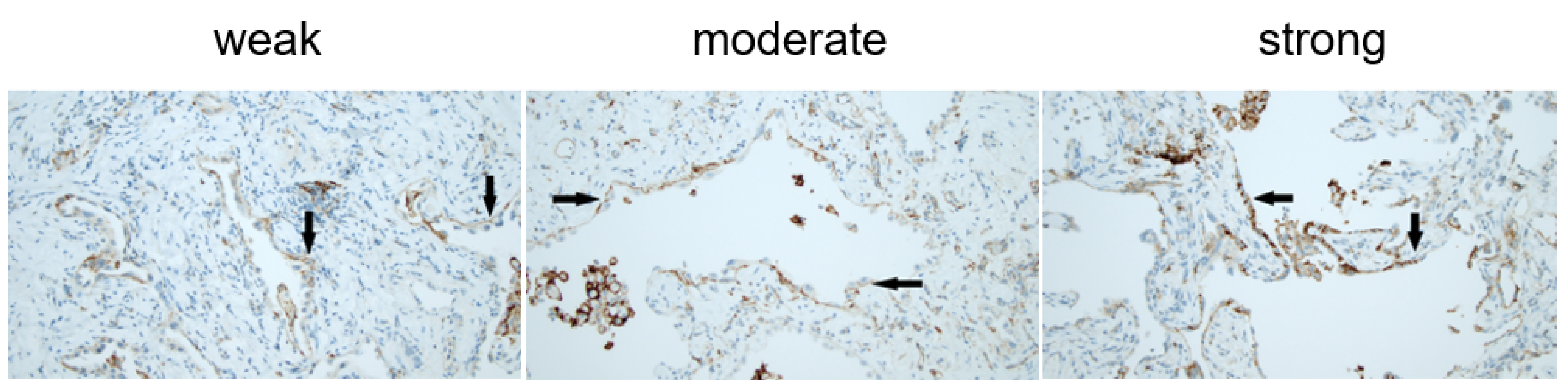
2.4. Pathology Method

All specimens were serially sectioned. The first section was stained with hematoxylin–eosin; the others were used for immunohistochemistry. The pathologic report followed previously published criteria [30]. A report for each sample was obtained including site, size of the specimen, characteristics for central versus peripheral sampling and the histopathologic pattern identified. The final diagnostic report was made including data for each sample. Evaluation of the PD-L1 protein expression was determined by use of the DAKO PD-L1 IHC 22C3 pharmDx kit (Agilent, Santa Clara, CA, USA). The slides were screened at low power, and the expression of membrane-bound PD-L1 (mPD-L1) in the epithelial cells present in fibrotic lung zones (excluding epithelial cells covering histologically normal bronchioles) was finally confirmed at high power (×40). The amount of positive mPD-L1 (mPD-L1pos) parenchymal epithelial cells (type II pneumocytes or bronchiolar cells) was graded using a semiquantitative approach: <25%, 25–49%, 50–74% and 75–100% of the epithelial alveolar and/ or bronchiolar cells. The intensity was graded in three categories (1 = weak, 2 = moderate, 3 = strong). The pathologist was blinded to the final diagnosis.

2.5. Statistics

Normally distributed data are presented as mean values ± standard deviation (SD), while data with a non-normal distribution are presented as median values with interquartile ranges (IQR) or total range. Data were checked for normality by histogram and the quantile–quantile plot. Continuous data were analyzed with the unpaired t-test or the Mann–Whitney test, as appropriate, according to their distribution. Categorical data are presented as proportions of the total population and analyzed with the chi2-test or Fisher’s exact test when the total number of one group was ≤5. A p-value of <0.05 was considered statistically significant. Stata IC (version 16.0, StataCorp, College Station, TX, USA) was used for statistical analysis.

3. Results

3.1. Demographics

Between September 2017 and May 2019, 102 patients were enrolled in the study. Four patients were excluded (three patients because biopsies were not obtained; one patient withdrew consent). IPF was diagnosed in 43 (44%) patients, and 55 (56%) patients were diagnosed with other ILDs than IPF (non-IPF), e.g., twenty with hypersensitivity pneumonitis (HP), seven with non-specific interstitial pneumonitis (NSIP), two with desquamative interstitial pneumonitis (DIP), one with eosinophilic pneumonia and twenty-five with other ILDs. Baseline characteristics are presented in Table 1. Higher age (p = 0.047) and traction bronchiectasis (p < 0.001) were identified more often in patients with IPF. Traction bronchiectasis was also found more frequently in mPD-L1pos IPF patients compared to the mPD-L1pos non-IPF patients (Table 2) (p = 0.003). When grouping the patients into mPD-L1pos and mPD-L1-negative (mPD-L1neg) (Table 3), baseline characteristics were not significantly different. TLco was significantly lower (p = 0.007) and GAP index 2 more frequent in the mPD-L1pos IPF group compared to the mPD-L1neg IPF group (see Table 4). When adjusting for age, gender and smoking status, the odds ratio for IPF when expressing mPD-L1 was 4.3 (CI: 1.3–14.3). The median number of days between first visit and the cryobiopsy procedure was 55 days (IQR: 27–82).

3.2. Specific Histological Lesions

The specific histological lesions identified are reported in Table 5. In the IPF group, 12 cases (28%) were mPD-L1pos in the epithelial alveolar and/or bronchiolar cells. In the non-IPF group, five cases were found (9%) (p = 0.015). Two of the non-IPF patients were diagnosed with HP, two with NSIP and one with DIP. The intensity of the mPD-L1 staining was weak (grade 1) in four of the patients and moderate (grade 2) in thirteen of the patients. In one patient, mPD-L1-positive cells were found in <25%, in six patients in 25–49%, in eight patients in 50–74% and in two patients in >75% of the cells (Figure 1). As expected, macrophages also expressed PD-L1.
When comparing the mPD-L1pos and mPD-L1neg patients, we found that fibroblast foci (p = 0.028) and a combination of patchy fibrosis and fibroblast foci p = 0.02) were more frequently detected in the mPD-L1pos patients. When comparing the IPF mPD-L1pos patients with the IPF mPD-L1neg patients, or the IPF mPD-L1pos with the non-IPF mPD-L1pos patients, we found no differences in histological elementary lesions. We found no association between honeycombing and expression of mPD-L1. No association was found either between lymphocytosis in BAL (lymphocytes > 15%) and PD-L1pos patients (p = 0.97) or between percentages of macrophages in BAL and PD-L1pos patients (p = 0.36), nor did we find an association between C-reactive protein levels and PD-L1pos patients (p = 0.66).

3.3. One-Year Follow-Up

During the one-year follow-up, four patients died (two IPF mPD-L1neg, one IPF mPD-L1pos and one non-IPF mPD-L1neg). Four patients withdrew from further participation in the study (one with IPF and three non-IPF). There was a small but significant difference in the change in TLco at the one-year follow-up between the patients with IPF (−1.5 ± 8) and the non-IPF patients (5.2 ± 12) (p = 0.003). There were no other significant differences in PFT or distance in 6MWT when comparing patients with IPF to non-IPF patients, mPD-L1pos patients to mPD-L1neg patients, mPD-L1pos IPF patients to mPD-L1neg IPF patients or PD-L1pos IPF to PD-L1pos non IPF patients (Table 6). Of the patients diagnosed with IPF, 40 (93%) started antifibrotic treatment (24 patients pirfenidone, 16 patients nintedanib). Four of the five mPD-L1pos non-IPF patients were treated with immunosuppressants (steroids and azathioprine).

4. Discussion

The present study describes the expression of mPD-L1 in alveolar and bronchiolar epithelial cells in a subgroup of patients with IPF. The expression of PD-L1 and PD-1 in cells of the immune system in patients with IPF has so far only been reported in a few studies [25,26]. Although we also found mPD-L1 in a few patients with other ILDs, mPD-L1 was found significantly more often in patients with IPF. Interestingly, IPF patients who were mPD-L1pos had lower TLco, and more were never-smokers compared to the mPD-L1neg IPF patients. TLco seemed to be lower, though not statistically significant, in the combined group of IPF and non-IPF mPD-L1pos patients compared to the mPDL1neg patients. Based on our data, it could be speculated that the expression of mPD-L1 may be associated with a peculiar subset of both IPF and non-IPF ILD patients, mainly a subset of IPF patients in whom fibroblast foci, or a combination of patchy fibrosis and fibroblast foci, are more densely represented compared to the mPD-L1neg patients. There was no association between honeycombing changes and expression of mPD-L1. This could be explained by the size of the biopsies or the site where the biopsies were taken, taking into account that areas with honeycombing changes visualized on HRCT were deliberately avoided. Alternatively, it could be explained by the fact that epithelial cells expressing mPDL-1 are in a context of epithelial mesenchymal transition, as suggested by Yamaguchi M. et al. [31], therefore in zones where clear honeycomb changes have not fully developed.
Traction bronchiectasis, as a radiologic marker of fibrotic changes, was found more frequently in patients with IPF compared to non-IPF patients and also more often in mPD-L1pos IPF compared to mPD-L1pos non-IPF patients (Table 1 and Table 2). However, no difference was found when comparing mPD-L1pos to mPD-L1neg patients or within the group of IPF patients. We did not investigate the extent or severity of the fibrotic changes on HRCT to differentiate between mild, moderate and severe disease, which might have revealed differences between the groups.
During the one-year follow-up of the participants, there was a significant difference, although small, in the change in TLco in patients with IPF compared to non-IPF patients. Based on our results, we were unable to demonstrate that the expression of mPD-L1 was associated with a worse prognosis in patients with IPF or non-IPF ILD. This might be explained by the short duration of the follow-up period of only one year and because the majority of the patients with IPF (93%) were treated with antifibrotics. At present, we do not know if the presence of mPD-L1 is a key step in a subset of patients with IPF or a marker associated with disease development or progression. Moreover, we do not know if the expression has a protective role in preventing further progression and fibrosis or if it leads to more fibrotic changes.
In recent years, the discussion of lumping and splitting patients with fibrotic ILDs and IPF has been frequently debated. Several studies have suggested that these diseases share common traits with respect to clinical course and treatment response to antifibrotics, probably due to different triggers resulting in the initiation of common fibrotic pathways [32,33,34,35]. When looking at fibrotic diseases from this perspective, the occurrence of mPD-L1 could be associated with fibrotic changes and not IPF as a disease entity. The lack of difference in baseline characteristics and changes in PFT and 6MWT at the one-year follow-up of patients with IPF and non-IPF mPD-L1pos patients might support this hypothesis, although traction bronchiectasis was found more frequently in the IPF mPD-L1pos patients. However, the number of patients in each group was low, and thus we cannot draw any conclusions. On the other side, the uncovering of pathological processes leading to disease development and progression might identify ILD subtypes based on different pathways. Splitting the diseases opens up the possibilities of targeted treatment and precision medicine, as has been seen in cancers.
Until now, IPF has been regarded as a risk factor for lung toxicity with respiratory deterioration and acute exacerbations when considering anti-PD-1 or anti-PD-L1 immunotherapy in non-small cell lung cancer. The incidence of any grade of pneumonitis was found to be 1.3–3.6% in a meta-analysis by Khunger et al. [36]; in a recent real-life study, the incidence was as high as 9.5% [37]. However, immunotherapy has reportedly been given to few patients with IPF and lung cancer without worsening their respiratory condition or showing any signs of lung toxicity [38,39,40]. Thus, it could be hypothesized that treatment with anti-PD-L1 or anti-PD-1 immunotherapy could be a promising future treatment in patients with IPF and mPD-L1 expression. One of the most feared adverse events of immunotherapy in IPF patients would be drug-induced pneumonitis. Adverse effects if treated with immunotherapy have to be anticipated and need to be handled similarly to patients with cancer [41].
Identification of a subgroup of patients in which expression of PD-L1 on membranes of epithelial cells in the injured lung areas could incite clinical trials with drugs already used to treat a variety of malignant tumors.
There are limited data regarding PD-L1 and PD-1 expression in non-cancer patients. However, a recent study found an increased expression of PD-L1 in renal cells in a mouse after certain types of injuries [42].
Our study has several limitations. First, the length of the follow-up time might have been too short to observe major disease progression. Most of the patients with IPF were diagnosed with well-preserved FVC and were treated with antifibrotics, which affects disease progression. Second, we were not able to perform a power calculation, and this may have affected the subgroup analysis in mPD-L1pos IPF and non- IPF patients.
Our study also has several strengths. Cryobiopsies were performed in all patients by three experienced bronchoscopists, ensuring a high biopsy quality, and all patients were diagnosed after MDT conferences. Additionally, the pathologist (L.B.M.) was blinded to the final diagnosis, routinely assesses PD-L1 expression in lung cancer and is familiar with this analysis using a semiquantitative approach. All subjects, apart from one patient, agreed to participate in the study, which minimized selection bias.

5. Conclusions

We demonstrated that the expression of mPD-L1 in epithelial cells in the alveoli and bronchiolar cells present in fibrotic areas was evident in a subgroup of patients with IPF. Larger studies with longer follow-up time are needed to explore the role of mPD-L1 in patients with IPF.

Author Contributions

Recruitment and provision of study materials or patients, S.K.-W.; collection and assembly of data, L.B.M. and S.K.-W.; data analysis and interpretation, E.B., V.P., L.B.M. and S.K.-W.; writing, E.B., V.P., L.B.M. and S.K.-W. All authors have read and agreed to the published version of the manuscript.

Funding

The study was funded by the Danish Lung Association.

Institutional Review Board Statement

The study protocol was approved by the Central Denmark Region Committee on Health Research Ethics (Identifier: 1-10-72-29-17) and the Danish Data Protection Agency (Identifier: 1-16-02-643-16). The trial is registered in clinicaltrials.gov (Identifier: NCT04187079).

Informed Consent Statement

Informed consent was obtained from all participants.

Data Availability Statement

All data used during the study are available on reasonable request.

Conflicts of Interest

E.B. has received honoraria for lectures and presentations from Boehringer Ingelheim, Roche, AstraZeneca and Galapagos; support for attending meetings and/or travel from Boehringer Ingelheim and Roche; and participation on a safety monitoring board or advisory board for Boehringer Ingelheim and Roche. V.P. has received honoraria for lectures and presentations from Boehringer Ingelheim, Roche, Ambu and Erbe; support for attending meetings and/or travel from Boehringer Ingelheim and Roche; and participation on a safety monitoring board or advisory board for Boehringer Ingelheim and Roche. L.B.M. has no conflict of interest. S.K.W. has received honoraria for lectures and presentations from Boehringer Ingelheim and Roche and support for attending meetings and/or travel from Boehringer Ingelheim and Roche.

References

  1. Brown, S.-A.W.; Dobelle, M.; Padilla, M.; Agovino, M.; Wisnivesky, J.P.; Hashim, D.; Boffetta, P. Idiopathic Pulmonary Fibrosis and Lung Cancer. A Systematic Review and Meta-analysis. Ann. Am. Thorac. Soc. 2019, 16, 1041–1051. [Google Scholar] [CrossRef]
  2. Jafarinezhad, A.; YektaKooshali, M.H. Lung cancer in idiopathic pulmonary fibrosis: A systematic review and meta-analysis. PLoS ONE 2018, 13, e0202360. [Google Scholar] [CrossRef] [PubMed]
  3. Tzouvelekis, A.; Gomatou, G.; Bouros, E.; Trigidou, R.; Tzilas, V.; Bouros, D. Common Pathogenic Mechanisms Between Idiopathic Pulmonary Fibrosis and Lung Cancer. Chest 2019, 156, 383–391. [Google Scholar] [CrossRef] [PubMed]
  4. Caliò, A.; Lever, V.; Rossi, A.; Gilioli, E.; Brunelli, M.; Dubini, A.; Tomassetti, S.; Piciucchi, S.; Nottegar, A.; Rossi, G.; et al. Increased frequency of bronchiolar histotypes in lung carcinomas associated with idiopathic pulmonary fibrosis. Histopathology 2017, 71, 725–735. [Google Scholar] [CrossRef] [PubMed]
  5. Hata, A.; Nakajima, T.; Matsusaka, K.; Fukuyo, M.; Nakayama, M.; Morimoto, J.; Ito, Y.; Yamamoto, T.; Sakairi, Y.; Rahmutulla, B.; et al. Genetic alterations in squamous cell lung cancer associated with idiopathic pulmonary fibrosis. Int. J. Cancer 2021, 148, 3008–3018. [Google Scholar] [CrossRef] [PubMed]
  6. Taskar, V.; Coultas, D. Exposures and idiopathic lung disease. Semin. Respir. Crit. Care Med. 2008, 29, 670–679. [Google Scholar] [CrossRef]
  7. Taskar, V.S.; Coultas, D.B. Is idiopathic pulmonary fibrosis an environmental disease? Proc. Am. Thorac. Soc. 2006, 3, 293–298. [Google Scholar] [CrossRef]
  8. Spira, A.; Beane-Ebel, J.; Shah, V.; Liu, G.; Schembri, F.; Yang, X.; Palma, J.; Brody, J.S. Effects of cigarette smoke on the human airway epithelial cell transcriptome. Proc. Natl. Acad. Sci. USA 2004, 101, 10143–10148. [Google Scholar] [CrossRef] [Green Version]
  9. Carloni, A.; Poletti, V.; Fermo, L.; Bellomo, N.; Chilosi, M. Heterogeneous distribution of mechanical stress in human lung: A mathematical approach to evaluate abnormal remodeling in IPF. J. Theor. Biol. 2013, 332, 136–140. [Google Scholar] [CrossRef] [PubMed]
  10. Yang, I.V.; Fingerlin, T.E.; Evans, C.M.; Schwarz, M.I.; Schwartz, D.A. MUC5B and Idiopathic Pulmonary Fibrosis. Ann. Am. Thorac. Soc. 2015, 12 (Suppl. 2), S193–S199. [Google Scholar]
  11. Moore, C.; Blumhagen, R.Z.; Yang, I.V.; Walts, A.; Powers, J.; Walker, T.; Bishop, M.; Russell, P.; Vestal, B.; Cardwell, J.; et al. Resequencing Study Confirms That Host Defense and Cell Senescence Gene Variants Contribute to the Risk of Idiopathic Pulmonary Fibrosis. Am. J. Respir. Crit. Care Med. 2019, 200, 199–208. [Google Scholar] [CrossRef]
  12. Kaur, A.; Mathai, S.K.; Schwartz, D.A. Genetics in Idiopathic Pulmonary Fibrosis Pathogenesis, Prognosis, and Treatment. Front. Med. 2017, 4, 154. [Google Scholar] [CrossRef]
  13. Renzoni, E.; Poletti, V.; Mackintosh, J.A. Disease pathology in fibrotic interstitial lung disease:is it all about usual interstitial pneumonia? Lancet 2021, 398, 1437–1449. [Google Scholar] [CrossRef]
  14. Chilosi, M.; Poletti, V.; Zamò, A.; Lestani, M.; Montagna, L.; Piccoli, P. Aberrant Wnt/beta-catenin pathway activation in idiopathic pulmonary fibrosis. Am. J. Pathol. 2003, 162, 1495–1502. [Google Scholar] [CrossRef]
  15. Chilosi, M.; Carloni, A.; Rossi, A.; Poletti, V. Premature lung aging and cellular senescence in the pathogenesis of idiopathic pulmonary fibrosis and COPD/emphysema. Transl. Res. 2013, 162, 156–173. [Google Scholar] [CrossRef] [PubMed]
  16. Chilosi, M.; Zamò, A.; Doglioni, C.; Reghellin, D.; Lestani, M.; Montagna, L.; Pedron, S.; Ennas, M.G.; Cancellieri, A.; Murer, B.; et al. Migratory marker expression in fibroblast foci of idiopathic pulmonary fibrosis. Respir. Res. 2006, 7, 95. [Google Scholar] [CrossRef] [Green Version]
  17. Nie, R.-C.; Zhao, C.-B.; Xia, X.-W.; Luo, Y.-S.; Wu, T.; Zhou, Z.-W.; Yuan, S.-Q.; Wang, Y.; Li, Y.-F. The Efficacy and Safety of PD-1/PD-L1 Inhibitors in Combination with Conventional Therapies for Advanced Solid Tumors: A Meta-Analysis. BioMed Res. Int. 2020, 2020, 1–10. [Google Scholar] [CrossRef] [PubMed]
  18. Okazaki, T.; Honjo, T. PD-1 and PD-1 ligands: From discovery to clinical application. Int. Immunol. 2007, 19, 813–824. [Google Scholar] [CrossRef] [PubMed] [Green Version]
  19. Sznol, M.; Chen, L. Antagonist antibodies to PD-1 and B7-H1 (PD-L1) in the treatment of advanced human cancer. Clin. Cancer Res. 2013, 19, 1021–1034. [Google Scholar] [CrossRef] [Green Version]
  20. Larsen, T.V.; Hussmann, D.; Nielsen, A.L. PD-L1 and PD-L2 expression correlated genes in non-small-cell lung cancer. Cancer Commun. 2019, 39, 30. [Google Scholar] [CrossRef] [PubMed] [Green Version]
  21. Yearley, J.H.; Gibson, C.; Yu, N.; Moon, C.; Murphy, E.; Juco, J. PD-L2 Expression in Human Tumors: Relevance to Anti-PD-1 Therapy in Cancer. Clin. Cancer Res. 2017, 23, 3158–3167. [Google Scholar] [CrossRef] [PubMed] [Green Version]
  22. Kalluri, R.; Weinberg, R.A. The basics of epithelial-mesenchymal transition. J. Clin. Investig. 2009, 119, 1420–1428. [Google Scholar] [CrossRef] [PubMed] [Green Version]
  23. Asgarova, A.; Asgarov, K.; Godet, Y.; Peixoto, P.; Nadaradjane, A.; Boyer-Guittaut, M. PD-L1 expression is regulated by both DNA methylation and NF-kB during EMT signaling in non-small cell lung carcinoma. Oncoimmunology 2021, 7, e1423170. [Google Scholar] [CrossRef] [Green Version]
  24. Salton, F.; Volpe, M.; Confalonieri, M. Epithelial–Mesenchymal Transition in the Pathogenesis of Idiopathic Pulmonary Fibrosis. Medicina 2019, 55, 83. [Google Scholar] [CrossRef] [PubMed] [Green Version]
  25. Jovanovic, D.; Milenkovic, M.R.; Stevuljevic, J.K.; Markovic, J.; Ceriman, V.; Kontic, M.; Trifunovic, V.S. Membrane PD-L1 expression and soluble PD-L1 plasma levels in idiopathic pulmonary fibrosis—a pilot study. J. Thorac. Dis. 2018, 10, 6660–6669. [Google Scholar] [CrossRef] [Green Version]
  26. Celada, L.J.; Kropski, J.A.; Herazo-Maya, J.D.; Luo, W.; Creecy, A.; Abad, A.T.; Chioma, O.S.; Lee, G.; Hassell, N.E.; Shaginurova, G.I.; et al. PD-1 up-regulation on CD4+ T cells promotes pulmonary fibrosis through STAT3-mediated IL-17A and TGF-β1 production. Sci. Transl. Med. 2018, 10. [Google Scholar] [CrossRef] [PubMed] [Green Version]
  27. Ni, K.; Liu, M.; Zheng, J.; Wen, L.; Chen, Q.; Xiang, Z.; Lam, K.-T.; Liu, Y.; Chan, G.C.-F.; Lau, Y.-L.; et al. PD-1/PD-L1 Pathway Mediates the Alleviation of Pulmonary Fibrosis by Human Mesenchymal Stem Cells in Humanized Mice. Am. J. Respir. Cell Mol. Biol. 2018, 58, 684–695. [Google Scholar] [CrossRef] [PubMed]
  28. Stanojevic, S.; Graham, B.L.; Cooper, B.; Thompson, B.R.; Carter, K.W.; Francis, R.W.; Hall, G. Official ERS technical standards: Global Lung Function Initiative reference values for the carbon monoxide transfer factor for Caucasians. Eur. Respir. J. 2017, 50, 1700010. [Google Scholar] [CrossRef]
  29. Kronborg-White, S.; Sritharan, S.S.; Madsen, L.B.; Folkersen, B.; Voldby, N.; Poletti, V.; Rasmussen, T.R.; Bendstrup, E. Integration of cryobiopsies for interstitial lung disease diagnosis is a valid and safe diagnostic strategy—Experiences based on 250 biopsy procedures. J. Thorac. Dis. 2021, 13, 1455–1465. [Google Scholar] [CrossRef] [PubMed]
  30. Ravaglia, C.; Rossi, G.; Tomassetti, S.; Dubini, A.; Piciucchi, S.; Chilosi, M. Report Standardization in Transbronchial Lung Cryobiopsy. Arch. Pathol. Lab. Med. 2019, 143, 416–417. [Google Scholar] [CrossRef] [Green Version]
  31. Yamaguchi, M.; Hirai, S.; Tanaka, Y.; Sumi, T.; Miyajima, M.; Mishina, T.; Yamada, G.; Otsuka, M.; Hasegawa, T.; Kojima, T.; et al. Fibroblastic foci, covered with alveolar epithelia exhibiting epithelial–mesenchymal transition, destroy alveolar septa by disrupting blood flow in idiopathic pulmonary fibrosis. Lab. Investig. 2016, 97, 232–242. [Google Scholar] [CrossRef] [PubMed] [Green Version]
  32. Flaherty, K.R.; Wells, A.U.; Cottin, V.; Devaraj, A.; Walsh, S.L.; Inoue, Y.; Richeldi, L.; Kolb, M.; Tetzlaff, K.; Stowasser, S.; et al. Nintedanib in Progressive Fibrosing Interstitial Lung Diseases. N. Engl. J. Med. 2019, 381, 1718–1727. [Google Scholar] [CrossRef] [Green Version]
  33. Distler, O.; Highland, K.B.; Gahlemann, M.; Azuma, A.; Fischer, A.; Mayes, M.D.; Raghu, G.; Sauter, W.; Girard, M.; Alves, M.; et al. Nintedanib for Systemic Sclerosis—Associated Interstitial Lung Disease. N. Engl. J. Med. 2019, 380, 2518–2528. [Google Scholar] [CrossRef] [PubMed]
  34. Behr, J.; Neuser, P.; Prasse, A.; Kreuter, M.; Rabe, K.; Schade-Brittinger, C.; Wagner, J.; Günther, A. Exploring efficacy and safety of oral Pirfenidone for progressive, non-IPF lung fibrosis (RELIEF)—A randomized, double-blind, placebo-controlled, parallel group, multi-center, phase II trial. BMC Pulm. Med. 2017, 17, 122. [Google Scholar] [CrossRef] [Green Version]
  35. Maher, T.M.; Corte, T.J.; Fischer, A.; Kreuter, M.; Lederer, D.J.; Molina-Molina, M.; Axmann, J.; Kirchgaessler, K.-U.; Samara, K.; Gilberg, F.; et al. Pirfenidone in patients with unclassifiable progressive fibrosing interstitial lung disease: A double-blind, randomised, placebo-controlled, phase 2 trial. Lancet Respir. Med. 2020, 8, 147–157. [Google Scholar] [CrossRef]
  36. Khunger, M.; Rakshit, S.; Pasupuleti, V.; Hernandez, A.V.; Mazzone, P.; Stevenson, J. Incidence of Pneumonitis with Use of Programmed Death 1 and Programmed Death-Ligand 1 Inhibitors in Non-Small Cell Lung Cancer: A systematic review and meta-analysis of trials. Chest 2017, 152, 271–281. [Google Scholar] [CrossRef] [PubMed] [Green Version]
  37. Atchley, W.T.; Alvarez, C.; Saxena-Beem, S.; Schwartz, T.A.; Ishizawar, R.C.; Patel, K.P. Immune Checkpoint Inhibitor-Related Pneumonitis in Lung Cancer: Real-World Incidence, Risk Factors, and Management Practices Across Six Health-Care Centers in North Carolina. Chest 2021, 160, 731–742. [Google Scholar] [CrossRef]
  38. Duchemann, B.; Didier, M.; Pailler, M.-C.; Brillet, P.-Y.; Kambouchner, M.; Uzunhan, Y. Can nivolumab be used safely in idiopathic pulmonary fibrosis? Rev. Mal. Respir. 2019, 36, 209–213. [Google Scholar] [CrossRef] [PubMed]
  39. Ide, M.; Tanaka, K.; Sunami, S.; Asoh, T.; Maeyama, T.; Tsuruta, N.; Nakanishi, Y.; Okamoto, I. Durable response to nivolumab in a lung adenocarcinoma patient with idiopathic pulmonary fibrosis. Thorac. Cancer 2018, 9, 1519–1521. [Google Scholar] [CrossRef]
  40. Fujimoto, D.; Yomota, M.; Sekine, A.; Morita, M.; Morimoto, T.; Hosomi, Y.; Ogura, T.; Tomioka, H.; Tomii, K. Nivolumab for advanced non-small cell lung cancer patients with mild idiopathic interstitial pneumonia: A multicenter, open-label single-arm phase II trial. Lung Cancer 2019, 134, 274–278. [Google Scholar] [CrossRef] [PubMed]
  41. Delaunay, M.; Prévot, G.; Collot, S.; Guilleminault, L.; Didier, A.; Mazières, J. Management of pulmonary toxicity associated with immune checkpoint inhibitors. Eur. Respir. Rev. 2019, 28, 190012. [Google Scholar] [CrossRef] [PubMed]
  42. Hakroush, S.; Kopp, S.B.; Tampe, D.; Gersmann, A.-K.; Korsten, P.; Zeisberg, M.; Tampe, B. Variable Expression of Programmed Cell Death Protein 1-Ligand 1 in Kidneys Independent of Immune Checkpoint Inhibition. Front. Immunol. 2021, 11, 3546. [Google Scholar] [CrossRef] [PubMed]
Figure 1. Membranous PD-L1 expression in parenchymal epithelial cells. PD-L1 expression in epithelial cells (arrows). Macrophages are positive and represent an internal control. Medium power.
Jcm 10 05562 g001
Table 1. Demographic characteristics of patients with IPF and without IPF.
IPF Patients
43 (44%)
Non-IPF Patients
55 (56%)
Age (median, range)73 (48–79) *70 (40–78) *
Gender (male) 26 (60%)34 (62%)
Smoking %
-
Never
3536
-
Ex
5651
-
Smoker
913
Packyears (SD)30.21 ± 1925.24 ± 18
FVC % of predicted (SD)93.60 ± 1992.73 ± 21
TLco % of predicted (median, IQR)60 (49–70)55 (49–66)
6MWT distance (m) (SD)466 ± 96491 ± 99
Desaturation ≥ 4 % during 6MWT (SD)35 (83%) **35 (71%) **
Traction bronchiectasis 41 (95%) *35 (64%) *
GAP index
-
1
34 (79%)46 (84%)
-
2
9 (21%)9 (16%)
IPF: idiopathic pulmonary fibrosis; FVC: forced vital capacity; SD: standard deviation; TLco: lung transfer factor for carbon monoxide; IQR: interquartile range; 6MWT: six-minute walk test; m: meters; GAP: gender–age–physiology. * Significantly different (p < 0.05). ** Forty-two IPF patients and forty-nine non-IPF patients performed 6MWT.
Table 2. Demographic characteristics of patients with PD-L1-positive membrane staining with IPF and without IPF.
IPF mPD-L1pos
n = 12
Non-IPF mPD-L1pos
n = 5
Age (median, range)76 (67–77)72 (64–75)
Gender (male)6 (50%)3 (60%)
Smoking %
-
Never
5840
-
Ex
3340
-
Smoker
820
Packyears (SD)31 ± 1537.67 ± 27
FVC % of predicted (SD)92.31 ± 1999.83 ± 11
TLco% of predicted (median, IQR)51 (46–61)53 (50–61)
6MWT distance (m) (SD)470 ± 86506 ± 19
Desaturation ≥ 4 % during 6MWT 9 (75%)3 (60%)
Traction bronchiectasis 12 (100%) *2 (40%) *
GAP index
-
1
7 (58%)5 (100%)
-
2
5 (42%)0
IPF: idiopathic pulmonary fibrosis; mPD-L1pos: patients positive for membrane-bound programmed death-ligand one; mPD-L1neg: patients negative for membrane-bound programmed death-ligand one; FVC: forced vital capacity; SD: standard deviation; TLco: lung transfer factor for carbon monoxide; IQR: interquartile range; 6MWT: six-minute walk test; m: meters; GAP: gender–age–physiology. * Significantly different (p < 0.05).
Table 3. Demographic characteristics of patients (IPF and non-IPF) with PD-L1-positive and PD-L1-negative membrane staining.
mPD-L1pos
n = 17
mPD-L1neg
n = 81
Age (median, range)68 (48–79)68 (40–79)
Gender (male)9 (53%)51 (63%)
Smoking %
-
Never
5332
-
Ex
3557
-
Smoker
1211
Packyears (SD)33.5 ± 1826.53 ± 21
FVC, % of predicted (SD)93.29 ± 1790.52 ± 20
TLco, % of predicted (median, IQR)51 (47–61)60 (49–68)
6MWT distance (m) (SD)481 ± 74479 ± 103
Desaturation ≥ 4 % during 6MWT12 (71%) **58 (82%) **
Traction bronchiectasis 14 (82%)62 (77%)
GAP index
-
1
12 (71%)68 (84%)
-
2
5 (29%)13 (16%)
mPD-L1pos: patients positive for membrane-bound programmed death-ligand one; mPD-L1neg: patients negative for membrane-bound programmed death-ligand one; FVC: forced vital capacity; SD: standard deviation; TLco: lung transfer factor for carbon monoxide; IQR: interquartile range; 6MWT: six-minute walk test; m: meters; GAP: gender–age–physiology. ** All mPD-L1pos patients and 74 mPD-L1neg patients performed 6MWT.
Table 4. Demographic characteristics of patients with IPF and PD-L1-positive and PD-L1-negative membrane staining.
IPF mPD-L1pos
n = 12
IPF mPD-L1neg
n = 31
Age (median, range)76 (67–77)72 (64–75)
Gender (male)6 (50%)20 (64%)
Smoking %
-
Never
5826
-
Ex
3364
-
Smoker
810
Packyears31 ± 1530.03 ± 21
FVC, % of predicted (SD)91.33 ± 1894.03 ± 22
TLco, % of predicted (median, IQR)51 (46–61) *63 (55–72) *
6MWT distance (m) (SD)470 ± 86464 ± 101
Desaturation ≥ 4 % during 6MWT 9 (75%) **26 (87%) **
Traction bronchiectasis 12 (100%)29 (94%)
GAP index
-
1
7 (58%) *27 (87%) *
-
2
5 (42%) *4 (13%) *
IPF: idiopathic pulmonary fibrosis; mPD-L1pos: patients positive for membrane-bound programmed death-ligand one; mPD-L1neg: patients negative for membrane-bound programmed death-ligand one; FVC: forced vital capacity; SD: standard deviation; TLco: lung transfer factor for carbon monoxide; IQR: interquartile range; 6MWT: six-minute walk test; m: meters; GAP: gender–age–physiology. * Significantly different (p < 0.05). ** All mPD-L1pos and 30 of the mPD-L1neg IPF patients performed 6MWT.
Table 5. Histological lesions.
IPF Patients
n = 43
Non- IPF Patients
n = 55
p-Value
mPD-L112 (28%)5 (9%)0.015
gPD-L133 (76%)48 (89%)0.172
Fibroblast foci17 (40%)13 (24%)0.09
Honeycombing9 (21%)2 (4%)0.007
Patchy fibrosis41 (95%)43 (78%)0.016
Patchy fibrosis + fibroblast foci17 (40%)12 (22%)0.057
mPD-L1pos
n = 17
mPD-L1neg
n = 81
p-Value
Fibroblast foci9 (53%)21 (26%)0.028
Honeycombing2 (12%)9 (11%)0.938
Patchy fibrosis17 (100%)67 (83%)0.064
Patchy fibrosis + fibroblast foci9 (53%)20 (25%)0.02
IPF mPD-L1pos
n = 12
Non-IPF mPD-L1pos
n = 5
p-Value
Fibroblast foci5 (42%)4 (80%)0.149
Honeycombing2 (17%)00.331
Patchy fibrosis12 (100%)5 (100%)
Patchy fibrosis + fibroblast foci5 (42%)4 (80%)0.149
IPF mPD-L1pos
n = 12
IPF mPD-L1neg
n = 31
p-Value
Fibroblast foci5 (42%)12 (39%)0.859
Honeycombing2 (17%)7 (23%)0.669
Patchy fibrosis12 (100%)29 (94%)0.368
Patchy fibrosis + fibroblast foci5 (42%)12 (39%)0.859
IPF: idiopathic pulmonary fibrosis; mPD-L1: membrane-bound programmed death-ligand one; gPD-L1: granular-bound programmed death-ligand one; mPD-L1pos: patients positive for membrane-bound programmed death-ligand one; mPD-L1neg: patients negative for membrane-bound programmed death-ligand one.
Table 6. Changes over one year.
IPF
n = 41
Non-IPF
n = 50
p-Value
Change FVC % (SD)−3.3 ± 10−1.64 ± 130.49
Change TLco % (SD)−1.5 ± 85.2 ± 120.003
Change 6MWTD (m) median (IQR)−12 (−42–13)−25 (−59–36)0.14
mPD-L1pos
n = 15
mPD-L1neg
n = 76
p-Value
Change FVC % (SD)−1.3 ± 11−2.6 ± 120.68
Change TLco % (SD)0.6 ± 92.5 ± 110.53
Change 6MWTD (m), median (IQR)−42 (−97–16)−15 (−50–29)0.9
IPF mPD-L1pos
n = 11
IPF mPD-L1neg
n = 30
p-Value
Change FVC % (SD)−2 ± 8 −3.8 ± 110.61
Change TLco %(SD)−1 ± 7−1.6 ± 80.83
Change 6MWTD (m), median (IQR)−4 (−97–4)−13 (−35–19)0.86
IPF mPD-L1pos
n = 11
Non- IPF mPD-L1pos
n = 4
p-Value
Change FVC % (SD)−2 ± 8 0.75 ± 180.34
Change TLco % (SD)−1 ± 75 ± 130.13
Change 6MWTD (m), median (IQR) −4 (−97–4)1 (−121–41)0.73
IPF: idiopathic pulmonary fibrosis; FVC: forced vital capacity; TLco: lung transfer factor for carbon monoxide; 6MWTD: six-minute walk test distance; IQR: interquartile range; mPD-L1pos: patients positive for membrane-bound programmed death-ligand one; mPD-L1neg: patients negative for membrane-bound programmed death-ligand one.
Publisher’s Note: MDPI stays neutral with regard to jurisdictional claims in published maps and institutional affiliations.

