


J. Clin. Med. 2026, 15(7), 2514; https://doi.org/10.3390/jcm15072514 (registering DOI) - 25 Mar 2026
Abstract
Background/Objectives: Abnormal muscle tone and impaired motor control commonly limit gait recovery after stroke. Robot-assisted gait training has been introduced to augment conventional rehabilitation; however, its effects on stage-based motor recovery, functional ambulation, and muscle tone during the subacute phase remain unclear. Methods:
[...] Read more.
Background/Objectives: Abnormal muscle tone and impaired motor control commonly limit gait recovery after stroke. Robot-assisted gait training has been introduced to augment conventional rehabilitation; however, its effects on stage-based motor recovery, functional ambulation, and muscle tone during the subacute phase remain unclear. Methods: This prospective, single-center, randomized controlled trial enrolled 30 patients with subacute stroke who received robot-assisted gait training plus conventional rehabilitation (R-BoT Plus group, n = 15) or conventional rehabilitation alone (control group, n = 15) over 4 weeks. The primary outcome was the change in Brunnstrom recovery stage of the lower extremities (BRS-LE). Secondary outcomes included Functional Ambulation Category (FAC), Fugl–Meyer Assessment for the Lower Extremity (FMA-LE), clinical spasticity measures (Modified Ashworth Scale and Modified Tardieu Scale), and muscle mechanical properties (MyotonPRO). Exploratory analyses were conducted to examine the associations between changes in stage-based motor recovery (ΔBRS-LE), functional ambulation (ΔFAC), and MyotonPRO parameters. Within-group changes were assessed using the Wilcoxon signed-rank test. Between-group effects were primarily evaluated using baseline-adjusted ANCOVA with HC3 robust standard errors, with Wilcoxon rank-sum tests on change scores as sensitivity analyses. Associations between changes in clinical outcomes and MyotonPRO parameters were evaluated using Spearman’s rank correlation coefficient (ρ). Results: BRS-LE (p = 0.014) and functional ambulation (p = 0.041) were significantly improved in the R-BoT Plus group. Changes in FMA-LE and clinical spasticity measures did not differ significantly between groups. Quantitative myotonometry revealed selective muscle- and parameter-specific changes. No robust correlations were observed between MyotonPRO parameters and changes in BRS-LE. Conclusions: The addition of robot-assisted gait training to conventional rehabilitation was associated with greater improvements in stage-based lower-limb motor recovery and functional ambulation in patients with subacute stroke. In contrast, cumulative impairment scores and conventional clinical spasticity measures demonstrated limited changes between groups. Quantitative muscle mechanical assessment revealed selective muscle-specific adaptations, supporting its role as a complementary tool for mechanistic characterization rather than as a surrogate marker of motor recovery. Future studies incorporating dose-matched designs and longer follow-up periods are warranted to clarify the independent and long-term effects of robot-assisted gait training.
Full article
(This article belongs to the Special Issue Advancements in Gait Rehabilitation: Innovative Approaches and Clinical Insights)
►
Show Figures
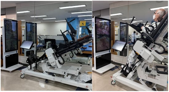
Figure 1


