2.1. Effects of cadmium in the growth, morphology and ultrastructure of Trichoderma harzianum
With the aim of selecting a standard medium for the physiological and biochemical experiments an initial study related to different growth media was performed.
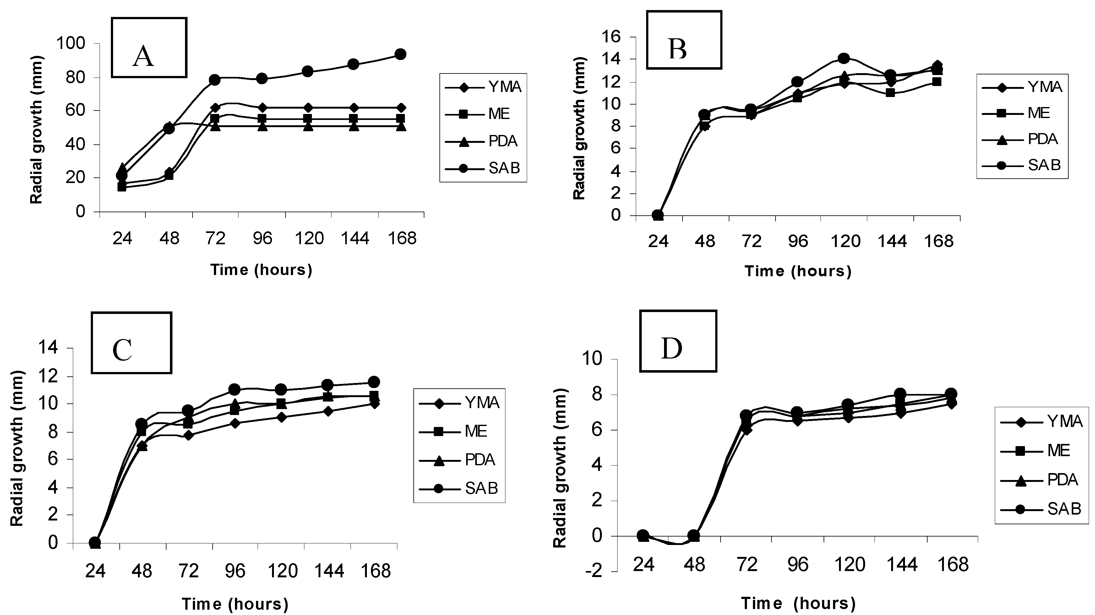
Figure 1A-D shows results for the radial growth obtained for the cultivation of
Trichoderma harzianum in Yeast Mold Agar, Malt Extract, Sabouraud and Potato Dextrose Agar media, in absence and presence of cadmium, at concentrations of 1, 2 and 3 mM, respectively.
From an analysis of graphics one can infer that the cultivation in the absence of metal in different media, results in different growth profiles, estimated by colony radial expansion. However, comparing the different media, higher radial growth was noted for cultivation in the Sabouraud medium. This study evaluated the percentage of relative growth of the organism for each test condition in relation to control. The results presented reveal the influence of cadmium on the radial growth of colonies of Trichoderma harzianum. The cultivation in different media resulted in variations of colony radial expansion, and revealed the inhibitory effect of the metal, which is directly related to its concentration.
Figure 1.
Radial growth of Trichoderma harzianum in different culture media, aiming to select a standard medium for the physiological and biochemical investigations. A- Control ; B- Treated with cadmium 1 mM; C- Treated with cadmium 2mM; D- Treated with cadmium 3mM. YMA (Yeast Malt Agar); ME (Malt Extract); PDA (Potato Dextrose Agar) and SAB (Sabouraud Dextrose Agar).
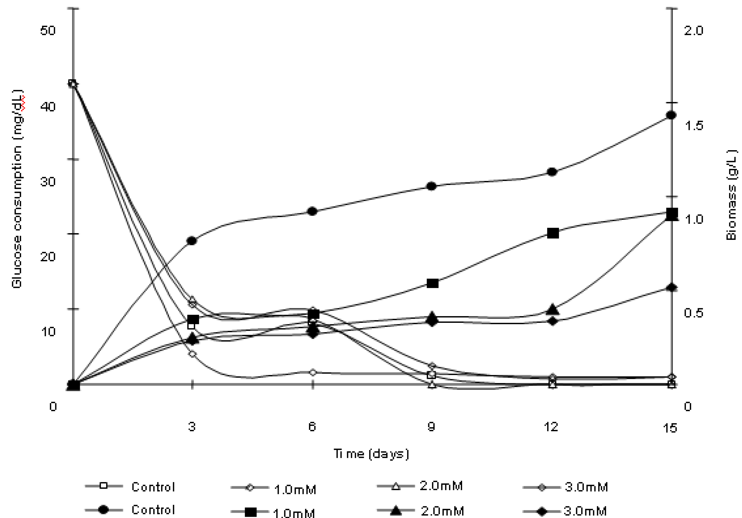
Figure 2 shows the results of the biomass production of
Trichoderma harzianum cultures in the presence and absence of cadmium. The presence of the metal induces a significant reduction of growth, as determined by the cellular biomass, in relation to control culture. A decrease in the biomass production proportional to the concentration of metal used was verified. It was observed that after 3 days a growth of biomass in control and treated samples occurred. Notwithstanding, treatment induced a reduction in biomass production, which amounted to 46.31%, 32.62% and 29.99% of the control culture production for concentrations of 1, 2 and 3 mM, respectively. At the end of the trial period for the culture treated with 1mM of cadmium, biomass obtained corresponded to 64.89% of control culture. For the treatments with 2 mM and 3 mM cadmium, the biomass corresponded to 63% and 36.83% of the mycelia mass obtained for control culture, respectively.
Figure 2.
Trichoderma harzianum biomass production and glucose consumption in response to cadmium exposure.
Figure 2.
Trichoderma harzianum biomass production and glucose consumption in response to cadmium exposure.
The behavior of the hyphae morphology, control and treated with cadmium, was evaluated through optical microscopy. The results are shown in
Figure 3A-D.
Figure 3.
Morphological behavior of Trichoderma harzianum. A. Control culture; B. Culture treated with 1 mM of cadmium, C. Culture treated with 2 mM of cadmium and D. Culture treated with 3 mM of cadmium. 400×.
Figure 3.
Morphological behavior of Trichoderma harzianum. A. Control culture; B. Culture treated with 1 mM of cadmium, C. Culture treated with 2 mM of cadmium and D. Culture treated with 3 mM of cadmium. 400×.
In control culture, the presence of chambered hyphae, with hyaline thin walls, is seen. Of note is the homogeneously stained and dense cytoplasm (
Figure 3A). Furthermore, samples treated with cadmium showed smaller radial expansion, proportional to the concentration of metal in the culture medium. The cultivation in the presence of cadmium also prompted the emergence of intense septation and branching in hyphae. Samples submitted to the highest concentration of cadmium, showed the largest branch and septation of hyphae.
In parallel, it is possible to perceive that the cytoplasm of hyphae grown in the presence of cadmium seems to be heterogeneous and granular in appearance. The cultivation in medium containing the heavy metal induced a most intense cytoplasmic granulation (
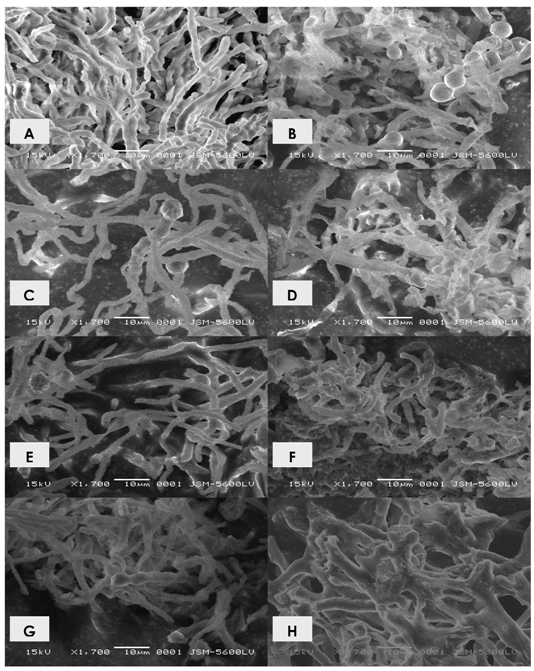
Figure 3B, 3C and 3D, 1 mM, 2 mM and 3 mM of cadmium, respectively). Through the ultrastructural study it was possible to verify the emergence of variations in the fine structure of the organism submitted to cultivation in the presence of cadmium. The results are shown in
Figure 4A-F.
Figure 4.
Electronmicrographs of Trichoderma harzianum. A. Control sample 3 days; B. Control sample 15 days; C. Sample grown in 1 mM of cadmium 3 days; D. Sample grown in 1 mM of cadmium in 15 days; E. Sample grown in 2 mM of cadmium 3 days; F. Sample grown in 2 mM of cadmium 15 days; G. Sample grown in 3 mM of cadmium 3 days; H. Sample grown in 3mM of cadmium in 15 days. 1.700×.
For the control samples with 3-days of cultivation it was verified the presence of abundant and homogeneous mycelium, with elongated hyphae, chambered, in the form of sticks (
Figure 4A). For samples with 15 days of cultivation, many reproductive structures were observed. The mycelium exhibited low electron density (
Figure 4B). Moreover, samples treated with 1 mM of cadmium, 3 days showed more scarce mycelium, twisted, intense branching, increased electron density; shortened hyphae were also viewed (
Figure 4C-D). Similar fine structure was observed in the samples treated with 2 mM of cadmium. However, higher electron density, patterns of branching and shortening of hyphae were observed for samples treated with 1 mM (
Figure 4E-F). For samples grown in the presence of 3 mM,
Figure 4G-H, the intensity of the changes mentioned were greater. Additionally, hyphae with 15 days of cultivation had become more twisted and thick that those treated with 1 mM and 2 mm of cadmium. An intense change in the branching pattern of hyphae was noted. The observed changes were directly related to the concentration of metal.
2.2. Polyphosphate behavior of Trichoderma harzianum in the presence of cadmium
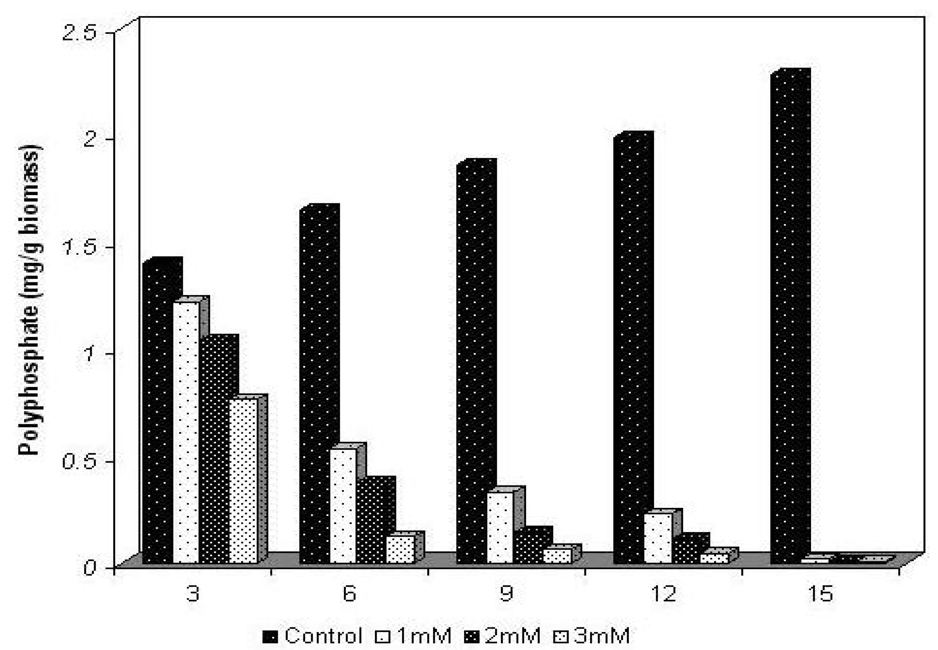
The physiological behavior for the content of polyphosphate of
Trichoderma harzianum, cultivated in the absence and presence of 1, 2 and 3 mM concentrations of cadmium, during intervals of 3, 6, 9, 12 and 15 days is shown in
Figure 5.
For the control sample, in the absence of the metal, a progressive increase in the cellular polyphosphate content occurred. Values corresponding to 1.4 mg/g biomass, 1.65 mg/g biomass, 1.86 mg/g biomass, 1.987 mg/g biomass and 2.28 mg/g biomass were obtained during the experimental period. By the end of the experiment an increase of approximately 81% in the content of polyphosphate was reached, resulting in the ability of synthesis and accumulation of polyphosphate by the mycelium of isolate of Trichoderma harzianum tested.
Figure 5.
The physiological behavior of Trichoderma harzianum polyphosphate, cultivated in the absence and presence of cadmium.
Figure 5.
The physiological behavior of Trichoderma harzianum polyphosphate, cultivated in the absence and presence of cadmium.
On the other hand, in cultures treated with 1 mM, 2 mM and 3 mM of cadmium, reductions of 13%, 25% and 45% respectively, in the content of polyphosphate, were observed within the first 3 days of cultivation in relation to control culture, although an increase in biomass production had occurred. At the end of the trial period the content of total cellular polyphosphate exhibited a reduction of approximately 99% for all cultures grown in the presence of the metal.
2.5. Discussion
The application of biomaterials for metal removal has attracted the attention of numerous researchers. Bacteria, fungi and algae, as well as products derived from such organisms, have the potential to remove many chemicals [
1,
4,
16,
22,
23].
The removal of heavy metals or their stabilization is the first steps towards detoxification of contaminated environments. Thus, the remediation of environments contaminated with heavy metals is a complex problem and has attracted the attention of many researchers and industries. Conventional physical-chemical methods to remove heavy metals in several environments include chemical reduction, electro-chemical treatment, ion exchange, and precipitation and evaporation recovery. However, such processes exhibit significant disadvantages, such as incomplete removal, high energy and reagent consumptions, besides generation of other toxic products the high cost of recovering them [
24,
25,
26].
Among the procedures used by microorganisms for metal detoxification can be cited: precipitation as phosphates, carbonates and sulfides; volatilization by methyl groups, physical exclusion by electronegative components in membranes and polymeric extracellular substances, efflux systems dependent on energy and intracellular sequestration by cysteine-rich protein of low molecular weight [
1,
2,
4]. In general, the resistance includes a variety of strategies to deal with toxic concentrations of metals in the environment [
7,
27]; such strategies exist to prevent the entry of the metal into the cell or to actively pump out the metal from the cell [
1,
4,
5].
Morphological changes as well as variations in cellular growth patterns of eukaryotes and prokaryotes are cited as effects induced by contact with cadmium. The intensity of responses is related to the time of contact and the metal concentration. Such changes are possibly related to changes in the structure/permeability of the cytoplasmic membrane, producing gradual alterations in morphology and loss of metabolic activity [
6,
9,
15,
16,
27,
28,
29,
30].
The metabolic and genetic diversity of fungi has been exploited for many years with the aim of obtaining industrial and biotechnological products in response to chemical and physical conditions of their environment [
25,
29]. Additionally, fungi have been used for the treatment of industrial and urban waste and residues. The potential of this usage lies in their molecular arsenal, and biomass produced in response to the environmental contamination [
4,
5,
22].
The toxic effects of cadmium on microorganisms are well documented and derive from many mechanisms. For example, the binding of cadmium to sulphydryl groups leads to protein dysfunction. In parallel, the binding of cadmium to nucleotides leads to the collapse of the DNA molecule. Such effects undoubtedly result in a prolonged lag phase in cell growth, decrease in cell density or death [
4,
8,
16,
19,
32,
33].
A comparison between the concentrations used in this research, considered high for microorganisms, and the data reported by Kim
et al., [
31], testing the growth of
Bacillus in presence of different metals, shows that only an isolate was able to grow in 2 mM and 3 mM of lead, copper and zinc. Furthermore, the authors showed that concentrations between 0.9 mM and 1.9mM of cadmium completely inhibited the growth of the bacteria. In addition, the authors demonstrated a relationship between the decrease in total protein content and the presence of metals in the culture medium.
Some reports point out the relationship between components of the culture medium, such as sources of carbon, nitrogen and phosphate, and their utilization processes during growth in the presence of heavy metals. Generally, the microorganisms evaluated exhibited higher reduction of biomass when cultivated in the presence of metal ions, even in low concentrations, occurring inhibition of growth and induction of metal accumulation [
6,
7,
22,
26].
On the other hand, some data show that metals such as cadmium are usually toxic for microorganisms, even at very low concentrations, which results in a reduction of the biomass production for different microorganisms [
1,
2,
31].
Lopez and Vazquez [
26] demonstrated that the presence of cadmium inhibited cell growth of
Trichoderma atroviride in 50% to concentrations lower than 1 mM. Additionally, different variations of growth were observed in the presence of cadmium in cultures of
Rhodotorula sp. Variations in the duration of the stages of cell growth and the production of biomass were viewed in different concentrations of the metal ion, usually lower than 1 mM [
23,
29]. In this study, however, it was possible to verify that the
Trichoderma harzianum isolate evaluated showed high potential for tolerance/resistance in the presence of cadmium, in concentrations of 1 mM, 2 mM, and 3 mM.
The literature shows that microorganisms with the ability to accumulate polyphosphate may be used in bioremediation of sewage contaminated with heavy metals, as several studies show the combination of polyphosphate granules with cations and heavy metals. In addition, the apparent relationship between polyphosphate and the increase of resistance or tolerance of some microorganisms to heavy metals strengthens its biotechnological potential in removal of these elements [
17,
18,
20,
27,
34].
Some studies assert the action of polyphosphate in precipitation of metals, being the polymer a potential candidate in procedures for removal of polluting agents according to their ability of chelating for divalent metal [
17,
18,
21,
33,
34].
It has been proposed that the cells use polyphosphate to detoxify only when the metal ions are internalized. However, recent evidence suggests that the polyphosphate is degraded during growth in the presence of metals [
17,
18,
33]. The ability of synthesis and degradation of polyphosphate, determined in prokariots is important for the emerging phenomenon of tolerance to heavy metals, in contrast with the data that suggests that only a high intracellular polyphosphate content determines the phenomenon [
17].
Alvarez and Jerez [
35] support the model of detoxification of heavy metals, in which the metal ions stimulate the hydrolysis of polyphosphate, and the complex metals-polyphosphates are transported out of the cell possibly as a functional mechanism of microbial tolerance/resistance to the presence of these pollutants.
The decline in the content of polyphosphate in cells grown in the presence of cadmium indicates an increase in the cell energy demand, which would determine the possible role of the polymer as a reserve, being degraded during cell growth period.
The isolate of Trichoderma harzianum used in this study was able to accumulate polyphosphate over growth. In the presence of different concentrations of cadmium a significant degradation of polyphosphate occurred, which may indicate its use in remediation of water contaminated with heavy metals in sewage treatment plants in the process of removal of phosphate and heavy metals.
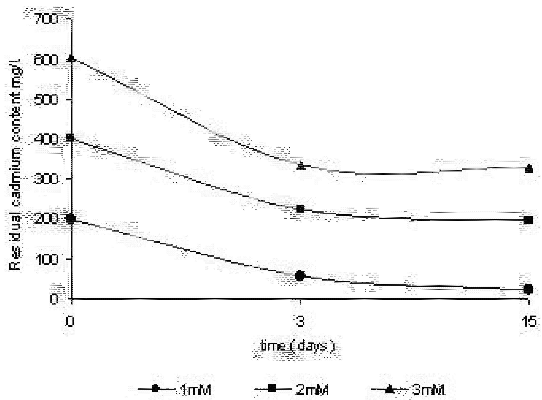
Data from literature on cadmium removal from culture media indicate that the process for most of the organisms tested, exhibits two stages: an initial one in which the sorption is characterized to be rapid and occurs in a short period of time, followed by a second stage, with a lower speed, and that contributes only a little to the total metal sorption [
2,
6,
22]. Kappor and Viraraghavan [
5] also reported that a higher rate of metal removal occurs in the early stages of growth.
Wang
et al. [
29] demonstrated the ability of growth in concentrations of cadmium above 5 mM in a strain of
Pseudomonas aeruginosa, isolated from deep marine waters. The organism was able to remove over 99% of cadmium of the means of cultivation, during the exponential growth phase.
Data gathered from this study corroborate those of literature in relation to the behaviour of removal in
Trichoderma harzianum. The removal of metal ions by fungi can offer an alternative method for biological remediation. Fungi are applied in a large number of industrial processes, which can serve as a constant and economic source of biomass. Additionally, such organisms can easily grow through fermentation techniques of low cost [
5]. Thus, researches on biosorption in fungi may represent an economic means for the treatment of areas contaminated with heavy metals.
Many species of fungi show potential for removal of cadmium.
Aspergillus terreus, Aspergillus flavus, Cladosporium cladosporioides, Fusarium oxysporum, Giocladium roseum, Penicillium spp.,
Mucor rouxii, Helminthosporium sp,
Talaromyces helicus, Trichoderma koningii, Trichoderma harzianum, and
Saccharomyces cerevisiae, isolated from a polluted industrial area, were all efficient in the removal of the metal in aqueous solution. Thus, these data show the ability to remove the metal ion cell for cell biomass through mechanisms that do not depend on the active metabolism [
5,
28,
32,
36,
37].
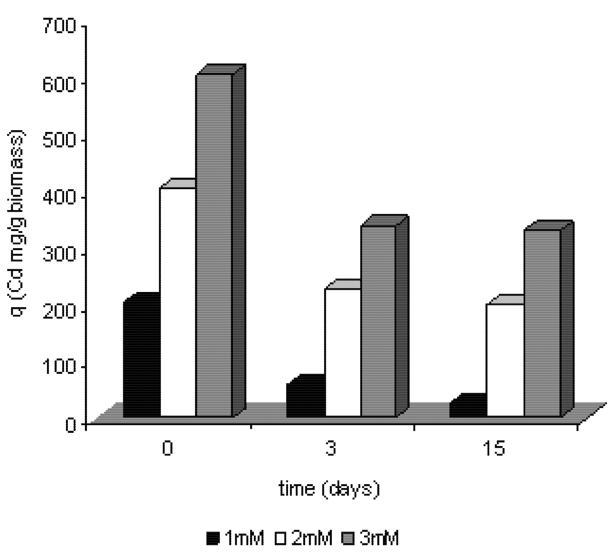
The values obtained for the efficiency of removal of cadmium vary depending on the concentrations of the metal used in the experiments. For example, Malik [
22] reported the ability of isolates of
Giocladium roseum to remove cadmium with a performance of 184 mg per gram of biomass produced. Moreover, Kapoor and Viraraghavan [
5] published values ranging between 0.4 mg/g and 71 mg/g.
The results of this paper, compared with the literature, showed a promising capacity for removal of cadmium by the mycelium of Trichoderma harzianum during its growth in the presence of the metal, considering the high concentrations used.
Additionally, the tests also show the ability to remove the cadmium from the culture medium, meaning that the process depends on the active metabolism and cellular growth as a result of tolerance shown by the organism to the presence of the metal.
Although a reduction in the production of biomass has been found, indicating the effect of metal on cellular growth, total cadmium removed by the cells increased, being related to the concentration of metal used. Thus, this study shows for the first time that the isolate of Trichoderma harzianum used is capable of growing at high concentrations of cadmium, and consequently, the microorganism can potentially be used in bioremediation of systems contaminated with that metal.


