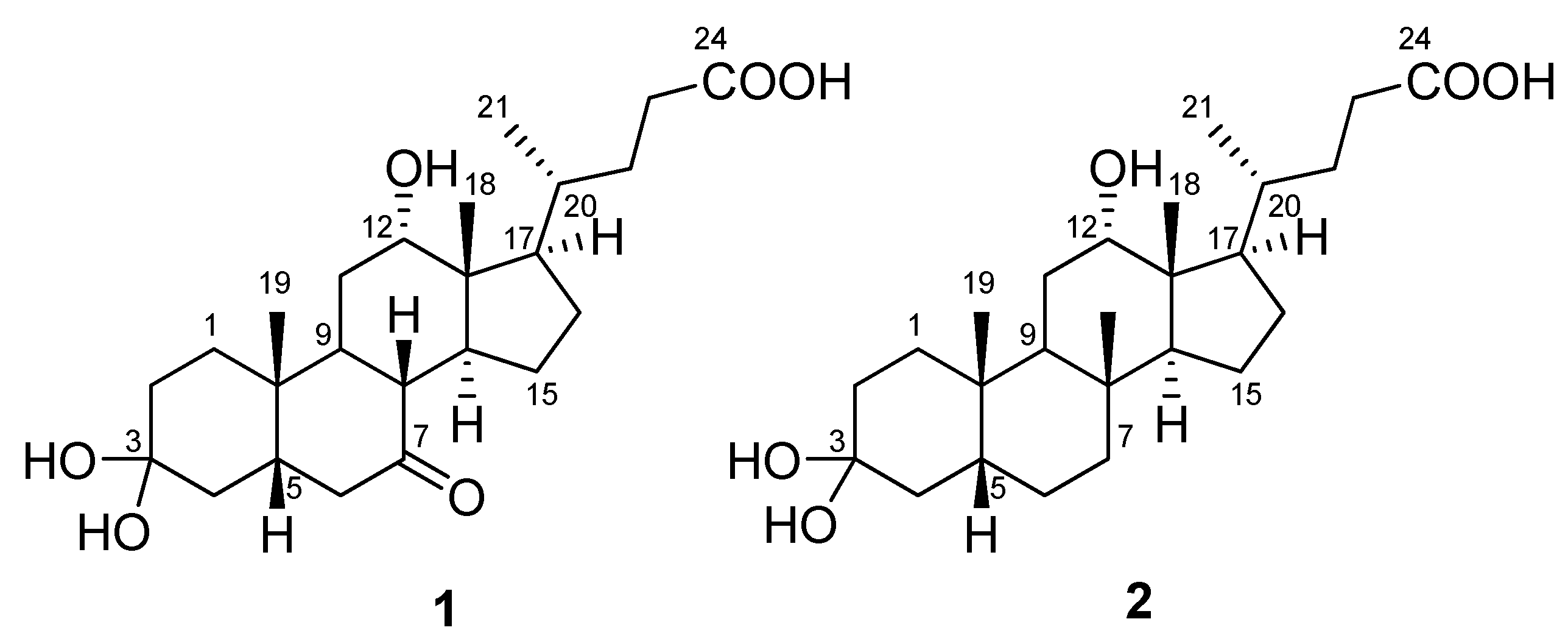
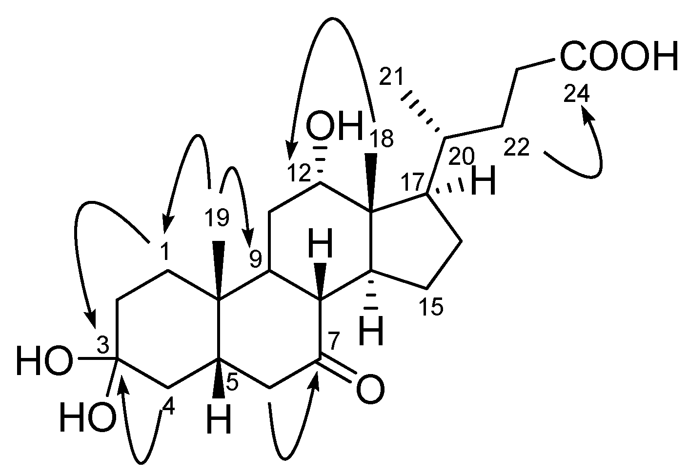
Two New Cholic Acid Derivatives from the Marine Ascidian-Associated Bacterium Hasllibacter halocynthiae

Abstract
:1. Introduction

2. Results and Discussion

| Position | 1 | 2 | ||||
|---|---|---|---|---|---|---|
| δH mult (J, Hz) a | δC b | δH mult (J, Hz) a | δC b | |||
| 1 | 1.67 m, 1.33 m | 33.2 | CH2 | 1.62 m, 1.15 m | 34.2 | CH2 |
| 2 | 1.75 m, 1.38 m | 28.0 | CH2 | 1.88 m, 1.25 m | 28.1 | CH2 |
| 3 | 101.7 | C | 102.5 | C | ||
| 4 | 1.72 m, 1.26 m | 35.8 | CH2 | 1.83, 1.57 m | 34.6 | CH2 |
| 5 | 2.02 m | 45.9 | CH | 1.50 m | 41.3 | CH |
| 6 | 2.97 dd (12.5, 6.0), 1.84 m | 46.1 | CH2 | 1.72 m, 1.48 m | 28.4 | CH2 |
| 7 | 215.1 | C | 1.86 m, 1.29 m | 28.8 | CH2 | |
| 8 | 2.56 dd (11.5, 11.5) | 50.9 | CH | 1.47 m | 37.4 | CH |
| 9 | 2.23 m | 37.4 | CH | 1.87 m | 34.4 | CH |
| 10 | 36.2 | C | 35.6 | C | ||
| 11 | 1.77 m, 1.54 m | 30.9 | CH2 | 1.53 m | 30.2 | CH2 |
| 12 | 3.98 br t (3.0) | 73.0 | CH | 3.96 br t (3.0) | 74.2 | CH |
| 13 | 47.7 | C | 47.7 | C | ||
| 14 | 1.98 m | 42.1 | CH | 1.60 m | 49.5 | CH |
| 15 | 2.15 m, 1.01 m | 25.5 | CH2 | 1.62 m, 1.10 m | 25.0 | CH2 |
| 16 | 1.90 m, 1.29 m | 28.9 | CH2 | 1.44 m, 1.13 m | 27.5 | CH2 |
| 17 | 1.80 m | 47.3 | CH | 1.84 m | 48.3 | CH |
| 18 | 0.72 s | 13.4 | CH3 | 0.71s | 13.3 | CH3 |
| 19 | 1.23 s | 23.4 | CH3 | 0.94 s | 23.8 | CH3 |
| 20 | 1.41 m | 36.7 | CH | 1.43 m | 36.9 | CH |
| 21 | 1.01 d (6.5) | 17.8 | CH3 | 1.01 d (6.5) | 17.7 | CH3 |
| 22 | 1.79 m, 1.36 m | 32.4 | CH2 | 1.79 m, 1.33 m | 32.5 | CH2 |
| 23 | 2.35 ddd (15.0, 9.5, 5.0) | 32.0 | CH2 | 2.35 ddd (15.5, 10.0, 5.5) | 32.2 | CH2 |
| 2.26 m | 2.22 ddd (7.0, 9.5, 16.0) | |||||
| 24 | 176.6 | C | 178.3 | C | ||


3. Experimental Procedures
3.1. General
3.2. Collection, Isolation and Identification of Strain KME 002T
3.3. Cultivation and Extraction
3.4. Separation and Purification of Two New Bile Acid Derivatives, Compounds 1 and 2
 : +19.0 (c 0.10, EtOH); IR (film) vmax 3479, 3056, 2952, 1714, 736 cm−1; 1H- and 13C-NMR spectra: see Table 1; HRFABMS [M–H2O+Na]+ m/z 427.2451 (calcd. for C24H36O5Na, 427.2460). : +25.0 (c 0.10, EtOH); IR (film) vmax 3444, 2939, 2868, 1700, 1206, 1140 cm−1; 1H- and 13C-NMR spectra: see Table 1; HRFABMS [M–H2O+Na]+m/z 413.2662 (calcd. for C24H38O4Na, 413.2668).
4. Conclusions
Acknowledgements
- Sample Availability: Samples of the compounds are available from the authors.
References
- Azumi, K.; Yokosawa, H.; Ishii, S. Halocyamines: Novel antimicrobial tetrapeptide-like substances isolated from the hemocytes of the solitary ascidian Halocynthia. roretzi. Biochemistry 1990, 29, 159–165. [Google Scholar]
- Tsukamoto, S.; Kato, H.; Hirota, H.; Fusetani, N. Antibacterial and antifungal sulfated alkane and alkenes from the Hepatopancreas of the ascidian Halocynthia. roretzi. J. Nat. Prod. 1994, 57, 1606–1609. [Google Scholar] [CrossRef]
- Konishi, I.; Hosokawa, M.; Sashima, T.; Kobayashi, H.; Miyashita, K. Halocynthiaxanthin and fucoxanthinol isolated from Halocynthia. roretzi induce apoptosis in human leukemia, breast and colon cancer cells. Comp. Biochem. Physiol. C Toxicol. Pharmacol. 2006, 142, 53–59. [Google Scholar]
- Kim, S.H.; Yang, H.O.; Sohn, Y.C.; Kwon, H.C. Aeromicrobium. halocynthiae sp. nov., A taurocholic acid–producing bacterium isolated from the marine ascidian Halocynthia. roretzi. Int. J. Syst. Evol. Microbiol. 2010, 60, 2793–2798. [Google Scholar]
- Kim, S.H.; Yang, H.O.; Kwon, H.C. Hasllibacter. halocynthiae gen. nov., sp. nov., A nutriacholic acid–producing bacterium isolated from the marine ascidian Halocynthia. roretzi. Int. J. Syst. Evol. Microbiol. 2012, 62, 624–631. [Google Scholar]
- Kim, Y.-O.; Park, S.Y.; Nam, B.-H.; Kang, S.-J.; Hur, Y.B.; Lee, S.-J.; Oh, T.-K.; Yoon, J.-H. Ruegeria. halocynthiae sp. nov., Isolated from sea squirt Halocynthia. roretzi. Int. J. Syst. Evol. Microbiol. 2012, 62, 925–930. [Google Scholar]
- Kim, Y.-O.; Kong, H.J.; Park, S.Y.; Kang, S.-J.; Kim, W.-J.; Kim, K.-K.; Oh, T.-K.; Yoon, J.-H. Roseovarius. halocynthiae sp. nov., Isolated from sea squirt Halocynthia. roretzi. Int. J. Syst. Evol. Microbiol. 2012, 62, 931–936. [Google Scholar]
- Brinkhoff, T.; Giebel, H.-A.; Simon, M. Diversity, ecology, and genomics of the Roseobacter. clade: A short overview. Arch. Microbiol. 2008, 189, 531–539. [Google Scholar]
- Martens, T.; Gram, L.; Grossart, H.-P.; Kessler, D.; Mueller, R.; Simon, M.; Wenzel, S.C.; Brinkhoff, T. Bacteria of the Roseobacter. clade show potential for secondary metabolite production. Microb. Ecol. 2007, 54, 31–42. [Google Scholar] [CrossRef]
- Paul, V.J.; Ritson-Williams, R.; Sharp, K. Marine chemical ecology in benthic environments. Nat. Prod. Rep. 2011, 28, 345–387. [Google Scholar] [CrossRef]
- Gram, L.; Melchiorsen, J.; Bruhn, J.B. Antibacterial activity of marine culturable bacteria collected from a global sampling of ocean surface waters and surface swabs of marine organisms. Mar. Biotechnol. 2010, 12, 439–451. [Google Scholar] [CrossRef]
- Mukhopadhyay, S.; Maitra, U. Chemistry and biology of bile acids. Curr. Sci. 2004, 87, 1666–1683. [Google Scholar]
- Chiang, J.Y.L. Bile Acid Metabolism. In Molecular Pathology of Liver Diseases; Monga, S.P.S., Ed.; Springer: Pittsburgh, PA, USA, 2011; pp. 165–179. [Google Scholar]
- Park, S.C.; Kim, C.J.; Uramoto, M.; Yun, H.I.; Yoon, K.H.; Oh, T.K. Antibacterial substance produced by Streptococcus faecium under anaerobic culture. Biosci.Biotechnol. Biochem. 1995, 59, 1966–1967. [Google Scholar] [CrossRef]
- Maneerat, S.; Nitoda, T.; Kanzaki, H.; Kawai, F. Bile acids are new products of a marine bacterium, Myroides. sp. strain SM1. Appl. Michrobiol. Biotechnol. 2005, 67, 679–683. [Google Scholar] [CrossRef]
- Kim, D.; Lee, J.S.; Kim, J.; Kang, S.-J.; Yoon, J.-H.; Kim, W.G.; Lee, C.H. Biosynthesis of bile acids in a variety of marine bacterial taxa. J. Microbiol. Biotechnol. 2007, 17, 403–407. [Google Scholar]
- Li, H.; Shinde, P.B.; Lee, H.J.; Yoo, E.S.; Lee, C.-O.; Hong, J.; Choi, S.H.; Jung, J.H. Bile acid derivatives from a sponge-associated bacterium Psychrobacter. sp. Arch.Pharm.Res. 2009, 32, 857–862. [Google Scholar] [CrossRef]
- Bortolini, O.; Fantin, G.; Fogagnolo, M.; Mari, L. Two-way enantioselective control in the epoxidation of alkenes with the keto bile acid-Oxone system. Tetrahedron 2006, 62, 4482–4490. [Google Scholar] [CrossRef]
- Waterhous, D.V.; Barnes, S.; Muccio, D.D. Nuclear magnetic resonance spectroscopy of bile acids. Development of two-dimensional NMR methods for the elucidation of proton resonance assignments for five common hydroxylated bile acids, and their parent bile acid, 5/3 cholanic acid. J. Lip. M Res. 1985, 26, 1068–1078. [Google Scholar]
- Kamekura, M.; Osterhelt, D.; Wallace, R.; Anderson, P.; Kushner, D.J. Lysis of halobacteria in bacto-peptone by bile acids. Appl. Environ. Microbiol. 1988, 54, 990–995. [Google Scholar]
© 2012 by the authors; licensee MDPI, Basel, Switzerland. This article is an open-access article distributed under the terms and conditions of the Creative Commons Attribution license (http://creativecommons.org/licenses/by/3.0/).
Share and Cite
Kim, S.H.; Shin, Y.K.; Sohn, Y.C.; Kwon, H.C. Two New Cholic Acid Derivatives from the Marine Ascidian-Associated Bacterium Hasllibacter halocynthiae. Molecules 2012, 17, 12357-12364. https://doi.org/10.3390/molecules171012357
Kim SH, Shin YK, Sohn YC, Kwon HC. Two New Cholic Acid Derivatives from the Marine Ascidian-Associated Bacterium Hasllibacter halocynthiae. Molecules. 2012; 17(10):12357-12364. https://doi.org/10.3390/molecules171012357
Chicago/Turabian StyleKim, Sung Hun, Yun Kyung Shin, Young Chang Sohn, and Hak Cheol Kwon. 2012. "Two New Cholic Acid Derivatives from the Marine Ascidian-Associated Bacterium Hasllibacter halocynthiae" Molecules 17, no. 10: 12357-12364. https://doi.org/10.3390/molecules171012357