2.1. Pre-Screen for Toxicity in Two Selected Model Organisms
The first task of this study has been the selection of relevant model systems. As human cells are rather difficult (and expensive) to culture, a more simple, easy-to-use model organism is required for a general pre-screening of activity in order to identify the most likely candidates for high activity and to assign a low priority to any entirely inactive agents. Ideally, such a simple system suitable for pre-screening should also be useful for subsequent intracellular diagnostics. Here, yeast first comes to mind. Indeed, yeast has already been employed successfully as a model system to study the toxicity of inorganic selenium compounds, such as SeO
32− and SeO
42−. Since the response of yeast cells to these two selenium salts is well-documented, both have been used here for comparison [
9,
10,
11,
12]. As yeast is a single cell organism, however, another, also readily available organism has been used as a “control”, namely the nematode
Steinernema feltiae. The latter represents an agriculturally relevant nematode which is not pathogenic, is easy to cultivate and generally provides reproducible and reliable results in an intact, multicellular living organism [
13,
14].
Figure 2 shows the impact of selected selenium and tellurium compounds on the survival of
Steinernema feltiae (
Figure 2a) and
Saccharomyces cerevisiae (
Figure 2b,c). In the case of yeast, two independent assays have been employed, one using an agar spotting method (
Figure 2b), the other a cell survival assay based on propidium iodide (PI) staining and counting of dead cells immediately after treatment with the compounds (
Figure 2c). Some of the substances tested seem to affect both, the survival of the nematodes and the growth of yeast, such as compound
5 (and to some extent compound
3), whilst the others (such as compounds
6 and
7) are only toxic against either yeast or nematodes. Interestingly, these results identify compound
5 as being particularly active. In the past, compound
5 and structurally closely related tellurium compounds have been studied extensively in human cancer cell lines and there have been identified as being amongst the most powerful, redox-modulating cytotoxic chalcogen agents available so far [
3,
15]. The results obtained in our simple nematode- and yeast-based assays therefore confirm this pronounced toxicity and also support the idea that such simple assays may be useful in pre-screening and subsequently pre-selecting compounds.
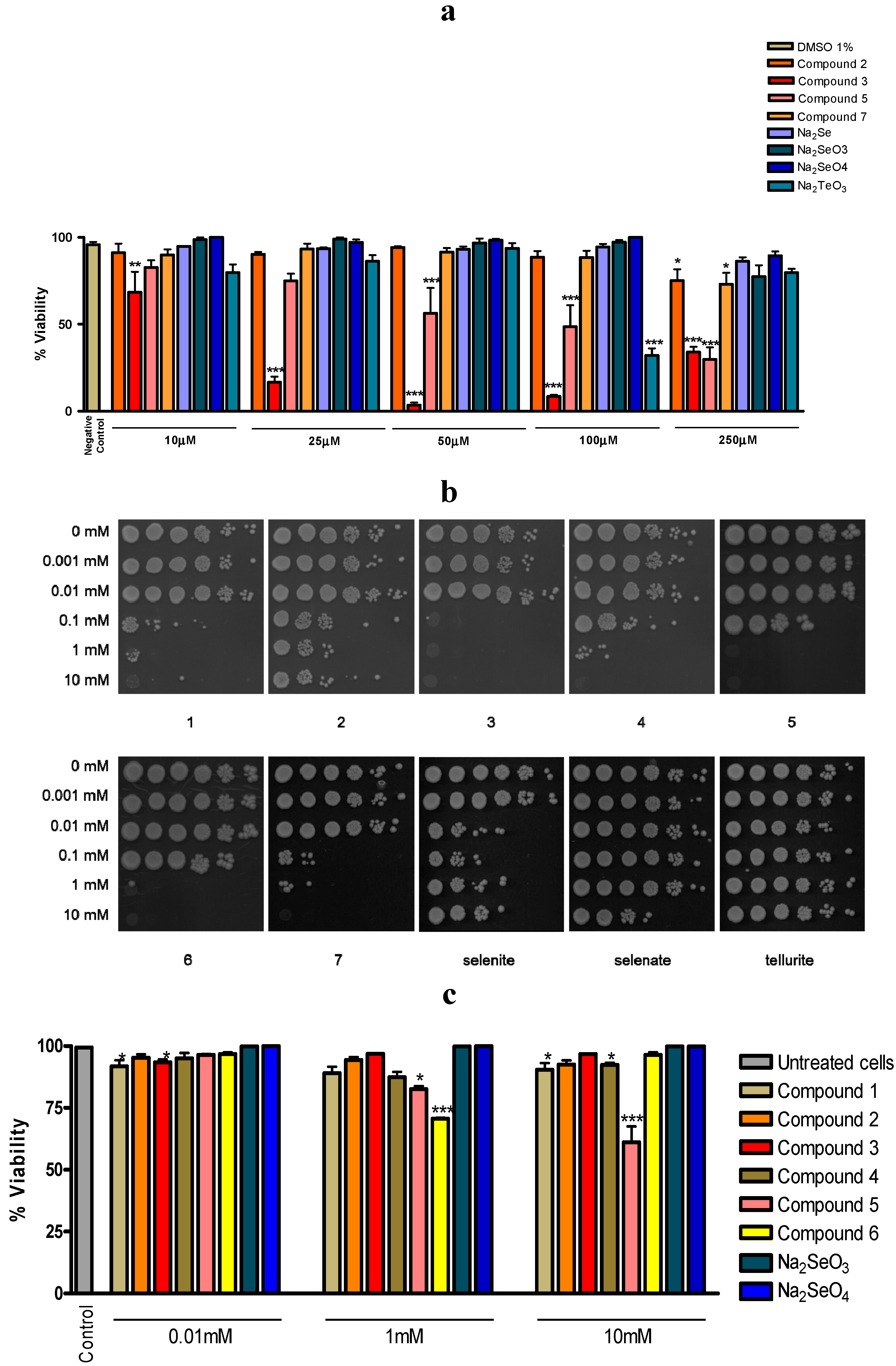
Figure 2.
Most selenium and tellurium compounds investigated exhibit a pronounced toxicity established in two independent pre-screens based on a nematode and yeast. Panel a: Toxicity against the agricultural nematode Steinernema feltiae; Panel b: Toxicity against the Saccharomyces cerevisiae strain BY4741 using the spot test. Cells were treated with increasing concentrations of compounds (0.1 mM to 10 mM) for 3 h. After treatment, they were collected by centrifugation, washed with, and resuspended in, physiological saline, diluted, and spotted onto YPD plates to determine cell survival; Panel c: Quantification of dead Saccharomyces cerevisiae strain BY4741 cells by staining with PI and flow cytometry. Cells were treated with increasing concentrations of compounds, collected by centrifugation, washed with, and resuspended in, physiological saline. They were then stained with PI and stained, dead cells were quantified by flow cytometry. The values represent means with SD error bars from two separate experiments, and statistical significances have been calculated using a one-way ANOVA followed by Bonferroni’s multiple comparison test or Kruskal-Wallis followed by Dunn’s multiple comparison test. * denotes values significantly different from the control. * p < 0.05; ** p < 0.01; *** p < 0.001. See text for experimental details.
![Molecules 19 12258 g002]()
Nonetheless, there are exceptions which caution against the orthodox use of just one assay system, especially yeast. It seems that yeast is particularly sensitive to certain selenium compounds. Whilst the results obtained for the selenium compound
3 are comparable in yeast,
Steinernema feltiae and to some extent even in human macrophage cell culture, other compounds, such as selenite, seem to affect yeast but not so much
Steinernema feltiae (
Figure 2a,b) [
3]. Hence, whilst most compounds show a similar toxicity in the nematode and yeast assay, which more or less corresponds to their cytotoxicity in human cell lines, and whilst SeO
32− is clearly a special case with a particular chemical reactivity (see below), there can be notable exceptions. It is therefore advisable to use at least two pre-screens for biological activity in tandem, such as a combination of nematodes and yeast,
i.e., a combination which seems to cover a wider range of activities, just to ensure that particular, perhaps also exceptional activities, do not escape our attention. Nonetheless, the inherent limitations still associated with employing a non-mammalian combination of organisms must be kept in mind. It is possible, for instance, that there are distinct differences in the uptake, metabolic pathways and excretion for selenium and tellurium compounds in yeast, worms and mammalian systems, and that such differences may form the basis for an exceptional toxicity of selenium compounds in yeast. It is also possible that yeast is not exceptionally sensitive to selenium compounds, but rather resilient against poisoning by tellurium compounds. Indeed, there are various tellurium-tolerant fungi which incorporate tellurium into sulfur-containing amino acids to form tellurocysteine and telluromethionine [
16]. In any case, once a more general activity has been detected, it is certainly necessary to study the relevant compounds in more sophisticated test systems and in considerably more detail. This, of course, will ultimately also include the use of mammalian and eventually human model systems.
2.2. From “Activity” to Mechanistic Considerations: Microscopy, Chemogenetic Screening and “Intracellular Diagnostics”
The results obtained in
Steinernema feltiae and the
Saccharomyces cerevisiae BY4741 strain together have confirmed a significant toxicity of some of the selenium and tellurium compounds, such as compounds
3,
5,
6 and
7, which is reflected in human cell cultures [
2,
3]. These assays seem to be able to distinguish between certain compounds that are active and hence interesting, and others which are not. Nonetheless, such a pre-selection tool mainly addresses the question if compounds are eventually active, and not necessarily why they are active.
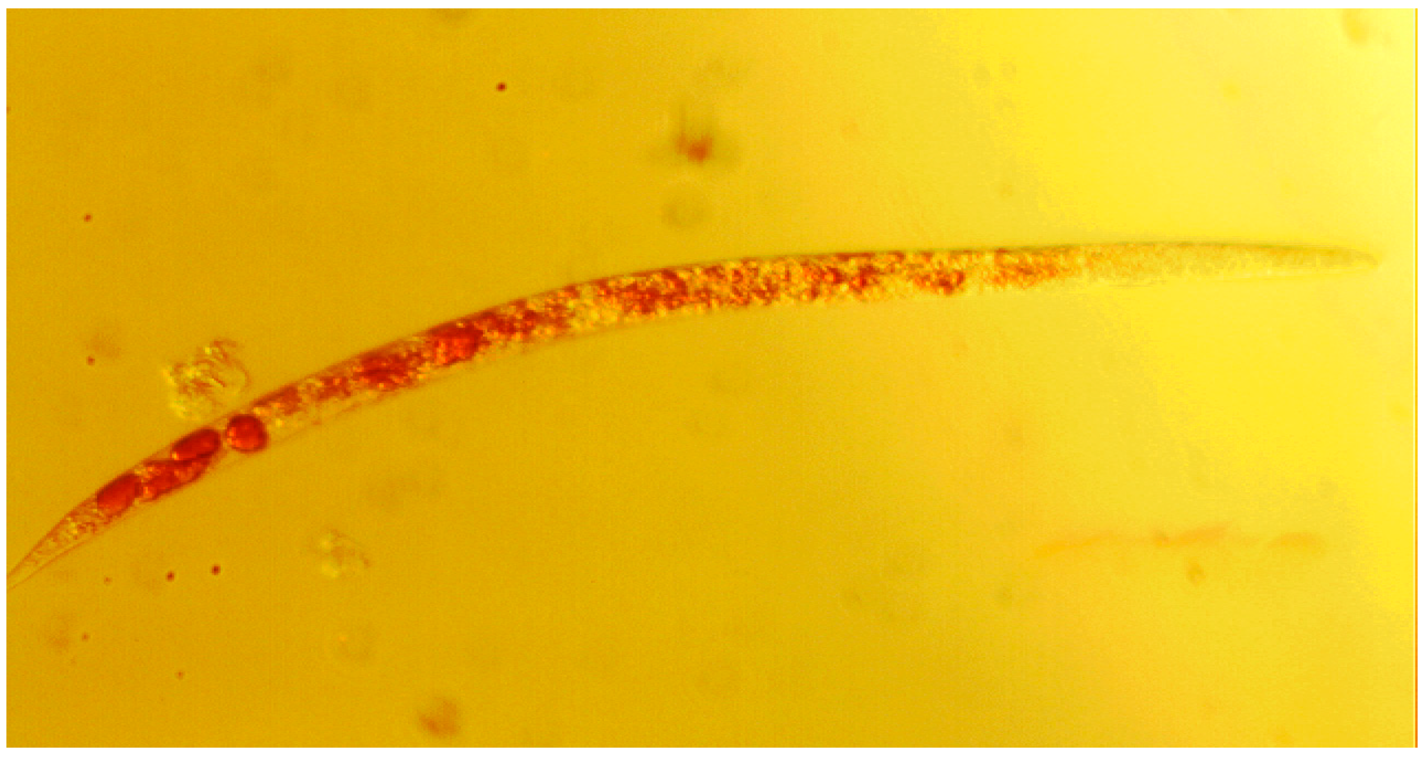
The next step therefore needs to consider the possible and/or likely causes for such toxicity. Here, both model organisms offer certain additional insights into the effects of such compounds on the living organism. To begin with, the nematode is an ideal organism to study under the light microscope at rather modest magnification (e.g., 10-fold). It is a comparably simple organism and transparent, which enables us to study the distribution of (colored) compounds inside the nematode. As
Figure 3 illustrates, compound
5 has apparently entered the nematode and has become enriched inside. This is a clear indication that the compound has been taken up and is distributed across the organism. Besides this simple but relevant “uptake” and “distribution” experiment, more detailed studies are also possible using nematodes such as
Steinernema feltiae or
Caenorhabditis elegans. The precise distribution of compounds can be studied, the organs and types of cells affected inside the nematode can be determined (e.g., cuticle, digestive tract) and such nematodes can even be stained and monitored under the fluorescence microscope.
Figure 3.
Microscopic image of a representative Steinernema feltiae nematode showing the enrichment of compound 5 (red color inside the nematode). 10-fold magnification on a TR 200 VWR International microscope.
Figure 3.
Microscopic image of a representative Steinernema feltiae nematode showing the enrichment of compound 5 (red color inside the nematode). 10-fold magnification on a TR 200 VWR International microscope.
Nonetheless, the nematode-based whole organism model cannot provide mechanistic information on the cellular level. Here, the yeast system offers a powerful alternative. As all of the compounds used in this study are known to be redox active based on structural considerations, in vitro redox assays and electrochemical investigations, a “redox link” is likely. To study this particular link, yeast endows the researcher with a set of tools which can be employed alone or in combination to address the “why” question in a eukaryotic organism. Here, yeast is particularly suited to uncover redox-related mechanisms, as redox-protecting systems are well conserved between yeast and mammals. Human cells, for instance, feature one additional (extracellular) superoxide dismutase to the cytosolic and mitochondrial one also existing in yeast. A similar situation is found in the case of catalase, where human cells possess one catalase enzyme. Nevertheless, additional ROS detoxifying components (such as peroxidases) may differ between mammals and yeast, and metabolic activity providing reducing equivalents for redox-maintenance generally is higher in yeast than in mammalian cell cultures. Still, many results that are obtained in the yeast system can be transferred to mammals with minor contingencies.
At the same time, yeast lends itself to extensive chemogenetic screening. As part of this approach, various yeast mutants lacking specific proteins and enzymes are employed and the toxicity observed for these mutants is then compared to the toxicity recorded in the corresponding wild type. Mutants that are more sensitive (or resistant) to a given compound when compared to the wild type may hint towards a possible link between the particular protein/enzyme lacking in the mutant and its role in metabolism or defense, and the compound in question. As knock-out mutants in all non-essential genes can be generated in yeast easily, or can be obtained from commercial suppliers such as EUROSCARF, chemogenetic screening is comparably easy to perform. This approach in some aspects also mirrors investigations concerned with the development of drug/pesticide resistance, where the resistance to a given compound provides hints of the pathways involved based on the overexpression of certain proteins. In both cases, such hints may then be used to guide further investigations by an array of cell-based analytical techniques we have branded as “intracellular diagnostics” [
17].
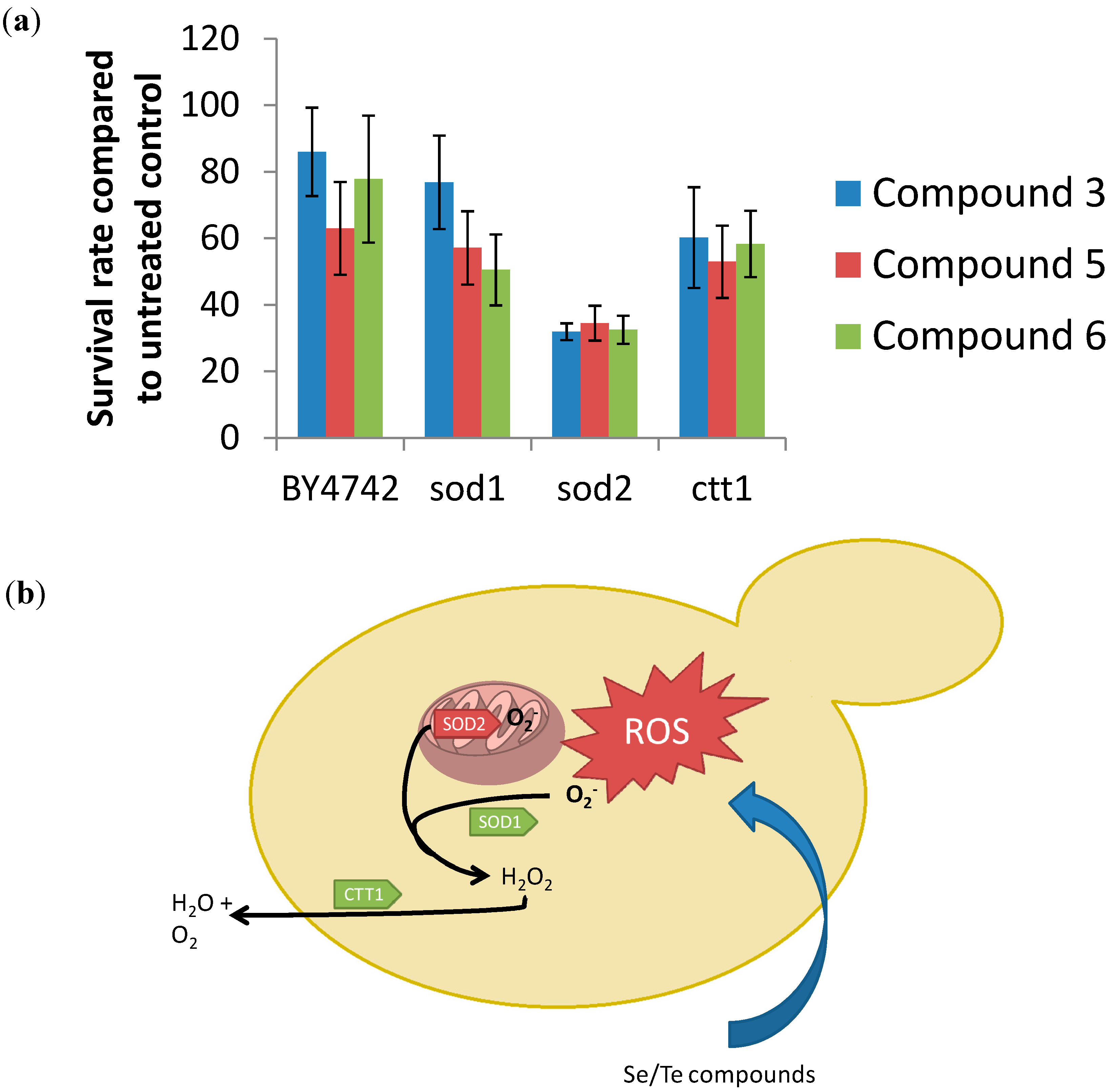
Indeed, we have recently shown that such a basic chemogenetic screen in yeast focusing on mutants that lack one (or several) antioxidant enzymes is rather revealing [
3]. Besides the wild type strain BY4742, mutants deficient in Sod1 (superoxide dismutase 1), Sod2 and Ctt1 (cytosolic catalase) have been considered. As
Figure 4 illustrates, a particularly high toxicity of compounds
3,
5 and
6 against the
sod2 mutant strain can be observed. This strain shows a survival rate of just 40% for these compounds, whilst under the same conditions, the other “redox impaired” mutants, such as the
sod1 or the
ctt1 mutant, show revival rates of 60% and higher. Such differences support the notion of a distinct redox link and also provide a strong hint that mitochondrially-based Sod2 and/or its substrate, namely O
2•−, are somehow involved in the biological action of these selenium and tellurium compounds.
To investigate this redox hypothesis further, we have therefore employed a range of selective fluorescent dyes which belong to the emerging toolkit of intracellular diagnostics and, as these dyes do not lyse or kill the cell, can be used to monitor intracellular events even in a time resolved manner. Out of the arsenal of dyes available to date, we have evidently selected a range of “redox indicators” to begin with (in the following such diagnostic dyes are highlighted in bold) [
4,
18,
19].
As the chemogenetic screen shown in
Figure 4 has already pointed towards the involvement of ROS, namely in form of O
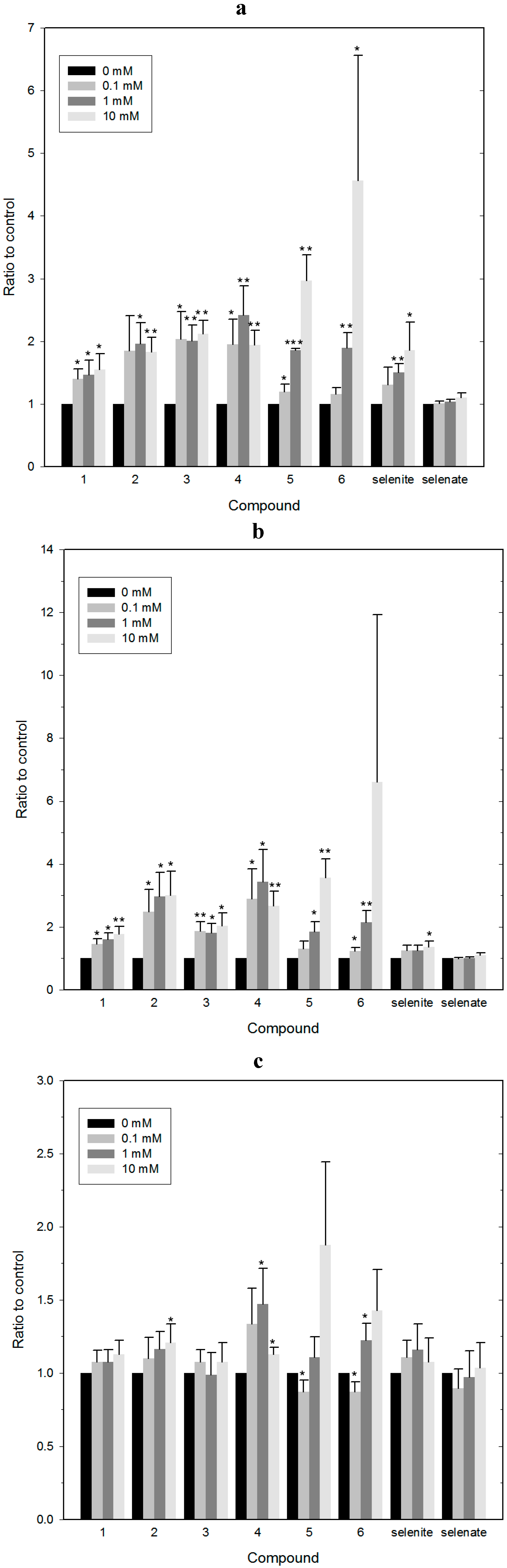
2•−, and since raised concentrations of oxidative stressors could indeed cause non-tolerable intracellular redox imbalances and cell death, we have therefore measured the total ROS levels in BY4741 yeast cells treated with the synthetic Se- and Te-containing compounds first, using 2',7'-dichlorodihydrofluorescein diacetate (DCFDA) as an indicator dye and the two inorganic selenium salts as benchmark. As shown in
Figure 5a, synthetic Se- and Te-containing organic redox modulators generate high levels of ROS, in some instances already at compound concentrations of 100 µM. Whilst these results are rather revealing, it should be emphasized that the use of DCFDA as a general stain for ROS provides initial information regarding the redox state of the cell (primarily its cytosol), yet the use of this indicator dye is not entirely unproblematic. DCFDA is hydrolyzed by certain esterases as it enters the cell and the intracellular concentration of its oxidized, fluorescent form therefore also depends on esterase activity—and not only on the concentration of ROS. In addition, DCFDA reacts with some ROS (mainly peroxides, H
2O
2), but not with all ROS and in any case not with the same kinetics. This may impact on the ability of DCFDA to monitor ROS levels in the cell adequately and in a differentiated manner.
Figure 4.
Chemogenetic screening of selected compounds using BY4742 wild type and the corresponding mutant strains of Saccharomyces cerevisiae. This screen indicates that the mutant deficient in the antioxidant enzyme Sod2 is particularly sensitive to the most active chalcogen compounds studied. Whilst no firm conclusions can be drawn, a redox link between the compounds on one side, and mitochondrial superoxide/superoxide dismutase and cell death on the other is likely. See text for experimental details. Panel a: Chemogenetic screening of yeast mutants impaired in ROS-detoxification in the BY4742 background compared with the BY4742-wild type. The results suggest an enhanced susceptibility of a Δsod2 mutant against compounds 3, 5 and 6; Panel b: Scheme illustrating the mode of action of selenium- and tellurium containing compounds tested. The compounds seem to trigger the formation of superoxide in the mitochondria, since a mutant of a mitochondrial superoxide-dismutase is hypersusceptible to the tested compounds (indicated in red) mutants of the cytosolic superoxide-dismutase and catalase show no difference to the wild type regarding the resistance to the tested compounds.
![Molecules 19 12258 g004]()
Interestingly, there is a notable absence of any major difference between Se- and Te-containing redox modulators. Furthermore, SeO
32− and SeO
42− do not seem to induce raised ROS levels. It therefore appears that the quinone moiety is mostly responsible for the increase in ROS concentrations observed, whilst the presence of either selenium or tellurium has only a minor impact. This finding is hardly surprising, as the quinone moiety is well known to undergo redox cycling in the presence of O
2 (hence generating O
2•−) and has been “added” to the chalcogen moiety intentionally to act as a ROS generator [
20,
21].
To return to our findings from the chemogenetic screen and hence to gain a deeper insight into which particular ROS may be involved in the toxicity of redox modulators in yeast, we have employed a selective assay for mitochondrial O
2•− based on the fluorescent indicator dye MitoSOX Red.
Figure 5b indicates that all synthetic Se- and Te-containing compounds tested also raise intracellular levels of mitochondrial O
2•−.
Figure 5.
Estimating intracellular redox changes by employing selective, redox sensitive fluorescent dyes. Panel a: Production of ROS. Cells were treated with increasing concentrations of selected selenium and tellurium compounds and the total ROS production after treatment was estimated by staining with DCFDA; Panel b: Production of O2•−. Cells were treated with increasing concentrations of compounds and the level of mitochondrial O2•− was estimated using MitoSOX Red as mitochondrial O2•− indicator;. Panel c: Production of 1O2. Cells were treated with increasing concentrations of compounds and intracellular levels of 1O2 were assessed using the Singlet Oxygen Sensor Green reagent. The values represent medians with SD error bars from at least four separate experiments. Statistical significances have been calculated using a one sample Student’s t test. * denotes values significantly different from the control. * p < 0.05; ** p < 0.01; *** p < 0.001.
Indeed, the general trend observed in
Figure 5a using the DCFDA stain can also be observed in
Figure 5b, with increases in the respective oxidant concentrations of around two- to three-fold. This indicates that most of the ROS formation observed is probably due to an (initial) formation of O
2•−. As before, quinone-containing compounds seem to be particularly active, whilst the inorganic salts (SeO
32− and SeO
42−) are not.
Besides O
2•−, singlet dioxygen
1O
2 is also sometimes formed in cells upon stimulation with redox agents and may represent another ROS type involved in the activity of the compounds under investigation. Indeed, there are several reasons to consider the presence of
1O
2 as part of this study. Firstly, most of the compounds used are colored and hence may exert a photosensitizing activity. Secondly, SeO
32− is known to cause DNA double-strand breaks (DSBs), and its chemistry is associated with the most potent types of ROS, such as the hydroxyl radical (
•OH) and
1O
2 (see below). And thirdly,
1O
2 represents a branch of ROS formation which runs independently of the mitochondrial O
2•− cascade or the ROS production by NADPH oxidases. As
Figure 5c indicates,
1O
2 levels, measured with the fluorescent dye Singlet Oxygen Sensor Green, did not increase considerably in the presence of the compounds. Only small, up to 1.5-fold increases were observed for compounds such as
4, and only at high compound concentrations of 10 mM, which are hardly relevant for potential biological applications. SeO
32− showed a very weak increase as well, rejecting the hypothesis that this particular compound causes DNA damage via the formation of high amounts of
1O
2.
Hence, it appears that the selenium and tellurium compounds investigated in yeast predominantly generate (mitochondrial) O
2•− compared to other ROS, a finding which agrees well with our chemogenetic screen and recent studies conducted in mammalian cells. This notion is also in line with the particular “chemistry” of such quinone-containing agents which is illustrated in
Figure 6. If O
2•− indeed is the ROS ultimately responsible for cellular damage (see below) and cell death, or if it is converted to H
2O
2 and to the
•OH radical—which in turn cause the damage and cell death observed—remains to be shown (see discussion below). Similarly, whilst we propose that there is a direct causal relationship between ROS production, cellular damage and cell death in case of many selenium and tellurium compounds, including inorganic selenium salts, one may speculate that such compounds themselves are fairly unreactive, yet may become metabolically converted to a common intermediate which subsequently triggers the formation of ROS. Such a metabolic conversion may be independent of the type and concentration of compound applied. Alternatively, these compounds may not generate ROS by themselves but interfere with either ROS-generating or ROS-detoxifying enzymes. They may, for instance, cause some hitherto unnoticed damage to the cell or to a particular cellular compartment (such as the cytoskeleton, the mitochondria, the endoplasmic reticulum), which would then result in an oxidative burst. Then again, such compounds may not generate ROS at all, either directly or indirectly, but may inhibit a ROS detoxifying system, such as a particular reductase, instead. Indeed, some recent studies have identified the human enzyme thioredoxin reductase as a potential target for selenium and especially also for tellurium compounds [
22,
23,
24].
In any case, a significant increase in intracellular concentrations of ROS upon application of certain selenium and tellurium compounds seems to occur consistently across different organisms. This raises the question which kind of damage such ROS subsequently cause inside the cell. Here, membranes, redox sensitive proteins of the cellular thiolstat and DNA represent prominent targets. As the effect on membranes and on proteins has been studied rather extensively before [
25,
26,
27,
28,
29], we have explored here the effects on DNA using an assay indicative of DSBs.
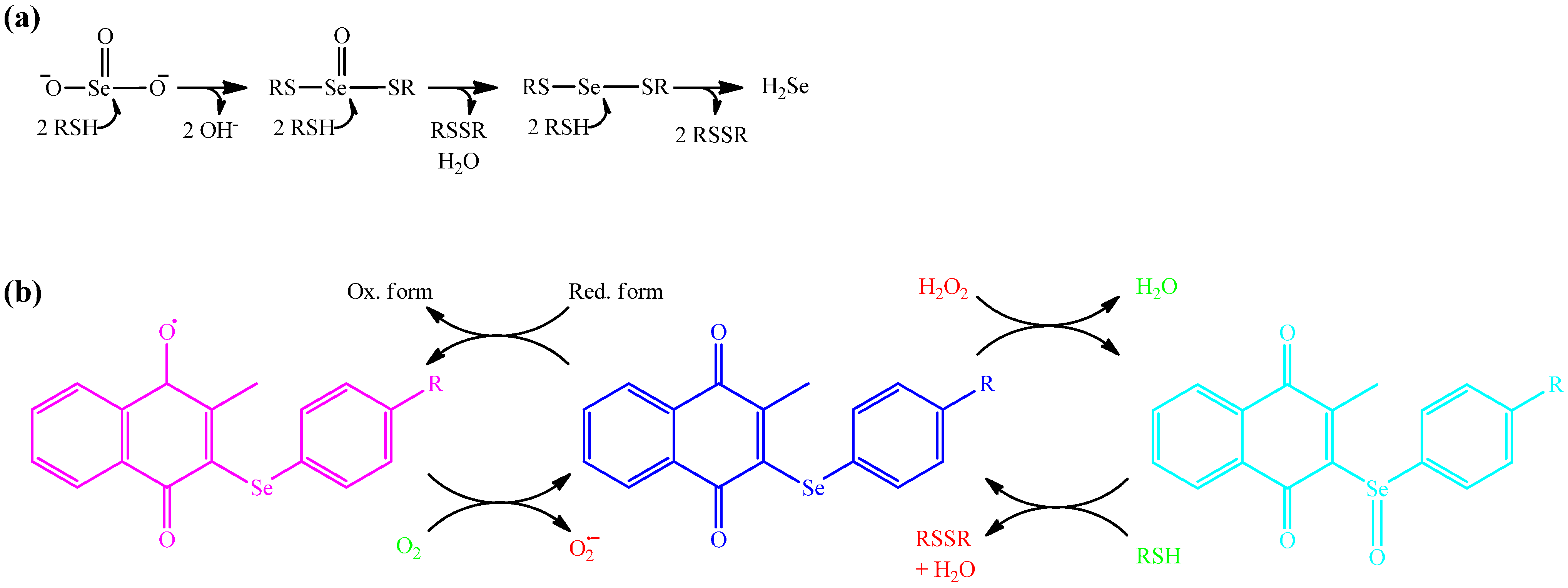
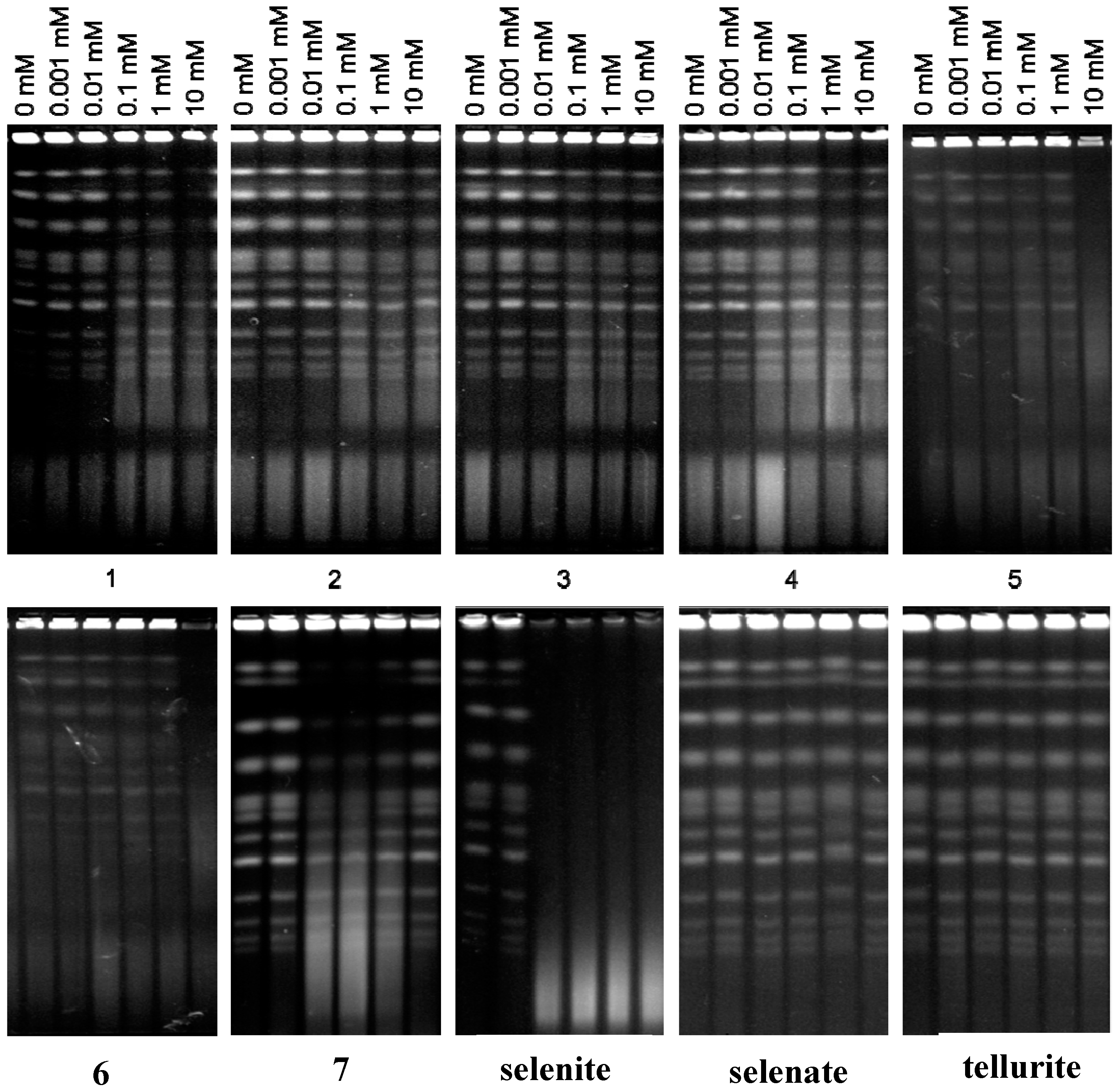
2.3. DNA as Target: The Special Role of SeO32−
Increased ROS levels are often capable of producing DSBs, and this particular damage may well also explain the notable toxicity associated with such agents [
30]. A possible induction of DSBs by the various selenium and tellurium compounds has therefore been investigated, using data for the inorganic selenium and tellurium salts for comparison. In line with our previous reports [
9,
10,
11], SeO
32− induces DSBs in yeast in a dose-dependent manner (
Figure 7). In stark contrast, neither SeO
42− nor any of the selenium or tellurium compounds tested (including TeO
32−) cause any significant DSBs. As there is no significant difference between the most and the least toxic compounds in this assay, it is unlikely that DSBs form a decisive part of the intracellular mechanism(s) underlying the rather pronounced toxicity observed in this study for some of the compounds, such as compounds
3,
5,
6 and
7.
Figure 6.
Schematic overview of the chemistry and reactivity of selected selenium and tellurium compounds under physiological conditions, i.e., in the presence of reducing thiols. Panel a: SeO32− reacts mostly spontaneously and rapidly with up to six thiol equivalents (including GSH and protein thiols) to generate first an unusual selenotrisulfide and ultimately a mixed selenodisulfide (selenylsulfide) and hydrogen selenide (H2Se). In contrast, neither SeO42− nor the organic selenium and tellurium compounds studied seem to exhibit this particular kind of chemistry; Panel b: Most of the organic compounds tested rather generate O2•− (probably due to the presence of a naphthoquinone moiety) or facilitate the reaction of existing ROS with protein thiols. Whilst this simple illustration cannot explain the entire biochemistry behind such compounds, it provides some hints why SeO32− and the organochalcogens tested are sometimes equally toxic, yet SeO32− reacts rather differently compared to most of the other compounds and, notably, also induces severe damage to DNA (and hence may not be the best choice of trace element supplement).
![Molecules 19 12258 g006]()
One should note, however, that among all of the compounds tested in this study, it is precisely SeO
32− which is used by people in practice. Here, selenite increasingly represents the selenium component of commercial trace element supplements (instead of selenomethionine which can be enriched in yeast cultivated in an appropriate source of selenium). As such nutritional supplements are freely available in supermarkets and their use is not controlled, considerable care should be taken. Whilst it is unlikely that SeO
32− reaches cellular DNA easily, it still has the potential to cause DSBs and hence also mutations which in the long term may result in the formation of cancer [
11].
Figure 7.
Induction of DSBs upon treatment with increasing concentrations of selenium and tellurium compounds. For analysis of DSBs, treated cells were embedded into low melting-point agarose to generate plugs, in which the cells were lysed. After lysis, the plugs were loaded into wells of the gel and chromosomal DNA was subjected to electrophoretic separation. After electrophoresis, the gels were stained with ethidium bromide, visualized using a UV transilluminator and photographed. In all cases, experiments were performed in triplicate and representative gels are shown.
The fact that SeO
32− seems to be fairly potent in causing or sustaining DNA damage, whilst many selenium and tellurium compounds—despite the fact that they up-regulate intracellular concentrations of ROS—do not, also deserves some comment. Selenite is a highly reactive inorganic form of selenium which reacts spontaneously—and rather rapidly—with many biological thiols, including GSH and numerous cysteine-containing proteins. This reaction is sketched out in
Figure 6a. It is often assisted by specific enzymes and results in some rather unusual chemical structures, and may ultimately end in the formation of disulfides, selenodisulfides (selenylsulfides), selenotrisulfides and H
2Se.
Whilst a direct “chemical attack” by SeO
32− on DNA is unlikely from a purely chemical point of view, SeO
32− is well posed to inhibit many (cysteine-containing) enzymes, including proteins and enzymes involved in DNA repair, and hence may increase the extent of DSBs in a more indirect manner, e.g., by inhibiting homologous recombination (HR). In fact, HR is one of the two main DSB repair mechanisms that has been shown previously to play a crucial role in repairing DSBs induced by SeO
32− [
10]. Here, one should notice that DSBs usually are not generated directly. Other types of damage may result in DSBs via DNA replication, attempted and aborted DNA repair, and by co-existence of adjacent single-strand breaks in opposite DNA strands.
It is doubtful that the Se- and Te-compounds investigated as part of this study can undergo the same kind of chemistry associated with SeO
32−. Consequently, a mechanism other than induction of DSBs seems to be responsible for the pronounced toxicity of synthetic Se- and Te-containing organic redox modulators used in the present study (see also
Figure 6b). Still, we have been able to demonstrate that those compounds generate considerable amounts of ROS, primarily O
2•−, and one may well speculate that such ROS can also induce or sustain DNA damage. It is therefore noteworthy that we could not find any DSBs despite an apparent increase in ROS levels.
In the future, it would therefore be worthwhile to consider other potential intracellular targets for the compounds investigated as part of study. Besides an increase in ROS concentrations, which may indirectly damage numerous biomolecules, a direct interaction with cysteine proteins of the thiolstat is likely. At the same time, considerable care should be taken with the uncontrolled public release and consumption of selenite, as SeO32− in a biological context seems to be a rather potent and aggressive selenium species to deal with.


 ,
, 


