
New Pregnane Glycosides from Gymnema sylvestre

Abstract
:1. Introduction

2. Results and Discussion
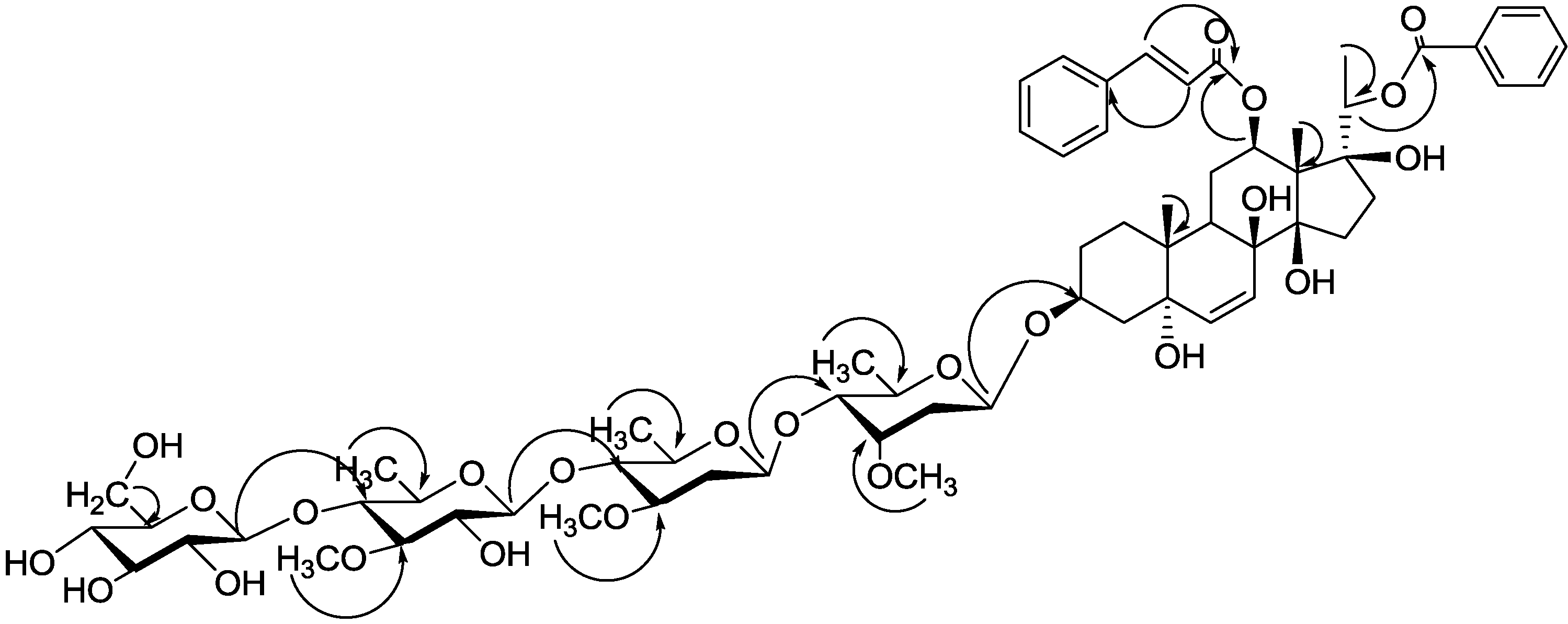
2.1. Isolation and Structure Elucidation

| NO. | δH (J in Hz) | |||||
|---|---|---|---|---|---|---|
| 1 (a) | 1 (a),(b) | 2 | 3 | 4 | 5 (a) | |
| 1a | 1.68 (m) | 1.59 (m) | 1.71 (m) | 1.73 (m) | 2.44 (m) | 1.69 (m) |
| 1b | 2.18 (m) | 1.21 (m) | 2.21 (m) | 2.22 (m) | 2.57 (m) | 2.17 (m) |
| 2a | 2.01 (m) | 1.63 (m) | 2.01 (m) | 2.03 (m) | 1.79 (m) | 2.02 (m) |
| 2b | 2.16 (m) | 1.26 (m) | 2.16 (m) | 2.16 (m) | 2.08 (m) | 2.14 (m) |
| 3 | 4.18 (m) | 3.06 (m) | 4.18 (m) | 4.20 (m) | 3.85 (m) | 4.20 (m) |
| 4a | 2.07 (m) | 1.79 (m) | 2.08 (m) | 2.08 (m) | 2.41 (m) | 2.07 (m) |
| 4b | 2.25 (m) | 1.64 (m) | 2.27 (m) | 2.27 (m) | 2.58 (m) | 2.27 (m) |
| 5-OH | 3.59 (s) | |||||
| 6 | 5.94 (d, 10.4 Hz) | 5.40 (d, 10.4 Hz) | 5.93 (d, 10.4 Hz) | 5.92 (d, 10.4 Hz) | 5.36 (m) | 5.95 (d, 10.4 Hz) |
| 7a | 6.26 (d, 10.4 Hz) | 5.63 (d,10.4 Hz) | 6.26 (d, 10.4 Hz) | 6.26 (d, 10.4 Hz) | 2.41 (m) | 6.25 (d, 10.4 Hz) |
| 7b | 2.53 (m) | |||||
| 8-OH | 4.09 (s) | |||||
| 9 | 2.41 (m) | 1.85 (m) | 2.41 (m) | 2.41 (m) | 1.78 (m) | 2.41 (m) |
| 11a | 2.20 (m) | 1.72 (m) | 2.23 (m) | 2.24 (m) | 2.05 (m) | 2.22 (m) |
| 11b | 2.43 (m) | 1.54 (m) | 2.49 (m) | 2.43 (m) | 2.37 (m) | 2.45 (m) |
| 12 | 5.38 (m) | 4.73 (m) | 5.35 (m) | 5.37 (m) | 5.25 (m) | 5.38 (m) |
| 14-OH | 5.23 (s) | |||||
| 15a | 2.00 (m) | 1.70 (m) | 1.48 (m) | 1.48 (m) | 1.80 (m) | 2.00 (m) |
| 15b | 2.19 (m) | 1.66 (m) | 2.17 (m) | 2.16 (m) | 2.15 (m) | 2.21 (m) |
| 16a | 2.01 (m) | 1.96 (m) | 1.48 (m) | 1.48 (m) | 1.81 (m) | 2.02 (m) |
| 16b | 2.10 (m) | 1.79 (m) | 2.03 (m) | 1.98 (m) | 2.14 (m) | 2.11 (m) |
| 17-OH | 5.30 (s) | |||||
| 18 | 2.20 (s) | 1.48 (s) | 2.18 (s) | 2.22 (s) | 2.18 (s) | 2.20 (s) |
| 19 | 1.52 (s) | 0.85 (s) | 1.57 (s) | 1.59 (s) | 1.32 (s) | 1.54 (s) |
| 20 | 5.28 (br q, 6.0 Hz) | 4.61 (br q, 6.0 Hz) | 5.11 (br q, 6.1 Hz) | 4.09 (br q, 6.4 Hz) | 5.28 (m) | 5.28 (br q, 6.2Hz) |
| 21 | 1.54 (d, 6.0 Hz) | 1.21 (d, 6.0 Hz) | 1.48 (d, 6.1 Hz) | 1.36 (d, 6.4 Hz) | 1.57 (d, 5.8 Hz) | 1.56 (d, 6.2 Hz) |
| Cinnamoyl moiety | ||||||
| 2' | 6.51 (d, 16 Hz) | 5.96 (d, 16 Hz) | 6.74 (d, 16 Hz) | 6.97 (d, 16 Hz) | 6.50 (d, 16 Hz) | 6.51 (d, 16 Hz) |
| 3' | 7.80 (d, 16 Hz) | 7.30 (d, 16 Hz) | 7.93 (d, 16 Hz) | 8.14 (d, 16 Hz) | 7.87 (d, 16 Hz) | 7.80 (d, 16 Hz) |
| 5', 9' | 7.35 (m) | 7.27 (m) | 7.66 (m) | 7.52 (m) | 7.35 (m) | 7.37 (m) |
| 6', 8' | 7.33 (m) | 7.10 (m) | 7.41 (m) | 7.32 (m) | 7.33 (m) | 7.31 (m) |
| 7' | 7.35 (m) | 7.27 (m) | 7.40 (m) | 7.33 (m) | 7.35 (m) | 7.37 (m) |
| (E)-2-Methyl-2-butenoyl or benzoyl moiety | ||||||
| 3'' | 8.21 (d, 7.6 Hz) | 7.90 (d, 7.2 Hz) | 7.00 (d, 7.3 Hz) | 8.23 (d, 7.2 Hz) | 8.21 (d, 7.2 Hz) | |
| 4'' | 7.30 (m) | 7.35 (m) | 1.51 (d, 7.3 Hz) | 7.39 (m) | 7.32 (m) | |
| 5'' | 7.50 (m) | 7.60 (m) | 1.78 (s) | 7.56 (m) | 7.53 (m) | |
| 6'' | 7.30 (m) | 7.35 (m) | 7.39 (m) | 7.32 (m) | ||
| 7'' | 8.21 (d, 7.6 Hz) | 7.90 (d, 7.2 Hz) | 8.23 (d, 7.2 Hz) | 8.21 (d, 7.2 Hz) | ||
| NO. | δC | |||||
|---|---|---|---|---|---|---|
| 1 (a) | 1 (a),(b) | 2 | 3 | 4 | 5 (a) | |
| 1 | 27.6 (t) | 26.6 (t) | 27.5 (t) | 27.6 (t) | 38.7 (t) | 27.6 (t) |
| 2 | 26.5 (t) | 25.2 (t) | 26.5 (t) | 26.5 (t) | 29.8 (t) | 26.5 (t) |
| 3 | 74.9 (d) | 73.3 (d) | 74.9 (d) | 74.9 (d) | 77.5 (d) | 74.9 (d) |
| 4 | 39.0 (t) | 37.4 (t) | 39.0 (t) | 39.0 (t) | 39.1 (t) | 39.1 (t) |
| 5 | 74.7 (s) | 72.6 (s) | 74.7 (s) | 74.8 (s) | 139.1 (s) | 74.7 (s) |
| 6 | 136.7 (d) | 135.4 (d) | 136.6 (d) | 136.1 (d) | 119.3 (d) | 136.7 (d) |
| 7 | 127.3 (d) | 124.0 (d) | 127.3 (d) | 127.6 (d) | 34.8 (t) | 127.3 (d) |
| 8 | 74.0 (s) | 72.6 (s) | 74.0 (s) | 73.8 (s) | 74.2 (s) | 74.0 (s) |
| 9 | 36.6 (d) | 34.9 (d) | 36.5 (d) | 36.6 (d) | 44.0 (d) | 36.6 (d) |
| 10 | 39.6 (s) | 38.3 (s) | 39.6 (s) | 39.6 (s) | 37.2 (s) | 39.6 (s) |
| 11 | 23.6 (t) | 22.3 (t) | 23.7 (t) | 23.6 (t) | 25.6 (t) | 23.6 (t) |
| 12 | 75.8 (d) | 74.2 (d) | 75.6 (d) | 75.9 (d) | 74.6 (d) | 75.8 (d) |
| 13 | 58.1 (s) | 56.8 (s) | 57.9 (s) | 58.1 (s) | 57.0 (s) | 58.1 (s) |
| 14 | 88.2 (s) | 87.1 (s) | 88.1 (s) | 88.9 (s) | 88.9 (s) | 88.2 (s) |
| 15 | 33.1 (t) | 32.1 (t) | 33.1 (t) | 33.3 (t) | 33.7 (t) | 33.1 (t) |
| 16 | 34.3 (t) | 33.5 (t) | 34.2 (t) | 33.5 (t) | 34.0 (t) | 34.3 (t) |
| 17 | 87.7 (s) | 86.5 (s) | 87.7 (s) | 87.9 (s) | 87.5 (s) | 87.7 (s) |
| 18 | 12.4 (q) | 11.6 (q) | 12.3 (q) | 12.7 (q) | 11.5 (q) | 12.5 (q) |
| 19 | 21.5 (q) | 20.9 (q) | 21.5 (q) | 21.6 (q) | 17.9 (q) | 21.6 (q) |
| 20 | 75.3 (d) | 73.9 (d) | 74.4 (d) | 70.4 (d) | 75.8 (d) | 75.3 (d) |
| 21 | 15.6 (q) | 15.0 (q) | 15.5 (q) | 19.6 (q) | 15.3 (q) | 15.6 (q) |
| Cinnamoyl moiety | ||||||
| 1' | 166.8 (s) | 165.5 (s) | 166.7 (s) | 167.0 (s) | 166.8 (s) | 166.8 (s) |
| 2' | 120.1 (d) | 118.9 (d) | 120.3 (d) | 119.6 (d) | 120.3 (d) | 120.2 (d) |
| 3' | 143.9 (d) | 143.2 (d) | 143.7 (d) | 145.2 (d) | 143.8 (d) | 143.9 (d) |
| 4' | 134.8 (s) | 133.9 (s) | 134.8 (s) | 134.9 (s) | 134.9 (s) | 134.8 (s) |
| 5', 9' | 128.5 (d) | 128.0 (d) | 128.5 (d) | 128.6 (d) | 128.5 (d) | 128.5 (d) |
| 6', 8' | 129.1 (d) | 128.8 (d) | 129.2 (d) | 129.2 (d) | 129.1 (d) | 129.1 (d) |
| 7' | 130.4 (d) | 130.1 (d) | 130.5 (d) | 130.5 (d) | 130.4 (d) | 130.4 (d) |
| (E)-2-Methyl-2-butenoyl or benzoyl moiety | ||||||
| 1'' | 165.6 (s) | 164.6 (s) | 166.7 (s) | 165.6 (s) | 165.6 (s) | |
| 2'' | 131.2 (d) | 130.3 (d) | 129.4 (s) | 131.2 (d) | 131.2 (d) | |
| 3'' | 130.2 (d) | 129.4 (d) | 137.7 (d) | 130.2 (d) | 130.2 (d) | |
| 4'' | 128.7 (d) | 128.5 (d) | 14.1 (q) | 128.7 (d) | 128.7 (d) | |
| 5'' | 133.2 (d) | 133.1 (d) | 12.2 (q) | 133.2 (d) | 133.2 (d) | |
| 6'' | 128.7 (d) | 128.5 (d) | 128.7 (d) | 128.7 (d) | ||
| 7'' | 130.2 (d) | 129.4 (d) | 130.2 (d) | 130.2 (d) | ||
| Sugar | NO. | δH (J in Hz) | ||||
|---|---|---|---|---|---|---|
| 1 (a) | 2 | 3 | 4 | 5 (a) | ||
| Cym | 1 | 5.17 (brd, 10.9 Hz) | 5.17 (brd, 10.9 Hz) | 5.17 (brd, 10.9 Hz) | 5.29 (brd, 10.9 Hz) | 5.17 (brd, 10.5 Hz) |
| 2a | 1.73 (m) | 1.73 (m) | 1.73 (m) | 1.89 (m) | 1.73 (m) | |
| 2b | 2.20 (m) | 2.20 (m) | 2.23 (m) | 2.31 (m) | 2.23 (m) | |
| 3 | 3.98 (m) | 3.98 (m) | 3.98 (m) | 3.98 (m) | 3.98 (m) | |
| 4 | 3.43 (m) | 3.43 (m) | 3.43 (m) | 3.43 (m) | 3.42 (m) | |
| 5 | 4.16 (m) | 4.16 (m) | 4.16 (m) | 4.16 (m) | 4.16 (m) | |
| 6 | 1.39 (d, 6.0 Hz) | 1.39 (d, 6.0 Hz) | 1.39 (d, 6.0 Hz) | 1.46 (d, 6.0 Hz) | 1.39 (d, 6.0 Hz) | |
| OMe | 3.52 (s) | 3.52 (s) | 3.52 (s) | 3.59 (s) | 3.52 (s) | |
| Ole | 1 | 4.68 (brd, 9.6 Hz) | 4.68 (brd, 9.6 Hz) | 4.68 (brd, 9.6 Hz) | 4.70 (brd, 9.6 Hz) | 4.68 (brd, 9.6 Hz) |
| 2a | 2.47 (m) | 2.47 (m) | 2.46 (m) | 2.49 (m) | 2.48 (m) | |
| 2b | 1.73 (m) | 1.73 (m) | 1.73 (m) | 1.76 (m) | 1.73 (m) | |
| 3 | 3.57 (m) | 3.57 (m) | 3.57 (m) | 3.57 (m) | 3.57 (m) | |
| 4 | 3.59 (m) | 3.59 (m) | 3.59 (m) | 3.59 (m) | 3.52 (m) | |
| 5 | 3.55 (m) | 3.58 (m) | 3.55 (m) | 3.51 (m) | 3.51 (m) | |
| 6 | 1.64 (d, 5.2 Hz) | 1.64 (d, 5.2 Hz) | 1.64 (d, 5.2 Hz) | 1.68 (d, 5.2 Hz) | 1.64 (d, 5.2 Hz) | |
| OMe | 3.50 (s) | 3.51 (s) | 3.51 (s) | 3.51 (s) | 3.51 (s) | |
| The | 1 | 4.88 (d, 7.8 Hz) | 4.88 (d, 7.6 Hz) | 4.88 (d, 7.6 Hz) | 4.88 (d, 7.6 Hz) | 4.88 (d, 7.6 Hz) |
| 2 | 3.90 (m) | 3.90 (m) | 3.90 (m) | 3.90 (m) | 3.90 (m) | |
| 3 | 3.67 (m) | 3.68 (m) | 3.67 (m) | 3.67 (m) | 3.69 (m) | |
| 4 | 3.83 (m) | 3.83 (m) | 3.83 (m) | 3.83 (m) | 3.88 (m) | |
| 5 | 3.75 (m) | 3.77 (m) | 3.75 (m) | 3.75 (m) | 3.75 (m) | |
| 6 | 1.75 (d, 5.6 Hz) | 1.76 (d, 5.6 Hz) | 1.75 (d, 5.6 Hz) | 1.75 (d, 5.6 Hz) | 1.75 (d, 5.6 Hz) | |
| OMe | 3.91 (s) | 3.82 (s) | 3.82 (s) | 3.91 (s) | 3.94 (s) | |
| Glc | 1 | 5.09 (d, 7.8 Hz) | 5.09 (d, 7.8 Hz) | 5.09 (d, 7.8 Hz) | 5.09 (d, 7.8 Hz) | 5.14 (d, 7.8 Hz) |
| 2 | 4.02 (m) | 4.02 (m) | 4.02 (m) | 4.02 (m) | 4.04 (m) | |
| 3 | 4.28 (m) | 4.28 (m) | 4.28 (m) | 4.28 (m) | 4.25 (m) | |
| 4 | 4.31 (m) | 4.31 (m) | 4.30 (m) | 4.31 (m) | 4.23 (m) | |
| 5 | 3.93 (m) | 3.93 (m) | 3.93 (m) | 3.93 (m) | 3.98 (m) | |
| 6a | 4.30 (m) | 4.30 (m) | 4.30 (m) | 4.30 (m) | 4.32 (m) | |
| 6b | 4.50 (m) | 4.50 (m) | 4.50 (m) | 4.50 (m) | 4.54 (m) | |
| Glc | 1 | 5.20 (d, 7.8 Hz) | 5.20 (d, 8.5 Hz) | 5.20 (d, 8.5 Hz) | 5.20 (d, 8.5 Hz) | |
| 2 | 4.10 (m) | 4.10 (m) | 4.10 (m) | 4.10 (m) | ||
| 3 | 4.23 (m) | 4.23 (m) | 4.23 (m) | 4.23 (m) | ||
| 4 | 4.19 (m) | 4.16 (m) | 4.19 (m) | 4.19 (m) | ||
| 5 | 4.04 (m) | 4.04 (m) | 4.04 (m) | 4.04 (m) | ||
| 6a | 4.30 (m) | 4.32 (m) | 4.30 (m) | 4.31 (m) | ||
| 6b | 4.53 (m) | 4.53 (m) | 4.53 (m) | 4.53 (m) | ||
| Sugar | NO. | δC | |||||
|---|---|---|---|---|---|---|---|
| 1 (a) | 2 | 3 | 4 | 5 (a) | |||
| Cym | 1 | 97.7 (d) | 97.6 (d) | 97.7 (d) | 96.3 (d) | 97.7 (d) | |
| 2 | 36.7 (t) | 36.7 (t) | 36.7 (t) | 37.2 (t) | 36.7 (t) | ||
| 3 | 77.7 (d) | 77.6 (d) | 77.7 (d) | 77.8 (d) | 77.7 (d) | ||
| 4 | 83.0 (d) | 83.0 (d) | 83.0 (d) | 83.2 (d) | 83.0 (d) | ||
| 5 | 68.9 (d) | 68.9 (d) | 68.9 (d) | 68.8 (d) | 68.9 (d) | ||
| 6 | 18.5 (q) | 18.4 (q) | 18.5 (q) | 18.4 (q) | 18.5 (q) | ||
| OMe | 58.7 (q) | 58.6 (q) | 58.7 (q) | 58.8 (q) | 58.7 (q) | ||
| Ole | 1 | 101.8 (d) | 101.8 (d) | 101.8 (d) | 101.8 (d) | 101.8 (d) | |
| 2 | 37.5 (t) | 37.5 (t) | 37.5 (t) | 37.6 (t) | 37.5 (t) | ||
| 3 | 79.1 (d) | 79.1 (d) | 79.2 (d) | 79.2 (d) | 79.1 (d) | ||
| 4 | 83.2 (d) | 83.2 (d) | 83.2 (d) | 83.4 (d) | 83.1 (d) | ||
| 5 | 71.9 (d) | 71.8 (d) | 71.8 (d) | 71.8 (d) | 71.9 (d) | ||
| 6 | 18.6 (q) | 18.6 (q) | 18.6 (q) | 18.6 (q) | 18.6 (q) | ||
| OMe | 57.3 (q) | 57.3 (q) | 57.3 (q) | 57.3 (q) | 57.4 (q) | ||
| The | 1 | 103.9 (d) | 104.0 (d) | 103.9 (d) | 103.9 (d) | 104.0 (d) | |
| 2 | 74.9 (d) | 74.9 (d) | 74.8 (d) | 74.9 (d) | 74.8 (d) | ||
| 3 | 86.3 (d) | 86.3 (d) | 86.3 (d) | 86.3 (d) | 86.2 (d) | ||
| 4 | 83.3 (d) | 83.3 (d) | 83.4 (d) | 83.4 (d) | 83.2 (d) | ||
| 5 | 71.9 (d) | 71.8 (d) | 71.9 (d) | 71.9 (d) | 71.9 (d) | ||
| 6 | 18.7 (q) | 18.7 (q) | 18.7 (q) | 18.7 (q) | 18.7 (q) | ||
| OMe | 60.6 (q) | 60.6 (q) | 60.6 (q) | 60.6 (q) | 60.6 (q) | ||
| Glc | 1 | 104.5 (d) | 104.5 (d) | 104.6 (d) | 104.6 (d) | 104.8 (d) | |
| 2 | 75.3 (d) | 75.3 (d) | 75.3 (d) | 75.3 (d) | 75.8 (d) | ||
| 3 | 76.8 (d) | 76.8 (d) | 76.8 (d) | 76.8 (d) | 78.6 (d) | ||
| 4 | 81.5 (d) | 81.5 (d) | 81.5 (d) | 81.5 (d) | 71.9 (d) | ||
| 5 | 76.2 (d) | 76.2 (d) | 76.2 (d) | 76.2 (d) | 78.1 (d) | ||
| 6 | 62.3 (t) | 62.2 (t) | 62.3 (t) | 62.3 (t) | 63.0 (t) | ||
| Glc | 1 | 104.9 (d) | 104.9 (d) | 104.9 (d) | 104.9 (d) | ||
| 2 | 74.7 (d) | 74.7 (d) | 74.9 (d) | 74.7 (d) | |||
| 3 | 78.2 (d) | 78.2 (d) | 78.2 (d) | 78.2 (d) | |||
| 4 | 71.5 (d) | 71.5 (d) | 71.5 (d) | 71.4 (d) | |||
| 5 | 78.4 (d) | 78.4 (d) | 78.4 (d) | 78.4 (d) | |||
| 6 | 62.4 (t) | 62.3 (t) | 62.4 (t) | 62.4 (t) | |||


2.2. Biological Activity Assay
| Sample | Concentration | OD | Inhibition Ratio |
|---|---|---|---|
| (mg/mL) | Average ± RSD | (%) | |
| Negetive control | 0 | 1.8573 ± 0.0129 | |
| Compound 1 | 1.02 | 1.7661 ± 0.0077 | 4.9 |
| Compound 2 | 1.07 | 1.7860 ± 0.0033 | 3.8 |
| Compound 3 | 1.11 | 1.7356 ± 0.0025 | 6.6 |
| Compound 4 | 1.16 | 1.7901 ± 0.0029 | 3.6 |
| Acarbose | 0.50 | 1.3654 ± 0.0026 | 26 |
3. Experimental
3.1. General Procedures
3.2. Plant Materials
3.3. Extraction and Isolation
3.4. Isolated Compounds
3.5. Enzymatic Hydrolysis of Compound 1
3.6. α-Glucosidase Inhibitory Effect
4. Conclusions
Supplementary Materials
Acknowledgments
Author Contributions
Conflicts of Interest
References
- Hua, Q. The exploitation and utilization of Gymnema sylvestre. Wild Plant Resour. China 1993, 3, 35–36. [Google Scholar]
- Jiangsu college of traditional Chinese medicine. The Dictionary of Traditional Chinese Medicine; Shanghai Science and Technology Press: Shanghai, China, 1977; Volume 1, pp. 1246–1247. [Google Scholar]
- Campbell, T.N.; Roberts, J.R.; Frank, H.H. Medicinal plants used by the Choctaw, Chickasaw, and Creek Indians in the early nineteenth century. J. Wash. Acad. Sci. 1951, 41, 285–290. [Google Scholar]
- Baskaran, K.; Kizar, A.B.; Radha, S.K.; Shanmugasundaram, E.R. Antidiabetic effect of a leaf extract from Gymnema sylvestre in non-insulin-dependent diabetes mellitus patients. J. Ethnopharmacol. 1990, 30, 295–300. [Google Scholar] [CrossRef] [PubMed]
- Sugihara, Y.; Nojima, H.; Matsuda, H.; Murakami, T.; Yoshikawa, M.; Kimura, I. Antihyperglycemic effects of gymnemic acid IV, a compound derived from Gymnema sylvestre leaves in streptozotocin-diabetic mice. J. Asian. Nat. Prod. Res. 2000, 2, 321–327. [Google Scholar] [CrossRef] [PubMed]
- Miyoshi, M.; Imoto, T.; Kasagi, T. Antieurodonitic effect of various fractions extracted from the leaves of Gymnema sylvestre. Yonago Igaku Zasshi 1987, 38, 127–137. [Google Scholar]
- Komalavalli, N.; Rao, M.V. In vitro micropropagation of Gymnema sylvestre: A multipurpose medicinal plant. Plant Cell Tissue Organ Cult. 2000, 61, 97–105. [Google Scholar] [CrossRef]
- Kumar, V.; Bhandari, U.; Tripathi, C.D.; Khanna, G. Anti-obesity effect of Gymnema sylvestre extract on high fat diet-induced obesity in Wistar rats. Drug Res. (Stuttg. Ger.) 2013, 63, 625–632. [Google Scholar] [CrossRef]
- Reddy, R.M.I.; Latha, P.B.; Vijaya, T.; Rao, D.S. The saponin-rich fraction of a Gymnema sylvestre R.Br. aqueous leaf extract reduces cafeteria and high-fat diet-induced obesity. Z. Naturforsch. C Biosci. 2012, 67, 39–46. [Google Scholar] [CrossRef]
- Preuss, H.G.; Bagchi, D.; Bagchi, M.; Rao, C.V.S.; Dey, D.K.; Satyanarayana, S. Effects of a natural extract of (−)-hydroxycitric acid (HCA-SX) and a combination of HCA-SX plus niacin-bound chromium and Gymnema sylvestre extract on weight loss. Diabetes Obes. Metab. 2004, 6, 171–180. [Google Scholar] [CrossRef] [PubMed]
- Daisy, P.; Eliza, P.; Mohanmed, F.K. A novel dihydroxy gymnemic triacetate isolated from Gymnema sylvestre possessing normoglycemic and hypolipidemic activity on STZ induced diabetic rats. J. Ethnopharmacol. 2009, 126, 339–344. [Google Scholar] [CrossRef] [PubMed]
- Yoshikawa, K.; Amimoto, S.; Arihara, K.; Matsuura, K. Structure studies of new antisweet constituents from Gymnema sylvestre. Tetrahedron Lett. 1989, 30, 1103–1106. [Google Scholar] [CrossRef]
- Maeda, M.; Iwashita, T.; Kurihara, Y. Studies on taste modifiers. II. Purification and structure determination of gymnemic acids, antisweet active principle from Gymnema sylvestre leaves. Tetrahedron Lett. 1989, 30, 1547–1550. [Google Scholar] [CrossRef]
- Yoshikawa, K.; Kondo, Y.; Arihara, S.; Matsuura, K. Antisweet natural products. IX. Structures of gymnemic acids XV-XVIII from Gymnema sylvestre R. Br. V. Chem. Pharm. Bull. 1993, 41, 1730–1738. [Google Scholar] [CrossRef]
- Kiuchi, F.; Liu, H.M.; Tsuda, Y. Two new gymnemic acid congeners congtaining a hexulo pyranside moiety. Chem. Pharm. Bull. 1990, 38, 2326–2328. [Google Scholar] [CrossRef]
- Yoshikawa, K.; Nakagawa, M.; Yamamoto, R.; Arihara, S.; Matsuura, K. Antisweet natural products V. Structures of Gymnemic acids VIII-XIII from Gymnema sylvestre R. Br. Chem. Pharm. Bull. 1992, 40, 1779–1782. [Google Scholar] [CrossRef]
- Yoshikawa, K.; Arihara, S.; Matsuura, K. A new type antisweet principles occurring in Gymnem sylvestre (M). Tetrahedron Lett. 1991, 32, 789–792. [Google Scholar] [CrossRef]
- Yoshikawa, K.; Amimoto, K.; Arihara, S.; Matsuura, K. Gymnemic acid V, VI and VII from Gur-Ma, the leaves of Gymnema sylvestre R. Br. Chem. Pharm. Bull. 1989, 37, 852–854. [Google Scholar] [CrossRef]
- Miyatake, K.; Takenaka, S.; Fujimoto, T.; Kensho, G.; Priya, S.U.; Kirihata, M.; Ichimoto, I.; Yoshihisa, N. Isolation of Conduritol A from Gymnema sylvestre and its effects against intest-inal glucose absorption in rats. Biosci. Biotechonol. Biochem. 1993, 57, 2184–2185. [Google Scholar] [CrossRef]
- Liu, X.; Ye, W.; Yu, B.; Zhao, S.; Wu, H.; Che, C. Two new flavonol glycosides from Gymnema sylvestre and Euphorbia ebracteolata. Carbohydr. Res. 2004, 339, 891–895. [Google Scholar] [CrossRef] [PubMed]
- Kamei, K.; Takano, R.; Miyasaka, A.; Imoto, T.; Hara, S. Amino acid sequence of sweet taste suppressing peptide (gurmarin) from the leaves of Gymnema sylvestre. J. Biochem. (Tokyo) 1992, 111, 109–112. [Google Scholar]
- Yoshiyuki, S.; Stxyzo, I.; Yumie, M.; Kazuya, N.; Sumiko, S.; Hiroyuki, N. Studies biochem on hyaluronidase inhibitor of Gyrnnema sylvestre R. Br. Eisei Kagaku 1990, 36, 314–319. [Google Scholar] [CrossRef]
- Rao, G.S.; Sinsheimer, J.E.; Mcihenny, H.M. Structure of gymnamine, a trace alkaloid from Gymnema sylvestre leaves. Chem. Ind. 1972, 13, 537–538. [Google Scholar]
- Sinsheimer, J.E.; McIlhenny, H.M. Constituents from Gymnema sylvestre leaves. II. Nitrogenous compounds. J. Pharm. Sci. 1967, 56, 732–736. [Google Scholar] [CrossRef] [PubMed]
- Gupta, S.S.; Seth, C.B.; Variyar, M.C. Experimental studies on pituitary-diabetes Part I Inhibitory effect of a few Ayurvedic antidiabetic remedies on antterior pituitary extract induced hyperglycemia in albino rats. J. Med. Res. 1962, 50, 73–81. [Google Scholar]
- Anupam, B.; Malay, C.H. Hypolipidaemic and antiatherosclerotic effects of oral Gymnema sylvestre R. Br. leaf extract in albino rats fed on a high fat diet. Phytother. Res. 1994, 8, 118–120. [Google Scholar] [CrossRef]
- Yoshikawa, M.; Murakami, T.; Kadoya, M.; Li, Y.H.; Murakami, N.; Yamahara, J.; Matsuda, H. Medicinal foodstuffs. IX. The inhibitors of glucoseabsorption from the leaves of Gymnema sylvestre R. Br. (Asclepiadaceae): Structures of gymnemosides a and b. Chem. Pharm. Bull. 1997, 45, 1671–1676. [Google Scholar] [CrossRef] [PubMed]
- Shimizu, K.; Iino, A.; Nakajima, J.; Tanaka, K.; Nakajyo, S.; Urakawa, N.; Atsuchi, M.; Wada, T.; Yamashita, C. Suppression of glucose absorption by some fractions extracted from Gymnema sylvestre leaves. J. Vet. Med. Sci. 1997, 59, 245–251. [Google Scholar] [CrossRef] [PubMed]
- Yoshioka, S.; Imoto, T.; Miyoshi, M.; Kasagi, T.; Kawahara, R.; Hiji, Y. Anti-diabetic effects of the extracts from the leaves of Gymnema sylvestre. Inhibitory effect of gymnemic acids on glucose absorption in the small intestine. WakanIyaku Zasshi 1996, 13, 300–303. [Google Scholar]
- Yoshikawa, M.; Murakami, T.; Matsuda, H. Medicinal foodstuffs. X. Structures of new triterpene glycosides, gymnemosides -c, -d, -e, and -f, from the leaves of Gymnema sylvestre R. Br.: Influence of gymnema glycosides on glucose uptake in rat small intestinal fragments. Chem. Pharm. Bull. 1997, 45, 2034–2038. [Google Scholar] [CrossRef] [PubMed]
- Yoshikawa, K.; Okada, N.; Kann, Y.; Matsuchika, K.; Arihara, S. Steroidal glycosides, from the fresh stem of Stephanotis lutchensis var. Iaponica (Asclepiadaceae). Chemical structures of stephanosides K-Q. Chem. Pharm. Bull. 1996, 44, 2243–2248. [Google Scholar] [CrossRef] [PubMed]
- Yoshikawa, K.; Matsuchika, K.; Takahashi, K.; Tanaka, M.; Arihara, S.; Chang, H.C.; Wang, J.D. Pregnane Glycosides, Gymnepregosides G-Q from the roots of Gymnema alternifolium. Chem. Pharm. Bull. 1999, 47, 798–804. [Google Scholar] [CrossRef] [PubMed]
- Yoshikawa, K.; Matsuchika, K.; Arihara, S.; Chang, H.C.; Wang, J.D. Pregnane Glycosides, Gymnepregosides A-F from the roots of Gymnema alternifolium. Chem. Pharm .Bull. 1998, 46, 1239–1243. [Google Scholar] [CrossRef] [PubMed]
- Vleggaar, R.; van Heerden, F.R.; Anderson, L.A.P.; Erasmus, G.R. Toxic constituents of the asclepiadaceae. Structure elucidation of sarcovimiside A–C, pregnane glycosides of Sarcostemma viminale. J. Chem. Soc. Perkin Trans. 1 1993, 483–487. [Google Scholar] [CrossRef]
- Li, X.Y.; Sun, H.X.; Ye, Y.P.; Chen, F.Y.; Pan, Y.J. C-21 steroidal glycosides from the roots of Cynanchum chekiangense and their immunosuppressive activities. Steroids 2006, 71, 61–66. [Google Scholar] [CrossRef] [PubMed]
- Hamed, A.I.; Sheded, M.G.; Shaheen, A.E.S.M.; Hamada, F.A.; Pizza, C.; Piacente, S. Polyhydroxypregnane glycosides from Oxystelmaesculentum var. alpini. Phytochemistry 2004, 65, 975–980. [Google Scholar] [CrossRef] [PubMed]
- Ma, X.X.; Wang, D.; Zhang, Y.J.; Yang, R.C. Identification of new qingyangshengenin and caudatin glycosides from the roots of Cynanchum otophyllum. Steroids 2011, 76, 1003–1009. [Google Scholar] [CrossRef] [PubMed]
- Bai, H.; Li, W.; Koike, K.; Satou, T.; Chen, Y.J.; Nikaido, T. Cynanosides A–J, ten novel pregnane glycosides from Cynanchum atratum. Tetrahedron 2005, 61, 5797–5811. [Google Scholar] [CrossRef]
- Wang, Y.Q.; Yan, X.Z.; Gong, S.S.; Fu, W.H. Two new C-21 steroidal glycosides from Cynanchum aurichulatum. Chin. Chem. Lett. 2002, 13, 543–546. [Google Scholar]
- Liu, Y.B.; Tang, W.Z.; Yu, S.S.; Qu, J.; Liu, J.; Liu, Y. Eight new C-21 steroidal glycosides from Dregea sinensis var. corrugate. Steroids 2007, 72, 514–523. [Google Scholar] [CrossRef]
- Pawara, S.R.; Shuklab, Y.J.; Khana, S.I.; Avulaa, B.; Khana, I.A. New oxypregnane glycosides from appetite suppressant herbal supplement Hoodia gordonii. Steroids 2007, 72, 524–534. [Google Scholar] [CrossRef] [PubMed]
- Massarani, S.M.A.; Bertrand, S.; Nievergelt, A.; Shafae, A.M.E.; Howiriny, T.A.A.; Musayeib, N.M.A.; Cuendet, M.; Wolfender, J.L. Acylated pregnane glycosides from Caralluma sinaica. Phytochemistry 2012, 79, 129–140. [Google Scholar] [CrossRef] [PubMed]
- Ma, X.X.; Jiang, F.T.; Yang, Q.X.; Liu, X.H.; Zhang, Y.J.; Yang, C.R. New pregnane glycosides from the roots of Cynanchum otophyllum. Steroids 2011, 76, 778–786. [Google Scholar]
- Juan, J.A.; Franklin, B.; Kelly, K.; Barbara, N.T. Verticillosides A-M: Polyxygenated pregnane glycosides from Asclepias verticillata L. Phtochemistry 2012, 78, 179–189. [Google Scholar] [CrossRef]
- Hou, W.L.; Li, Y.F.; Zhang, Q.; Wei, X.; Peng, A.H.; Chen, L.J.; Wei, Y.Q. Triterpene acids isolated from Lagerstroemia speciosa leaves as α-glucosidase inhibitors. Phytother. Res. 2009, 23, 614–618. [Google Scholar] [CrossRef] [PubMed]
- Sample Availability: Samples of the compounds 1–5 are available from the authors.
© 2015 by the authors. Licensee MDPI, Basel, Switzerland. This article is an open access article distributed under the terms and conditions of the Creative Commons Attribution license ( http://creativecommons.org/licenses/by/4.0/).
Share and Cite
Xu, R.; Yang, Y.; Zhang, Y.; Ren, F.; Xu, J.; Yu, N.; Zhao, Y. New Pregnane Glycosides from Gymnema sylvestre. Molecules 2015, 20, 3050-3066. https://doi.org/10.3390/molecules20023050
Xu R, Yang Y, Zhang Y, Ren F, Xu J, Yu N, Zhao Y. New Pregnane Glycosides from Gymnema sylvestre. Molecules. 2015; 20(2):3050-3066. https://doi.org/10.3390/molecules20023050
Chicago/Turabian StyleXu, Rui, Yu Yang, Yang Zhang, Fengxia Ren, Jinlong Xu, Nengjiang Yu, and Yimin Zhao. 2015. "New Pregnane Glycosides from Gymnema sylvestre" Molecules 20, no. 2: 3050-3066. https://doi.org/10.3390/molecules20023050