Article

Evaluation of New Dihydrophthalazine-Appended 2,4-Diaminopyrimidines against Bacillus anthracis: Improved Syntheses Using a New Pincer Complex

by
Nagendra Prasad Muddala
1,
Baskar Nammalwar
1,
Subhashini Selvaraju
1,
Christina R. Bourne
2,
Mary Henry
3,
Richard A. Bunce
1,*,
K. Darrell Berlin
1,
Esther W. Barrow
3 and
William W. Barrow
3
1
Department of Chemistry, Oklahoma State University, 107 Physical Sciences, Stillwater, OK 74078, USA
2
Department of Chemistry and Biochemistry, University of Oklahoma, 101 Stephenson Parkway, Norman, OK 73019, USA
3
Department of Veterinary Pathobiology, Oklahoma State University, 250 McElroy Hall, Stillwater, OK 74078, USA
*
Author to whom correspondence should be addressed.
Molecules 2015, 20(4), 7222-7244; https://doi.org/10.3390/molecules20047222
Submission received: 20 March 2015 / Revised: 14 April 2015 / Accepted: 15 April 2015 / Published: 21 April 2015
(This article belongs to the Section Medicinal Chemistry)

Abstract

:
The synthesis and evaluation of ten new dihydrophthalazine-appended 2,4-diaminopyrimidines as potential drugs to treat Bacillus anthracis is reported. An improved synthesis utilizing a new pincer catalyst, dichlorobis[1-(dicyclohexylphosphanyl)-piperidine]palladium(II), allows the final Heck coupling to be performed at 90 °C using triethylamine as the base. These milder conditions have been used to achieve improved yields for new and previously reported substrates with functional groups that degrade or react at the normal 140 °C reaction temperature. An analytical protocol for separating the S and R enantiomers of two of the most active compounds is also disclosed. Finally, the X-ray structure for the most active enantiomer of the lead compound, (S)-RAB1, is given.

Graphical Abstract

1. Introduction

Bacillus anthracis, a Gram-positive, non-motile bacterium distributed as spores, is the etiologic agent responsible for deadly anthrax in both humans and animals [1,2]. The Center for Disease Control and Prevention (CDC) lists B. anthracis spores as a category A agent which can spread to individuals via gastrointestinal, cutaneous, injection, or inhalation routes and is considered a serious bioweapon threat [3,4,5]. Due to the worldwide emergence of antibiotic resistance, these pathogens can be engineered against current available drugs for mass destruction purposes [6,7,8,9]. Thus, there is an imminent need to develop new antibiotics to counteract this organism in case of a bioterror event [10].
In the last 50 years, inhibition of dihydrofolate reductase enzyme (DHFR) in the folate pathway has been a focus for the development of various antibacterial drugs [11,12]. With the advent of the drug trimethoprim (TMP), which demonstrated higher inhibition (5 log10) of bacterial DHFR compared to mammalian DHFR, many researchers targeted these enzymes to develop medicinal agents with better pharmacokinetics [11].
Some bacteria have proven resistant to TMP due to encoded differences in the chromosomal DHFR sequence, which makes it an ineffective drug. B. anthracis is one such bacterium, which presumably has innate resistance to TMP due to the poor binding affinity of the drug with DHFR. Due to the increased concern regarding bioterrorism and engineered drug-resistant B. anthracis strains, a new method to treat this bacterium has been sought. Over the last five years, our research group has developed substituted dihydrophthalazine-appended 2,4-diaminopyrimidines (DAP inhibitors) as modified TMP derivatives for effective inhibition of this organism [13,14,15]. These structures have shown inhibition of B. anthracis at 0.5–2 μg/mL concentrations. In pursuit of identifying a drug with enhanced activity and bioavailability, we previously developed first and second generation DAP inhibitors. In the current project, a family of compounds with changes at the R1 position of the ring system has been prepared and studied (see Figure 1). It was observed earlier that any alteration of R1, at the C-1 stereocenter of the dihydrophthalazine unit, tended to modulate interaction of the protein surface with the surrounding solvent, and thus, the inhibitory activity [13,15]. Alternatively, changes at the R2, R3 and R4 positions on the ring disrupted the compound orientation in the binding pocket, which resulted in attenuated potency [14,16]. Numerous compounds with changes at the R1 position were synthesized and evaluated, but derivatives with sensitive functional groups at this position proved challenging to prepare. Such groups often underwent decomposition at high temperatures leading to an increased impurity profile and decreased yields of the targeted products. Hence, isolation and purification remained tedious with respect to these substrates. To overcome this difficulty, a highly active, reliable, versatile, functional group tolerant catalyst, which could perform the final Heck coupling reaction at lower temperature was sought.
Figure 1. Modification of DAP inhibitors.
Molecules 20 07222 g001
Over the past decade, a number of palladium pincer complexes with high temperature and moisture tolerance have been used in various Heck couplings [17,18,19]. These pincer complexes have played an important role in cross-coupling aryl/alkyl halides with alkenes to create new C-C bonds [20]. The recent development of pincer complexes has permitted milder reaction conditions, better substrate scope, and lower catalyst loading to afford cleaner products in higher yields. Frech and coworkers have synthesized dichlorobis[1-(dicyclohexylphosphanyl)piperidine]palladium(II) (Pd pincer-II, see Figure 2), which performs arylations of olefins using a wide variety of substrates, including activated, unactivated and deactivated systems, as well as heterocyclic aryl bromides and various substituted olefins [21,22]. This aminophosphine palladium complex offers a superior conversion rate by forming palladium nanoparticles and functions as a homogeneous catalyst. Hence, we chose the Pd pincer-II over the Pd pincer-I [18,23,24] catalyst to carry out Heck couplings at lower temperatures compared with conventional palladium catalysts, which generally required 120–140 °C.
Figure 2. Pd catalysts for Heck coupling.
Molecules 20 07222 g002

2. Results and Discussion

2.1. Chemistry

To address the issue of functional group tolerance at R1, the newly developed pincer catalyst (Pd pincer-II) [21,22] was evaluated along with the previously reported commercial Pd pincer-I catalyst [18,23,24], Pd(OAc)2, and (PPh3)2PdCl2. Experiments involving the coupling of 1 with 2a to give 3a using various catalysts, bases, and solvents at different temperatures are summarized in Table 1.
Table 1. Reaction optimization. Molecules 20 07222 i001
Catalyst (mol %)SolventBase aTemp (°C)Time (h)Yield b (%)
Pd(OAc)2 (5.0)DMFNEP902435
(PPh3)2PdCl2 (5.0)DMFNEP902422
(PPh3)2PdCl2 (5.0)DMFTEA902420
Pd pincer-I (0.06)DMFNEP1401881
Pd pincer-II (0.054)DMFNEP1401285
Pd pincer-I (0.06)DMFTEA901867
Pd pincer-II (0.054)DMFTEA90888 c
Pd pincer-I (0.06)DMFK2CO39024NR
Pd pincer-II (0.054)DMFK2CO39024NR
Pd pincer-I (0.06)DMFNaOAc90246
Pd pincer-II (0.054)DMFNaOAc9024NR
Pd pincer-I (0.06)DMSOTEA1201872
Pd pincer-II (0.054)DMSOTEA1201874
Pd pincer-I (0.06)dioxaneTEA11024NR
Pd pincer-II (0.054)dioxaneTEA11024NR
Pd pincer-I (0.06)MeCNNEP9024NR
Pd pincer-II (0.054)MeCNNEP9024NR
Pd pincer-I (0.06)NMPNEP1302412
Pd pincer-II (0.054)NMPNEP1302420
Pd pincer-I (0.06)THFNEP14024NR
Pd pincer-II (0.054)THFNEP14024NR
a NMP = N-methylpyrrolidinone; NEP = N-ethylpiperidine; TEA = triethylamine; b NR = no reaction; c Optimized conditions.
The results revealed that 0.054 mol % of the Pd pincer-II complex gave high yields of the coupled product using TEA as the base in DMF at 90 °C. By comparison, other promoters required greater loading (up to 5 mol %), a stronger base (NEP) and higher temperature (140 °C) [18,25]. Although the Pd pincer-I complex catalyzed the coupling at a loading of 0.06 mol % using TEA at 90 °C, the reaction did not undergo complete conversion, even after extended reaction times. For all catalysts, the choice of base and solvent proved crucial. For example, the reaction did not proceed with bases such K2CO3 or NaOAc, or with solvents such as dioxane, THF, MeCN, or NMP. Moderate conversions were obtained in DMF and DMSO using NEP, but cross coupling of iodide 1 with the substituted alkene at the R1 position competed with the desired coupling process.
The synthesis of our DAP inhibitors 3ar is outlined in Scheme 1. The overall structures are comprised of three rings: namely, a 2,4-diaminopyrimidine, linked by a one-carbon bridge to a central dimethoxyaromatic ring, which in turn, is tethered via an acrylamide chain to N-2 of a C-1-substituted dihydrophthalazine. Precursor 1, consisting of a 2,4-diaminopyrimidine and a dimethoxybenzyl ring, was synthesized by a two-step sequence using a previously reported strategy [15]. The substituted dihydrophthalazines 2 were obtained by addition of organolithium/magnesium reagents to phthalazine (4) in THF to generate racemic adducts 5ap, which were N-acylated with acryloyl chloride and TEA to furnish 2ap [26]. Precursors 2q and 2r were generated via a two-step process involving Bi(OTf)3 promoted hydrolysis of tert-butyl ester 2p in benzene to give acid 6, followed by re-esterification with ethyl and methyl alcohol, respectively, using catalytic Bi(OTf)3 [13]. Final Heck coupling of 1 and 2ar using Pd pincer-II then delivered the desired targets 3ar (Table 2).
Scheme 1. Synthesis of DAP inhibitors.
Molecules 20 07222 g005
After the optimized conditions were identified, the method was employed to install a broad range of groups at R1 to validate the reaction scope. A selection of alkenyl- (3ac), alkyl- (3de), cycloalkyl- (3fh), heteroaryl- (3jo) and ester- (3pr) substituted derivatives was prepared (see Table 2). For all cases, the reactants underwent coupling at lower temperature (90 °C) with milder base (TEA) and in shorter times (8–12 h) compared to the standard conditions (140 °C, NEP, 16 h). Compounds 3a, d, f, j, k, p, q, r were previously obtained in lower yields under the standard protocol, which made the purification more tedious.
Table 2. Product yields.
CpdR1Time (h) (h)Yield (%)
3aCH=C(CH3)2888
3bCH2-CH=CH2881
3cCH=CH2885
3d aCH2CH2CH3886
3en-C5H111290
3fc-C3H51084
3gc-C4H71087
3hc-C5H91092
3iC(CH3)31283
3jfuran-2-yl890
3kthiophen-2-yl881
3l1-methylindol-2-yl1075
3mbenzofuran-2-yl880
3nbenzothiophen-2-yl1078
3obenzothiazol-2-yl1072
3pCH2CO2C(CH3)3883
3qCH2CO2CH2CH3880
3rCH2CO2CH3882
a This compound has been previously designated as RAB1 [6].

2.2. Enantiomer Resolutions for (±)-3a and (±)-3d and X-ray Studies of (S)-3d

The addition of R1 at C-1 of the dihydrophthalazine ring creates a stereocenter, and thus, generates a racemic mixture of the target molecules. Co-crystallization of racemic 3a and 3d with the B. anthracis DHFR exhibited a preference for binding the (S)-enantiomers, which was revealed by X-ray studies; the (R)-isomers were not observed [6,27]. Previously, Chiral Technologies (West Chester, PA, USA) was engaged to resolve the enantiomers of 3a and 3d. The separation of 50 mg of material was accomplished under supercritical conditions with a 4.6 mm × 100 mm Chiralpak® ADH column (5 µm particle size) using 40% EtOH/0.2% Et2NH/60% CO2 as the mobile phase at 35 °C [28]. For the current study, a new in-house method was developed to resolve these enantiomers under non-supercritical conditions using a 10 mm × 250 mm Chiralpak® IA preparative HPLC column (5 µm). This column has greater stability than the Chiralpak® ADH column, tolerates more solvents, and permits separation of a wider range of compounds. In the current work, the Chiralpak® IA column, eluted with MeCN/MeOH/Et2NH (50:50:0.1) at 23 °C, permitted the resolution of 20 mg of the target molecule per run with 99% purity. The separation yielded the (S)- and (R)-isomers, in sufficient quantities for evaluation against bacterial targets and also for generating crystals suitable for X-ray diffraction studies.
The X-ray structure of (S)-3d, previously designated as (S)-RAB1, is shown in Figure 3. Figure 3A shows a single molecule of the compound and Figure 3B shows the arrangement of molecules within the unit cell. The single molecule shows an angular structure, which minimizes steric interactions between functional groups on the three rings. In the unit cell, the dihydrophthalazine and dimethoxyaromatic subunits from two molecules align head-to-tail to maximize two π-stacking interactions between the electron-poor acrylamide in one molecule and the electron-rich dimethoxyaromatic ring of the other. The 2,4-diaminopyrimidine rings position themselves above and below the cage created by this stacking interaction. Additionally, the two proximal structures align to take advantage of intermolecular H-bonding interactions between the acrylamide carbonyl of one molecule and the C-4 amino group of the 2,4-diaminopyrimidine of the other. Finally, as elemental analysis has suggested, there are 2.5 water molecules in the unit cell and two of these are observed to be associated with the highly polar diaminopyrimidine rings.
Figure 3. X-ray structure of (S)-3d [(S)-RAB1]: (A) Single molecule; (B) Unit cell
Molecules 20 07222 g003

2.3. Biology

The biological activities for the new subset of DAP-based inhibitors are summarized in Table 3. These activities were evaluated employing whole cell bacterial cultures using the standard Clinical Laboratory Standards Institute guidelines. The values are reported in minimum inhibitory concentrations (MIC) in μg/mL [29]. The enzymatic activities were evaluated using purified B. anthracis DHFR protein in a standardized assay yielding the concentration required to inhibit the half maximal enzyme activity rates. This IC50 value (data not given) was used in combination with the substrate affinity of the DHFR enzyme, in this case the KM for dihydrofolate, to derive the inhibition constants (Ki) reported in Table 3.
Table 3. MIC values of the inhibitors against B. anthracis and Ki values for B. anthracis DHFR.
CpdR1MIC (μ/mL) B. anthracisKi (nM ± SEM) B. anthracis DHFR
(R)-3a *(R)-CH=C(CH3)282631
(S)-3a *(S)-CH=C(CH3)20.54.4 ± 0.2
(RS)-3bCH2-CH=CH248.2 ± 0.3
(RS)-3cCH=CH21–23.2 ± 0.2
(R)-3d *(R)-CH2CHCH38>2870
(S)-3d *(S)-CH2CHCH30.25–0.55.0 ± 0.5
(RS)-3en-C5H1145.5 ± 0.2
(RS)-3gc-C4H745.5 ± 0.2
(RS)-3hc-C5H945.2 ± 0.2
(RS)-3iC(CH3)386.4 ± 0.2
(RS)-3l1-methylindol-2-yl>3287.6 ± 0.9
(RS)-3mbenzofur-2-yl>3248.8 ± 0.5
(RS)-3nbenzothiophen-2-yl>32111.6 ± 0.9
(RS)-3obenzothiazol-2-yl>3266.9 ± 0.5
* Data obtained from earlier work for comparison [27].
Comparison of the single crystal X-ray structure of (S)-3d (Figure 3A) with its pose in the DHFR binding site (Figure 4A) shows a very similar conformation. Thus, the enzyme permits the substrate to adopt a low energy conformation when bound with the protein, similar to its preferred solid-state minimum. The R1 group at the C-1 dihydrophthalazine stereocenter serves as an interface between the substrate-binding pocket and the surrounding solvent. Figure 4B,C demonstrate the position of the R1 group of (S)-3d ((S)-RAB1) within the binding pocket of the B. anthracis dihydrofolate reductase (BaDHFR) protein and its proximity to the surrounding medium.
Earlier studies indicated that R1 interacts effectively with surrounding amino acid residues in B. anthracis DHFR such as Lys33, Thr35, Leu41, Pro56, and Arg58 [15,27]. Derivatives with smaller groups at R1, such as (RS)-3ah, adopted planar conformations within the binding pocket that have more optimal interactions with the guanidinium moiety of Arg58, and hence we observed increased activity for these compounds. Additionally, compounds containing alkenes (RS)-3a-c have a restricted motion within the pocket, and this also leads to higher activity.
Larger aryl groups at the R1 position were previously prepared and studied by Nammalwar and co-workers [13,15]. It was surmised that aryl substituents protruded into solvent space outside the binding pocket, resulting in lesser inhibitory properties. A similar situation was presumed for compounds 3l-o. These derivatives exhibited lower potency and this was attributed to the larger size and the spatial orientation of R1 groups, which forced the molecules to move outside the binding pocket. These observations demonstrated that the protrusion of more hydrophobic R1 groups beyond the confines of the protein binding site should be energetically unfavorable and render these compounds as weaker inhibitors.
Figure 4. The inhibitor series adopts a low energy conformation prior to binding, and retains this conformation when buried in the DHFR substrate-binding pocket. (A) Superposition of (S)-3d [(S)-RAB1] from single crystal analysis (cyan) with that observed when bound by the BaDHFR protein (yellow). Note the highly similar conformation, with differences in torsional rotation to optimize the DAP ring placement, and the induced bend of the dihydrophthalazine from planar (cyan) to 107° to 117° (yellow), critical for interactions with the protein site [6]; (B) View of (S)-RAB1 bound in the BaDHFR binding site, with the protein van der Waals surface colored by electrostatic potential; (C) Highly buried position of (S)-RAB1 in the BaDHFR binding site, with part of the protein van der Waals surface shown by outline to permit visualization of the inhibitor (NADPH also visible, green). Note the single crystal (cyan) version of (S)-RAB1 adopts a highly similar orientation even in the absence of the DHFR protein. Both (B) and (C) illustrate the limited volume for R1 modifications, with favorable occupancy by up to one ring structure, but extension beyond this results in protrusion into the solvent region.
Molecules 20 07222 g004

3. Experimental Section

3.1. General Information

Commercial anhydrous N,N-dimethylformamide (DMF) and dimethylsulfoxide (DMSO) were stored under dry nitrogen and transferred by syringe into reactions when needed. Tetrahydrofuran (THF) was dried over potassium hydroxide pellets and distilled from lithium aluminium hydride prior to use. Other commercial reagents [bismuth(III) triflate (Bi(OTf)3), butyllithium (n- and t-BuLi), magnesium sulfate (MgSO4), triethylamine (TEA)] and solvents (acetonitrile (MeCN), dichloromethane (DCM), ethyl acetate (EtOAc), hexanes, methanol (MeOH)) were used as received. Aqueous wash solutions (saturated sodium chloride (NaCl) and ammonium chloride (NH4Cl)) were used in work-up procedures.
All reactions were run under dry nitrogen in oven-dried glassware. Reactions were monitored by thin layer chromatography (TLC) on silica gel GF plates (Analtech, No. 21521, Newark, DE, USA) and visualized using a hand-held UV lamp. Preparative column chromatography was carried out on 63–200 µm silica gel (Sorbent Technologies, Norcross, GA, USA). Melting points were determined using a 200 W MEL-TEMP apparatus (Cambridge, MA, USA) and were uncorrected. FT-IR spectra were run as DCM solutions between NaCl disks. Unless otherwise indicated, 1H and 13C-NMR spectra were measured at 400 MHz and 100 MHz, respectively, in the indicated solvent. Chemical shifts (δ) are referenced to internal (CH3)4Si and coupling constants (J) are given in Hz. Elemental analyses (±0.4%) were performed by Atlantic Microlabs, Inc. (Norcross, GA, USA).

3.2. 2,4-Diamino-5-(5-iodo-3,4-dimethoxybenzyl)pyrimidine (1)

This compound was prepared from 5-iodovanillin in 70% yield on a 0.40-mol scale according to the literature procedure [15], mp 217–218 °C (lit. [15] mp 217–218 °C). The spectral data matched those reported [15].

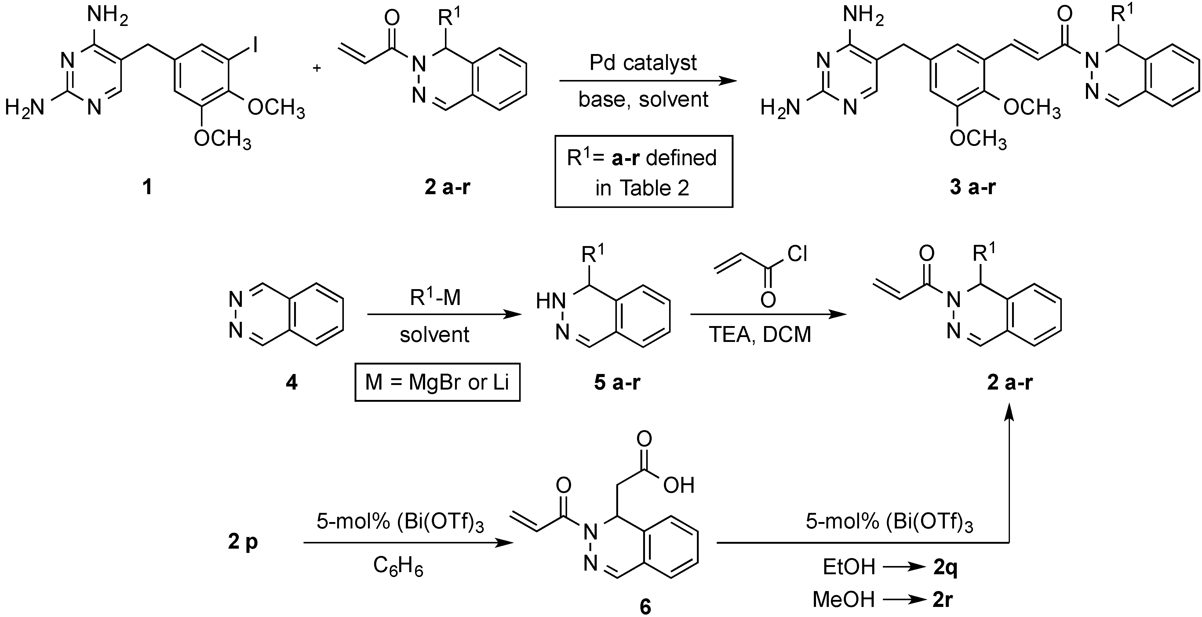
3.3. Preparation of Phthalazin-2(1H)-yl]-2-propen-1-ones 2ar

(±)-1-[1-(2-Methyl-1-propen-1-yl)phthalazin-2(1H)-yl]-2-propen-1-one (2a). This compound was prepared from 4 and 2-methyl-1-propenylmagnesium bromide in 62% yield on a 15.4-mmol scale according to the literature procedure [15]. The spectral data matched those reported [15].
(±)-1-(1-Allylphthalazin-2(1H)-yl)-2-propen-1-one (2b). A stirred solution of 4 (2.50 g, 19.2 mmol) in dry THF (60 mL) was treated dropwise with a solution of allylmagnesium bromide (12.4 mL, 24.9 mmol, 2 M in THF) for 30 min at 0 °C. The reaction mixture was warmed to room temperature and stirred for 1 h. After completion, the reaction mixture was treated with saturated NH4Cl (50 mL) and extracted with EtOAc (3 × 100 mL). The combined organic extracts were washed with saturated NaCl (50 mL), dried (MgSO4), filtered, and concentrated under vacuum to give 3b as a dark brown, viscous liquid. The crude 5b was dissolved in DCM (70 mL) and TEA (5.87 mL, 41.7 mmol) was added, followed by dropwise addition of acryloyl chloride (1.35 mL, 16.7 mmol) at 0 °C. The reaction mixture was stirred at this temperature for 2 h. The mixture was then treated with saturated NaCl (100 mL), the organic layer was separated, and the aqueous layer was extracted with DCM (2 × 50 mL). The combined extracts were washed with saturated NaCl (50 mL), dried (MgSO4), filtered, and concentrated to afford the crude product. This material was purified on a silica gel column eluted with hexanes–EtOAc (7:3) to afford 2b (2.47 g, 57%) as a viscous, yellow liquid. IR: 1662 cm−1; 1H-NMR (CDCl3): δ 7.60 (s, 1H), 7.43 (td, J = 7.4, 1.4 Hz, 1H), 7.35 (td, J = 7.6, 1.2 Hz, 1H), 7.30 (dd, J = 17.1, 10.5 Hz, 1H), 7.28 (d, J = 7.6, 0.6 Hz, 1H), 7.15 (dd, J = 7.6, 0.6 Hz, 1H), 6.42 (dd, J = 17.1, 2.1 Hz, 1H), 5.91 (t, J = 6.4 Hz, 1H), 5.78 (dd, J = 10.5, 2.1 Hz, 1H), 5.68 (ddt, J = 17.1, 10.1, 7.3 Hz, 1H), 4.98 (dm, J = 10.2 Hz, 1H), 4.90 (dm, J = 17.0 Hz, 1H), 2.41 (m, 2H); 13C-NMR (CDCl3): δ 166.3, 142.2, 133.2, 132.7, 131.4, 128.4, 128.1, 127.1, 126.6, 125.6, 123.9, 118.7, 51.1, 39.8.
(±)-1-(1-Vinylphthalazin-2(1H)-yl)-2-propen-1-one (2c). This compound was prepared via the procedure described for 2b using 4 (2.50 g, 19.2 mmol) and vinylmagnesium bromide (11.5 mL, 23.0 mmol, 2.0 M in THF) in dry THF (60 mL) to afford 3c, followed by acylation using TEA (5.3 mL, 37.8 mmol) and acryloyl chloride (1.20 mL, 15.1 mmol) in DCM (70 mL) to give 2c (2.36 g, 58%) as a colorless, viscous liquid. IR: 1665 cm−1; 1H-NMR (CDCl3): δ 7.59 (s, 1H), 7.45 (td, J = 7.4, 0.8 Hz, 1H), 7.40–7.28 (m, 3H), 7.21 (dd, J = 7.6, 0.4 Hz, 1H), 6.51 (dd, J = 17.4, 2.1 Hz, 1H), 6.37 (d, J = 4.9 Hz, 1H), 5.81 (m, 2H), 5.10 (dd, J = 10.3, 0.4 Hz, 1H), 4.86 (d, J = 17.4 Hz, 1H); 13C-NMR (CDCl3): δ 166.2, 141.7, 134.7, 131.9, 131.7, 128.8, 128.4, 126.9, 126.8, 125.9, 123.8, 116.2, 53.0.
(±)-1-(1-Propylphthalazin-2(1H)-yl)-2-propen-1-one (2d). This compound was prepared in 70% yield from 4 (2.00 g, 15.4 mmol) and propylmagnesium chloride (8.45 mL, 16.9 mmol, 2.0 M in ether) in dry THF (50 mL) to afford 5d, followed by acylation using TEA (1.82 g, 2.50 mL, 18.0 mmol) and acryloyl chloride (1.40 g, 1.26 mL, 15.5 mmol) in DCM (70 mL) to afford 2d according to the literature procedure [15]. The spectral data matched those reported [15].
(±)-1-(1-Pentylphthalazin-2(1H)-yl)-2-propen-1-one (2e). This compound was prepared via the procedure described for 2b from 4 (2.50 g, 19.2 mmol) and n-pentylmagnesium bromide (18.4 mL, 18.4 mmol, 1 M in THF) in dry THF (60 mL) to afford 5e, followed by acylation using TEA (3.27 g, 4.51 mL, 32.4 mmol) and acryloyl chloride (1.05 mL, 12.9 mmol) in DCM (70 mL) to give 2e (3.05 g, 62%) as an off-white solid, mp 52–54 °C. IR: 1664 cm−1; 1H-NMR (CDCl3): δ 7.61 (s, 1H), 7.42 (td, J = 7.4, 1.1 Hz, 1H), 7.33 (m, 2H), 7.26 (d, J = 7.0 Hz, 1H), 7.15 (dd, J = 7.4, 0.7 Hz, 1H), 6.47 (dd, J = 17.2, 2.4 Hz, 1H), 5.83 (t, J = 6.7 Hz, 1H), 5.77 (dd, J = 10.5, 2.4 Hz, 1H), 1.63 (m, 2H), 1.21 (m, 6H), 0.81 (t, J = 6.6 Hz, 3H); 13C-NMR (CDCl3): δ 166.1, 142.3, 134.0, 131.3, 128.2, 127.9, 127.1, 126.4, 125.6, 123.8, 51.2, 34.9, 31.4, 24.4, 22.3, 13.8.
(±)-1-(1-Cyclopropylphthalazin-2(1H)-yl)-2-propen-1-one (2f). This compound was prepared in 87% yield from 4 (2.00 g, 15.4 mmol) and cyclopropylmagnesium chloride (33.8 mL, 16.9 mmol, 0.5 M in THF) in dry THF (50 mL) to give 5f, followed by acylation using TEA (1.86 g, 2.56 mL, 18.4 mmol) and acryloyl chloride (1.39 g, 1.25 mL, 15.4 mmol) in DCM (70 mL) to afford 2f according to the literature procedure [13]. The spectral data matched those reported [13].
(±)-1-(1-Cyclobutylphthalazin-2(1H)-yl)-2-propen-1-one (2g). This compound was prepared via the procedure described for 2b using 4 (2.50 g, 19.2 mmol) and cyclobutylmagnesium bromide (24.9 mL, 24.9 mmol, 1 M in THF) in dry THF (60 mL) to give 5g, followed by acylation using TEA (3.25 g, 4.48 mL, 32.2 mmol) and acryloyl chloride (1.16 g, 1.04 mL, 12.8 mmol) in DCM (70 mL) to afford 2g (3.04 g, 66%) as a yellow liquid. IR: 1663 cm−1; 1H-NMR (CDCl3): δ 7.61 (s, 1H), 7.44 (td, J = 7.4, 1.5 Hz, 1H), 7.35 (td, J = 7.4, 1.5 Hz, 1H), 7.31 (dd, J = 17.2, 10.5 Hz, 1H), 7.29 (m, 1H), 7.16 (dd, J = 7.5, 0.7 Hz, 1H), 6.48 (dd, J = 17.2, 2.0 Hz, 1H), 5.83 (d, J = 8.4 Hz, 1H), 5.77 (dd, J = 10.5, 2.0 Hz, 1H), 2.60 (sextet, J = 8.4 Hz, 1H), 1.98 (m, 1H), 1.90–1.73 (m, 2H), 1.67 (m, 3H); 13C-NMR (CDCl3): δ 166.5, 143.8, 132.6, 131.3, 128.2, 128.0, 127.1, 126.5, 125.6, 124.0, 54.5, 40.2, 25.6, 24.9, 17.6.
(±)-1-(1-Cyclopentylphthalazin-2(1H)-yl)-2-propen-1-one (2h). This compound was prepared via the procedure described for 2b from 4 (2.50 g, 19.2 mmol) and cyclopentylmagnesium bromide (24.9 mL, 24.9 mmol, 1 M in THF) in dry THF (60 mL) to give 5h, followed by acylation using TEA (3.78 g, 5.20 mL, 37.4 mmol) and acryloyl chloride (1.35 g, 1.21 mL, 14.9 mmol) in DCM (70 mL) to afford 2h (2.73 g, 56%) as a viscous, colorless liquid. IR: 1663 cm−1; 1H-NMR (CDCl3): δ 7.69 (s, 1H), 7.43 (td, J = 7.4, 1.4 Hz, 1H), 7.35 (td, J = 7.4, 1.4 Hz, 1H), 7.32 (dd, J = 17.2, 10.5 Hz, 1H), 7.28 (dd, J = 7.4, 1.4 Hz, 1H), 7.18 (dd, J = 7.4, 0.5 Hz, 1H), 6.46 (dd, J = 17.2, 2.1 Hz, 1H), 5.80 (d, J = 8.6 Hz, 1H), 5.77 (dd, J = 10.5, 2.1 Hz, 1H), 2.12 (m, 1H), 1.61 (m, 3H), 1.42 (m, 4H), 1.23 (m, 1H); 13C-NMR (CDCl3): δ 166.3, 143.6, 133.5, 131.2, 128.3, 128.0, 127.1, 126.8, 125.4, 124.2, 54.1, 45.2, 29.3, 28.7, 24.3, 24.2.
(±)-1-(1-(tert-Butyl)phthalazin-2(1H)-yl)-2-propen-1-one (2i). A stirred solution of 4 (2.00 g, 17.6 mmol) in 50 mL of dry THF was treated dropwise with a solution of t-BuLi (16.9 mL, 16.9 mmol, 1.0 M in heptanes) over a period of 15 min at −78 °C. The reaction was stirred at this temperature for 1 h and then slowly warmed to room temperature and stirred for an additional 30 min. The reaction mixture was added to cold saturated NH4Cl (50 mL) and extracted with EtOAc (3 × 50 mL). The combined organic extracts were washed with saturated NaCl (50 mL), dried (MgSO4), filtered, and concentrated under vacuum to give 5i as a dark brown liquid. The crude product was dissolved in DCM (100 mL), and TEA (4.00 g, 5.50 mL, 39.6 mmol) was added, followed by dropwise addition of acryloyl chloride (1.44 g, 1.29 mL, 15.9 mmol) at 0 °C. The reaction mixture was stirred at this temperature for 2 h. The mixture was then added to saturated NaCl (100 mL), the organic layer was separated, and the aqueous layer was extracted with DCM (2 × 50 mL). The combined extracts were washed with saturated NaCl (50 mL), dried (MgSO4), filtered, and concentrated to afford the crude product. The crude product was purified on a silica gel column eluted with hexanes–EtOAc (7:3) to afford 2i as a viscous, yellow liquid (2.79 g, 75%). IR: 1666 cm−1; 1H-NMR (CDCl3): δ 7.65 (s, 1H), 7.46 (td, J = 7.4, 1.4 Hz, 1H), 7.37 (td, J = 7.4, 1.4 Hz, 1H), 7.34 (dd, J = 17.1, 10.1 Hz, 1H), 7.29 (dd, J = 7.4, 0.8 Hz, 1H), 7.19 (d, J = 7.8 Hz, 1H), 6.43 (dd, J = 17.1, 2.3 Hz, 1H), 5.78 (s, 1H), 5.75 (dd, J = 10.1, 2.3 Hz, 1H), 0.86 (s, 9H); 13C-NMR (CDCl3): δ 167.0, 144.6, 133.4, 131.0, 130.6, 128.6, 128.2, 128.0, 127.4, 125.4, 125.1, 58.1, 39.4, 26.6, 25.5.
(±)-1-(1-(Furan-2-yl)phthalazin-2(1H)-yl)-2-propen-1-one (2j). This compound was prepared in 72% yield from 4 (2.00 g, 15.4 mmol), furan-2-yllithium [from furan (1.20 g, 17.6 mmol) and n-BuLi (7.30 mL, 18.3 mmol, 2.5 M in hexanes)] in dry THF (50 mL) to give 5j, followed by acylation using TEA (2.37 g, 3.26 mL, 23.5 mmol) and acryloyl chloride (1.59 g, 1.43 mL, 17.6 mmol) in DCM (70 mL) to afford 2j according to the literature procedure [13]. The spectral data matched those reported [13].
(±)-1-[1-(Thiophen-2-yl)phthalazin-2(1H)-yl]-2-propen-1-one (2k). This compound was prepared in 60% yield from 4 (2.00 g, 15.4 mmol), thiophen-2-ylmagnesium bromide [from 2-bromothiophene (1.77 g, 1.69 mL, 21.0 mmol) and magnesium (0.69 g, 28.4 mmol)] in dry THF (50 mL) to give 5k. The crude 5k was acylated using TEA (2.80 g, 3.86 mL, 27.7 mmol) and acryloyl chloride (1.90 g, 1.71 mL, 21.0 mmol) in DCM (70 mL) to afford 2k according to the literature procedure [13]. The spectral data matched those reported [13].
(±)-1-[1-(1-Methyl-1H-indol-2-yl)phthalazin-2(1H)-yl]-2-propen-1-one (2l). To a stirred solution of 1-methylindole (1.50 g, 11.4 mmol) in dry THF (25 mL) was added dropwise n-BuLi (6.86 mL, 17.2 mmol, 2.5 M in hexanes) over a period of 30 min at −78 °C. The solution was warmed to −25 °C, and stirring was continued at this temperature for 1 h. The reaction mixture was cooled to −78 °C, and a solution of 4 (1.48 g, 11.4 mmol) in dry THF (20 mL) was added dropwise over 30 min. The reaction mixture was stirred at this temperature for 2 h. The mixture was poured into saturated NH4Cl (100 mL) and extracted with EtOAc (3 × 50 mL). The combined organic extracts were washed with saturated NaCl (50 mL), dried (MgSO4), filtered, and concentrated under vacuum to give 5l as a light yellow liquid. The crude product 5l was dissolved in DCM (100 mL), and TEA (2.08 g, 2.87 mL, 20.6 mmol) was added, followed by dropwise addition of acryloyl chloride (0.81 g, 0.73 mL, 8.95 mmol) at 0 °C. The reaction mixture was stirred at this temperature for an additional 2 h. The aqueous layer was added to saturated NaCl (50 mL), and the organic layer was separated. The aqueous layer was extracted with DCM (2 × 30 mL), and the combined organic extracts were washed with saturated NaCl (50 mL), dried (MgSO4), filtered, and concentrated to afford the crude product. The product was purified on a silica gel column eluted with hexanes–EtOAc (7:3) to afford 2l (3.01 g, 62%) as a light yellow solid, mp 69–71 °C. IR: 1657 cm−1; 1H-NMR (CDCl3): δ 7.79 (s, 1H), 7.42–7.34 (complex, 5H), 7.22 (dd, J = 17.2, 10.5 Hz, 1H), 7.18 (s, 1H), 7.16 (m, 2H), 7.00 (t, J = 7.6 Hz, 1H), 6.45 (dd, J = 7.2, 2.0 Hz, 1H), 5.89 (s, 1H), 5.75 (dd, J = 10.5, 2.0 Hz, 1H), 3.99 (s, 3H); 13C-NMR (CDCl3): δ 166.0, 143.3, 138.9, 137.4, 132.3, 132.1, 129.0, 128.6, 127.2, 127.0, 126.7, 126.0, 123.7, 122.0, 120.5, 119.6, 109.6, 103.3, 46.6, 30.6.
(±)-1-[1-(Benzofuran-2-yl)phthalazin-2(1H)-yl]-2-propen-1-one (2m). The compound was prepared using benzofuran-2-yllithium [from benzofuran (2.00 g, 16.9 mmol) and n-BuLi (6.8 mL, 17.0 mmol, 2.5 M in hexanes)] and 4 (2.20 g, 16.9 mmol) in dry THF (75 mL) to give 5m. The crude 5m was acylated using TEA (3.05 g, 4.20 mL, 30.2 mmol) and acryloyl chloride (1.09 g, 0.97 mL, 12.0 mmol) in DCM (120 mL) to afford 2m (2.37 g, 51%) as a light yellow solid, mp 55–57 °C. IR: 1657 cm−1; 1H-NMR (CDCl3): δ 7.84 (d, J = 7.8 Hz, 1H), 7.63 (dd, J = 7.8, 0.6 Hz, 1H), 7.56 (d, J = 8.2 Hz, 1H), 7.48–7.40 (complex, 3H), 7.36 (td, J = 7.5, 1.2 Hz, 2H), 7.27 (t, J = 7.5 Hz, 1H), 7.23 (s, 1H), 7.23 (d, J = 7.5 Hz, 1H), 6.54 (dd, J = 17.1, 2.1 Hz, 1H), 5.84 (dd, J = 10.4, 2.0 Hz, 1H), 5.02 (s, 1H); 13C-NMR (CDCl3): δ 166.4, 154.9, 151.1, 141.8, 131.7, 131.0, 139.1, 138.1, 137.7, 126.5, 126.3, 126.0, 125.4, 123.6, 123.3, 121.5, 111.5, 108.0, 42.0.
(±)-1-[1-(Benzo[b]thiophen-2-yl)phthalazin-2(1H)-yl]-2-propen-1-one (2n). The compound was prepared using benzothiophen-2-yllithium [from benzothiophene (2.00 g, 14.9 mmol) and n-BuLi (6.0 mL, 15.0 mmol, 2.5 M in hexanes)] and 4 (1.94 g, 14.9 mmol) in dry THF (75 mL) to give 5n. The crude 5n was acylated using TEA (2.86 g, 3.94 mL, 28.3 mmol) and acryloyl chloride (1.02 g, 0.92 mL, 11.3 mmol) in DCM (120 mL) to afford 2n (2.94 g, 62%) as a light yellow solid, mp 69–71 °C. IR: 1663 cm−1; 1H-NMR (CDCl3): δ 7.86 (m, 1H), 7.81 (m, 2H), 7.70 (s, 1H), 7.51 (t, J = 7.6 Hz, 1H), 7.46–7.36 (complex, 5H), 7.32 (d, J = 7.2 Hz, 1H), 6.54 (dd, J = 17.2, 1.8 Hz, 1H), 5.87 (dd, J = 10.3, 1.8 Hz, 1H), 5.05 (s, 1H); 13C-NMR (CDCl3): δ 166.4, 146.4, 139.8, 139.7, 139.0, 131.8, 131.7, 129.2, 128.2, 126.6, 126.1, 125.8 (2C), 125.7, 124.6, 124.4, 124.2, 122.2, 42.2.
(±)-1-[1-(Benzo[d]thiazol-2-yl)phthalazin-2(1H)-yl]-2-propen-1-one (2o). The compound was prepared using benzothiazol-2-yllithium [from (2.00 g, 14.8 mmol) and n-BuLi (6.5 mL, 16.3 mmol, 2.5 M in hexanes)] and 4 (1.92 g, 14.8 mmol) in dry THF (75 mL) to give 5o. The crude 5o was acylated using TEA (2.86 g, 3.94 mL, 28.3 mmol) and acryloyl chloride (1.02 g, 0.92 mL, 11.3 mmol) in DCM (120 mL) to afford 5o (3.20 g, 68%) as a light yellow solid, mp 68–70 °C. IR: 1663 cm−1; 1H-NMR (CDCl3): δ 7.93 (d, J = 8.2 Hz, 1H), 7.75 (dt, J = 8.0, 0.6 Hz, 1H), 7.70 (s, 1H), 7.56 (d, J = 7.6 Hz, 1H), 7.51 (td, J = 7.4, 1.4 Hz, 1H), 7.45–7.27 (complex, 6H), 6.56 (dd, J = 17.4, 2.0 Hz, 1H), 5.87 (dd, J = 10.5, 2.0 Hz, 1H); 13C-NMR (CDCl3): δ 169.5, 166.6, 153.0, 141.5, 135.2, 132.2, 130.3, 129.9, 129.3, 127.7, 126.5, 126.3, 125.9, 125.2, 123.6, 123.2, 121.5, 53.2.
(±)-t-Butyl 2-(2-acryloylphthalazin-2(1H)-yl)acetate (2p). This compound was prepared in 87% yield from tert-butyl acetate (2.67 g, 3.08 mL, 23.0 mmol), n-BuLi (7.7 mL, 19.3 mmol, 2.5 M in hexanes), 4 (2.99 g, 23.0 mmol), TEA (1.86 g, 2.56 mL, 18.4 mmol) and acryloyl chloride (1.39 g, 1.25 mL, 15.4 mmol) according to the literature procedure [13]. The spectral data matched those reported [13].
(±)-2-(2-Acryloylpthalazin-2(1H)-yl)acetic acid (6). This compound was prepared in 94% yield from 2p (1.50 g, 5.00 mmol), and Bi(OTf)3 (0.164 g, 0.25 mmol, 5 mol %) in benzene (25 mL) according to the literature procedure [13]. The spectral data matched those reported [13].
(±)-Ethyl 2-(2-acryloylphthalazin-2(1H)-yl)acetate (2q). This compound was prepared in 95% yield from 6 (1.00 g, 4.10 mmol), and Bi(OTf)3 (0.134 g, 0.20 mmol, 5 mol %) in ethanol (25 mL) according to the literature procedure [13]. The spectral data matched those reported [13].
(±)-Methyl 2-(2-acryloylphthalazin-2(1H)-yl)acetate (2r). This compound was prepared in 95% yield from 6 (1.00 g, 4.10 mmol), Bi(OTf)3 (0.134 g, 0.20 mmol, 5 mol %) in CH3OH (25 mL) according to the literature procedure [13]. The spectral data matched those reported [13].

3.4. Preparation of Drug Candidates 3ar

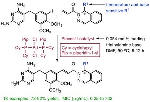
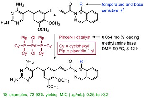
(±)-(E)-3-{5-[(2,4-Diaminopyrimidin-5-yl)methyl]-2,3-dimethoxyphenyl}-1-[1-(2-methyl-1-propen-1-yl)phthalazin-2(1H)yl]-2-propen-1-one (3a). To a stirred solution of 1 (1.00 g, 2.59 mmol) in dry DMF (10 mL) was added a solution of 
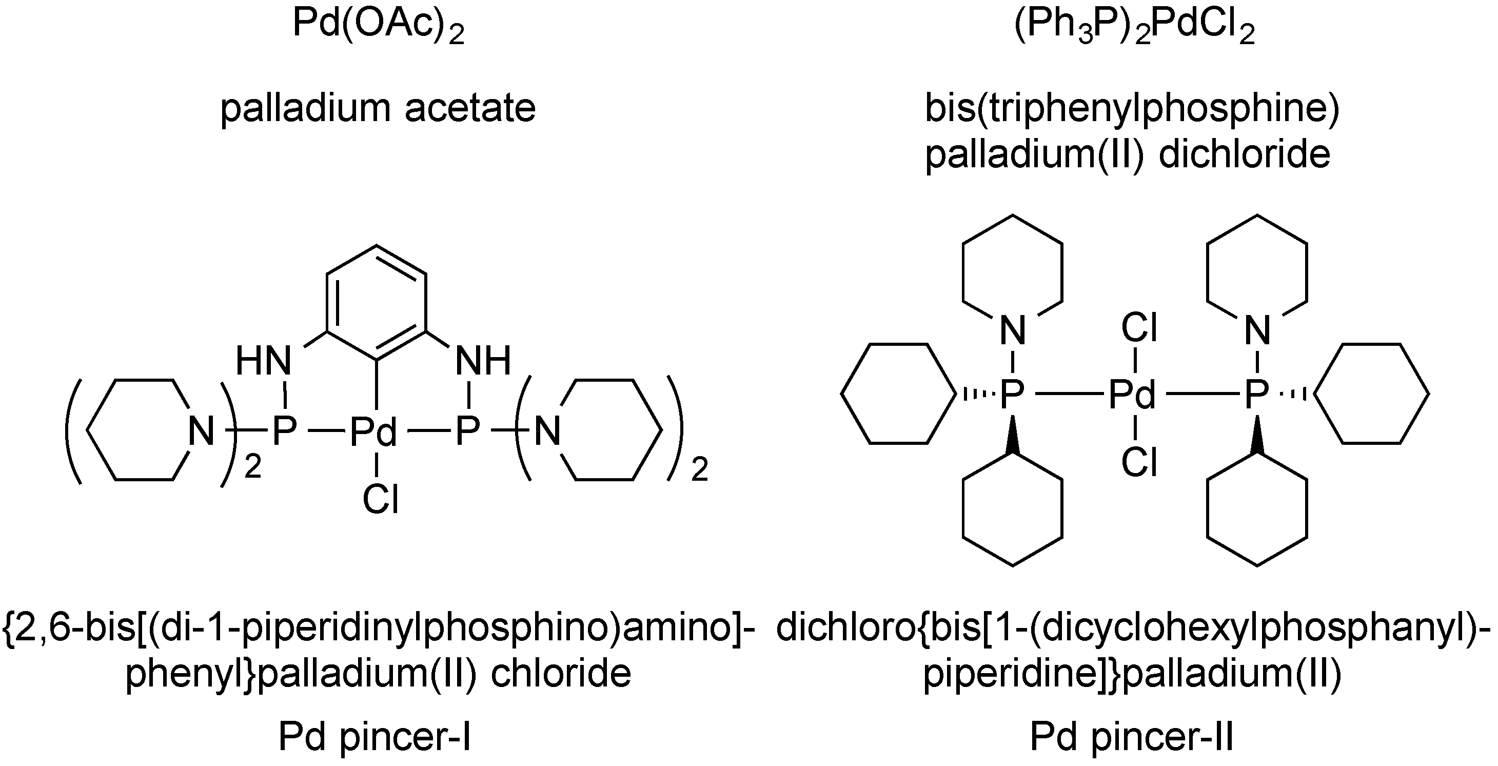
2a (0.684 g, 2.85 mmol) in DMF (2 mL), followed by TEA (0.313 g, 0.431 mL, 3.10 mmol), and the Pd pincer-II catalyst (1 mg, 0.0014 mmol). The reaction was heated at 90 °C for 8 h and then cooled using an ice bath. The product was purified by directly pouring the crude reaction mixture onto a 50 cm × 2.5 cm silica gel chromatography column slurry packed with DCM. Impurities were eluted using DCM, and the final product was collected using DCM/MeOH/TEA (95:4:1). Evaporation of the solvent gave a yellow solid, which was dried under high vacuum for 2 h. MeOH (5 mL) was added to dissolve the crude product, followed by ether (10 mL), and the mixture was cooled for 4 h to crystallize the product. The product was filtered and dried under vacuum to afford 3a as a yellow solid (1.18, 88%). The melting point and spectral data matched those in the literature [15].
(±)-(E)-1-(1-Allylphthalazin-2(1H)-yl)-3-{5-[(2,4-diaminopyrimidin-5-yl)methyl]-2,3-dimethoxy-phenyl}-2-propen-1-one (3b). This compound was prepared as above using 1 (2.00 g, 5.18 mmol), 2b (1.55 g, 6.73 mmol), TEA (0.680 g, 0.937 mL, 6.73 mmol), and the Pd pincer-II catalyst (2 mg, 0.0028) in dry DMF (15 mL) to give 3b (2.03 g, 81%) as a white solid, mp 215–217 °C. IR: 3418, 3123, 3122, 1657, 1641, 1598, 1564 cm−1; 1H-NMR (DMSO-d6): δ 7.91 (s, 1H), 7.86 (d, J = 16.2 Hz, 1H), 7.62 (d, J = 16.2 Hz, 1H), 7.60 (s, 1H), 7.56–7.37 (complex, 4H), 7.24 (s, 1H), 6.99 (s, 1H), 6.19 (br s, 2H), 5.95 (t, J = 6.3 Hz, 1H), 5.74 (br s, 2H), 5.66 (ddt, J = 17.4, 10.5, 6.3 Hz, 1H), 4.94 (dm, J = 10.5 Hz, 1H), 4.86 (dm, J = 17.4 Hz, 1H), 3.79 (s, 3H), 3.74 (s, 3H), 3.60 (s, 2H), 2.36 (m, 2H); 13C-NMR (DMSO-d6): δ 165.6, 162.3, 162.2, 155.8, 152.5, 146.0, 142.5, 136.6, 133.2, 132.9, 131.7, 128.3, 127.8, 126.6, 126.0, 123.7, 118.4, 118.3, 117.9, 114.8, 105.7, 60.8, 55.7, 54.9, 50.4, 32.4 (1 aromatic/alkene C unresolved); Anal. Calcd for C27H28N6O3·1.3 H2O: C, 63.84; H, 6.07; N, 16.54. Found: C, 63.84; H, 5.69; N, 16.28.
(±)-(E)-3-{5-[(2,4-Diaminopyrimidin-5-yl)methyl]-2,3-dimethoxyphenyl}-1-(1-vinylphthalazin-2(1H)-yl)-2-propen-1-one (3c). This compound was prepared as above using 1 (1.60 g, 4.15 mmol), 2c (1.14 g, 5.38 mmol), TEA (0.543 g, 0.750 mL, 5.38 mmol) and the Pd pincer-II catalyst (1.5 mg, 0.0021 mmol) in dry DMF (15 mL) to give 3c (1.65 g, 85%) as a white solid, mp 210–212 °C. IR: 3354, 3169, 1638, 1593, 1567 cm−1; 1H-NMR (DMSO-d6): δ 7.91 (s, 1H), 7.89 (d, J = 16.0 Hz, 1H), 7.68 (d, J = 16.0 Hz, 1H), 7.60–7.44 (complex, 5H), 7.32 (d, J = 1.3 Hz, 1H), 7.25 (br s, 2H), 7.04 (d, J = 1.4, 1H), 6.73 (br s, 2H), 6.40 (d, J = 4.5 Hz, 1H), 5.80 (ddd, J = 1.5, 10.2, 4.5 Hz, 1H), 5.08 (d, J = 10.2 Hz, 1H), 4.78 (d, J = 17.0 Hz, 1H), 3.81 (s, 3H), 3.76 (s, 3H), 3.65 (s, 2H); 13C-NMR (DMSO-d6): δ 165.6, 163.2, 157.7, 152.6, 146.8, 146.3, 142.1, 136.9, 135.4, 135.0, 132.0, 131.7, 128.6, 127.9, 126.9, 126.3, 123.5, 118.8, 117.9, 115.3, 115.0, 107.6, 60.8, 55.9, 52.5, 31.9; Anal. Calcd for C26H26N6O3·4.6 H2O·0.1 C2H5OH: C, 56.21; H, 5.32; N, 15.01. Found: C, 56.25; H, 4.99; N, 15.04.
(±)-(E)-3-{5-[(2,4-Diaminopyrimidin-5-yl)methyl]-2,3-dimethoxyphenyl}-1-(1-propylphthalazin-2(1H)-yl)-2-propen-1-one (3d). This compound was prepared as above using 1 (1.00 g, 2.59 mmol), 2d (0.701 g, 3.10 mmol), TEA (0.313 g, 0.432 mL, 3.10 mmol), and the Pd pincer-II catalyst (1 mg, 0.0014 mmol) in dry DMF (15 mL) to give 3d (1.08 g, 86%) as a white solid. The melting point and spectral data matched those in the literature [15].
(±)-(E)-3-{5-[(2,4-Diaminopyrimidin-5-yl)methyl]-2,3-dimethoxyphenyl}-1-(1-pentylphthalazin-2(1H)-yl)-2-propen-1-one (3e). This compound was prepared as above using 1 (1.60 g, 4.15 mmol), 2e (1.39 g, 5.35 mmol), TEA (0.500 g, 0.689 mL, 4.95 mmol), and the Pd pincer-II catalyst (1.5 mg, 0.0021 mmol) in dry DMF (15 mL) to give 3e (1.91 g, 90%) as a white solid, mp 212–213 °C. IR: 3363, 3173, 1638, 1590, 1560 cm−1; 1H-NMR (DMSO-d6): δ 7.92 (s, 1H), 7.86 (d, J = 16.4 Hz, 1H), 7.64 (d, J = 16.4 Hz, 1H), 7.54 (s, 1H), 7.52 (m, 2H), 7.43 (td, J = 7.4, 0.8 Hz, 1H), 7.38 (d, J = 1.6 Hz, 1H), 7.28 (d, J = 1.6 Hz, 1H), 7.08 (br s, 2H), 7.02 (d, J = 1.5 Hz, 1H), 6.58 (br s, 2H), 5.82 (t, J = 6.7 Hz, 1H), 3.79 (s, 3H), 3.74 (s, 3H), 3.63 (s, 2H), 1.53 (m, 2H), 1.16 (m, 6H) 0.77 (t, J = 6.6 Hz, 3H); 13C-NMR (DMSO-d6): δ 165.6, 163.1, 158.3, 152.6, 148.2, 146.2, 142.8, 136.5, 135.2, 133.6, 131.7, 128.3, 127.9, 126.5, 126.1, 123.6, 118.7, 118.0, 114.9, 107.3, 60.8, 55.8, 50.5, 34.5, 32.0, 30.9, 24.0, 21.9, 13.8; Anal. Calcd for C29H34N6O3·3.9 H2O·0.3 C2H5OH: C, 59.94; H, 6.42; N, 14.21. Found: C, 59.61; H, 6.42; N, 14.21.
(±)-(E)-1-(1-Cyclopropylphthalazin-2(1H)-yl)-3-{5-[(2,4-diaminopyrimidin-5-yl)methyl]-2,3-dimethoxyphenyl}-2-propen-1-one (3f). This compound was prepared as above using 1 (1.00 g, 2.59 mmol), 2f (0.645 g, 2.80 mmol), TEA (0.313 g, 0.432 mL, 3.10 mmol), and the Pd pincer-II catalyst (1 mg, 0.0014 mmol) in dry DMF (15 mL) to give 3f (1.05 g, 84%) as a white solid. The melting point and spectral data matched those in the literature [13].
(±)-(E)-1-(1-Cyclobutylphthalazin-2(1H)-yl)-3-{5-[(2,4-diaminopyrimidin-5-yl)methyl]-2,3-dimethoxyphenyl}-2-propen-1-one (3g). This compound was prepared as above using 1 (1.50 g, 3.89 mmol), 2g (1.21 g, 5.04 mmol), TEA (0.510 g, 0.702 mL, 5.05 mmol), and the Pd pincer-II catalyst (1.5 mg, 0.0021 mmol) in dry DMF (15 mL) to give 3g (1.68 g, 87%) as a white solid, mp 125–127 °C. IR: 3397, 3272, 1642, 1605, 1562 cm−1; 1H-NMR (DMSO-d6): δ 7.93 (s, 1H), 7.85 (d, J = 16.0 Hz, 1H), 7.64 (d, J = 16.0 Hz, 1H), 7.59 (s, 1H), 7.52 (m, 2H), 7.44 (m, 2H), 7.25 (s, 1H), 7.00 (s, 1H), 6.39 (br s, 2H), 5.92 (br s, 2H), 5.86 (d, J = 8.2 Hz, 1H), 3.79 (s, 3H), 3.74 (s, 3H), 3.60 (s, 2H), 2.54 (sextet, J = 8.2 Hz, 1H), 1.84 (m, 3H), 1.63 (m, 3H); 13C-NMR (DMSO-d6): δ 165.9, 162.4, 161.4, 154.0, 152.5, 146.0, 143.3, 136.6, 136.3, 132.1, 131.7, 128.3, 127.8, 126.6, 126.1, 123.8, 118.4, 117.9, 114.8, 106.1, 60.8, 55.8, 53.8, 32.3, 25.1, 24.2, 17.1; Anal. Calcd for C28H30N6O3·1.6 H2O: C, 60.27; H, 6.31; N, 14.84. Found: C, 60.08; H, 6.07; N, 15.01.
(±)-(E)-1-(1-Cyclopentylphthalazin-2(1H)-yl)-3-{5-[(2,4-diaminopyrimidin-5-yl)methyl]-2,3-dimethoxyphenyl}-2-propen-1-one (3h). This compound was prepared as above using 1 (1.50 g, 3.89 mmol), 2h (1.28 g, 5.04 mmol), TEA (0.510 g, 0.702 mL, 5.05 mmol), and the Pd pincer-II catalyst (1.5 mg, 0.0021 mmol) in dry DMF (15 mL) to give 3h (1.83 g, 92%) as a white solid, mp 190–192 °C. IR: 3338, 3174, 1637, 1594, 1560 cm−1; 1H-NMR (DMSO-d6, 400 MHz): δ 7.96 (s, 1H), 7.81 (d, J = 16.1 Hz, 1H), 7.60 (d, J = 16.1 Hz, 1H), 7.52 (s, 1H), 7.49 (m, 1H), 7.42 (d, J = 7.4 Hz, 2H), 7.38 (d, J = 7.4, 1H), 7.23 (d, J = 1.6 Hz, 1H), 6.97 (d, J = 1.6 Hz, 1H), 6.86 (br s, 2H), 6.37 (br s, 2H), 5.74 (d, J = 8.4 Hz, 1H), 3.76 (s, 3H), 3.70 (s, 3H), 3.58 (s, 2H), 2.10 (sextet, J = 8.4 Hz, 1H), 1.50 (m, 3H), 1.36 (m, 3H), 1.22 (m, 2H); 13C-NMR (DMSO-d6): δ 165.4, 162.9, 159.2, 152.6, 149.7, 146.1, 144.1, 136.6, 135.5, 133.1, 131.6, 128.3, 127.9, 126.9, 125.9, 124.1, 118.6, 118.0, 114.9, 107.0, 60.8, 55.8, 53.4, 44.6, 32.1, 29.0, 28.2, 24.1, 24.0; Anal. Calcd for C29H32N6O3·3.5 H2O: C, 60.32; H, 5.84; N, 14.55. Found: C, 60.19; H, 5.79; N, 14.57.
(±)-(E)-1-(1-tert-Butylphthalazin-2(1H)-yl)-3-{5-[(2,4-diaminopyrimidin-5-yl)methyl]-2,3-dimethoxyphenyl}-2-propen-1-one (3i). This compound was prepared as above using 1 (2.20 g, 5.70 mmol), 2i (1.77 g, 7.32 mmol), TEA (0.748 g, 1.03 mL, 7.41 mmol), and the Pd pincer-II catalyst (2 mg, 0.0028 mmol) in dry DMF (15 mL) to give 3i (2.36 g, 83%) as a white solid, mp 228–230 °C. IR: 3354, 3174, 1637, 1590, 1561 cm−1; 1H-NMR (DMSO-d6): δ 7.97 (s, 1H), 7.84 (d, J = 16.0 Hz, 1H), 7.71 (d, J = 16.0 Hz, 1H), 7.56 (td, J = 7.5, 1.6 Hz, 1H), 7.53 (m, 2H), 7.48 (td, J = 7.4, 1.2 Hz, 1H), 7.46 (br s, 2H), 7.39 (d, J = 7.8 Hz, 1H), 7.32 (d, J = 1.5 Hz, 1H), 7.02 (d, J = 2.0 Hz, 1H), 6.92 (br s, 2H), 5.79 (s, 1H), 3.80 (s, 3H), 3.73 (s, 3H), 3.64 (s, 2H), 0.80 (s, 9H); 13C-NMR (DMSO-d6): δ 166.7, 163.4, 156.8, 152.6, 146.2, 145.1, 144.7, 136.3, 134.6, 131.3, 129.8, 128.5, 128.4, 128.0, 125.5, 125.1, 118.9, 118.4, 114.9, 107.9, 60.8, 53.7, 55.9, 39.0, 31.8, 26.4 (3C); Anal. Calcd for C28H32N6O3·2.4 H2O·1.1 C2H5OH: C, 56.50; H, 5.96; N, 14.21. Found: C, 56.31; H, 5.74; N, 13.93.
(±)-(E)-3-{5-[(2,4-Diaminopyrimidin-5-yl)methyl]-2,3-dimethoxyphenyl}-1-[1-(furan-2-yl)phthalazin-2(1H)-yl]-2-propen-1-one (3j). This compound was prepared as above using 1 (1.00 g, 2.59 mmol), 2j (0.785 g, 3.12 mmol), TEA (0.313 g, 0.432 mL, 3.10 mmol), and the Pd pincer-II catalyst (1 mg, 0.0014 mmol) in dry DMF (15 mL) to give 3j (1.19 g, 90%) as a white solid. The melting point and spectral data matched those in the literature [13].
(±)-(E)-3-{5-[(2,4-Diaminopyrimidin-5-yl)methyl]-2,3-dimethoxyphenyl}-1-[1-(thiophen-2-yl)phthalazin-2(1H)-yl]-2-propen-1-one (3k). This compound was prepared as above using 1 (1.00 g, 2.59 mmol), 2k (0.836 g, 3.12 mmol), TEA (0.313 g, 0.432 mL, 3.10 mmol), and the Pd pincer-II catalyst (1 mg, 0.0014 mmol) in dry DMF (15 mL) to give 3k (1.10 g, 81%) as a white solid. The melting point and spectral data matched those in the literature [13].
(±)-(E)-3-{5-[(2,4-Diaminopyrimidin-5-yl)methyl]-2,3-dimethoxyphenyl}-1-[1-(1-methyl-1H-indol-2-yl)phthalazin-2(1H)-yl]-2-propen-1-one (3l). This compound was prepared as above using 1 (1.30 g, 3.37 mmol), 2l (1.27 g, 4.04 mmol), TEA (0.440 g, 0.60 mL, 4.36 mmol), and the Pd pincer-II catalyst (1.3 mg, 0.0018 mmol) in dry DMF (15 mL) to give 3l (1.45 g, 75%) as a white solid, mp 178–180 °C. IR: 3345, 3150, 1634, 1602, 1562 cm−1; 1H-NMR (DMSO-d6): δ 8.12 (s, 1H), 7.86 (d, J = 16.0 Hz, 1H), 7.63 (d, J = 16.0 Hz, 1H), 7.62 (d, J = 7.8 Hz, 1H), 7.55–7.48 (complex, 4H), 7.44 (d, J = 7.2 Hz, 1H), 7.36 (d, J = 7.8 Hz, 1H), 7.27 (d, J = 1.5 Hz, 1H), 7.26 (s, 1H), 7.11 (td, J = 7.4, 0.8 Hz, 1H), 7.02 (br s, 2H), 7.01 (d, J = 1.5 Hz, 1H), 6.95 (td, J = 7.4, 0.8 Hz, 1H), 6.52 (br s, 2H), 5.92 (s, 1H), 4.05 (s, 3H), 3.78 (s, 3H), 3.71 (s, 3H), 3.61 (s, 2H); 13C-NMR (DMSO-d6): δ 165.2, 163.0, 158.5, 152.6, 148.5, 146.2, 142.8, 140.3, 137.2, 136.6, 135.3, 132.3, 128.7, 127.8, 126.8, 126.7, 123.1, 121.6, 120.0, 119.4, 118.7, 117.8, 115.1, 110.1, 107.2, 100.9, 60.8, 55.8, 46.3, 32.0, 30.5 (2 aromatic C unresolved); Anal. Calcd for C33H31N7O3·4.1 H2O: C, 61.70; H, 5.28; N, 14.92. Found: C, 61.75; H, 5.13; N, 15.25.
(±)-(E)-1-[1-(Benzofuran-2-yl)phthalazin-2(1H)-yl]-3-{5-[(2,4-diaminopyrimidin-5-yl)methyl]-2,3-dimethoxyphenyl}-2-propen-1-one (3m). This compound was prepared as above using 1 (1.80 g, 4.66 mmol), 2m (1.83 g, 6.06 mmol), TEA (0.615 g, 0.85 mL, 6.06 mmol), and the Pd pincer-II catalyst (2 mg, 0.0028 mmol) in dry DMF (15 mL) to give 3m (2.09 1.51 g, 80%) as a light yellow solid, mp 222–224 °C. IR: 3345, 3150, 1634, 1602, 1562 cm−1; 1H-NMR (DMSO-d6): δ 7.83 (d, J = 16.0 Hz, 1H), 7.83 (m, 2H), 7.73 (d, J = 16.0 Hz, 1H), 7.73 (obscured, 1H), 7.61 (s, 1H), 7.58 (t, J = 7.4 Hz, 1H), 7.53 (s, 1H), 7.52–7.42 (complex, 4H), 7.33 (t, J = 7.4 Hz, 1H), 7.17 (d, J = 1.7 Hz, 1H), 7.05 (d, J = 1.7 Hz, 1H), 6.18 (br s, 2H), 5.68 (br s, 2H), 5.05 (s, 1H), 3.78 (s, 3H), 3.76 (s, 3H), 3.60 (s, 2H); 13C-NMR (DMSO-d6): δ 166.1, 162.3, 162.1, 155.5, 154.4, 152.5, 150.5, 146.1, 141.2, 137.6, 136.6, 132.1, 131.3, 128.5, 127.8, 127.7, 126.8, 126.0, 125.7, 123.6, 123.2, 122.1, 119.2, 117.8, 115.2, 111.6, 108.7, 105.8, 60.7, 55.8, 42.0, 32.4; Anal. Calcd for C32H28N6O4: C, 68.56; H, 5.03; N, 14.92. Found: C, 68.30; H, 4.95; N, 14.92.
(±)-(E)-1-[1-(Benzo[b]thiophen-2-yl)phthalazin-2(1H)-yl]-3-{5-[(2,4-diaminopyrimidin-5-yl)methyl]-2,3-dimethoxyphenyl}-2-propen-1-one (3n). This compound was prepared as above using 1 (1.60 g, 4.15 mmol), 2n (1.71 g, 5.38 mmol), TEA (0.543 g, 0.75 mL, 5.38 mmol), and the Pd pincer-II catalyst (1.5 mg, 0.0021 mmol) in dry DMF (15 mL) to give 3n (1.86 g, 78%) as a yellow solid, mp 256–258 °C. IR: 3347, 3180, 1637, 1601, 1559 cm−1; 1H-NMR (DMSO-d6): δ 8.27 (d, J = 6.4 Hz, 1H), 8.10–7.30 (complex, 10H), 7.15 (s, 2H), 6.18 (br s, 2H), 5.76 (s, 1H), 5.65 (br s, 2H), 5.06 (s, 2H), 3.84 (s, 3H), 3.80 (s, 3H), 3.67 (s, 2H); 13C-NMR (DMSO-d6): δ 165.9, 162.9, 158.8, 152.6, 149.1, 146.3, 144.5, 142.5, 139.1, 138.5, 137.6, 135.4, 132.4, 131.5, 129.3, 127.7, 127.3, 126.5, 124.6, 124.5, 123.8, 123.5, 122.4 (2C), 118.8, 117.6, 115.1, 107.1, 60.8, 55.8, 49.6, 32.0; Anal. Calcd for C32H28N6O3S·2.1 H2O: C, 62.55; H, 5.18; N, 13.14. Found: C, 62.72; H, 4.91; N, 13.06.
(±)-(E)-1-[1-(Benzo[d]thiazol-2-yl)phthalazin-2(1H)-yl]-3-{5-[(2,4-diaminopyrimidin-5-yl)methyl]-2,3-dimethoxyphenyl}-2-propen-1-one (3o). This compound was prepared as above using 1 (1.50 g, 3.89 mmol), 2o (1.61 g, 5.04 mmol), TEA (0.510 g, 0.70 mL, 5.05 mmol), and the Pd pincer-II catalyst (1 mg, 0.0014 mmol) in dry DMF (15 mL) to give 3o (1.61 g, 72%) as a white solid, mp 145–147 °C. IR: 3339, 3202, 1660, 1614, 1564 cm−1; 1H-NMR (DMSO-d6): δ 8.04 (s, 1H), 8.03 (obscured dm, 1H), 7.95 (d, J = 16.0 Hz, 1H), 7.91 (dd, J = 7.4, 0.8 Hz, 1H), 7.74 (d, J = 16.0 Hz, 1H), 7.63 (td, J = 7.4, 1.2 Hz, 1H), 7.60 (m, 2H), 7.54 (td, J = 7.4, 0.8 Hz, 1H), 7.47 (td, J = 7.4, 0.8 Hz, 1H), 7.45 (s, 1H), 7.40 (td, J = 7.4, 1.2 Hz, 1H), 7.34 (d, J = 1.6 Hz, 1H), 7.04 (d, J = 1.6 Hz, 1H), 6.66 (br s, 2H), 6.17 (br s, 2H), 5.77 (s, 1H), 3.81 (s, 3H), 3.75 (s, 3H), 3.63 (s, 2H); 13C-NMR (DMSO-d6): δ 170.1, 168.1, 162.6, 160.3, 152.5, 152.2, 151.9, 146.3, 141.9, 137.8, 136.0, 134.7, 132.3, 130.1, 129.5, 127.8, 127.6, 126.7, 126.4, 125.5, 123.2, 122.8, 122.4, 118.6, 117.3, 115.2, 106.5, 60.8, 55.8, 52.5, 32.2; Anal. Calcd for C31H27N7O3S·3.0 H2O·0.5 C2H5OH: C, 58.70; H, 5.12; N, 14.08. Found: C, 58.49; H, 4.81; N, 14.15.
(±)-tert-Butyl (E)-2-[2-(3-{5-[(2,4-diaminopyrimidin-5-yl)methyl]-2,3-dimethoxyphenyl}acryloyl)-1,2-dihydrophthalazin-1-yl]acetate (3p). This compound was prepared as above using 1 (1.00 g, 2.59 mmol), 2p (0.935 g, 3.12 mmol), TEA (0.313 g, 0.432 mL, 3.10 mmol), and the Pd pincer-II catalyst (1 mg, 0.0014 mmol) in dry DMF (15 mL) to give 3p (1.20 g, 83%) as a white solid. The melting point and spectral data matched those in the literature [13].
(±)-Ethyl (E)-2-[2-(3-{5-[(2,4-diaminopyrimidin-5-yl)methyl]-2,3-dimethoxyphenyl}acryloyl)-1,2-dihydrophthalazin-1-yl]acetate (3q). This compound was prepared as above using 1 (1.00 g, 2.59 mmol), 2q (0.849 g, 3.12 mmol), TEA (0.313 g, 0.432 mL, 3.10 mmol), and the Pd pincer-II catalyst (1 mg, 0.0014 mmol) in dry DMF (15 mL) to give 3q (1.10 g, 80%) as a white solid. The melting point and spectral data matched those in the literature [13].
(±)-Methyl (E)-2-[2-(3-{5-[(2,4-diaminopyrimidin-5-yl)methyl]-2,3-dimethoxyphenyl}acryloyl)-1,2-dihydrophthalazin-1-yl-acetate (3r). This compound was prepared as above using 1 (1.00 g, 2.59 mmol), 2r (0.805 g, 3.12 mmol), TEA (0.313 g, 0.432 mL, 3.10 mmol), and the Pd pincer-II catalyst (1 mg, 0.0014 mmol) in dry DMF (15 mL) to give 3r (1.10 g, 82%) as a white solid. The melting point and spectral data matched those in the literature [13].

3.5. Enantiomer Resolutions for (±)-3a and (±)-3d

Chiral HPLC enantiomer separations for (±)-3a and (±)-3d were performed using a Waters 600 series pump in conjunction with a Waters Model 2487 (Milford, MA, USA). The Empower 3 software (Orlando, FL, USA) was used for instrument control, data acquisition, and data analysis. A Chiralpak® IA column (10 mm × 250 mm id) from Chiral Technologies (West Chester, PA, USA) was used for all of the analyses. The mobile phase consisted of MeCN–MeOH–Et2NH (50:50:0.1), and the samples were dissolved in the mobile phase. All HPLC separations were performed at 25 °C with 3 mL/min flow rate, while detection was monitored at 230 nm. For semi-preparative analyses, a 5 mL sample with a concentration of 2 mg/mL was injected. In both cases the (S) enantiomer eluted first.

3.6. X-ray Structure for (S,E)-3-{5-[(2,4-Diaminopyrimidin-5-yl)methyl]-2,3-dimethoxyphenyl}-1-(1-propylphthalazin-2(1H)-yl)-2-propen-1-one [(S)-(3d), (S)-RAB1]

A colorless, plate-shaped crystal of dimensions 0.340 × 0.140 × 0.050 mm was selected for structural analysis. Intensity data for this compound were collected using a diffractometer with a Bruker APEX ccd area detector [30,31] and graphite-monochromated Mo Kα radiation (λ = 0.71073 Å). The sample was cooled to 100(2) K. Cell parameters were determined from a non-linear least squares fit of 2159 peaks in the range 2.50° < θ < 24.72°. A total of 18456 data were measured in the range 1.312 < θ < 25.998° using φ and ω oscillation frames. The data were corrected for absorption by the empirical method [32] giving minimum and maximum transmission factors of 0.971 and 0.996. The data were merged to form a set of 9928 independent data with R(int) = 0.0596 and a coverage of 99.9%.
The triclinic space group P1 was determined by statistical tests and verified by subsequent refinement. The structure was solved by direct methods and refined by full-matrix least-squares methods on F2 [33]. The positions of hydrogens bonded to carbons and nitrogens were initially determined by geometry and were refined using a riding model. Hydrogens bonded to oxygens were located on a difference map, and their positions were refined with a riding model. Non-hydrogen atoms were refined with anisotropic displacement parameters. Hydrogen atom displacement parameters were set to 1.2 (1.5 for methyl) times the isotropic equivalent displacement parameters of the bonded atoms. A total of 676 parameters were refined against 9 restraints and 9928 data to give wR(F2) = 0.1912 and S = 0.968 for weights of w = 1/[σ2(F2) + (0.0960P)2], where P = [Fo2 + 2Fc2]/3. The final R(F) was 0.0665 for the 5384 observed, [F > 4σ(F)], data. The largest shift/s.u. was 0.001 in the final refinement cycle. The final difference map had maxima and minima of 0.486 and −0.417 e/Å3, respectively. The absolute structure was determined by refinement of the Flack parameter [34]. The three polar axis restraints were taken from Flack and Schwarzenbach [35].

3.7. Biological Potency Measurements

Measurements of the MIC and the Ki utilized a racemic mixture, unless otherwise noted, of each compound, as described earlier [8,13,27,36]. In brief, MIC values were based on standardized cultures of B. anthracis Sterne strain as prescribed by the CLSI [29]. Evaluation of growth utilized spectrophotometric values of turbidity at 600 nm and on visual inspection for assessment of bacterial growth. The lowest concentration that yielded no growth after 18 h incubation was assigned as the MIC. Evaluation of the enzymatic activity and inhibition utilized purified DHFR protein cloned from B. anthracis Sterne strain and expressed recombinantly in E. coli BL21 (DE3) cells. The reaction was reconstituted, including the NADPH co-factor, and was initiated by the addition of the dihydrofolate substrate. The reaction was carried out at 30 °C, and the linear rate was monitored for 2.8 min. These rates were plotted as a function of inhibitor concentration, and the 50% activity point was calculated using a 4-parameter curve fit. These IC50 values were converted to Ki values using the Cheng-Prusoff equation [37].

4. Conclusions

A newly developed protocol, employing dichlorobis[1-(dicyclohexylphosphanyl)piperidine]-palladium(II) (Pd pincer-II), allowed for the Heck synthesis of potential drugs 3ar under milder conditions, which gave higher yields for substrates with sensitive/reactive functional groups. The catalyst was highly active and enabled the reaction to be performed at much lower temperatures and in shorter reaction times with low catalyst loading. Single crystal analysis of the most active enantiomer of the lead compound revealed an orientation in the unit cell highly similar to that observed for this isomer seated in the DHFR protein binding site. This could be a highly favorable property in terms of the energetics of binding, as it seems that this inhibitor requires only limited re-orientation to conform to the optimum position needed for binding to the enzyme substrate site. Biological studies demonstrated that derivatives with smaller groups at R1 showed greater activity, with planar groups such as allyl (3b) and vinyl (3c) exhibiting the highest potency both in inhibition of B. anthracis culture growth and in the DHFR enzymatic assay. On the other hand, larger heteroaromatic substituents, such as those in 3lo, tempered the inhibition of the whole cell growth. These results reinforced previous findings on the role of size and nature of functional groups that could fit into the binding pocket. An in-house separation protocol for resolution of the S and R enantiomers was developed for both 3a and 3d. Screening indicated that the S isomers produced greater inhibition at both the whole cell growth and the enzymatic level with the DHFR protein.

Acknowledgments

The authors gratefully acknowledge support of this work by the National Institutes of Allergy and Infectious Diseases (R01-AI090685) of the NIH/NIAID and the Sitlington Chair in Infectious Diseases, both to W.W.B. NSF (BIR-9512269), the Oklahoma State Regents for Higher Education, the W. M. Keck Foundation, Conoco, Inc, and the College of Arts and Sciences at Oklahoma State University provided funding for the NMR spectrometers of the Oklahoma State-Wide Shared NMR Facility. NSF (CHE-0130835) and the University of Oklahoma provided funds to purchase of the X-ray instrument and computers at the University of Oklahoma Chemical Crystallography Laboratory. We are also pleased to acknowledge Douglas R. Powell (University of Oklahoma, Norman, OK, USA) for his skill and effort in acquiring the X-ray structure. Finally, the authors wish to thank Christian M. Frech (Zürich University of Applied Sciences, Universität Zürich, Zürich, Switzerland) for a generous sample of dichlorobis[1-(dicyclohexylphosphanyl)piperidine]palladium(II) (the Pd pincer-II) catalyst.

Author Contributions

N.P.M., B.N., R.A.B. and K.D.B. performed the compound synthesis work. S.S. performed the separation of enantiomers using chiral HPLC. C.R.B., M.H., E.W.B. and W.W.B. performed the biological screening. R.A.B. and C.R.B. wrote the paper, but all authors read and approved the final version of the manuscript before submission.

Supplementary Materials

Supplementary materials can be accessed at: https://www.mdpi.com/1420-3049/20/04/7222/s1.
Electronic Supplementary Information (ESI) available: CCDC 1052963 contains supplementary crystallographic data for (S)-RAB1 [(S)-3d], see doi:10.3390/molecules20047222. These data can be obtained free of charge via http://www.ccdc.cam.ac.uk/conts/retrieving.html (or from the CCDC, 12 Union Road, Cambridge CB2 1EZ, UK; Fax: +44 1223 336033; E-mail: [email protected]).

Conflicts of Interest

The authors declare no conflict of interest.

References

  1. Boucher, H.W.; Talbot, G.H.; Bradley, J.S.; Edwards, J.E.; Gilbert, D.; Rice, L.B.; Scheld, M.; Spellberg, B.; Bartlett, J. Bad bugs, no drugs: No ESKAPE! An update from the infectious diseases society of America. Clin. Infect. Dis. 2009, 48, 1–12. [Google Scholar] [CrossRef]
  2. Beierlein, J.M.; Anderson, A.C. New developments in vaccines, inhibitors of anthrax toxins, and antibiotic therapeutics for Bacillus anthracis. Curr. Med. Chem. 2011, 18, 5083–5094. [Google Scholar] [CrossRef] [PubMed]
  3. Proliferation of Weapons of Mass Destruction: Assessing the Risks. Available online: http://www.au.af.mil/au/awc/awcgate/ota/9341.pdf (accessed on 13 March 2014).
  4. Twenhafel, N.A. Pathology of inhalational anthrax animal models. Vet. Pathol. 2010, 47, 819–830. [Google Scholar] [CrossRef] [PubMed]
  5. Riedel, S. Anthrax: A continuing concern in the era of bioterrorism. Proc. (Bayl. Univ. Med. Cent.) 2005, 18, 234–243. [Google Scholar]
  6. Bourne, C.R.; Bunce, R.A.; Bourne, P.C.; Berlin, K.D.; Barrow, E.W.; Barrow, W.W. Crystal structure of Bacillus anthracis dihydrofolate reductase with the dihydrophthalazine-based trimethoprim derivative RAB1 provides a structural explanation of potency and selectivity. Antimicrob. Agents Chemother. 2009, 53, 3065–3073. [Google Scholar] [CrossRef] [PubMed]
  7. D’Costa, V.M.; King, C.E.; Kalan, L.; Morar, M.; Sung, W.W.L.; Schwarz, C.; Froese, D.; Zazula, G.; Calmels, F.; Debruyne, R.; et al. Antibiotic resistance is ancient. Nature 2011, 477, 457–461. [Google Scholar]
  8. Barrow, E.W.; Dreier, J.; Reinelt, S.; Bourne, P.C.; Barrow, W.W. In vitro efficacy of new antifolates against trimethoprim-resistant Bacillus anthracis. Antimicrob. Agents Chemother. 2007, 51, 4447–4452. [Google Scholar] [CrossRef] [PubMed]
  9. Fischbach, M.A.; Walsh, C.T. Antibiotics for emerging pathogens. Science 2009, 325, 1089–1093. [Google Scholar] [CrossRef] [PubMed]
  10. Odendaal, M.W.; Pieterson, P.M.; Devos, V. The antibiotic-sensitivity patterns of Bacillus anthracis isolated from the Kruger National Park. Onderstepoort J. Vet. 1991, 58, 17–19. [Google Scholar]
  11. Wright, D.L.; Anderson, A.C. Antifolate agents: A patent review (2006–2010). Exp. Opin. Ther. Pat. 2011, 21, 1293–1308. [Google Scholar] [CrossRef]
  12. Schweitzer, B.I.; Dicker, A.P.; Bertino, J.R. Dihydrofolate reductase as a therapeutic target. FASEB J. 1990, 4, 2441–2452. [Google Scholar] [PubMed]
  13. Nammalwar, B.; Muddala, N.P.; Bourne, C.R.; Henry, M.; Bourne, P.C.; Bunce, R.A.; Barrow, E.W.; Berlin, K.D.; Barrow, W.W. Synthesis and biological evaluation of 2,4-diaminopyrimidine-based antifolate drugs against Bacillus anthracis. Molecules 2014, 19, 3231–3246. [Google Scholar] [CrossRef] [PubMed]
  14. Nammalwar, B.; Bourne, C.R.; Wakeham, N.; Bourne, P.C.; Barrow, E.W.; Muddala, N.P.; Bunce, R.A.; Berlin, K.D.; Barrow, W.W. Modified 2,4-diaminopyrimidine-based dihydrofolate reductase inhibitors as potential drug scaffolds against Bacillus anthracis. Bioorg. Med. Chem. 2015, 23, 203–211. [Google Scholar] [CrossRef] [PubMed]
  15. Nammalwar, B.; Bunce, R.A.; Berlin, K.D.; Bourne, C.R.; Bourne, P.C.; Barrow, E.W.; Barrow, W.W. Synthesis and biological activity of substituted 2,4-diaminopyrimidines that inhibit Bacillus anthracis. Eur. J. Med. Chem. 2012, 54, 387–396. [Google Scholar] [CrossRef] [PubMed]
  16. Nammalwar, B.; Bourne, C.R.; Bunce, R.A.; Wakeham, N.; Bourne, P.C.; Ramnarayan, K.; Mylvaganam, S.; Berlin, K.D.; Barrow, E.W.; Barrow, W.W. Inhibition of bacterial dihydrofolate reductase by 6-alkyl-2,4-diaminopyrimidines. ChemMedChem 2012, 7, 1974–1982. [Google Scholar] [CrossRef] [PubMed]
  17. Oberholzer, M.; Gerber, R.; Frech, C.M. Mizoroki–Heck Reactions catalyzed by dichloro{bis[1-(dicyclohexylphosphanyl)piperidine]}palladium: Palladium nanoparticle formation promoted by water-Induced ligand degradation. Adv. Synth. Catal. 2012, 354, 627–641. [Google Scholar] [CrossRef]
  18. Nammalwar, B.; Bunce, R.A.; Berlin, K.D.; Bourne, C.R.; Bourne, P.C.; Barrow, E.W.; Barrow, W.W. Comparative study of the Frech catalyst with two conventional catalysts in the Heck synthesis of 2,4-diaminopyrimidine-based antibiotics. Org. Prep. Proced. Int. 2013, 45, 66–71. [Google Scholar] [CrossRef] [PubMed]
  19. Morales-Morales, D. Pincer complexes. Applications in catalysis. Rev. Soc. Quim. Mex. 2004, 48, 338–346. [Google Scholar]
  20. Selander, N.; Szabó, K.J. Catalysis by palladium pincer complexes. Chem. Rev. 2010, 111, 2048–2076. [Google Scholar] [CrossRef] [PubMed]
  21. Bolliger, J.L.; Frech, C.M. Dichlorobis(aminophosphine) complexes of palladium: Highly convenient, reliable and extremely active Suzuki–Miyaura catalysts with excellent functional group tolerance. Chem. Eur. J. 2010, 16, 4075–4081. [Google Scholar] [CrossRef] [PubMed]
  22. Bolliger, J.L.; Frech, C.M. [Pd(Cl)2{P(NC5H10)(C6H11)2}2]—A highly effective and extremely versatile palladium-based Negishi catalyst that efficiently and reliably operates at low catalyst loadings. Chem. Eur. J. 2010, 16, 11072–11081. [Google Scholar] [CrossRef] [PubMed]
  23. Bolliger, J.L.; Blacque, O.; Frech, C.M. Short, facile, and high-yielding synthesis of extremely efficient pincer-type Suzuki catalysts bearing aminophosphine substituents. Angew. Chem. Int. Ed. 2007, 46, 6514–6517. [Google Scholar] [CrossRef]
  24. Bolliger, J.L.; Frech, C.M. Highly convenient, clean, fast, and reliable Sonogashira coupling reactions promoted by aminophosphine-based pincer complexes of palladium performed under additive- and amine-free reaction conditions. Adv. Synth. Catal. 2009, 351, 891–902. [Google Scholar] [CrossRef]
  25. Nammalwar, B.; Bunce, R.A.; Berlin, K.D.; Bourne, C.R.; Bourne, P.C.; Barrow, E.W.; Barrow, W.W. Microwave-assisted Heck synthesis of substituted 2,4-diaminopyrimidine-based antibiotics. Org. Prep. Proced. Int. 2012, 44, 281–287. [Google Scholar] [CrossRef] [PubMed]
  26. Guerry, P.; Hubschwerlen, C.; Jolidon, S.; Specklin, J.-L.; Wyss, P.-C. Preparation of N-[(2,4-diaminopyrimidinylmethyl)cinnamoyl]phthalazines as bactericides. Chem. Abstr. International Patent WO9839328A1; 11 September 1998, 1998. [Google Scholar]
  27. Bourne, C.R.; Wakeham, N.; Nammalwar, B.; Tseitin, V.; Bourne, P.C.; Barrow, E.W.; Mylvaganam, S.; Ramnarayan, K.; Bunce, R.A.; Berlin, K.D.; et al. Structure-activity relationship for enantiomers of potent inhibitors of B. anthracis dihydrofolate reductase. BBA Proteins Proteomics 2013, 1834, 46–52. [Google Scholar]
  28. Bunce, R.A.; Berlin, K.D.; Bourne, C.R.; Bourne, P.C.; Barrow, E.W.; Barrow, W.W. Identification and enantiomeric separation of (+)- and (–)-(E)-3-[5-(2,4-diaminopyrimidin-5-ylmethyl)-2,3-dimethoxyphenyl]-1-(1-propyl-1H-phthalazin-2-yl)propenone. Proc. Okla. Acad. Sci. 2010, 90, 139–142. [Google Scholar]
  29. Clinical and Laboratory Standards Institute (CLSI). Methods for dilution antimicrobial susceptibility test for bacteria that grow aerobically, In Approved Standard, 8th ed.; CLSI document M07-A8; CLSI: Wayne, PA, USA, 2009. [Google Scholar]
  30. APEX2; Bruker AXS, Inc.: Madison, WI, USA, 2007.
  31. SAINT; Bruker AXS, Inc.: Madison, WI, USA, 2007.
  32. SADABS; Bruker AXS, Inc.: Madison, WI, USA, 2002.
  33. Sheldrick, G.M. Crystal structure refinement with SHELXL. Acta Crystallogr. Sect. C Struct. Chem. 2015, 71, 3–8. [Google Scholar] [CrossRef]
  34. Parsons, S.; Flack, H.D.; Wagner, T. Use of intensity quotients and differences in absolute structure refinement. Acta Crystallogr. B 2013, 69, 249–259. [Google Scholar] [CrossRef]
  35. Flack, H.D.; Schwarzenbach, D. On the use of least-squares restraints for origin fixing in polar space-groups. Acta Crystallogr. A 1988, 44, 499–506. [Google Scholar] [CrossRef]
  36. Bourne, C.R.; Wakeham, N.; Webb, N.; Nammalwar, B.; Bunce, R.A.; Berlin, K.D.; Barrow, W.W. The structure and competitive substrate inhibition of dihydrofolate reductase from Enterococcus faecalis reveal restrictions to cofactor docking. Biochemistry 2014, 53, 1228–1238. [Google Scholar] [CrossRef] [PubMed]
  37. Cheng, Y.; Prusoff, W.H. Relationship between inhibition constant (K1) and concentration of inhibitor which causes 50 per cent inhibition (I50) of an enzymatic reaction. Biochem. Pharmacol. 1973, 22, 3099–3108. [Google Scholar] [CrossRef] [PubMed]
  • Sample Availability: Samples of the compounds are not available from the authors.

