Article

Thiourea-Modified TiO2 Nanorods with Enhanced Photocatalytic Activity

Key Laboratory of Catalysis and Materials Science of the State Ethnic Affairs Commission & Ministry of Education, South-Central University for Nationalities, Wuhan 430074, China
*
Author to whom correspondence should be addressed.
Molecules 2016, 21(2), 181; https://doi.org/10.3390/molecules21020181
Submission received: 5 December 2015 / Revised: 24 January 2016 / Accepted: 28 January 2016 / Published: 1 February 2016

Abstract

:
Semiconductor TiO2 photocatalysis has attracted much attention due to its potential application in solving the problems of environmental pollution. In this paper, thiourea (CH4N2S) modified anatase TiO2 nanorods were fabricated by calcination of the mixture of TiO2 nanorods and thiourea at 600 °C for 2 h. It was found that only N element was doped into the lattice of TiO2 nanorods. With increasing the weight ratio of thiourea to TiO2 (R) from 0 to 8, the light-harvesting ability of the photocatalyst steady increases. Both the crystallization and photocatalytic activity of TiO2 nanorods increase first and then decrease with increase in R value, and R2 sample showed the highest crystallization and photocatalytic activity in degradation of Brilliant Red X3B (X3B) and Rhodamine B (RhB) dyes under visible light irradiation (λ > 420 nm). The increased visible-light photocatalytic activity of the prepared N-doped TiO2 nanorods is due to the synergistic effects of the enhanced crystallization, improved light-harvesting ability and reduced recombination rate of photo-generated electron-hole pairs. Note that the enhanced visible photocatalytic activity of N-doped nanorods is not based on the scarification of their UV photocatalytic activity.

Graphical Abstract

1. Introduction

In recent years, intensive studies have been reported to on the preparation of TiO2 due to its potential application in environmental remediation [1,2,3,4,5]. One of the most significant scientific and commercial advances to date has been the development of visible-light active TiO2 photocatalytic materials [6]. It is generally recognized that the band gap excitation of TiO2 results in the generation of conduction band electrons (ecb) and valence band holes (hvb+), which is followed by generation of various reactive oxidative species (ROSs) such as hydroxyl radicals (•OH) and superoxide radicals (O2) [7]. Using O2 as an oxidant, a variety of organic compounds can be degraded into CO2 and the corresponding inorganic ions due to the attack of the ROSs in solution. However, the efficiency achieved so far with the TiO2-based system is still not high enough to enable practical application due to the quick recombination of photo-genreated ecb and hvb+, without initiating the chemical reactions with surface adsorbates [8].
Recently, great effort has been dedicated to fabrication of 1 dimensional (1D) TiO2 nanomaterials such as nanotubes [9,10,11], nanorods [12,13,14,15] nanowires [16] and nanofibers [3,17,18,19,20] due to their unique structures. It is believed that 1D structure can facilitate the quick separation of photo-generated electrons and holes along opposite direction, retarding the recombination of electron-hole pairs, and therefore enhancing the photocatalytic activity of TiO2 nanomaterials. They, however, can still only be activated by UV light (band gap 3.2 eV). As a result, visible-light driven 1D TiO2 nanomaterials are highly desired.
Doping TiO2 with metal or nonmetal atoms is an effective way to extend the absorption of TiO2 from UV to the visible region [6,11,21,22,23,24,25,26,27]. For example, Gang et al. [27] reported the preparation of the visible light responsive N-doped anatase TiO2 sheets with dominant {001} facets by hydrothermal treatment of TiN-HF mixed solution. The study of Manthina et al. [28] showed that the band edge positions of the TiO2 and ZnO can been shifted by doping with Zr4+ and Co2+, respectively. Doping moved the conduction band minimum of ZnO to a more positive potential than that of the TiO2, enabling electron transfer from dye-sensitized TiO2 nanoparticles to the underlying ZnO nanorods for efficient charge collection.
Recently, we have successfully fabricated Bi, C and N co-doped TiO2 nanoparticles by a sol-gel method. Although the prepared TiO2 nanoparticles are visible-light responsive, they showed poor crystallization [29]. Our group also fabricated C, N and S codoped TiO2 hollow microspheres (TiO2-HMSs) by calcination of the mixture of TiO2-HMSs and cysteine at 300 °C for 2 h [30]. According to the study of Zhang et al. [31], the photoreactivity of visible-light responsive C, N and S co-dopd TiO2 nanoparticles, prepared by calcination the mixture of TiO2 and thiourea at 300 °C, which exhibited stronger photo-absorption in the visible light region and higher dopant content, indicating its potential for higher visible-light photocatalytic activity. However, according to the report of Zhu et al. [32], surface hybridization of TiO2 particles with graphite-like carbon may be formed during calcination of TiO2 in the presence of organic materials, where high migration efficiency of photoinduced electrons at the graphite-like carbon/TiO2 interface is response for the enhanced photocatalytic activity.
In this paper, we report the preparation of 1D N-doped TiO2 nanorods by simply calcining TiO2 nanorods in the presence of thiourea, which is one of the commonly used TiO2 dopant precursors [6]. The effect of weight ratio of thiourea to TiO2 (R) on the structure and photocatalytic activity of TiO2 nanorods was systematically studied. To exclude the formation of surface hybridized graphite-like carbon, the calcination temperature was set to 600 °C, because almost all of the organics can be removed by calcination at high temperature under air.

2. Results and Discussion

2.1. XRD

XRD was used to investigate the phase structure and crystallite size of the TiO2 samples with different dopant levels. Figure 1 shows XRD patterns of the TiO2 samples calcined at 600 °C with different mass ratio (R) of thiourea to TiO2 nanorods. It can be seen that all the samples were pure anatase phase with a peak at 2θ = 25.3° corresponding to the (101) plane diffraction of anatase TiO2 [23]. With increasing R value from 0 to 2, the peak intensities of anatase increased, indicating an enhancement of crystallization (from 1.0 to 1.69). Meanwhile, the width of the (101) plane diffraction peaks became narrower, showing an increase in anatase crystallite size (from 22.2 to 25.8 nm) (see Table 1). Improved crystallization means less deflects, which favors for the enhanced photocatalytic activity of TiO2. However, both the crystallization (from 1.69 to 1.21) and crystalline size (from 25.8 to 22.9 nm) of TiO2 samples were found to decrease with further increase in the R value from 2 to 8.
Table 1. Nitrogen adsorption and XRD characterization results of the photocatalysts.
SampleR aPore Volume (cm3·g−1)Average Pore Size (nm)SBET (m2·g−1)Average Crystalline Size (nm)Relative Crystallinity b
R000.09510.635.922.21
R110.08210.531.525.01.37
R220.11412.735.825.81.69
R440.10713.432.022.91.17
R880.09711.434.222.91.21
a Weight ratio of thiourea to TiO2 before calcination; b Relative crystallinity: the relative intensity of the diffraction peak from the anatase (101) plane (R0 is used as reference).
Figure 1. XRD patterns of TiO2 nanorods prepared in the presence of different R (weight ratio of thiourea to TiO2).
Molecules 21 00181 g001

2.2. Morphology of Titanates

Figure 2 shows the TEM images of sodium titanates (Figure 2a,b) and corresponding hydrogen titanates (Figure 2c,d). Consistent with the reports in the literature [16], the prepared sodium titanates are nanowires with length of more than 10 um and diameter of about 50 nm. After treatment with H2SO4, however, the length of hydrogen titanates decreases to less than 10 um. This is due to acid induced transformation from sodium titanates to hydrogen titanates.
Figure 2. TEM images of sodium titanates before acid wash (a,b), and anatase TiO2 nanorods after hydrothermal treatment by H2SO4 (c,d).
Molecules 21 00181 g002

2.3. Morphology of Thiourea-Modified TiO2 Nanorods

Figure 3 shows the TEM and SEM images of the thiourea-modified TiO2 sample (R2). It can be clearly seen that the morphology of thiourea-modified TiO2 sample is similar to hydrogen titanates nanorods precursor. However, carefully view showed that some nanoparticles were also formed after modification (Figure 3b,d), possibly due to the phase transformation from hydrogen titanates to anatase TiO2 after calcination at 600 °C. High-resolution TEM image (inset of Figure 3b) clearly shows the lattice spacing of 0.35 nm, corresponding to the (001) planes of anatase TiO2, further confirming the high crystallization of R2 TiO2 nanorods. Judging from the clearly boundary of TiO2 nanocrystals shown in high-resolution TEM image of R2 (inset of Figure 3b), the possibility of the formation of carbon shell (graphitic carbon) can be excluded.

2.4. Diffused Reflectance Spectroscopy

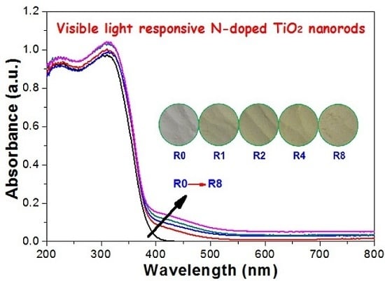
Usually, doping obviously influences light absorption characteristics of TiO2 [29]. Therefore, the optical property of the pristine and thiourea-modified TiO2 nanorods were measured by UV-vis diffuse reflectance spectra. As displayed in Figure 4, all of these samples display the typical absorption with an intense transition in the UV region of the spectra, which is assigned to the intrinsic band gap absorption of TiO2 due to the electron transitions from the valence band to conduction band (O2p → Ti3d) [30]. The pure TiO2 (R0) shows no absorption above its fundamental absorption edge (around 400 nm). In contrast, the absorption spectra of the thiourea-modified TiO2 samples show enhanced absorption in the visible region. The inset in Figure 4 shows the corresponding photos of pristine and thiourea-modified TiO2 samples. It can be seen that the color of TiO2 sample becomes deeper with an increase in R value, which is consistent with the diffused reflectance spectroscopy. Undoubtedly, these results reveal that the nonmetal elements are indeed incorporated into the lattice of TiO2, forming two phase structures (pristine and doped anatase TiO2 nanorods). Judging from the orange color of the samples (inset of Figure 4), a black graphitic carbon shell on the surface of TiO2 nanorods is unlikely to be formed, which is consistent with the high-resolution TEM characterization result (inset of Figure 3b).
Figure 3. TEM images (a,b) and SEM images (c,d) of thiourea-modified TiO2 nanorods (R2), Inset of (b) showing the high-resolution TEM image of R2 TiO2 nanorod.
Molecules 21 00181 g003
Figure 4. Diffused reflectance spectroscopy and the corresponding optical digital images (inset) of the prepared photocatalysts.
Molecules 21 00181 g004

2.5. XPS Analysis

Figure 5 compares the XPS survey spectra of pristine (R0) and thiourea-modified TiO2 nanorods (R2). As demonstrated by XPS, both samples contain Ti, O and C elements. However, small amounts of N and S elements were also detected for thiourea-modified TiO2 nanorods.
Figure 5. XPS survey spectra of pristine R0 (a) and thiourea-modified TiO2 nanorods of R2 (b).
Molecules 21 00181 g005
Figure 6 shows the high-resolution XPS spectra of C 1s, N 1s and S 2p regions for thiourea-modified TiO2 nanorods, respectively. From Figure 6A, it can be seen that the spectrum of C 1s can be deconvoluted into three peaks, one strong peak and two week peaks at binding energies of 284.6, 286.6 and 288.6 eV, implying three different chemical environments of carbon in the R2 sample. The peak around 284.6 eV is usually assigned to carbon adsorbed on the surface of the photocatalyst as a contaminant, namely C-C bonds, which cannot be eliminated. The peak at 286.6 eV can be ascribed to the existence of C-O bonds, as carbonate species. The peak observed at binding energy of 288.6 eV is attributed to O-C=O bonds [11]. No peak was observed at a binding energy of about 282 eV, corresponding to the incorporation of C atoms in form of Ti-C bond, reflecting that C element was not doped into the lattice of TiO2 nanorods [30]. The pertaining peaks in the XPS spectrum may be due to the presence of adventitious carbon species.
Figure 6B shows the corresponding high-resolution XPS spectrum of the N 1s region taken from R2 sample. The curve of the N 1s region can be deconvoluted into three peaks. The small peak (399.2 eV) is attributed to the Ti-N (at. 11.4%). The other two peaks at about 400.5 and 401.8 eV are assigned to NH3 (at. 70.8%) and NH4+ (at. 18.1%) adsorbed on the surface of TiO2, respectively [30].
Figure 6C shows the high-resolution XPS spectrum of the S 2p region for R2 sample. It can be seen that the peak of S 2p centers at binding energy of 168.9 eV, which can be attributed to the S (+VI), which is assigned to the SO42− ions adsorbed on the surface of TiO2 sample. No obvious signal of the doped S element in the form of Ti-S at binding energy of about 165 eV can be found from the high-resolution XPS spectrum [30]. The failure doping of S element into the lattice of TiO2 nanorods may be due to the fact that the diameter of S2− (1.7 Å) ion is larger than that of O2− (1.22 Å) [33].
According to the above XPS results, it can be concluded that only N element was in situ doped into the lattice of R2 TiO2 nanorods during calcination. The composition of the detected elements by XPS for R0 (pristine TiO2 nanorods), R2 and R8 samples are listed in Table 2. It shows that the content of the N and S elements increases with increasing R value. However, the content of carbon does increase with increase in R, further confirming that the interference of adventitious hydrocarbon is from the XPS instrument itself.
Figure 6. High-resolution XPS spectra of the C 1s (A); N 1s (B) and S 2p (C) regions of R2 sample.
Molecules 21 00181 g006
Table 2. XPS characterization results of the photocatalysts.
SampleMolar Ratio
C/TiN/TiS/TiO/Ti
R02.85--2.74
R22.020.0790.1063.28
R83.690.1030.1233.81
When compared with our previous reported C, N and S co-doped TiO2-HMSs which were prepared by calcination of TiO2-HMSs in the presence of cysteine at 300 °C [30], the contents of C, N and S in 600 °C-calcined TiO2 nanorods are much lower. This reflects that it is much harder to dope nonmetal ions into the lattice of TiO2 at high temperature because C and S elements can be easily oxidized into CO2 and SO2 in air atmosphere. In addition, the electronegativity of N (3.04) is larger than that of C (2.55) and S (2.58), which also results in an easier N-doping due to the stronger interaction between Ti and N.
It has been reported that anatase titania nanobelts were prepared via hydrothermal processing and subsequent heat treatment in NH3 [34]. However, the present method has the merit of being simple and easy to scale up.

2.6. Nitrogen Adsorption

Figure 7 shows the nitrogen sorption isotherms and the corresponding pore size distribution curves of R2 sample. It can be seen that the isotherm is of types IV (BDDT classification) [35]. At high relative pressure range from 0.8 to 1.0, the isotherm exhibits a hysteresis loop of type H2 associated with the ink bottle pores, indicating that the powders contain mesopores (2–50 nm). The corresponding pore size distribution curve of R2 exhibits a wide pore size distribution with the average pore diameters about 12.7 nm.
Figure 7. Nitrogen adsorption-desorption isotherm and the corresponding pore size distributions (inset) of R2 sample.
Molecules 21 00181 g007
Table 2 shows the effects of R value on the surface properties of the TiO2 nanorods. It can be seen that all these samples show similar BET surface areas and pore diameters, reflecting that modification of TiO2 nanorods by thiourea has not changed their physical structures.

2.7. Photocatalytic Activity

To study the effect of thiourea modification on the photocatalytic activity of TiO2 nanorods, both degradation of X3B (Figure 8A) and RhB (Figure 8B) dye under visible irradiation (Figure 8) and formation of •OH radicals in solution (Figure 9) under UV irradiation were performed. From Figure 8A, it can be seen that pristine R0 shows the smallest adsorption to X3B (only 4.12%), while R2 sample shows the highest adsorption (9.13%). It has been well documented that preliminary adsorption of substrate on the catalyst surface is a prerequisite for highly efficient oxidation [36]. The stronger adsorptive ability of R2 should facilitate the enhancement of its photocatalytic activity. Inset of Figure 8 compares the degradation rate constant of X3B in different photocatalysts. It can be clearly seen that R2 sample shows the highest photocatalytic activity (0.017 min−1), which is two times higher than pristine TiO2 nanorods (0.0071 min−1 for R0). This is consistent with their adsorption capabilities. Similar results were also obtained in photocatalytic degradation of RhB dye. The R2 sample shows the highest degradation ratio of RhB (80.13%), which is almost two times higher than the R0 sample (only 47.27%).
Figure 8. Photocatalytic degradation profiles of X3B (A) and RhB (B) under visible-light irradiation in the presence of different TiO2 nanorods photocatalyst (inset: comparison of the corresponding photocatalytic activity).
Molecules 21 00181 g008
Figure 9. PL spectral changes (A) observed during irradiation of the R2 sample in 5.0 × 10⁻³ mol·L−1 solution of coumarin (excitation at 332 nm) under UV irradiation, and the comparison of formation rate constant of hydroxyl radicals in the different photocatalyst (B).
Molecules 21 00181 g009
Recently, it has been proved that several non- or weakly luminescent test molecules, such as terephthalic acid [37,38] and coumarin [39,40] produce strongly luminescent compounds with •OH radical, and these molecules can be applied for evaluation of the relative photocatalytic activity of the photocatalyst. Here coumarin is used as a probe to evaluate the photocatalytic activity of TiO2 nanorods. Figure 9A shows the typical PL spectral changes observed during illumination of the suspensions of R2 sample. It is observed that the PL intensity of photo-generated 7-hydroxycoumarin at 450 nm (excited at 332 nm) increases with irradiation time, obeying a pseudo-zero order reaction rate equation in kinetics. Figure 9B compares the •OH radical formation rate constant in different photocatalyst. It can be seen that thiourea shows little effect on the UV photocatalytic activity of TiO2 nanorods, confirming that the enhanced visible photocatalytic activity of thiourea-modified TiO2 nanorods is not based on the sacrifice of their UV photocatalytic activity.

2.8. Photoluminescence Spectrum Analysis

Photoluminescence (PL) analysis is commonly used to analyze the recombination rate of photo-generated electron-hole of semiconductor [41]. Herein, we conduct PL measurement for the pristine (R0) and thiourea-modified TiO2 sample (R2), respectively. It can be seen that the emission spectra shapes of R0 and R2 are similar (Figure 10). The strong peak at about 397 nm is attributed to the emission of band gap transition [30]. When compared with pure TiO2 nanorods (R0), the intensity of PL signal for R2 is much lower. This is due to the reduction of the radiative recombination process, that is, the lower the recombination, the weaker the PL signals are. Consequently, it is understandable that thiourea-modified TiO2 nanorods show superior photocatalytic activity to that of pristine TiO2 nanorods (R0).
Figure 10. PL spectra of pristine R0 (a) and N-doped TiO2 nanorods R2 (b).
Molecules 21 00181 g010

2.9. Photocurrent Response

The value of photocurrent can indirectly reflect the semiconductor’s ability to generate and transferr photo-generated charge carriers under irradiation [30]. The photocurrent responses of pure and thiourea-modified TiO2 nanorods were tested in several on-off cycles (Figure 11). A prompt generation of photocurrents are observed and with good reproducibility when the ITO/TiO2 electrodes are illuminated. While the lamp is off, the value of photocurrent for all the ITO/TiO2 samples are instantaneously close to zero. It can be clearly seen that the photocurrent value increases first and then decreases with increase in the R value, and R2 sample shows the highest photocurrent value, which is consistent with the photocatalytic activity of the samples (Figure 8). The photocatalytic activity of TiO2 is highly related to the number of the separated photo-generated charge carriers. Nonmetal doping, in particular nitrogen doping, can be incorporated as a substitutional or insterstitial state in the TiO2 lattice, which results in the visible-light activity [6]. From the photocurrent, it can be seen that high concentrated N-doping can cause the formation of photo-generated electron-hole recombination center, which is detrimental to the photocatalytic activity of TiO2 nanorods. Therefore, it is understandable that R2 shows superior photocatalytic activity, although R8 possesses the highest visible-light harvesting ability (Figure 4).
Figure 11. Comparison of the photocurrent response.
Molecules 21 00181 g011

3. Experimental Section

3.1. Preparation

3.1.1. Titanates

In our experiments, the hydrothermal method was employed for the synthesis of titanate nanostructures, which involved a primary reaction between a concentrated NaOH solution and titanium dioxide [16,42]. Briefly, 1.0 g P25 TiO2 (Degussa, Shanghai Pharm, China) was dispersed in an 80 mL aqueous solution of NaOH (10 mol·L−1), which was then transferred into a 100 mL Teflon-lined autoclave and then oven-heated at 200 °C for 24 h. After cooling to room temperature, the white precipitates were filtrated through a membrane filter (pore size, 0.45 μm) and rinsed with distilled water until the pH value of the filtrate is about 7. The obtained white 1-D sodium titanates cake was re-dispersed in 80 mL of HCl solution (0.1 mol·L−1) under magnetic stir. 24 h later, the resulted hydrogen titanates precipitates were washed with deionized water for several times and dried in oven at 80 °C for 6 h.

3.1.2. TiO2 Nanorods Precursors

To transformation of the prepared hydrogen titanates to anatase TiO2 nanorods, the obtained hydrogen titanates powders were re-dispersed into 80 mL of H2SO4 solution (0.2 mol·L−1), which were then heated at 100 °C for 12 h. Similarly, the obtained white precipitates were collected and dried at 80 °C after wash with deionized water.

3.1.3. N-doped TiO2 Nanorods

Certain amount of thiourea (0–3.2 g) was mixed with 0.4 g of the as-prepared TiO2 nanorods precursors. The mixture was ground and calcined at 600 °C for 2 h in a furnace. According to this method, the mass ratio of thiourea to TiO2 nanorods precursors (R) varies from 0 to 8.0. For simplicity, the prepared sample was named as Rx, where x represents R value, the mass ratio of thiourea to TiO2 nanorods precursors. R0 refers to the calcined TiO2 nanorods in the absence of thiourea (Table 1).

3.2. Characterization

The X-ray diffraction (XRD) patterns of the samples were obtained on a D8 advance X-ray diffractometer (Germany Bruker, Madison, WI, USA) using Cu Kα radiation at a scan rate (2θ) of 0.05 s−1. The voltage and applied current were 40 kV and 80 mA, respectively. The morphology of the photocatalyst was observed on a transmission electron microscopy (TEM) (Tecnai G20, Hillsboro, OR, USA) using an acceleration voltage of 200 kV and a field emission scanning electron microscope (SEM) (Hitach, Tokyo, Japan) with an acceleration voltage of 20 kV, respectively. Nitrogen adsorption-desorption isotherms were obtained on an ASAP 2020 (Micromeritic Instruments, Atlanta, GA, USA) nitrogen adsorption apparatus. All the samples were degassed at 200 °C prior to Brunauer-Emmett-Teller (BET) measurements. The BET specific surface area (SBET) was determined by a multipoint BET method using the adsorption data in the relative pressure P/P0 range of 0.05–0.30. The desorption isotherm was used to determine the pore size distribution by using the Barret-Joyner-Halenda (BJH) method. The nitrogen adsorption volume at P/P0 = 0.994 was used to determine the pore volume and average pore size. UV-vis diffuse reflectance spectroscopy (DRS) was carried out on a Hitachi U-3010 UV-vis spectrophotometer (Hitachi, Tokyo, Japan). BaSO4 was the reference sample. X-ray photoelectron spectroscopy (XPS) measurement was done using Multilab 2000 XPS system (ThermoVG Scientific, East Grinstead, West Sussex, UK) with a monochromatic Mg Kα source and a charge neutralizer, all the binding energies were referenced to the C 1s peak at 284.6 eV of the surface adventitious carbon. Photoluminescence (PL) spectra were measured at room temperature on a Fluorescence Spectrophotometer (F-7000, Hitachi, Tokyo, Japan). The excitation wavelength was 315 nm, the scanning speed was 1200 nm·min−1, and the PMT voltage was 700 V. The width of excitation slit and emission slit were both 5.0 nm.

3.3. Photocatalytic Activity

3.3.1. Visible Photoctalytic Activity

A 350-W Xe lamp (Lanshen electronics, Shanghai, China) used as the light source was positioned above a cylindrical Pyrex vessel surrounded by a jacket with circulating water. A cutoff filter was used to remove wavelengths less than 420 nm completely and to ensure irradiation only by visible light. Brilliant Red X-3B (X3B) [43,44,45], an anionic azo dye, is used as a probe molecule. During the photocatalytic reaction, the reactor was mechanically stirred at a constant rate. The concentration of TiO2 was 1.0 g·L−1, and the initial concentration of X3B was 1.0 × 10⁻⁴ mol·L−1. Before irradiation, the suspensions were sonicated first for 5 min, and then were shaken overnight in the dark to established the adsorption-desorption equilibrium. At given intervals of irradiation, small aliquots were withdrawn by a syringe, and filtered through a membrane (pore size 0.45 μm). The concentration of X3B remaining in the filtrate was then analyzed by an Agilent 8451 spectrometer (Aglent Technologies, Palo Alto, CA, USA) at 510 nm.
Photocatalytic activity degradation of Rhodamine B (RhB) dye was also performed similar to the degradation of X3B. The initial concentration of RhB for degradation is 1.0 × 10⁻⁵ mol·L−1, and the wavelength for analysis of RhB is 554 nm.

3.3.2. UV Photocatalytic Activity

The UV photocatalytic activity of the photocatalyst was evaluated by a photoluminescence (PL) technique using coumarin as a probe molecule, which readily reacted with •OH radicals to produce highly fluorescent product, 7-hydroxycoumarin, under UV irradiation [39]. The suspensions of TiO2(1.0 g·L−1) containing coumarin (0.5 mmol·L−1) is mixed under magnetic stirring, and then was shaken overnight. At given intervals of irradiation, small aliquots were withdrawn by a syringe, and filtered through a membrane (pore size 0.45 μm). Solution after filtration was analyzed on a Hitachi F-7000 fluorescence spectrophotometer by the excitation with the wavelength of 332 nm.

3.4. Photocurrent

Photocurrent measurements were carried out on an Electrochemical Station (CHI660, Chenhua Instrument, Shanghai, China). The Xe lamp with cutoff filter was used for excitation of the ITO/TiO2 electrode. These measurements were carried out with a standard three-electrode assembly. An ITO/TiO2 electrode, Pt plate, and Ag/AgCl electrode were used as the working, counter, and reference electrodes, respectively. The ITO/TiO2 electrode was prepared using a doctor-blade method. Na2SO4 (0.4 mol·L−1) is used as the electrolyte and is saturated with air.

4. Conclusions

Enhanced visible photocatalytic activity of TiO2 nanorods were successfully studied through thiourea modification of TiO2 by calcination the mixture of TiO2 nanorods and thiourea at 600 °C. Only N was doped into the lattice of TiO2 nanorods due to the greater electronegativity of N (3.04) than that of C (2.55) and S (2.58). The enhanced photocatalytic activity of the R2 sample is due to the synergistic effects of improved crystallization and increased light-harvesting ability, which results in the reduced recombination of photo-generated electron-hole pairs. The enhanced visible photocatalytic activity of thiourea-modified TiO2 nanorods is not based on the sacrifice of their UV photocatalytic activity.

Acknowledgments

This work was supported by the Program for New Century Excellent Talents in University (NCET-12-0668), National Natural Science Foundation of China (21373275 & 20977114).

Author Contributions

The list authors contributed to this work as follows: Kangle Lv conceived and designed the experiments, Xiaofeng Wu and Shun Fang performed the experiments and analyzed the data, Yang Zheng wrote the paper, Jie Sun and Kangle Lv polished the paper. Kangle Lv acquired funding for the research. All authors read and approved the final manuscript.

Conflicts of Interest

The authors declare no conflict of interest.

References

  1. Yu, J.G.; Low, J.X.; Xiao, W.; Zhou, P.; Jaroniec, M. Enhanced photocatalytic CO2-reduction activity of anatase TiO2 by coexposed {001} and {101} facets. J. Am. Chem. Soc. 2014, 136, 8839–8842. [Google Scholar] [CrossRef] [PubMed]
  2. Schneider, J.; Matsuoka, M.; Takeuchi, M.; Zhang, J.L.; Horiuchi, Y.; Anpo, M.; Bahnemann, D.W. Understanding TiO2 photocatalysis: Mechanisms and materials. Chem. Rev. 2014, 114, 9919–9986. [Google Scholar] [CrossRef]
  3. Yang, D.J.; Liu, H.W.; Zheng, Z.F.; Yuan, Y.; Zhao, J.C.; Waclawik, E.R.; Ke, X.B.; Zhu, H.Y. An efficient photocatalyst structure: TiO2(B) nanofibers with a shell of anatase nanocrystals. J. Am. Chem. Soc. 2005, 131, 17885–17893. [Google Scholar] [CrossRef] [PubMed]
  4. Liu, Y.; Chen, L.F.; Hu, J.C.; Li, J.L.; Richards, R. TiO2 nanoflakes modified with gold nanoparticles as photocatalysts with high activity and durability under near UV irradiation. J. Phys. Chem. C 2010, 114, 1641–1645. [Google Scholar] [CrossRef]
  5. Lv, K.L.; Cheng, B.; Yu, J.G.; Liu, G. Fluorine ions-mediated morphology control of anatase TiO2 with enhanced photocatalytic activity. Phys. Chem. Chem. Phys. 2012, 14, 5349–5362. [Google Scholar] [CrossRef] [PubMed]
  6. Pelaez, M.; Nolan, N.T.; Pillai, S.C.; Seery, M.K.; Falaras, P.; Kontos, A.G.; Dunlop, P.S.M.; Hamilton, J.W.J.; Byrne, J.A.; O’Shea, K.; et al. A review on the visible light active titanium dioxide photocatalysts for environmental applications. Appl. Catal. B Environ. 2012, 125, 331–349. [Google Scholar] [CrossRef]
  7. Xu, Y.M.; Lv, K.L.; Xiong, Z.G.; Leng, W.H.; Du, W.P.; Liu, D.; Xue, X.J. Rate enhancement and rate inhibition of phenol degradation over irradiated anatase and rutile TiO2 on the addition of naf: New insight into the mechanism. J. Phys. Chem. C 2007, 111, 19024–19032. [Google Scholar] [CrossRef]
  8. Sun, Q.; Xu, Y.M. Evaluating intrinsic photocatalytic activities of anatase and rutile TiO2 for organic degradation in water. J. Phys. Chem. C 2010, 114, 18911–18918. [Google Scholar] [CrossRef]
  9. Liu, Y.B.; Zhou, B.X.; Li, J.H.; Gan, X.J.; Bai, J.; Cai, W.M. Preparation of short, robust and highly ordered TiO2 nanotube arrays and their applications as electrode. Appl. Catal. B 2009, 92, 326–332. [Google Scholar] [CrossRef]
  10. Sun, J.; Yan, X.; Lv, K.L.; Sun, S.; Deng, K.J.; Du, D.Y. Photocatalytic degradation pathway for azo dye in TiO2/UV/O3 system: Hydroxyl radical versus hole. J. Mol. Catal. A 2013, 367, 31–37. [Google Scholar] [CrossRef]
  11. Mollavali, M.; Falamaki, C.; Rohani, S. Preparation of multiple-doped TiO2 nanotube arrays with nitrogen, carbon and nickel with enhanced visible light photoelectrochemical activity via single-step anodization. Int. J. Hydrog. Energy 2015, 40, 12239–12252. [Google Scholar] [CrossRef]
  12. Li, J.M.; Xu, D.S. Tetragonal faceted-nanorods of anatase TiO2 single crystals with a large percentage of active {100} facets. Chem. Commun. 2010, 46, 2301–2303. [Google Scholar]
  13. Yu, H.G.; Yu, J.G.; Cheng, B.; Lin, J. Synthesis, characterization and photocatalytic activity of mesoporous titania nanorod/titanate nanotube composites. J. Hazard. Mater. 2007, 147, 581–587. [Google Scholar] [CrossRef] [PubMed]
  14. Yu, J.G.; Xiang, Q.J.; Zhou, M.H. Preparation, characterization and visible-light-driven photocatalytic activity of Fe-doped titania nanorods and first-principles study for electronic structures. Appl. Catal. B 2009, 90, 595–602. [Google Scholar] [CrossRef]
  15. Kim, H.S.; Lee, J.W.; Yantara, N.; Boix, P.P.; Kulkarni, S.A.; Mhaisalkar, S.; Gratzel, M.; Park, N.G. High efficiency solid-state sensitized solar cell-based on submicrometer rutile TiO2 Nanorod and CH3NH3PbI3 perovskite sensitizer. Nano Lett. 2013, 13, 2412–2417. [Google Scholar] [CrossRef] [PubMed]
  16. Mao, Y.B.; Wong, S.S. Size- and Shape-dependent transformation of nanosized titanate into analogous anatase titania nanostructures. J. Am. Chem. Soc. 2006, 128, 8217–8226. [Google Scholar] [CrossRef] [PubMed]
  17. Yuan, R.S.; Fu, X.Z.; Wang, X.C.; Liu, P.; Wu, L.; Xu, Y.M.; Wang, X.X.; Wang, Z.Y. Template synthesis of hollow metal oxide fibers with hierarchical architecture. Chem. Mater. 2006, 18, 4700–4705. [Google Scholar] [CrossRef]
  18. Wang, F.C.; Liu, C.H.; Liu, C.W.; Chao, J.H.; Lin, C.H. Effect of Pt loading order on photocatalytic activity of Pt/TiO2 nanofiber in generation of H2 from neat ethanol. J. Phys. Chem. C 2009, 113, 13832–13840. [Google Scholar] [CrossRef]
  19. Cao, T.P.; Li, Y.J.; Wang, C.H.; Shao, C.L.; Liu, Y.C. A facile in situ hydrothermal method to SrTiO3/TiO2 nanofiber heterostructures with high photocatalytic activity. Langmuir 2011, 27, 2946–2952. [Google Scholar] [CrossRef] [PubMed]
  20. Formo, E.; Lee, E.; Campbell, D.; Xia, Y. Functionalization of electrospun TiO2 nanofibers with pt nanoparticles and nanowires for catalytic applications. Nano Lett. 2008, 8, 668–672. [Google Scholar]
  21. Asahi, F.; Morikawa, T.; Ohwaki, T.; Aoki, K.; Taga, Y. Visible-light photocatalysis in nitrogen-doped titanium oxides. Science 2001, 293, 269–271. [Google Scholar] [CrossRef] [PubMed]
  22. Zheng, R.Y.; Guo, Y.; Jin, C.; Xie, J.L.; Zhu, Y.X.; Xie, Y.C. Novel thermally stable phosphorus-doped TiO2 photocatalyst synthesized by hydrolysis of TiCl4. J. Mol. Catal. A 2010, 319, 46–51. [Google Scholar] [CrossRef]
  23. Xiang, Q.J.; Yu, J.G.; Jaroniec, M. Nitrogen and sulfur co-doped TiO2 nanosheets with exposed {001} facets: Synthesis, characterization and visible-light photocatalytic activity. Phys. Chem. Chem. Phys. 2011, 13, 4853–4861. [Google Scholar] [CrossRef] [PubMed]
  24. Si, L.L.; Huang, Z.A.; Lv, K.L.; Tang, D.G.; Yang, C.J. Facile preparation of Ti3+ self-doped TiO2 nanosheets with dominant {001} facets using zinc powder as reductant. J. Alloys Compd. 2014, 601, 88–93. [Google Scholar] [CrossRef]
  25. Yu, J.G.; Li, Q.; Liu, S.W.; Jaroniec, M. Ionic-liquid-assisted synthesis of uniform fluorinated B/C-codoped TiO2 nanocrystals and their enhanced visible-light photocatalytic activity. Chem. Eur. J. 2013, 19, 2433–2441. [Google Scholar] [CrossRef] [PubMed]
  26. Cai, J.H.; Huang, Z.A.; Lv, K.L.; Sun, J.; Deng, K.J. Ti powder-assisted synthesis of Ti3+ self-doped TiO2 nanosheets with enhanced visible-light photoactivity. RSC Adv. 2014, 4, 19588–19593. [Google Scholar] [CrossRef]
  27. Liu, G.; Yang, H.G.; Wang, X.W.; Cheng, L.; Pan, J.; Lu, G.Q.; Cheng, H.M. Visible light responsive nitrogen doped anatase TiO2 sheets with dominant {001} facets derived from TiN. J. Am. Chem. Soc. 2009, 131, 12868–12869. [Google Scholar] [CrossRef] [PubMed]
  28. Manthina, V.; Agrios, A.G. Band edge engineering of composite photoanodes for dye-sensitized solar cells. Electrochim. Acta 2015, 169, 416–423. [Google Scholar] [CrossRef]
  29. Lv, K.L.; Zuo, H.S.; Sun, J.; Deng, K.J.; Liu, S.C.; Li, X.F.; Wang, D.Y. (Bi, C and N) codoped TiO2 nanoparticles. J. Hazard. Mater. 2009, 161, 396–401. [Google Scholar] [CrossRef] [PubMed]
  30. Lv, K.L.; Hu, J.C.; Li, X.H.; Li, M. Cysteine modified anatase TiO2 hollow microspheres with enhanced visible-light-driven photocatalytic activity. J. Mol. Catal. A 2012, 356, 78–84. [Google Scholar] [CrossRef]
  31. Zhang, G.S.; Zhang, Y.C.; Nadagouda, M.; Han, C.; O’Shea, K.; El-Sheikh, S.M.; Ismail, A.A.; Dionysiou, D.D. Visible light-sensitized S, N and C co-doped polymorphic TiO2 forphotocatalytic destruction of microcystin-LR. Appl. Catal. B Environ. 2014, 144, 614–621. [Google Scholar] [CrossRef]
  32. Zhang, L.W.; Fu, H.B.; Zhu, Y.F. Efficient TiO2 photocatalysts from surface hybridization of TiO2 particles with graphite-like carbon. Adv. Funct. Mater. 2008, 18, 2180–2189. [Google Scholar] [CrossRef]
  33. Periyat, P.; Pillai, S.C.; McCormack, D.E.; Colreavy, J.C.; Hinder, S.J. Improved high-temperature stability and sun-light-driven photocatalytic activity of sulfur-doped anatase TiO2. J. Phys. Chem. C 2008, 112, 7644–7652. [Google Scholar] [CrossRef] [Green Version]
  34. Wang, J.; Tafen, D.N.; Lewis, J.P.; Hong, Z.L.; Manivannan, A.; Zhi, M.J.; Li, M.; Wu, N.Q. Origin of photocatalytic activity of nitrogen-doped TiO2 nanobelts. J. Am. Chem. Soc. 2009, 131, 12290–12297. [Google Scholar] [CrossRef] [PubMed]
  35. Yu, J.G.; Su, Y.R.; Cheng, B. Template-free fabrication and enhanced photocatalytic activity of hierarchical macro-/mesoporous titania. Adv. Funct. Mater. 2007, 17, 1984–1990. [Google Scholar] [CrossRef]
  36. Xu, Y.M.; Langford, C.H. UV- or visible-light-induced degradation of X3B on TiO2 nanoparticles: The influence of adsorption. Langmuir 2001, 17, 897–902. [Google Scholar] [CrossRef]
  37. Lin, Y.-C.; Chien, T.E.; Lai, P.C.; Chiang, Y.H.; Li, K.L.; Lin, J.L. TiS2 transformation into S-doped and N-doped TiO2 with visible-light catalytic activity. Appl. Surf. Sci. 2015, 359. [Google Scholar] [CrossRef]
  38. Lv, K.L.; Yu, J.G.; Deng, K.J.; Li, X.H.; Li, M. Effect of phase structures on the formation rate of hydroxyl radicals on the surface of TiO2. J. Phys. Chem. Solids 2010, 71, 519–522. [Google Scholar] [CrossRef]
  39. Wang, Z.Y.; Lv, K.L.; Wang, G.H.; Deng, K.J.; Tang, D.G. Study on the shape control and photocatalytic activity of high-energy anatase titania. Appl. Catal. B 2010, 100, 378–385. [Google Scholar] [CrossRef]
  40. Huang, Z.A.; Wang, Z.Y.; Lv, K.L.; Zheng, Y.; Deng, K.J. Transformation of TiOF2 cube to a hollow nanobox assembly from anatase TiO2 nanosheets with exposed {001} facets via solvothermal strategy. ACS Appl. Mater. Interfaces 2013, 5, 8663–8669. [Google Scholar] [CrossRef] [PubMed]
  41. Huang, Z.A.; Sun, Q.; Lv, K.L.; Zhang, Z.H.; Li, M.; Li, B. Effect of contact interface between TiO2 and g-C3N4 on the photoreactivity of g-C3N4/TiO2 photocatalyst: (001) vs. (101) facets of TiO2. Appl. Catal. B 2015, 164, 420–427. [Google Scholar] [CrossRef]
  42. Zhu, H.Y.; Lan, Y.; Gao, X.P.; Ringer, S.P.; Zheng, Z.F.; Song, D.Y.; Zhao, J.C. Phase transition between nanostructures of titanate and titanium dioxides via simple wet-chemical reactions. J. Am. Chem. Soc. 2005, 127, 6730–6736. [Google Scholar] [CrossRef] [PubMed]
  43. Lv, K.L.; Xu, Y.M. Effects of Polyoxometalate and fluoride on adsorption and photocatalytic degradation of organic dye X3B on TiO2: The difference in the production of reactive species. J. Phys. Chem. B 2006, 110, 6204–6212. [Google Scholar] [CrossRef] [PubMed]
  44. Lan, J.F.; Wu, X.F.; Lv, K.L.; Si, L.L.; Deng, K.J. Fabrication of TiO2 hollow microspheres using K3PW12O40 as template. Chin. J. Catal. 2015, 36, 2237–2243. [Google Scholar] [CrossRef]
  45. Zheng, Y.; Cai, J.H.; Lv, K.L.; Sun, J.; Ye, H.P.; Li, M. Hydrogen peroxide assisted rapid synthesis of TiO2 hollow microspheres with enhanced photocatalytic activity. Appl. Catal. B 2014, 147, 789–795. [Google Scholar] [CrossRef]
  • Sample Availability: Samples of the N-doped TiO2 nanorods are available from the authors.