Article

Integrated Analysis of the Wood Oil from Xanthocyparis vietnamensis Farjon & Hiep. by Chromatographic and Spectroscopic Techniques

1
Université de Corse-CNRS, UMR 6134 SPE, Equipe Chimie et Biomasse, Route des Sanguinaires, 20000 Ajaccio, France
2
Institute of Ecology and Biological Resources, Vietnam Academy of Science and Technology, Cau Giay, Hanoi 10000, Vietnam
*
Author to whom correspondence should be addressed.
Molecules 2016, 21(7), 840; https://doi.org/10.3390/molecules21070840
Submission received: 26 May 2016 / Revised: 20 June 2016 / Accepted: 23 June 2016 / Published: 27 June 2016
(This article belongs to the Collection Recent Advances in Flavors and Fragrances)

Abstract

:
In order to get better knowledge about the volatiles produced by Xanthocyparis vietnamensis, a species recently discovered in Vietnam, its wood oil has been analyzed by a combination of chromatographic (GC, CC) and spectroscopic (GC-MS, 13C-NMR) techniques. Forty components that accounted for 87.9% of the oil composition have been identified. The composition is dominated by nootkatene (20.7%), 11,12,13-tri-nor-eremophil-1(10)-en-7-one (17.2%), γ-eudesmol (5.1%), nootkatone (4.7%), valencene (3.5%) and 13-nor-eremophil-1(10)-en-11-one (2.6%). The structure of two new compounds—10-epi-nor-γ-eudesmen-11-one and 12-hydroxy-isodihydroagarofuran—has been elucidated, while 11,12,13-tri-nor-eremophil-1(10)-en-7-ol is reported as a natural product for the first time. The composition of X. vietnamensis wood oil varied drastically from those of leaf oils, dominated by hedycaryol (34.4%), phyllocladene (37.8%) or by pimara-6(14)-15-diene (19.4%).

Graphical Abstract

1. Introduction

Xanthocyparis vietnamensis Farjon & Hiep (Cupressaceae), also known as Vietnamese gold cypress (Vietnamese name: Bách vàng), was discovered in 1999 in the calcareous mountains of Northern Vietnam (Quản Bạ, Hà Giang Province, Figure 1) [1]. Although found since that time in other localities (Hà Giang, Cao Bằng, Tuyên Quang provinces), it grows in a restricted area at an altitude of 1000–1600 m. The estimated number of adult trees is in the range 500–1000 individuals, therefore, X. vietnamensis remains among the endangered species [2]. This species has also been added to Group IA of the National List of Rare and Precious Flora and Fauna which prohibits any exploitation. An ex situ conservation program has been initiated and some restoration work undertaken [3]. This species grows associated with conifer species: Pseudotsuga sinensis, Nageia jneiryi, Podocarpus pilgeri, Calocedrus macrolepis, Taxils chinensis, Amentotaxus sp.
The discovery of this new species led to the proposal of a new genus in 2002, the genus Xanthocyparis [4]. From the taxonomic point of view, analysis of 54 characteristics demonstrated the proximity of Chamaecyparis nootkatensis (D. Don) Spach with X. vietnamensis and therefore the former species has been transferred to the genus Xanthocyparis and it is nowadays inventoried as Xanthocyparis nootkatensis (D. Don) Farjon & Harder. This denomination was agreed in 2011 by the International Association for Plant Taxonomy [5,6].
X. vietnamensis is an evergreen, medium-sized tree, 10–15 m in height, 0.5 m in diameter. Bark is smooth, thin, red or brown-red and fibrous. The originality of this tree is the occurrence, at the adult stage, of two types of leaves (needles and tortoiseshells) [7]. The tree bears both male and female cones, solitary at the top of the branches, coming to maturity in two years [4]. Female cones are nearly globose, 9–11 mm × 10–12 mm, black or dark brown when ripe. Seeds occur in February. Male cones are oval, 2.5–3.5 mm × 2.0–2.5 mm. The wood is odoriferous and of excellent quality and it is used by local craftsmen for house building and house furniture as well as for wood-fired heating [1].
Only two papers have reported on the chemical composition of X. vietnamensis essential oil (EO). Three oil samples have been obtained by vapor distillation of fresh leaves collected on trees cultivated in the botanical garden of Edinburgh (Scotland). The essential oil composition was dominated by sesquiterpenes and diterpenes like hedycaryol (1.4%–34.4%), phyllocladene (0.9%–37.8%), germacrene D (3.7%–7.9%) and sandaracopimara-8(14),15-diene (0.7%–8.9%) and it varied substantially from sample to sample. Monoterpenes present at appreciable contents were α-pinene (0.8%–14.9%) and myrcene (0.3%–7.9%) [8]. Another X. vietnamensis leaf oil sample isolated from trees growing wild in Vietnam (yield = 0.03% vs. dry material) exhibited a substantially different composition with germacrene D (14.6%), pimara-6(14)-15-diene (19.4%), the unusual cyclohexen-1,5-dienthenyl-3-ene (12.7%) and epi-bicyclosesquiphellandrene (6.4%) as main components [9].
The aim of the present work was to get a better knowledge of the volatiles produced by X. vietnamensis by analyzing an essential oil sample isolated from the wood of this species. Due to the complexity of this essential oil, analysis was conducted by a combination of chromatographic and spectroscopic techniques.

2. Results

Wood from X. vietnamensis has been collected in Hà Giang Province (Figure 1) and it produced by water-distillation a pale-yellow essential oil with a yield of 0.20% (v:m).

2.1. Analysis of X. vietnamensis Wood Oil by GC(RI), GC-MS and 13C-NMR

The oil sample has been analyzed by GC(RI), GC-MS and 13C-NMR without isolation of individual components following a computerized method developed at the University of Corsica [10,11,12]. In total, 28 compounds were identified accounting for 82.8% of the whole composition (Table 1). However, various components present at appreciable contents (up to 2.3%) remained unidentified. In detail and taking in mind that direct identification of a component of an EO by 13C-NMR without isolation may be achieved if the compound accounts at least for 0.3%–0.4% of the whole composition [10,11,12], 17 out of the 28 components have been identified by the three techniques, GC(RI), GC-MS and 13C-NMR. They belonged to various families of terpenes:
-
Monoterpenes bearing the p-menthane skeleton: p-cymene (5), p-cymenene (7), p-cymen-8-ol (8), terpinen-4-ol (9), α-terpineol (10) and carvacrol (12);
-
Sesquiterpenes bearing the bicyclo[4.4.0]decane skeleton: β-selinene (17), valencene (18), δ-cadinene (22), γ-eudesmol (28), τ-muurolol (30), β-eudesmol (33), α-eudesmol (35);
-
Sesquiterpenes with substituted six-membered ring: β-elemene (14), β-elemol (23). It could be pointed out that β-elemene, being identified by NMR at room temperature, is a metabolite of the plant and it is not an artifact resulting from the thermal rearrangement of germacrene A in the GC injector [13];
-
Overall, the two major components of the EO—nootkatene (21, 20.7%) and 11,12,13-tri-nor-eremophil-1(10)-en-7-one (15, 17.2%) bear the eremophilane skeleton (Figure 2).
Seven minor components have been identified by GC(RI) and GC-MS. They are: α-thujene (1), sabinene (2), myrcene (3), 1,4-cineole (4), limonene (6), carvacryl methyl oxide (11) and cadalene (37). Finally, and more surprisingly, four compounds, 13-nor-eremophil-1(10)-en-11-one (25, 2.6%), τ-cadinol (29, 0.7%), α-cadinol (34, 1.9%) and nootkatone (40, 4.7%) have been identified by GC(RI) and 13C-NMR. Matching against commercial and laboratory-made MS libraries did not provide acceptable fit. This probably means that every GC peak does not belong to a unique component. In contrast, the 13C-NMR chemical shifts fitted perfectly with those of the reference compounds compiled in our NMR library.

2.2. Fractionation of X. vietnamensis Essential Oil

Taking into account that various components remained unidentified and owing to its complexity, the EO was submitted to column chromatography (CC) and 10 fractions were obtained: F1 (hydrocarbon-containing fraction) and F2-F10 (oxygenated compounds-containing fractions). The 10 fractions have been analyzed by GC(RI) and 13C-NMR. The occurrence of various terpene hydrocarbons like sabinene (2), myrcene (3), limonene (6) and cadalene (37) in fraction F1 was confirmed by NMR. Moreover, two other sesquiterpenes have been identified: α-selinene (19) and γ-cadinene (20; 0.7% and 1.0% in F1, 0.3% each in EO). Similarly, two compounds were confirmed in fraction F2: carvacryl methyl oxide (11; NMR data from our library, 1.0% in fraction F2) and 1,4-cineole (4; 6.0% in fraction F2) by comparison of NMR data with those reported by Asakawa et al. [15]. Finally, fraction F2 contained also dehydrojinkoh-eremol (38; 2.0% in F2, 0.2% in EO), identified by comparison of NMR data with those reported by Ishihara et al. [16]. The retention index (RI = 1671) is in agreement with that reported by Tajuddin et al. (RI = 1673) [17]. Other components have been identified by GC(RI) and 13C-NMR in various fractions of CC: α-terpinyl acetate (13), eremophil-9-en-11-ol (26), eremoligenol (27), δ-cadinol (torreyol) (31), valerianol (32) and selina-11-en-4α-ol (36).
The chromatographic profile as well as the 13C-NMR spectrum of fractions F2, F8 and F9 demonstrated that every fraction contained an unidentified major component. Therefore, it was decided to combine column chromatography (CC on silica gel using a gradient of solvents pentane/diethyl ether) and the concept of “extraction” NMR [18,19] to identify the major components of every fraction.

2.3. Structure Elucidation of New Natural Compounds

2.3.1. Identification of 11,12,13-Tri-nor-eremophil-1(10)-en-7-ol (16)

Fraction F9 was subjected to CC on silica gel using a gradient of solvents (pentane/Et2O). Sub-fraction F9-7 contained a major component A (52.5%, 0.4% in EO; RIa/RIp = 1458/2173). The 13C-NMR spectrum of this fraction exhibited 12 signals with high intensities. Combination of the information provided by DEPT spectrum (2 C, 3 CH, 5 CH2 and 2 CH3), and by 1H and 13C chemical shift values (occurrence of one double bond C=CH and an alcohol function CH-OH), suggested the formula C12H20O, in agreement with the molecular ion in the MS (M+ = 180). Therefore, A is a bicyclic, mono unsaturated tri-nor-sesquiterpene alcohol. Otherwise, 2D NMR experiments, particularly long range proton-carbon connectivities in the HMBC spectrum, suggested the 11,12,13-tri-nor-eremophil-1(10)-en-7-ol structure for A (component 16 in Table 1, Figure 3). It could be pointed out that the chemical shifts of seven out of twelve carbons appeared very close to those of 11,12,13-tri-nor-eremophil-1(10)-en-7-one (15), one of the major components of the EO. The axial stereochemistry of H7 was ensured by the coupling constant values of its signal (H7, triplet of triplet, J = 11.3 and 4.2 Hz). A search in the computerized data library SciFinder revealed that the molecule has been already obtained by Revial et al. [20] in the course of their synthesis of valencenol. It appears that this compound is reported from natural sources for the first time.

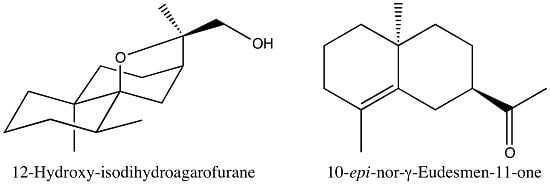
2.3.2. Identification of 10-epi-Nor-γ-eudesmen-11-one (24)

Sub-fraction F2.3, obtained by fractionation of F2 on silica gel, contained a main compound (B, 40.4%, 0.9% in EO; RIa/RIp = 1553/2039). The 13C-NMR spectrum of that fraction displayed 14 signals with high intensities that obviously belonged to B (Table 2). DEPT spectra differentiated four quaternary carbons (including a carbonyl carbon and two olefinic carbons), one CH, six CH2 and three CH3. Therefore, the formula C14H22O was deduced in agreement with the molecular ion M+ = 206 in the MS. Beside the carbonyl function, that belonged to an acetyl group according to chemical shift data, and one double bond, the structure of B contained two rings and therefore it formed a bicyclodecane framework. Two possibilities have been considered: the bicyclo[4.4.0]decane and bicyclo[5.3.0]decane skeletons. Although the 1H-NMR spectrum of the fraction was complex, the HSQC spectrum allowed the identification of protons that belonged to B. Then the long range heteronuclear connectivities reported in the Table 2 conducted to the structure of a nor-γ-eudesmenone (Figure 4). The dimethylbicyclo[4.4.0]decane framework was constructed starting from signals of hydrogens of methyl groups located on C4 and C10, respectively, and from the H7 (methine) hydrogen. The location of the acetyl group on C7 was confirmed by the connectivities of H12 with C7. Other heteronuclear connectivities were in agreement with the proposed structure. Moreover, the structure was confirmed by the proximity of the chemical shift values with those of γ-eudesmol and 10-epi-γ-eudesmol [21,22]. Unfortunately, the relative cis or trans stereochemistry of C10-Me and the acetyl group bore by C7 cannot be deduced from the through space H-H connectivities in the NOESY spectrum that appeared really poor. In both cases, the acetyl group should adopt an equatorial (or pseudo-equatorial) position. This point is confirmed by the multiplicity (doublet of triplets) and the coupling constant values (14.8 and 2.2 Hz) of equatorial H6 (2JH6eq-H6ax; 3JH6-H7 and 4JH6eq-H8eq, W stereochemistry) and it is in agreement with the occurrence of a correlation plot between H6eq and H13 in the NOESY spectrum.
Comparison of the chemical shift values of B with those of the γ-eudesmol epimers reported in the literature could be informative. Indeed, the chemical shift of carbon C7 that differs drastically in γ-eudesmol (50.56 ppm) and epi-γ-eudesmol (44.21 ppm) [21,22] cannot be taken into consideration since compound 24 bore an acetyl group instead of the “isopropyl alcohol” group. However, a significant difference was observed on the chemical shift value of olefinic quaternary carbon C4, 124.48 ppm for γ-eudesmol and 125.98 ppm for 10-epi-γ-eudesmol, the latter being close to the C7 of compound 24 (126.29 ppm). The trans stereochemistry of the acetyl group and C10-Me was corroborated by considering the chemical shifts of carbons C1 and C9 of 24 (39.99 and 38.29 ppm) and comparing with those of epi-γ-eudesmol (38.13 and 39.46 ppm) and those of γ-eudesmol (40.22 and 42.28 ppm). Proton NMR also favors the trans stereochemistry. Indeed, C4-Me/C10-Me chemical shift values of 24 (1.67/1.06 ppm) fit perfectly with those of epi-γ-eudesmol (1.66/1.06 ppm) compared with those of γ-eudesmol (1.60, 1.01 ppm). Finally, the multiplicity (dt) and coupling constants (14.8 and 2.2 Hz) of H6eq of 24 are in agreement with those of epi-γ-eudesmol (dt, 14.8 and 2.8 Hz) [21,22].
It should be mentioned that the C10 epimeric ketone has been reported by Marshall and Pike in the course of the stereoselective total synthesis of γ-eudesmol [23]. The relative cis stereochemistry of acetyl and methyl groups was deduced from mechanistic considerations and confirmed by MeLi addition to the keto group leading to γ-eudesmol. Although 1H-NMR data were not reported in detail, the chemical shifts of C4-Me and C10-Me (1.61 and 1.04 ppm) fit perfectly with those of γ-eudesmol (1.60 and 1.01 ppm), and therefore confirm the trans stereochemistry of C10-Me and acetyl group in 24.

2.3.3. Identification of 12-Hydroxy-isodihydroagarofuran (39)

Sub-fraction F8.15, obtained by fractionation of F8 on silica gel, contained a main component (C, 60.1%; 2.3% in EO; RIa/RIp = 1747/2452). The 13C-NMR spectrum of that fraction exhibited 15 signals with high intensities. DEPT spectra allowed the differentiation of three C, two CH, seven CH2 and three CH3. Chemical shift values suggested the occurrence of an oxide function (2 C at δ = 88.21 ppm and 82.91 ppm) and a primary alcohol function CH2-OH (δ = 69.29 ppm) corroborated by the occurrence of two doublets at 3.40 and 3.24 ppm (2J = 10.4 Hz) in the 1H spectrum. The molecule contained also three methyl groups (1H-NMR signals: a doublet and two singlets). The formula C15H26O2 was deduced from these data in agreement with the molecular ion M+ = 238 in the MS. Obviously, the molecule displays a tricyclic structure. Owing to the occurrence of a CH2-OH group, one of the three rings is oxygenated. This suggestion was confirmed by examination of long range correlations in the HMBC spectrum. The bicyclo[4.4.0]decane skeleton was constructed starting from methyls C14 and C15 and the dihydroagarofuran framework resulted inter alia from correlations of H7 and H13 with various carbons (Table 3). The primary alcohol function was assigned to carbons C12 or C13 on the basis of the correlation plot of the methylenic hydrogens with C7. It was attributed to C12, the γ-shielding effect of the hydroxyl group on C13 being in the range 5–6 ppm, as expected. At this stage, component 39 should be a 12-hydroxydihydroagarofuran (Figure 5). Two points should be specified: (i) the stereochemistry of the ring junction of the decalin substructure; (ii) the relative stereochemistry of methyls C14 and C15. Unfortunately, the NOESY spectrum was particularly poor and therefore no significant through space interaction between hydrogen atoms has been detected. The correct stereochemistry was obtained by comparison of the 13C-NMR and 1H-NMR chemical shifts of 39 with those of the four dihydroagarofuran isomers reported in the literature [24,25,26,27]. Indeed, the 13C-NMR chemical shifts of C2 (21.33 ppm) and C4 (32.23 ppm) of 39 differ substantially from those of trans-dihydroagarofuran and 4-epi-cis-dihydroagarofuran (17.0/17.7 and 40.5/43.1, respectively) [25,27]. In contrast they are close to those of isodihydroagarofuran and cis-dihydroagarofuran (21.4/21.6 ppm and 32.2/32.6 ppm), respectively [24,26]. Differentiation between the last two isomers has been achieved by comparison of the 1H chemical shifts of methyls C14 and C15, 1.01 and 0.85 for 39, much most closer to those of isodihydroagarofuran (1.00 and 0.88 ppm) than to those of cis-dihydroagarofuran (0.88 and 0.82 ppm). Therefore, compound 39 is 12-hydroxy-isodihydroagarofuran.

3. Discussion

Analysis of Vietnamese Xanthocyparis vietnamensis wood oil by a combination of chromatographic (GC, CC) and spectroscopic (GC-MS, 13C-NMR) techniques allowed the identification of 40 components that accounted for 87.9% of the oil composition. It is a sesquiterpene-rich oil whose composition is dominated by nootkatene (20.7%), a sesquiterpene hydrocarbon bearing the eremophilane skeleton and 11.12.13-tri-nor-eremophil-1(10)-en-7-one (17.2%). The eremophilane skeleton was also represented by valencene (3.5%), eremophil-9-en-11-ol (tr), eremoligenol (tr), dehydrojinkoheremol (0.2%), valerianol (tr), 13-nor-eremophil-1(10)-en-11-one (2.6%) and 11,12,13-tri-nor-eremophil-1(10)-en-7-ol (0.4%), the latter compound being found in Nature for the first time. Other components present at appreciable contents, including two new compounds, 10-epi-nor-γ-eudesmen-11-one and 12-hydroxyisodihydroagarofuran, displayed the bicyclo[4.4.0]decane framework of cadinane, eudesmane or selinane, Thirteen monoterpenes have also been identified (tr-4.1%; 14.8% in total).
The composition of X. vietnamensis wood oil varied drastically from those of leaf oils dominated either by hedycaryol (34.4%), phyllocladene (37.8%) (Edinburgh sample) [8] or by pimara-6(14)-15-diene (19.4%) and germacrene D (14.6%) (Vietnamese sample) [9]. Otherwise, nootkatene has been identified as a component of various essential oils, for instance Alaska cedar (Chamaecyparis nootkatensis) heartwood essential oil (20.1%) [28].

4. Materials and Methods

4.1. Plant Location and Essential Oil Isolation

Plant material (wood, about 4 kg) from X. vietnamensis has been collected in Quan Ba District. Hà Giang Province (Northern Vietnam, GPS coordinates: 23°09′N, 104°59′E, 1100–1200 m, Figure 1) and subjected to hydrodistillation for 3 h using a 5 L Clevenger-type apparatus yielding 8 mL of EO. The plant name was identified by Nguyen Sinh Khang by comparing morphological details. A voucher specimen has been deposited at IEBR under the reference: N.S. Khang 2014. A (HN).

4.2. Fractionation of Essential Oil

X. vietnamensis wood oil (2.2 g) was subjected to column chromatography (CC) on silica gel 250–500 µm, 35 g) using a gradient of solvents pentane/Et2O: 100/0 to 0/100. Ten fractions have been eluted: F1–F10. Fractions F2 (120 mg), F8 (466 mg) and F9 (345 mg) have been chromatographed once again (silica gel 250–500 µm) leading to sub-fractions F2.1–F2.6, F8.1–F8.18 and F9.1–F9.11 respectively. Sub-fraction F2.2 (18 mg, eluted with P:Et2O = 98:2) contained compound B = 24 (40.4%); sub-fraction F8.15 (43 mg, eluted with P:Et2O = 90:10) contained 39 (60.1%) and sub-fraction F9.7 (18 mg, eluted with P:Et2O = 80:20) contained 16 (52.5%). The three sub-fractions were subjected to spectroscopic analysis for structure elucidation.

4.3. Gas Chromatography (GC) Analysis

GC analyses were carried out with a Clarus 500 Autosystem (Perkin-Elmer, Courtaboeuf, France) apparatus equipped with two flame ionization detectors (FIDs) and fused capillary columns (50 m × 0.22 mm i.d. film thickness 0.25 μm). BP-1 (dimethylpolysiloxane) and BP-20 (polyethylene glycol). The carrier gas was helium with a linear velocity of 1.0 mL/min. The oven temperature was programmed from 60 °C to 220 °C at 2 °C/min and then held isothermal (20 min). Injector temperature was 250 °C (injection mode: split 1/60). Detector temperature: 250 °C.

4.4. Gas Chromatography-Mass Spectroscopy (GC/MS) Analysis

GC/MS analyses were carried out using an 7890A detector (quadrupole) (Agilent Technologies, Santa Clara, CA, USA) directly coupled to an Agilent Technology 5975C equipped with a fused-silica capillary column (30 m × 0.25 mm i.d. film thickness 0.25 μm). HP-MS 5% phenylmethylsiloxane. Carrier gas: helium at 1 mL/min; split 1:80; injection volume 0.2 μL. The injection port was set at 250 °C; the oven temperature was programmed from 60 °C to 250 °C at 4 °C/min (52.5 min). Significant quadrupole MS operating parameters: Ion source temperature 150 °C; electron impact ionization at 70 eV with scan mass range of 35–350 m/z.

4.5. 13C-NMR Analysis

13C-NMR analysis was performed on an AVANCE 400 Fourier Transform spectrometer (Bruker, Wissembourg, France) operating at 100.63 MHz for 13C equipped with a 5 mm probe in deuterated chloroform (CDCl3) with all shifts referred to internal tetramethylsilane (TMS). 13C-NMR spectra were recorded, at room temperature, with the following parameters: pulse width (PW) 4 μs (flip angle 45°); acquisition time 2.7 s for a 128 K data table with a spectral width (SW) of 24,000 Hz (240 ppm); CPD mode decoupling; digital resolution 0.183 Hz/pt. The number of accumulated scans ranged between 2000 and 3000 for each sample depending on the amount of oil available (18–40 mg in 0.5 mL of CDCl3). 2D NMR spectra were acquired using Bruker pulse programs.

4.6. Identification of Individual Components

Identification of the components was based on: (a) comparison of their GC retention indices (RI) on polar and apolar columns determined relative to the retention times of a series of n-alkanes with linear interpolation (Target Compounds software, version 6.3.2, Perkin-Elmer). with those of authentic compounds or literature data; (b) on computer matching with laboratory-made and commercial mass spectral libraries [14,29,30]; and (c) on comparison of the signals in the 13C-NMR spectra of essential oils with those of reference spectra compiled in the laboratory spectral library with the help of laboratory-developed software [10].

Acknowledgments

Vietnamese authors thank Nafosted for partial financial support (Project 106-NN.03-2013.42).

Author Contributions

F.T. and T.H.T. conceived and designed the experiments; O.B. N.S.K. and N.T.H. performed the experiments; A.B. and O.B. analyzed the data; T.M.H. contributed reagents/materials/analysis tools; J.C. coordination of project.

Conflicts of Interest

The authors declare no conflict of interest.

References

  1. Farjon, A. The discovery of a new genus of conifer in northern Vietnam. Dendrology 2002, 142–144. [Google Scholar]
  2. Thomas, P. IUCN Red List. Available online: http://threatenedconifers.rbge.org.uk/taxa/details/xanthocyparis-vietnamensis) (accessed on 7 July 2013).
  3. Missouri Botanical Garden—Plan Conservation. Available online: http://www.mobot.org/plantscience/ccsd/programs/cb/cbcloc/vietnam.shtml (accessed on 3 June 2013).
  4. Farjon, A. A new genus and species in Cupressaceae (Coniferales) from Northern Vietnam. Xanthocyparis vietnamensis. Novon 2002, 12, 179–189. [Google Scholar] [CrossRef]
  5. Little, D.P.; Schwarbach, A.E.; Adams, R.P.; Hsieh, C.F. The circumscription and phylogenetic relationships of Callitropsis and the newly described genus Xanthocyparis (Cupressaceae). Am. J. Bot. 2004, 91, 1872–1881. [Google Scholar] [CrossRef] [PubMed]
  6. Russell, J. A new conifer species affects taxonomic classification in the Cupressaceae. TIC Talk. 2012, 11, 3–4. [Google Scholar]
  7. Regalado, J.C., Jr.; Loc, P.K.; Hiep, N.T.; van Thao, T.; Averyanov, L.V. The Vietnamese Golden Cypress (Xanthocyparis vietnamensis) Conservation Status Assessment (CSA) and Conservation Action Plan (CAP). Fauna and Flora International. Hoang Lien Son Project 2006, 1–12. [Google Scholar]
  8. Adams, R.P.; Thomas, P.; Rushforth, K. The leaf essential oils of new conifer genus, Xanthocyparis: Xanthocyparis vietnamensis and X. nootkatensis. J. Essent. Oil Res. 2007, 19, 30–33. [Google Scholar] [CrossRef]
  9. Thai, T.H.; Hiep, N.T.; Hong, P.T.; Minh, D.T. Chemical composition of the essential oil of Xanthocyparis vietnamensis Farjon and Hiep from Vietnam. Vietnam J. Biol. 2007, 29, 92–94. [Google Scholar] [CrossRef]
  10. Tomi, F.; Bradesi, P.; Bighelli, A.; Casanova, J. Computer-aided identification of individual components of essential oils using carbon-13 NMR spectroscopy. J. Magn. Reson. Anal. 1995, 1, 25–34. [Google Scholar]
  11. Rezzi, S.; Bighelli, A.; Castola, V.; Casanova, J. Direct Identification and Quantitative Determination of Acidic and Neutral Diterpenes Using 13C-NMR Spectroscopy. J. Appl. Spectrosc. 2002, 56, 312–317. [Google Scholar] [CrossRef]
  12. Ouattara, Z.A.; Boti, J.B.; Ahibo, A.C.; Sutour, S.; Casanova, J.; Tomi, F.; Bighelli, A. The key role of 13C-NMR analysis in the identification of individual components of Polyalthia longifolia leaf oil. Flavour Fragr. J. 2014, 29, 371–379. [Google Scholar] [CrossRef]
  13. Ouattara, Z.A.; Boti, J.B.; Ahibo, A.C.; Casanova, J.; Tomi, F.; Bighelli, A. Analysis of Cleistopholis patens Leaf and Trunk Bark Oils Using Combined GC- Flame Ionisation Detection. GC-Retention Index. GC–MS and 13C-NMR. Phytochem. Anal. 2013, 24, 574–580. [Google Scholar] [CrossRef] [PubMed]
  14. König, W.A.; Hochmuth, D.H.; Joulain, D. Terpenoids and Related Constituents of Essential Oils; Library of MassFinder 2.1; Institute of Organic Chemistry: Hamburg, Germany, 2001. [Google Scholar]
  15. Asakawa, Y.; Matsuda, R.; Tori, M.; Hashimoto, T. Preparation of biologically active substances and animal and microbial metabolites from menthols, cineoles and kauranes. Phytochemistry 1988, 27, 3861–3869. [Google Scholar] [CrossRef]
  16. Ishihara, M.; Tsuneya, T.; Uneyama, K. Fragrant sesquiterpenes from Agarwood. Phytochemistry 1993, 33, 1147–1155. [Google Scholar] [CrossRef]
  17. Tajuddin, S.N.; Yusoff, M.M. Chemical composition of volatile oils of Aquilaria malaccensis (Thymelaeaceae) from Malaysia. Nat. Prod. Commun. 2010, 5, 1965–1968. [Google Scholar] [PubMed]
  18. Gerhard, U.; Thomas, S.; Mortishire-Smith, R. Accelerated metabolite identification by “extraction NMR”. J. Pharm. Biomed. Anal. 2003, 32, 531–538. [Google Scholar] [CrossRef]
  19. Hubert, P.; Tomi, F.; Casanova, J.; Bighelli, A. Direct identification of two major components of an essential oil using “Extraction NMR”. Anal. Chem. Lett. 2011, 1, 115–122. [Google Scholar] [CrossRef]
  20. Revial, G.; Jabin, I.; Redolfi, M.; Pfau, M. Enantioselective imine Michael reaction for the preparation of the (8′R,8a′S)-8,8a′-dimethyl-1′,3′,4′,7′,8′,8a′-hexahydrospiro[1,3-dioxolane-2,2′(6′H)naphtalen]-6′-one building block. A formal synthesis of (+)-valencenol. Tetrahedron Asym. 2001, 12, 1683–1688. [Google Scholar] [CrossRef]
  21. Raharivelomanana, P.; Bianchini, J.P.; Cambon, A.; Azzaro, M.; Faure, R. Two-dimensional NMR of sesquiterpenes. Complete assignment of 1H and 13C-NMR spectra of seven sesquiterpene alcohols from Neocallitropsis pancheri. Magn. Reson. Chem. 1995, 33, 233–235. [Google Scholar] [CrossRef]
  22. Raharivelomanana, P.; Bianchini, J.P.; Ramanoelina, A.R.P.; Rasoarahona, J.R.E.; Faure, R.; Cambon, A. Eudesmane sesquiterpenes from Laggera alata. Phytochemistry 1998, 47, 1085–1088. [Google Scholar] [CrossRef]
  23. Marshall, J.A.; Pike, M.T. The stereoselective total synthesis of racemic γ-eudesmol. Tetrahedron Lett. 1966, 7, 4989–4992. [Google Scholar] [CrossRef]
  24. Thomas, A.F.; Ozainne, M. The stereochemistry of dihydroagarofurans. Tetrahedron Lett. 1976, 17, 1717–1718. [Google Scholar] [CrossRef]
  25. Itokawa, H.; Morita, H.; Watanabe, K.; Mihashi, S.; Iitaka, Y. Agarofurane. Eudesmane and Eremophilane-type Sesquiterpenoids from Alpinia japonica (Thunb.) Miq. Chem. Pharm. Bull. 1985, 33, 1148–1153. [Google Scholar] [CrossRef]
  26. Southwell, I.A.; Tucker, D.J. Cis-dihydroagarofuran from Prostanthera sp. aff. ovalifolia. Phytochemistry 1993, 33, 857–862. [Google Scholar] [CrossRef]
  27. Cavalli, J.F.; Tomi, F.; Bernardini, A.F.; Casanova, J. Dihydroagarofurans: The fourth isomer isolated from Cedrelopsis grevei bark oil. Magn. Reson. Chem. 2004, 42, 709–711. [Google Scholar] [CrossRef] [PubMed]
  28. Khasawneh, M.A.; Xiong, Y.; Peralta-Cruz, J.; Karchesy, J.J. Biologically Important Eremophilane Sesquiterpenes from Alaska Cedar Heartwood Essential Oil and Their Semi-Synthetic Derivatives. Molecules 2011, 16, 4775–4785. [Google Scholar] [CrossRef] [PubMed]
  29. Adams, R.P. Identification of Essential Oil Components by Gas Chromatography/Mass Spectrometry, 4th ed.; Allured Publishing Corporation Company: Carol Stream, IL, USA, 2007. [Google Scholar]
  30. US National Institute of Standards and Technology. PC Version 1.7 of the NIST/EPA/NIH Mass Spectra Library; US National Institute of Standards and Technology: Norwalk, CT, USA, 1999.
  • Sample Availability: Samples of the compounds are not yet available from the authors.
Figure 1. Distribution map of Xanthocyparis vietnamensis Farjon & Hiep in Vietnam.
Molecules 21 00840 g001
Figure 2. Major components of X. vietnamensis wood oil: 15 = 11,12,13-tri-nor-Eremophil-1(10)-en-7-one; 18 = Valencene; 21 = Nootkatene; 28 = γ-Eudesmol; 33 = β-Eudesmol; 35 = α-Eudesmol; 40 = Nootkatone.
Molecules 21 00840 g002
Figure 3. 11,12,13-Tri-nor-eremophil-1(10)-en-7-ol (16).
Molecules 21 00840 g003
Figure 4. Structure of 10-epi-nor-γ-eudesmen-11-one (24).
Molecules 21 00840 g004
Figure 5. Structure of 12-hydroxyisodihydroagarofuran (39).
Molecules 21 00840 g005
Table 1. Chemical composition of Xanthocyparis vietnamensis wood oil.
No.ComponentsRIRIaRIp%Identification
1α-Thujene93292210150.1RI, MS
2Sabinene9739661124trRI, MS, 13C-NMR
3Myrcene98797911610.2RI, MS, 13C-NMR
41,4-Cineole-100311780.6RI, MS, 13C-NMR
5p-Cymene1015101112711.3RI, MS, 13C-NMR
6Limonene1025102012020.2RI, MS, 13C-NMR
7p-Cymenene1075107214362.4RI, MS, 13C-NMR
8p-Cymen-8-ol 1169115918424.1RI, MS, 13C-NMR
9Terpinen-4-ol1164116115973.0RI, MS, 13C-NMR
10α-Terpineol1176117116900.5RI, MS, 13C-NMR
11Carvacryl methyl oxide1226122316020.2RI, MS, 13C-NMR
12Carvacrol1278127522042.1RI, MS, 13C-NMR
13α-Terpinyl acetate1335133016850.1RI, 13C-NMR
14β-Elemene1389138615880.8RI, MS, 13C-NMR
1511,12,13-tri-nor-Eremophil-1(10)-en-7-one -1438199617.2RI, MS, 13C-NMR
1611,12,13-tri-nor-Eremophil-1(10)-en-7-ol (A)-145821730.4RI, 2D-NMR
17β-Selinene1486148017200.8RI, MS, 13C-NMR
18Valencene1494148717153.5RI, MS, 13C-NMR
19α-Selinene1494149017230.3RI, 13C-NMR
20γ-Cadinene1507149217500.3RI, 13C-NMR
21Nootkatene15121507181120.7RI, MS, 13C-NMR
22δ-Cadinene1520151217531.2RI, MS, 13C-NMR
23β-Elemol1541153220700.9RI, MS, 13C-NMR
2410-epi-nor-γ-Eudesmen-11-one (B)-155320390.9RI, 2D-NMR
2513-nor-Eremophil-1(10)-en-11-one-159421332.6RI, 13C-NMR
26Eremophil-9-en-11-ol (jinkoheremol)-16132206trRI, 13C-NMR
27Eremoligenol-16162172trRI, 13C-NMR
28γ-Eudesmol1618161721595.1RI, MS, 13C-NMR
29τ-Cadinol1633162521590.7RI, 13C-NMR
30τ-Muurolol1633162721760.7RI, MS, 13C-NMR
31δ-Cadinol (torreyol)-162921930.3RI, 13C-NMR
32Valerianol164716332207trRI, 13C-NMR
33β-Eudesmol1641163422203.7RI, MS, 13C-NMR
34α-Cadinol1643163622201.9RI, 13C-NMR
35α-Eudesmol1653163922103.3RI, MS, 13C-NMR
36Selin-11-en-4α-ol-164322410.3RI, 13C-NMR
37Cadalene1659165322040.3RI, MS, 13C-NMR
38Dehydro-jinkoheremol-167122140.2RI, 13C-NMR
3912-Hydroxy-isodihydroagarofuran (C)-174222452.3RI, 2D NMR
40Nootkatone1782177425014.7RI, 13C-NMR
Total 87.9
Order of elution and percentages are given on apolar column. RI: retention indices from literature [14]. RIa, RIp: retention indices measured on apolar (BP-1) and polar (BP-20) columns, respectively. tr: traces (<0.05%); nd: not determined. 13C-NMR (italic) = compounds identified in fractions of chromatography.
Table 2. NMR data of compound 24.
Cδ C (ppm)DEPTδ 1H (ppm)Multiplicity (J, Hz)HMBCCOSYNOESY
139.99CH21.42 (a)m1---
1.28 (b)m---
218.88CH21.53m---
332.96CH21.98 (a)m---
1.86 (b)m---
4126.29C-----
5131.73C-----
625.89CH21.96 (b)mC12--
3.01 (a)dt (14.8;2.2)C4; C5; C7; C8; C10H12H12; H13
749.02CH2.62mC5; C6; C8; C9H12H12
822.7CH22.02 (a)m--H8b
1.84 (b)mC7; C9; C11/C5; C6; C10H9H8a
938.29CH21.32sC1; C5; C7; C8; C10; C10-MeH8b-
1034.57C-----
11211.19C-----
1228.06CH32.14sC7; C11H6a; H7 H7; H6a
C4-Me19.55CH31.67sC3; C4; C5; C10-Me -H6a
C10-Me24.56CH31.06sC1; C5; C9; C10--
1 m = multiplet, s = singlet, d = doublet, t = triplet, (a), (b) = non equivalent protons.
Table 3. NMR data of 12-hydroxyisodihydroagarofurane (39).
CδC (ppm)DEPTδH (ppm)Multiplicity (J, Hz)HMBCCOSYNOESY
137.90CH21.12 (a)br s 1-
1.55 (b)m-
221.33CH21.44 (a)t (3.0)-
0.92 (b)d (7.0)C 15
332.08CH21.44 (a)t (3.0)-
1.35 (b)m-
432.23CH1.74mC7H15
588.21C-----
633.39CH21.98 (a)dd (11.7; 4.3)C4; C5; C7; C8; C10H6b; H8H12a
1.55 (b)br sC7; C8; C10; C11H6a
739.99CH1.87mC6; C8; C9; C12
824.84CH21.70mC9
936.12CH21.67 (a)mC14
1.20 (b)mC8
1038.83C-----
1182.91C-----
1269.29CH23.40 (a)d (10.4)C7; C11; C13H12a; H13H6a; H15
3.24 (b)d (10.4)C13H12b; H13
1317.76CH31.41sC7; C11; C12
1423.69CH31.01sC1; C5; C9; C10
1515.52CH30.85d (7.0)C3; C4; C5H4
1 br s = broad singlet, d = doublet, t = triplet, m = multiplet, (a), (b) = non equivalent protons.