Article

Porphyromonas gingivalis Lipopolysaccharide Induced Proliferation and Activation of Natural Killer Cells in Vivo

1
Department of Prosthodontics, Ninth People’s Hospital, College of Stomatology, Shanghai Jiao Tong University School of Medicine, Shanghai Key Laboratory of Stomatology, Shanghai 200011, China
2
Shanghai Public Health Clinical Center, Shanghai Medical College, Fudan University, Shanghai 201508, China
*
Author to whom correspondence should be addressed.
Molecules 2016, 21(8), 1086; https://doi.org/10.3390/molecules21081086
Submission received: 27 May 2016 / Revised: 15 August 2016 / Accepted: 16 August 2016 / Published: 19 August 2016
(This article belongs to the Special Issue Natural Products and Inflammation)

Abstract

:
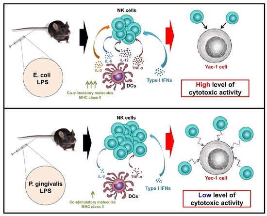
Porphyromonas gingivalis (P. gingivalis) lipopolysaccharide (LPS) promoted different innate immune activation than that promoted by Escherichia coli (E. coli) LPS. In this study, we examined the effect of P. gingivalis LPS on the proliferation and activation of natural killer (NK) cells in vivo and compared that function with that of E. coli LPS. Administration of P. gingivalis LPS to C57BL/6 mice induced stronger proliferation of NK cells in the spleen and submandibular lymph nodes (sLNs) and increased the number of circulating NK cells in blood compared to those treated with E. coli LPS. However, P. gingivalis LPS did not induce interferon-gamma (IFN-γ) production and CD69 expression in the spleen and sLN NK cells in vivo, and this was attributed to the minimal activation of the spleen and sLN dendritic cells (DCs), including low levels of co-stimulatory molecule expression and pro-inflammatory cytokine production. Furthermore, P. gingivalis LPS-treated NK cells showed less cytotoxic activity against Yac-1 target cells than E. coli LPS-treated NK cells. Hence, these data demonstrated that P. gingivalis LPS promoted limited activation of spleen and sLN NK cells in vivo, and this may play a role in the chronic inflammatory state observed in periodontal disease.

Graphical Abstract

1. Introduction

Porphyromonas gingivalis (P. gingivalis) is a gram-negative anaerobic bacterium implicated as one of the major pathogens contributing to the development of chronic inflammatory periodontal diseases [1]. Lipopolysaccharide (LPS) in the surface components of P. gingivalis interacts with the innate immune cell-expressed toll-like receptors (TLRs), which provokes the release of chemokines and cytokines [2,3,4]. Activation of innate immune cells—such as neutrophils, natural killer (NK) cells, macrophages, and dendritic cells (DCs)—accelerates inflammatory responses, which contributes to the clearance of the invading pathogens [5]. Previous studies have shown that P. gingivalis LPS promotes a different innate immune activation compared to that of Escherichia coli (E. coli) LPS [3,6]. Systemic administration of E. coli LPS induces a potent stimulus for tumor necrosis factor-alpha (TNF-α) production in the mouse, whereas P. gingivalis LPS has a minimal stimulatory capacity on TNF-α production [7]. Moreover, P. gingivalis LPS activates macrophages weakly compared to E. coli LPS [8]. In addition, in vitro studies have shown that the capacity of P. gingivalis LPS to stimulate DCs is much weaker than that of E. coli LPS [9,10]. Furthermore, P. gingivalis LPS promotes production of interleukin-1β (IL-1β), TNF-α, and IL-6 in the human monocyte cell line THP-1 dependent on the TLR2 and c-Jun N-terminal kinase (JNK) pathway, while the cytokine induction by E. coli LPS was primarily via TLR4-NF-kappaB and TLR4-p38MAPK pathway [11,12]. P. gingivalis LPS promotes T helper 2 (Th2) immune responses, whereas E. coli LPS induces Th1 polarization [10]. Although previous studies have shown that P. gingivalis LPS plays a different and lower capacity in immune regulation compared to that of E. coli LPS, the activation of spleen NK cells in vivo by the P. gingivalis and E. coli LPSs has not been compared.
NK cells are one of the three major lymphocyte subsets that play important roles in the innate immune responses against bacterial and viral infection [13]. As the major interferon-gamma (IFN-γ) producers in the early stages, NK cells contribute to the promotion of inflammation in bacterial infection and sepsis [14,15]. Conversely, IFN-γ production can be triggered in NK cells as a result of contact with and recognition of target cells, which consequently promotes cytolysis of the target cells [15]. IFN-γ production in NK cells is mediated by interactions with other innate immune cells, such as macrophages and DCs, or pro-inflammatory cytokines, notably interleukin-12 (IL-12) and IL-2 [15,16]. In addition, in periodontitis lesions, infiltration and activation of NK cells have been demonstrated in inflamed gingival tissues and periodontal tissue destruction [17,18,19].
DCs promote enhancement of the NK cell functions including IFN-γ production and cytotoxic activities [3,16,20,21]. After exposure to microbial stimuli, DCs undergo a maturation process, which is defined as upregulation of co-stimulatory molecule expression and pro-inflammatory cytokine production [22,23]. IL-2 and IL-12, produced by stimulated DCs, play a fundamental role in the activation of NK cells [3,21,24]. E. coli LPS has been well characterized as potent inducers of IL-2 and IL-12 production in DCs [25], which induces NK cell activation in vivo [3,24,26]. Moreover, P. gingivalis-stimulated bone marrow-derived DCs (BMDCs) promote IFN-γ production in NK cells in vitro [27]. However, the function of P. gingivalis LPS in spleen and submandibular lymph node (sLN) DC activation and the interaction of DC and NK cells in vivo, have not been studied.
Because P. gingivalis LPS and E. coli LPS exhibited different effects in the activation of DC, macrophage, and T cells, we hypothesized that the effect of P. gingivalis LPS and E. coli LPS in NK cell activation may be different. The current study was undertaken to test this hypothesis.

2. Results

2.1. P. Gingivalis LPS Promotes Proliferation of Spleen and sLN NK Cells but Not Blood NK Cells in Vivo

A previous study has shown that E. coli LPS promotes proliferation of NK cells in the spleen [20]. Therefore, we examined whether P. gingivalis LPS can also induce the proliferation of NK cells in vivo. C57BL/6 mice were treated intravenously (i.v.) with 1 mg/kg of P. gingivalis LPS or E. coli LPS for 18 h [28,29] Treatment with P. gingivalis LPS induced significant decreases in the number and frequency of CD3NK1.1+ cells in the spleen and sLNs compared to null-treatment controls (Figure 1A,B), whereas the number of CD3+NK1.1 cells were not changed by P. gingivalis LPS (Figure 1A,B). In contrast to the spleen and sLN NK cells, the number of blood NK cells were significantly increased by P. gingivalis LPS (Figure 1A lower panels and B right panels). Moreover, intranuclear staining of the Ki-67 antigen, a well-established measure of proliferative capacity, demonstrated that P. gingivalis LPS treatment substantially increased Ki-67 positive cells in the spleen and sLN CD3NK1.1+ cells (Figure 1C), whereas blood CD3NK1.1+ cells did not exhibit increased Ki-67 levels by P. gingivalis LPS (Figure 1C) E. coli LPS treatment led to a much stronger reduction in the cell number and frequency of CD3NK1.1+ cells in the spleen and sLNs (Figure 1A,B), but the number of blood circulating NK cells and levels of Ki-67 levels in the spleen and sLNs were significantly lower than those induced by P. gingivalis LPS (Figure 1A–C). Thus, these data suggest that P. gingivalis LPS induced a stronger proliferating effect in the spleen and sLN NK cells compared to those cells treated with E. coli LPS.

2.2. P. Gingivalis LPS Did Not Upregulate IFN-γ Production and CD69 Expression in NK Cells

Activated NK cells produce high levels of IFN-γ, and E. coli LPS has been well defined as a strong inducers of IFN-γ in NK cells [14,15]. We examined whether P. gingivalis LPS also promotes the production of IFN-γ in the spleen, sLNs, and blood NK cells in vivo. C57BL/6 mice were injected i.v. with 1 mg/kg of either P. gingivalis LPS or E. coli LPS. Six h after the injection, splenocytes were further incubated with a monensin solution for 4 h. Consistent with previous reports, E. coli LPS promoted marked increases in the IFN-γ-producing NK cells in the spleen, sLNs, and blood (Figure 2A), whereas P. gingivalis LPS did not induce this increase in the spleen, sLNs, and blood (Figure 2A). To further define IFN-γ production in NK cells, C57BL/6 mice were injected i.v. with 1 mg/kg of either P. gingivalis LPS or E. coli LPS. Six h after the injection, NK cells were isolated from the spleen and mRNA levels and production levels of IFN-γ were measured. The mRNA level of IFN-γ in the spleen NK cells were not increased by the P. gingivalis LPS treatment compared to the null-treatment control (Figure 2B), whereas the E. coli LPS treatment exhibited significant increases in the IFN-γ mRNA levels (Figure 2B) Consistent with the mRNA levels, the production levels of IFN-γ in the cultured medium of the isolated NK cells were not increased by the P. gingivalis LPS treatment, whereas E. coli LPS treated spleen NK cells produced high levels of IFN-γ (Figure 2C). In addition, the E. coli LPS treated mice, but not the P. gingivalis LPS treated mice, displayed an increased level of the CD69 expression, an early NK cell activation marker [30], on the surface of spleen, sLN, and blood NK cells compared to the null-treatment control (Figure 2D). Thus, these data suggest that P. gingivalis LPS is not able to induce full activation of spleen, sLN, and blood NK cells in vivo.

2.3. P. Gingivalis LPS Induced Minimal Activation of Spleen DCs

Our data indicated that P. gingivalis LPS did not fully induce spleen, sLN, and blood NK cell activation in vivo. This conclusion prompted us to examine in vivo DC activation by P. gingivalis LPS, because mature DCs interacted and induced NK cell activation. C57BL/6 mice were injected i.v. with 1 mg/kg of P. gingivalis LPS or E coli LPS for 12 h. The P. gingivalis LPS injection did not alter the population, frequency, or number of spleen and sLN DCs compared to the null-treatment mice (Figure 3A,B). However, treatment with E. coli LPS led to substantial decreases in the frequency and number of spleen and sLN DCs (Figure 3A,B). The expression levels of co-stimulatory molecules and MHC class II were considerably increased by the P. gingivalis LPS treatment in the spleen (Figure 3C) and sLN DCs (Figure 3D) compared to the null-treatment control mice. The levels of those molecules were significantly lower than the E. coli LPS-induced upregulation (Figure 3C,D). Thus, these data suggest that the effect of P. gingivalis LPS in the spleen and sLN DC activation in vivo was much weaker than those induced by E. coli LPS.

2.4. P. Gingivalis LPS Failed to Induce IL-2, IL-12 and IL-18 Production in Spleen DCs

In the literature, activated DCs have been reported to produce IL-2, IL-12, and IL-18, which induce IFN-γ production in NK cells [31,32,33]. We therefore assessed whether P. gingivalis LPS can induce the production of those cytokines in the spleen DCs, because the in vivo treatment of P. gingivalis LPS did not induce the production of IFN-γ in NK cells. C57BL/6 mice were injected i.v. with P. gingivalis LPS or E. coli LPS. Four hours after injection, splenocytes were further incubated with monensin for an additional 2 h and the intracellular IL-2, IL-6, IL-12p40, and TNF-α levels were measured in the spleen DCs. The E. coli LPS treatment in the mice led to a dramatic increase in the IL-2, IL-6, IL-12p40, and TNF-α-producing DCs in the spleen (Figure 4A). Unlike the E. coli LPS treatment, which substantially induced the production of all these cytokines, the P. gingivalis LPS treatment did not affect the production of the IL-2 or IL-12p40 in spleen DCs (Figure 4A). IL-6 and TNF-α levels were considerably increased by P. gingivalis LPS in the spleen DCs, although those levels were much lower than induced by E. coli LPS induced (Figure 4A).
Next, spleen DCs were purified by microbead-conjugated anti-CD11c antibodies (Milteni Biotec, Bergisch Gladbach, Germany) after 4 h of LPS stimulation. The isolated spleen DCs were measured for the mRNA levels of those cytokines and P. gingivalis LPS were determined not to induce increases in the IL-2, IL-12p40, and IL-18 mRNA levels in the spleen DCs compared to the null-treatment control DCs (Figure 4B), whereas the IL-6 and TNF-α mRNA levels were significantly increased by P. gingivalis LPS (Figure 4B). E. coli LPS-treated spleen DCs exhibited much higher mRNA levels of those cytokines than the null-treatment or P. gingivalis LPS-treated spleen DCs (Figure 4B). We also examined the production levels of those cytokines in the spleen DCs. Spleen DCs were isolated after 6 h of LPS treatment and were cultured in vitro for an additional 4 h. The cytokine concentration in the cultured medium was then measured. Consistent with mRNA levels, the concentration of IL-2, IL-12p40, and IL-18 in the cultured medium of P. gingivalis-treated spleen DCs did not increase (Figure 4C), whereas the E. coli LPS-treated spleen DCs substantially increased the concentration of cytokines in the medium compared to the null-treatment control DCs (Figure 4C). Meanwhile, the IL-6 and TNF-α concentrations in the P. gingivalis LPS-stimulated spleen DCs were considerably increased in comparison to the null-treatment controls. Thus, these data suggest that P. gingivalis LPS did not induce IL-2, IL-12, and IL-18 production in spleen DCs.

2.5. P. Gingivalis LPS Induced Minimal Levels of Cytotoxic Activity in Spleen NK Cells

The cytotoxic activities of spleen NK cells by P. gingivalis and E. coli LPS were examined. C57BL/6 mice were injected i.v. with 1 mg/kg P. gingivalis and E. coli LPS and 24 h later, they were injected again with the same amount of LPS. Twenty-four hours after the last injection, NK cells (effector cells) were isolated from the spleen and co-cultured with Yac-1 cells (target cells) for detection of MHC-negative target cell killing. The presence of P. gingivalis LPS-stimulated spleen NK cells led to significantly increased levels of cytotoxic activity compared to the null-treatment spleen NK cells (Figure 5A), whereas E. coli LPS-treated spleen NK cells exhibited greater cytolysis of Yac-1 cells than P. gingivalis LPS-induced cells (Figure 5A).
Because the cytotoxic activities of NK cells are dependent on type I IFNs [24], we next measured the levels of type I IFNs in the LPS-treated mice. The P. gingivalis LPS treatment promoted considerable increases in the mRNA levels of type I IFNs in the spleen and protein levels of type I IFNs in serum compared to the null-treatment controls (Figure 5B,C). In contrast, the E. coli LPS treatment substantially elevated the mRNA and protein levels of type I IFNs, and the levels were greater than that of the P. gingivalis LPS-induced levels (Figure 5B,C). Hence, these data demonstrated that P. gingivalis LPS promotes minimal levels of cytotoxic activity in spleen NK cells, attributed to low levels of type I IFN production.

3. Discussion

In this study, we demonstrated that P. gingivalis LPS and E. coli LPS elicited different effects on NK cell proliferation and activation in vivo. E. coli LPS induced activation of NK cells including IFN-γ production, CD69 expression, and cytotoxic activity, while P. gingivalis LPS induced minimal activation of NK cells. In contrast to the activation effect, P. gingivalis LPS promoted a greater proliferation in the spleen and sLN and increased the number of NK cells circulating in blood relative to E. coli LPS. The different effects of these two LPSs are consistent with several previous reports, which have investigated the macrophages and T cells [6,7,8]. Hence, the minimal activation effect of P. gingivalis LPS in the macrophages, T cells, and NK cells may be a potential reason for P. gingivalis-induced chronic inflammatory diseases. Further investigation will address this possibility and elucidate the specific role of the immune cells in P. gingivalis-induced chronic periodontitis.
We also determined that P. gingivalis LPS did not fully activate spleen and sLN DCs as indicated by the low levels of co-stimulatory molecule expression and pro-inflammatory cytokine production, while E. coli LPS promoted strong activation of those cells in vivo. Although previous studies demonstrated that P. gingivalis LPS induced DC activation in vivo [10], the activation efficiencies of E. coli and P. gingivalis LPSs were not compared. Moreover, a recent in vitro study also demonstrated that P. gingivalis LPS induced lower secretion levels of IL-12 and IFN-γ in BMDCs than those induced by E. coli LPS [9]. In addition, we also found that P. gingivalis LPS did not induce the changes in the spleen and sLN DC number, whereas the E. coli LPS treatment induced a markedly decreased number of spleen and sLN DCs. Because fully matured DCs undergo exhaustion, anergy, or apoptosis following T-cell activation [34,35,36], E. coli LPS might induce full activation but P. gingivalis LPS might not induce full activation of spleen DCs. Therefore, our results provide several lines of evidence that P. gingivalis LPS does not fully activate spleen and sLN DCs in vivo, and clearly demonstrate the unexplored in vivo function of P. gingivalis LPS in the spleen and sLN DCs.
Activated and matured DCs promote IFN-γ production in NK cells [21,24,26]. Consistent with previous reports, E. coli LPS induced DC activation and IFN-γ production in NK cells [21,24]. Moreover, activated DCs produced IL-2 and IL-12, which are crucial promoters of the production of IFN-γ in the NK cells [21,24,26]. In this study, we also found that E. coli LPS promotes IL-2 and IL-12 production in the spleen DCs in vivo. In contrast, P. gingivalis LPS did not induce IL-2 and IL-12 secretion in the spleen DCs although the DCs showed increased expression of co-stimulatory molecules. The data suggest that these cytokines may be essential for IFN-γ production in the NK cells in vivo, and upregulation of co-stimulatory molecule expression in DCs may not be required for IFN-γ production in NK cells. Our further investigation will determine the function of co-stimulatory molecules in the spleen DCs on the NK cells by LPS treatment using IL-2-, IL-12-, and IL-18-depleted mice, or a blockade antibody.
NK cells have the ability to kill MHC-negative targets [24,37]. Previous studies have demonstrated that the NK cell-mediated cytotoxic effect is dependent on type I IFNs [24,38,39]. We also found that P. gingivalis LPS induced upregulation of type I IFNs, and consequently promotes cytotoxic activities in the spleen NK cells against Yak-1 target cells. Type I IFNs can be produced by many immune cell types including NK cells, B cells, T cells, macrophages, and DCs [40,41]. Plasmacytoid DCs (pDCs) have been identified as a potent producer of type I IFNs in response to viral antigens; however, pDCs do not produce type I IFNs in the spleen in vivo in response to E. coli LPS [42]. Unlike the in vivo study, in vitro generated BMDCs produce type I IFNs after E. coli LPS stimulation [24]. Moreover, human and mouse macrophages derived in vitro from precursors can also produce type I IFNs by E. coli LPS [43,44]. Therefore, the precise contribution of type I IFN-producing immune cells in the response to E. coli or P. gingivalis LPS in the circumstances of in vivo still remains to be studied. Future studies will define the cellular and molecular mechanisms of type I IFN production in the immune cells by E. coli or P. gingivalis LPS stimulation in the mouse model in vivo.
NK cells are abundant in periodontitis lesions, and activation of NK cells has been linked to inflammation of periodontal tissue [19]. Infiltrated NK cells in inflamed gingival tissue have been observed [19]. Moreover, it has been shown that NK cells are increased in chronic periodontitis [17,18,19]. However, the activation of NK cells was not fully investigated in chronic periodontitis. Because P. gingivalis LPS did not induce full activation of NK cells in sLNs but promoted proliferation of NK cells in vivo, those inactivated NK cells may be not able to be clear the P. gingivalis bacterium and this may contribute to the persistence of P. gingivalis and development of chronic periodontitis. Further studies will define the state of NK cells in chronic periodontitis.
In summary, we found that P. gingivalis LPS induces proliferation of NK cells in the spleen and sLNs, but those NK cells did not produce IFN-γ nor expressed CD69. Moreover, P. gingivalis LPS treated NK cells promote low levels of cytotoxic activates compared to E. coli LPS. Those low levels of NK cell activation in response to P. gingivalis LPS was attributed to the minimal activation of DCs and type I IFN production.

4. Materials and Methods

4.1. Animals

C57BL/6 mice were purchased from the Shanghai Public Health Clinical Center, and kept under pathogen-free conditions. The mice were maintained in a room with controlled temperature (20–22 °C), humidity (50%–60%), and light (12 h:12 h), and free access to standard rodent chow and water. This study was performed in strict accordance with the recommendations in the Guide for the Care and Use of Laboratory Animals of the Shanghai Public Health Clinical Center. The protocol was approved by the Committee on the Ethics of Animal Experiments of the Fudan University (Permit Number: SYXK-2010-0098). Mice were sacrificed by CO2 inhalation euthanasia, and all efforts were made to minimize suffering.

4.2. Antibodies and Reagents

The cells were stained and analyzed on a FACSAria II (Becton Dickinson, San Diego, CA, USA), with dead cells excluded by DAPI staining. The following fluorescence-conjugated Abs were used: CD3 (17A2), NK1.1 (PK136), Ki-67 (16A8), CD69 (H1.2F3), CD11c (N418), CD40 (HM40-3), CD80 (16-10A1), CD86 (GL-1), MHC class II (M5/114.15.2), anti-IFN-γ (XMG1.2), IL-2 (JES6-5H4), IL-6 (MP5-20F3), IL-12p40 (C15.6), and TNF-α (MP6-XT22) and obtained from BioLegend (San Diego, CA, USA). The P. gingivalis LPS (LPS-PG; standard) and E. coli LPS (LPS-EK; ultrapure) used in this study were purchased from InvivoGen (San Diego, CA, USA).

4.3. NK Cell Analysis

The spleens were cut into small fragments and digested with 2% fetal bovine serum (FBS) containing collagenase for 20 min at room temperature. Cells from the digest were centrifuged to a pellet, and the pellet was re-suspended in 5 mL of a 1.077 histopaque (Sigma-Aldrich, St. Louis, MO, USA). Additional histopaque was layered below and EDTA-FBS was layered above the cell suspension, which was then centrifuged at 1700 g for 10 min. For blood NK cell analysis, the blood cells were layered above the 1.077 histopaque, and then centrifuged at 1700 g for 10 min. The light density fraction (<1.077 g/cm3) was collected and stained with fluorescence-labeled CD3 and NK1.1 monoclonal antibodies. The sLNs were placed in a grinder and processed with a tissue homogenizer. Tissue homogenates were filtered through a 100 μm nylon mesh, washed, and the erythrocytes were removed with an Ammonium-Chloride-Potassium (ACK) lysing buffer (Thermo Fisher Scientific, Waltham, MA, USA). The single cells were resuspended in culture medium and stained with fluorescence-labeled CD3 and NK1.1 monoclonal antibodies.

4.4. DC Analysis

Spleen and sLN DCs were analyzed as described elsewhere [23,45,46]. After density cut by 1.077 histopaque from digested spleen as shown in NK cell analysis, the light density fraction (<1.077 g/cm3) was collected and incubated for 30 min with the following FITC-conjugated monoclonal antibodies (mAbs): anti-CD3 (17A2), anti-Thy1.1 (OX-7), anti-B220 (RA3-6B2), anti-Gr1 (RB68C5), anti-CD49b (DX5), and anti-TER-119 (TER-119) (BioLegend, San Diego, CA, USA). The lineageCD11c+ cells were defined as DCs. Analysis was carried out on a FACS Aria II (Becton Dickinson, San Diego, CA, USA).

4.5. Cytotoxicity Assay

Activated spleen NK cells were isolated and co-cultured with Yac-1 cells from a mouse lymphoma cell line in V-bottomed 96-well plates in 200 μL of RPMI 1640 10% fetal calf serum for 24 h. 50,000 target Yac-1 cells were co-cultured in triplicate with 5000 (1:10), 25,000 (1:2), 50,000 (1:1), 250,000 (5:1), and 500,000 (10:1) NK cells, respectively. Cytotoxicity was determined by the lactate dehydrogenase (LDH) assay. The LDH assay was performed using a LDH Cytotoxicity Assay Kit (Roche, Basel, Switzerland) according to the manufacturer’s instructions.

4.6. Ex Vivo Cell Stimulation and Intracellular Cytokine Staining

As described in detail previously [45,47], single cell suspensions prepared from spleen were incubated in vitro for 4 h with monensin solution (Biolegend). For intracellular cytokine staining, cells were stained for surface molecules first, then fixed and permeabilized with Cytofix/Cytoperm buffer (eBioscience, San Diego, CA, USA) and subsequently incubated with anti-cytokine antibodies in Perm/Wash buffer (eBioscience) for 30 min. Control staining with isotype control IgGs was performed in all experiments.

4.7. Real-Time qPCR

Total RNA was reverse-transcribed into cDNA using Oligo (dT) and M-MLV reverse transcriptase (Promega, Madison, WI, USA). The cDNA was subjected to real-time PCR amplification (Qiagen, Hilden, Germany) for 40 cycles with annealing and extension temperature at 60 °C, on a LightCycler 480 Real-Time PCR System (Roche, Basel, Switzerland). Primer sequences are: mouse β-actin forward, 5′-TGGATGACGATATCGCTGCG-3′; reverse, 5′-AGGGTCAGGATACCTCTCTT-3′, IFN-α1 forward, 5′-ACCTCAGGAACAAGAGAGCC-3′; reverse, 5′-CTGCGGGAATCCAAAGTCCT-3′, IFN-β1 forward, 5′-TAAGCAGCTCCAGCTCCAAG-3′; reverse, 5′-CCCTGTAGGTGAGGTTGATC-3′,IL-2 forward, 5′-ATGAACTTGGACCTCTGCG-3′; reverse, 5′-GGGCTTGTTGAGATGATGC-3′, IL-6 forward, 5′-AACGATGATGCACTTGCAGA-3′; reverse, 5′-GAGCATTGGAAATTGGGGTA-3′, IL-12p40 forward, 5′-CACATCTGCTGCTCCACAAG-3′; reverse, 5′-CCGTCCGGAGTAATTTGGTG-3′, IL-18 forward, 5′-AGGACACTTTCTTGCTTGCC-3′; reverse, 5′-CCTCGGGTATTCTGTTATGGA-3′, TNF-α forward, 5′-CCTTTCACTCACTGGCCCAA-3′; reverse, 5′-AGTGCCTCTTCTGCCAGTTC-3′.

4.8. ELISA Assay

The IL-2, IL-6, IL-12p40, and TNF-α ELISA kits were purchased from Biolegend. The IFN-α and IFN-β ELISA kits were purchased from eBioscience. The concentration of proteins in the cultured medium and serum were measured in triplicate using standard ELISA kits, with standard cytokine preparations being used as the internal controls.

4.9. Statistical Analysis

Results were expressed as the mean ± standard error of the mean (SEM). Data sets were analyzed by one-way ANOVA using the Tukey multiple comparison test with GraphPad Prism 4. p values smaller than 0.05 were considered to be statistically significant.

5. Conclusions

In conclusion, the present study demonstrates that P. gingivalis LPS induced low levels of activation and high levels of proliferation in the spleen and sLN NK cells in vivo. The low levels of activation and increased number of NK cells observed may lead to the persistence and survival of P. gingivalis, resulting in P. gingivalis-induced chronic periodontal diseases.

Acknowledgments

We thank the Shanghai Public Health Clinical Center animal facility for maintaining the animals in this study. This study was supported by the Research Fund for International Young Scientists from the National Natural Science Foundation of China (Grant No. 81550110507). Yuhua Wang was supported by The National Natural Science Foundation of China (Grant No. 81500881) Shanghai Municipal Commission of Health and Family Planning (Grant No. 201540045). Wei Zhang was supported by the Budding Plan of the Shanghai Public Health Clinical Center (Grant No. 2015MY03).

Author Contributions

J.O.J. conceived and designed the experiments; Y.W., W.Z., and L.X. performed the experiments; J.O.J. analyzed the data and wrote the paper.

Conflicts of Interest

The authors declare no conflict of interest. The founding sponsors had no role in the design of the study; in the collection, analyses, or interpretation of data; in the writing of the manuscript, and in the decision to publish the results.

References

  1. Hussain, M.; Stover, C.M.; Dupont, A. P. gingivalis in Periodontal Disease and Atherosclerosis - Scenes of Action for Antimicrobial Peptides and Complement. Front. Immunol. 2015, 6, 45. [Google Scholar] [CrossRef] [PubMed]
  2. Berezow, A.B.; Ernst, R.K.; Coats, S.R.; Braham, P.H.; Karimi-Naser, L.M.; Darveau, R.P. The structurally similar, penta-acylated lipopolysaccharides of Porphyromonas gingivalis and Bacteroides elicit strikingly different innate immune responses. Microb. Pathog. 2009, 47, 68–77. [Google Scholar] [CrossRef] [PubMed]
  3. Fujimoto, Y.; Shimoyama, A.; Saeki, A.; Kitayama, N.; Kasamatsu, C.; Tsutsui, H.; Fukase, K. Innate immunomodulation by lipophilic termini of lipopolysaccharide; synthesis of lipid As from Porphyromonas gingivalis and other bacteria and their immunomodulative responses. Mol. BioSyst. 2013, 9, 987–996. [Google Scholar] [CrossRef] [PubMed]
  4. Jitprasertwong, P.; Charadram, N.; Kumphune, S.; Pongcharoen, S.; Sirisinha, S. Female sex hormones modulate Porphyromonas gingivalis lipopolysaccharide-induced Toll-like receptor signaling in primary human monocytes. J. Periodontal Res. 2015, 51, 395–406. [Google Scholar] [CrossRef] [PubMed]
  5. McGuire, V.A.; Arthur, J.S. Subverting Toll-Like Receptor Signaling by Bacterial Pathogens. Front. Immunol. 2015, 6, 607. [Google Scholar] [CrossRef] [PubMed]
  6. Andrukhov, O.; Ertlschweiger, S.; Moritz, A.; Bantleon, H.P.; Rausch-Fan, X. Different effects of P. gingivalis LPS and E. coli LPS on the expression of interleukin-6 in human gingival fibroblasts. Acta Odontol. Scand. 2014, 72, 337–345. [Google Scholar] [CrossRef] [PubMed]
  7. Liu, R.; Desta, T.; Raptis, M.; Darveau, R.P.; Graves, D.T. P. gingivalis and E. coli lipopolysaccharides exhibit different systemic but similar local induction of inflammatory markers. J. Periodontol. 2008, 79, 1241–1247. [Google Scholar] [CrossRef] [PubMed]
  8. Holden, J.A.; Attard, T.J.; Laughton, K.M.; Mansell, A.; O’Brien-Simpson, N.M.; Reynolds, E.C. Porphyromonas gingivalis lipopolysaccharide weakly activates M1 and M2 polarized mouse macrophages but induces inflammatory cytokines. Infect. Immun. 2014, 82, 4190–4203. [Google Scholar] [CrossRef] [PubMed]
  9. Su, H.; Yan, X.; Dong, Z.; Chen, W.; Lin, Z.T.; Hu, Q.G. Differential roles of Porphyromonas gingivalis lipopolysaccharide and Escherichia coli lipopolysaccharide in maturation and antigen-presenting functions of dentritic cells. Eur. Rev. Med. Pharmacol. Sci. 2015, 19, 2482–2492. [Google Scholar] [PubMed]
  10. Pulendran, B.; Kumar, P.; Cutler, C.W.; Mohamadzadeh, M.; van Dyke, T.; Banchereau, J. Lipopolysaccharides from distinct pathogens induce different classes of immune responses in vivo. J. Immunol. 2001, 167, 5067–5076. [Google Scholar] [CrossRef] [PubMed]
  11. Zhang, D.; Chen, L.; Li, S.; Gu, Z.; Yan, J. Lipopolysaccharide (LPS) of Porphyromonas gingivalis induces IL-1beta, TNF-alpha and IL-6 production by THP-1 cells in a way different from that of Escherichia coli LPS. Innate Immun. 2008, 14, 99–107. [Google Scholar] [CrossRef] [PubMed]
  12. Hajishengallis, G.; Martin, M.; Schifferle, R.E.; Genco, R.J. Counteracting interactions between lipopolysaccharide molecules with differential activation of toll-like receptors. Infect. Immun. 2002, 70, 6658–6664. [Google Scholar] [CrossRef] [PubMed]
  13. Degli-Esposti, M.A.; Smyth, M.J. Close encounters of different kinds: Dendritic cells and NK cells take centre stage. Nat. Rev. Immunol. 2005, 5, 112–124. [Google Scholar] [CrossRef] [PubMed]
  14. Artavanis-Tsakonas, K.; Riley, E.M. Innate immune response to malaria: Rapid induction of IFN-gamma from human NK cells by live Plasmodium falciparum-infected erythrocytes. J. Immunol. 2002, 169, 2956–2963. [Google Scholar] [CrossRef] [PubMed]
  15. Souza-Fonseca-Guimaraes, F.; Adib-Conquy, M.; Cavaillon, J.M. Natural killer (NK) cells in antibacterial innate immunity: Angels or devils? Mol. Med. 2012, 18, 270–285. [Google Scholar] [CrossRef] [PubMed]
  16. Gerosa, F.; Baldani-Guerra, B.; Nisii, C.; Marchesini, V.; Carra, G.; Trinchieri, G. Reciprocal activating interaction between natural killer cells and dendritic cells. J. Exp. Med. 2002, 195, 327–333. [Google Scholar] [CrossRef] [PubMed]
  17. Fujita, S.; Takahashi, H.; Okabe, H.; Ozaki, Y.; Hara, Y.; Kato, I. Distribution of natural killer cells in periodontal diseases: An immunohistochemical study. J. Periodontol. 1992, 63, 686–689. [Google Scholar] [CrossRef] [PubMed]
  18. Stelin, S.; Ramakrishan, H.; Talwar, A.; Arun, K.V.; Kumar, T.S. Immunohistological analysis of CD1a langerhans cells and CD57 natural killer cells in healthy and diseased human gingival tissue: A comparative study. J. Indian Soc. Periodontol. 2009, 13, 150–154. [Google Scholar] [PubMed]
  19. Kramer, B.; Kebschull, M.; Nowak, M.; Demmer, R.T.; Haupt, M.; Korner, C.; Perner, S.; Jepsen, S.; Nattermann, J.; Papapanou, P.N. Role of the NK cell-activating receptor CRACC in periodontitis. Infect. Immun. 2013, 81, 690–696. [Google Scholar] [CrossRef] [PubMed]
  20. Tsujimoto, H.; Uchida, T.; Efron, P.A.; Scumpia, P.O.; Verma, A.; Matsumoto, T.; Tschoeke, S.K.; Ungaro, R.F.; Ono, S.; Seki, S.; et al. Flagellin enhances NK cell proliferation and activation directly and through dendritic cell-NK cell interactions. J. Leukoc. Biol. 2005, 78, 888–897. [Google Scholar] [CrossRef] [PubMed]
  21. Cooper, M.A.; Fehniger, T.A.; Fuchs, A.; Colonna, M.; Caligiuri, M.A. NK cell and DC interactions. Trends Immunol. 2004, 25, 47–52. [Google Scholar] [CrossRef] [PubMed]
  22. Banchereau, J.; Steinman, R.M. Dendritic cells and the control of immunity. Nature 1998, 392, 245–252. [Google Scholar] [CrossRef] [PubMed]
  23. Jin, J.O.; Zhang, W.; Du, J.Y.; Wong, K.W.; Oda, T.; Yu, Q. Fucoidan can function as an adjuvant in vivo to enhance dendritic cell maturation and function and promote antigen-specific T cell immune responses. PLoS ONE 2014, 9, e99396. [Google Scholar] [CrossRef] [PubMed]
  24. Granucci, F.; Zanoni, I.; Pavelka, N.; van Dommelen, S.L.; Andoniou, C.E.; Belardelli, F.; Degli Esposti, M.A.; Ricciardi-Castagnoli, P. A contribution of mouse dendritic cell-derived IL-2 for NK cell activation. J. Exp. Med. 2004, 200, 287–295. [Google Scholar] [CrossRef] [PubMed]
  25. Huang, Q.; Liu, D.; Majewski, P.; Schulte, L.C.; Korn, J.M.; Young, R.A.; Lander, E.S.; Hacohen, N. The plasticity of dendritic cell responses to pathogens and their components. Science 2001, 294, 870–875. [Google Scholar] [CrossRef] [PubMed]
  26. Ferlazzo, G.; Pack, M.; Thomas, D.; Paludan, C.; Schmid, D.; Strowig, T.; Bougras, G.; Muller, W.A.; Moretta, L.; Munz, C. Distinct roles of IL-12 and IL-15 in human natural killer cell activation by dendritic cells from secondary lymphoid organs. Proc. Natl. Acad. Sci. USA 2004, 101, 16606–16611. [Google Scholar] [CrossRef] [PubMed]
  27. Kikuchi, T.; Willis, D.L.; Liu, M.; Purkall, D.B.; Sukumar, S.; Barbour, S.E.; Schenkein, H.A.; Tew, J.G. Dendritic-NK cell interactions in P. gingivalis-specific responses. J. Dent. Res. 2005, 84, 858–862. [Google Scholar] [CrossRef] [PubMed]
  28. Endo, Y.; Shibazaki, M.; Nakamura, M.; Takada, H. Contrasting effects of lipopolysaccharides (endotoxins) from oral black-pigmented bacteria and Enterobacteriaceae on platelets, a major source of serotonin, and on histamine-forming enzyme in mice. J. Infect. Dis. 1997, 175, 1404–1412. [Google Scholar] [CrossRef] [PubMed]
  29. Kim, Y.G.; Kang, J.H.; Kim, H.J.; Kim, H.J.; Kim, H.H.; Kim, J.Y.; Lee, Y. Bortezomib Inhibits Osteoclastogenesis and Porphyromonas gingivalis Lipopolysaccharide-induced Alveolar Bone Resorption. J. Dent. Res. 2015, 94, 1243–1250. [Google Scholar] [CrossRef] [PubMed]
  30. Werfel, T.; Boeker, M.; Kapp, A. Rapid expression of the CD69 antigen on T cells and natural killer cells upon antigenic stimulation of peripheral blood mononuclear cell suspensions. Allergy 1997, 52, 465–469. [Google Scholar] [CrossRef] [PubMed]
  31. Zelante, T.; Fric, J.; Wong, A.Y.; Ricciardi-Castagnoli, P. Interleukin-2 production by dendritic cells and its immuno-regulatory functions. Front. Immunol. 2012, 3, 161. [Google Scholar] [CrossRef] [PubMed]
  32. Miller, G.; Lahrs, S.; Dematteo, R.P. Overexpression of interleukin-12 enables dendritic cells to activate NK cells and confer systemic antitumor immunity. FASEB J. 2003, 17, 728–730. [Google Scholar] [CrossRef] [PubMed]
  33. Semino, C.; Angelini, G.; Poggi, A.; Rubartelli, A. NK/iDC interaction results in IL-18 secretion by DCs at the synaptic cleft followed by NK cell activation and release of the DC maturation factor HMGB1. Blood 2005, 106, 609–616. [Google Scholar] [CrossRef] [PubMed]
  34. Griffiths, K.L.; Tan, J.K.; O’Neill, H.C. Characterization of the effect of LPS on dendritic cell subset discrimination in spleen. J. Cell. Mol. Med. 2014, 18, 1908–1912. [Google Scholar] [CrossRef] [PubMed]
  35. Kurts, C.; Kosaka, H.; Carbone, F.R.; Miller, J.F.; Heath, W.R. Class I-restricted cross-presentation of exogenous self-antigens leads to deletion of autoreactive CD8(+) T cells. J. Exp. Med. 1997, 186, 239–245. [Google Scholar] [CrossRef] [PubMed]
  36. Tan, J.K.; O’Neill, H.C. Maturation requirements for dendritic cells in T cell stimulation leading to tolerance versus immunity. J. Leukoc. Biol. 2005, 78, 319–324. [Google Scholar] [CrossRef] [PubMed]
  37. Le Bouteiller, P.; Barakonyi, A.; Giustiniani, J.; Lenfant, F.; Marie-Cardine, A.; Aguerre-Girr, M.; Rabot, M.; Hilgert, I.; Mami-Chouaib, F.; Tabiasco, J.; et al. Engagement of CD160 receptor by HLA-C is a triggering mechanism used by circulating natural killer (NK) cells to mediate cytotoxicity. Proc. Natl. Acad. Sci. USA 2002, 99, 16963–16968. [Google Scholar] [CrossRef] [PubMed]
  38. Swann, J.B.; Hayakawa, Y.; Zerafa, N.; Sheehan, K.C.; Scott, B.; Schreiber, R.D.; Hertzog, P.; Smyth, M.J. Type I IFN contributes to NK cell homeostasis, activation, and antitumor function. J. Immunol. 2007, 178, 7540–7549. [Google Scholar] [CrossRef] [PubMed]
  39. Zhu, J.; Huang, X.; Yang, Y. A critical role for type I IFN-dependent NK cell activation in innate immune elimination of adenoviral vectors in vivo. Mol. Ther. J. Am. Soc. Gene Ther. 2008, 16, 1300–1307. [Google Scholar] [CrossRef] [PubMed]
  40. Coccia, E.M.; Battistini, A. Early IFN type I response: Larning from microbial evasion strategies. Semin. Immunol. 2015, 27, 85–101. [Google Scholar] [CrossRef] [PubMed]
  41. Stifter, S.A.; Feng, C.G. Interfering with immunity: Detrimental role of type I IFNs during infection. J. Immunol. 2015, 194, 2455–2465. [Google Scholar] [CrossRef] [PubMed]
  42. Asselin-Paturel, C.; Brizard, G.; Chemin, K.; Boonstra, A.; O’Garra, A.; Vicari, A.; Trinchieri, G. Type I interferon dependence of plasmacytoid dendritic cell activation and migration. J. Exp. Med. 2005, 201, 1157–1167. [Google Scholar] [CrossRef] [PubMed]
  43. Reimer, T.; Brcic, M.; Schweizer, M.; Jungi, T.W. Poly(I:C) and LPS induce distinct IRF3 and NF-kappaB signaling during type-I IFN and TNF responses in human macrophages. J. Leukoc. Biol. 2008, 83, 1249–1257. [Google Scholar] [CrossRef] [PubMed]
  44. Vadiveloo, P.K.; Vairo, G.; Hertzog, P.; Kola, I.; Hamilton, J.A. Role of type I interferons during macrophage activation by lipopolysaccharide. Cytokine 2000, 12, 1639–1646. [Google Scholar] [CrossRef] [PubMed]
  45. Zhang, W.; Cho, S.Y.; Xiang, G.; Min, K.J.; Yu, Q.; Jin, J.O. Ginseng Berry Extract Promotes Maturation of Mouse Dendritic Cells. PLoS ONE 2015, 10, e0130926. [Google Scholar] [CrossRef] [PubMed]
  46. Zhang, W.; Okimura, T.; Xu, L.; Zhang, L.; Oda, T.; Kwak, M.; Yu, Q.; Jin, J.O. Ascophyllan functions as an adjuvant to promote anti-cancer effect by dendritic cell activation. Oncotarget 2016, 7, 19284–19298. [Google Scholar] [CrossRef] [PubMed]
  47. Jin, J.O.; Han, X.; Yu, Q. Interleukin-6 induces the generation of IL-10-producing Tr1 cells and suppresses autoimmune tissue inflammation. J. Autoimmun. 2013, 40, 28–44. [Google Scholar] [CrossRef] [PubMed]
  • Sample Availability: Samples of the compounds are available from the authors.
Figure 1. P. gingivalis LPS enhanced the proliferation of spleen and submandibular lymph node (sLN) natural killer (NK) cells in vivo. C57BL/6 mice were injected intravenously (i.v.) with 1 mg/kg of P. gingivalis (P.g) LPS or E. coli (E.c) LPS for 18 h. (A) The percentage of CD3NK1.1+ and CD3+NK1.1 cells in the spleen, sLNs, and blood were analyzed by flow cytometry; (B) Absolute cell number of CD3NK1.1+ and CD3+NK1.1 cells within live cells in the spleen, sLNs, and blood are shown; (C) Intranuclear expression levels of Ki-67 in the CD3NK1.1+ cells in the spleen, sLNs, and blood are shown (left panel). The mean percentage of Ki-67 positive cells in the CD3NK1.1+ in the spleen, sLN, and blood were illustrated (right panel). All data are representative or the average of analyses of six samples from three independent experiments. * p < 0.05, ** p < 0.01.
Molecules 21 01086 g001
Figure 2. P. gingivalis LPS did not upregulate IFN-γ production and CD69 expression in the spleen and sLN NK cells. C57BL/6 mice were injected i.v. with 1 mg/kg of P.g LPS or E.c LPS. Six hours after injection, splenocytes were further cultured with monensin solution for 4 h. (A) Intracellular IFN-γ production levels in the CD3NK1.1+ cells in the spleen, sLNs, and blood were analyzed by flow cytometry; (B) C57BL6 mice were treated with the LPS for 6 h; subsequently NK cells were isolated from the spleen. The mRNA levels of IFN-γ were measured in the spleen NK cells; (C) Isolated NK cells were further incubated in the culture medium for 4 h. The IFN-γ concentration in the cultured medium was measured by ELISA; (D) The CD69 expression level in the spleen, sLN, and blood NK cells were analyzed 6 h after LPS treatment. All data are representative or the average of analyses of six samples from three independent experiments. ** p < 0.01.
Molecules 21 01086 g002
Figure 3. P. gingivalis LPS weakly activated spleen and sLN DCs. C57BL/6 mice were injected i.v. with 1 mg/kg of P.g LPS or E.c LPS for 12 h. (A) The percentage of lineageCD11c+ DCs in the spleen (upper panel) and sLN (lower panel) was analyzed by flow cytometry; (B) Absolute numbers of live, lineageCD11c+ cells in the spleen and sLN were shown; (C,D) Mean fluorescence intensity (MFI) of indicated surface molecules in the spleen (C) and sLN (D) was analyzed on a flow cytometry. All data are representative of or the average of analyses of six independent samples (two mice per experiment, total three independent experiments). * p < 0.05. ** p < 0.01.
Molecules 21 01086 g003
Figure 4. P. gingivalis LPS weakly induced pro-inflammatory cytokine production in spleen DCs. C57BL/6 mice were injected i.v. with 1 mg/kg of P.g LPS or E.c LPS. (A) Four hours after injection, the spleen was harvested from the mice and 1 × 10⁶ splenocytes were further incubated with monensin for an additional 2 h. Levels of intracellular cytokine production in the spleen DCs were analyzed by flow cytometry; (B) CD11c+ spleen DCs were isolated 6 h after injection, and measured to mRNA levels of pro-inflammatory cytokines were measured; (C) The isolated spleen DCs (0.1 × 10⁶) were cultured in 1 mL of RPMI-1640 for 4 h in vitro. The concentrations of indicated cytokines were measured in the cultured medium by ELISA. All data are representative of or the average of analyses of six individual mice in each group (two mice per experiment, total three independent experiments). * p < 0.05, ** p < 0.01.
Molecules 21 01086 g004
Figure 5. P. gingivalis LPS promoted a weak level of cytotoxic activity in the spleen NK cells. C57BL/6 mice were injected i.v. with 1 mg/kg of P.g LPS or E.c LPS and 24 h later, were injected again with the same amount of LPS. (A) Twenty-four hours after last treatment, NK cells were isolated from the spleen and co-cultured with Yac-1 cell by the indicated ratio for 6 h. Cytotoxicity was measured by the lactate dehydrogenase (LDH) assay. # p < 0.05 P. gingivalis LPS versus E. coli LPS group, * p < 0.05 None versus P. gingivalis LPS group, ** p < 0.01 None versus E. coli LPS group; (B) mRNA levels of IFN-α1 and IFN-β1 in splenocyte after 12 h of LPS treatment were measured; (C) The concentration of IFN-α and IFN-β in the serum was measured by ELISA. All data are representative of or the average of analyses of six individual mice in each group (two mice per experiment, total three independent experiments). * p < 0.05, ** p < 0.01.
Molecules 21 01086 g005