1. Introduction
Diabetes mellitus and its related complications are some of the most prevalent and serious metabolic disorders characterized by high blood-glucose levels. One of the therapeutic approaches for counteracting hyperglycemia is to block the absorption of glucose by inhibiting the activity of carbohydrate-hydrolyzing enzymes such as α-glucosidase in the digestive organs. Small intestinal α-glucosidases (EC 3.2.1.20) are key enzymes involved in dietary carbohydrate digestion in humans [
1]. Inhibitors of these enzymes may be effective in decreasing carbohydrate digestion and glucose absorption to suppress postprandial hyperglycemia [
2]. However, α-glucosidase inhibitors (acarbose, miglitol and voglibose) have been shown to exhibit many side effects. A known α-glucosidase inhibitor, acarbose, is known to cause side effects such as abdominal pain, distension and diarrhea [
3]. For this reason, many researchers have been trying to find a safe, potent and non-toxic α-glucosidase inhibitor from natural sources.
Protein tyrosine phosphatases (PTPases) are expressed in insulin-sensitive tissues (such as the liver, muscle and adipose tissue) and have a key role in the regulation of insulin signal transduction pathways [
4]. Therefore, it is considered a favorable target for the treatment of type 2 diabetes mellitus (T2DM) and obesity [
5]. Although several PTPases such as PTP-α, leukocyte antigen-related tyrosine phosphatase (LAR) and SH2-domain-containing phosphotyrosine phosphatase (SHP2) have been implicated in the regulation of insulin signaling, there is substantial evidence supporting PTP1β as the critical PTP controlling the insulin signaling pathway. PTP1β can interact with and dephosphorylate the activated insulin receptor (IR) as well as insulin receptor substrate (IRS) proteins [
6].
Advanced glycation end products (AGEs) are formed from the non-enzymatic glycation of reducing sugars with the amino group of proteins. The subsequent products are Schiff bases that are then rearranged to more stable ketoamines [
7]. It was proposed that accelerated chemical modification of proteins by glucose during hyperglycemia contributes to the pathogenesis of diabetes and the formation and accumulation of AGEs will induce oxidative stress and affect extracellular and intracellular function structure in many different cell types [
8]. Therefore, AGEs are associated to increased oxidative stress and it would have deleterious effects on various cellular functions, and is thought to contribute to the pathogenesis of various diabetic complications (neuropathy, nephropathy, and retinopathy) [
9].
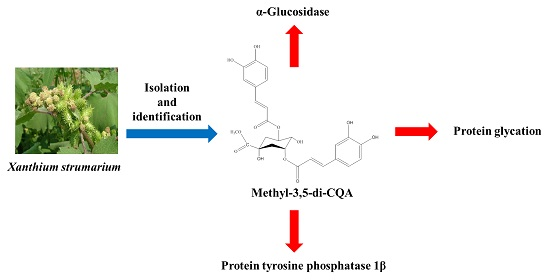
The fruit of
Xanthium strumarium has been used in traditional medicine in Korea for the treatment of chronic rhinitis, headache, cough and atopic dermatitis [
10]. It has also been reported to possess curative effects against inflammation, allergic rhinitis, infection and cancer [
11]. Various compounds have been isolated from this plant including caffeoylquinic acid (CQA) derivatives, carboxyatractyloside, xanthanol, isoxantanolm hydroquinone, alkaloids and thiazinedione [
12]. Recently, our research team reported that CQA derivatives and phenolic constituents were isolated from the MeOH extract of
X. strumarium. Furthermore, these compounds showed a strong inhibitory effect on aldose reductase [
13]. Therefore, we investigated the inhibitory effect of
X. strumarium on α-glucosidase, PTP1β, AGEs and ABTS
+ to evaluate its potential for the prevention and treatment of diabetes mellitus.
3. Discussion
X. strumarium is a traditional herb medicine and its ethnomedicinal uses including to control blood sugar in diabetic patients. EzazulHaque et al. has reported the antihyperglycemic activity of
X. strumarium in vivo [
14].
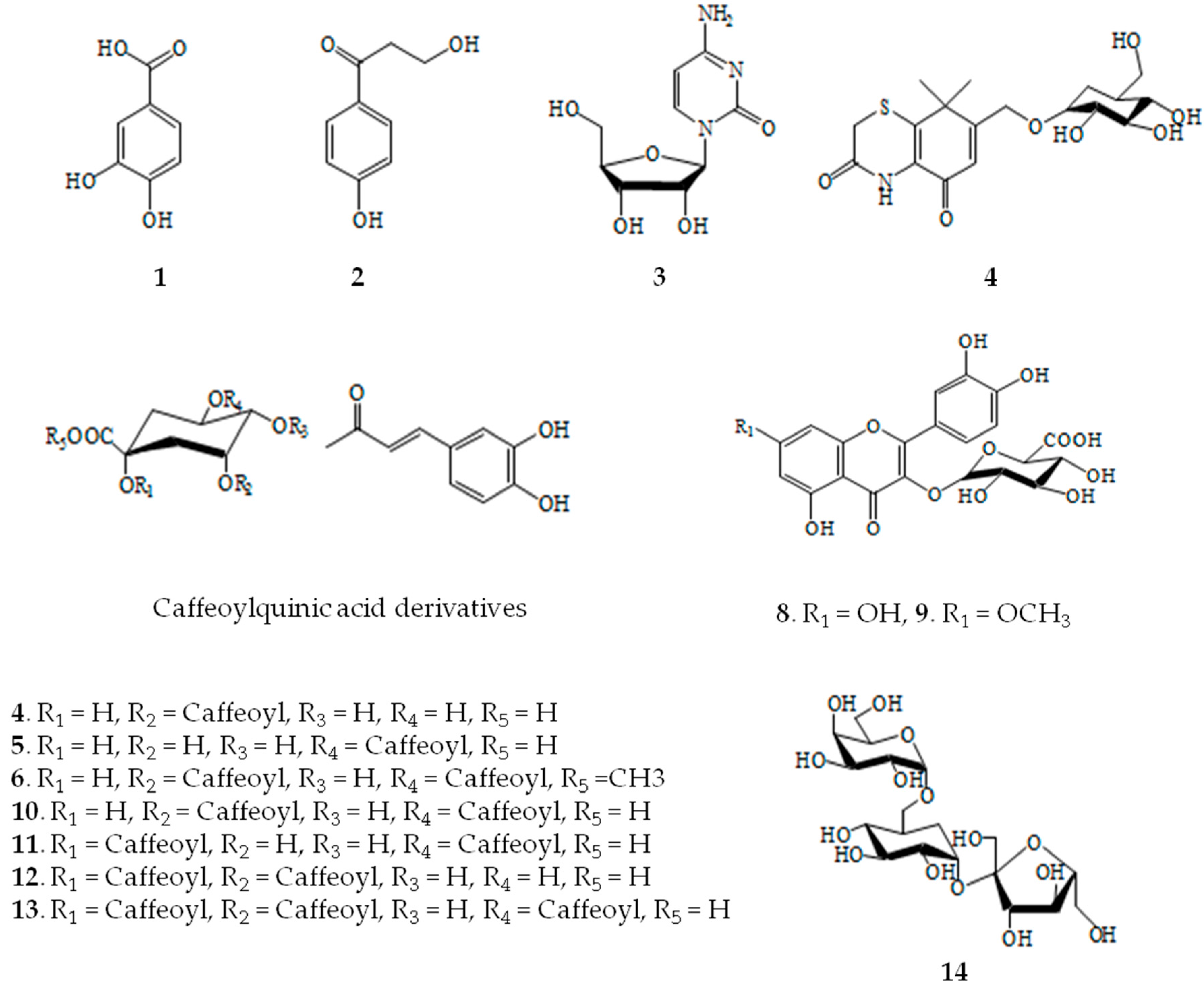
The compound 6, isolated from X. strumarium, is an ester derivative which is difference from the other compounds. Of the tested compounds, 6, which contains a methyl ester at quinic acid moiety, exhibited the most potent inhibitory activity with an IC50 value of 82.79 μM on AGEs, 1.88 μM on PTP1β, 18.42 μM on α-glucosidase, and 6.03 μM on ABTS+, respectively. The compound 10 only exhibited α-glucosidase inhibitory activity, whereas compounds 11–13, which include two/three caffeoyl groups and different position at quinic acid moiety, showed no inhibitory activity against AGEs, PTP1β, and ABTS+, respectively. The present study demonstrated that the methyl esters of the isolates were stronger inhibitors of anti-diabetic implying the importance of esterification for inhibitory potency.
Previous investigations into the anti-diabetic effects of CQA derivatives by Chen et al. reported that 3,4-di-
O-CQA (187.2 μM), 4,5-di-
O-CQA (130.8 μM), methyl 3,4-di-
O-CQA (12.23 μM), and methyl 4,5-di-
O-CQA (13.08 μM) isolated from the aerial parts of
Gynura divaricate showed the inhibitory effects against α-glucosidase and compared to that of the positive control (acarbose 867.4 μM). This literature indicated that the inhibitory activities of methyl-CQA derivatives were almost ten times higher than CQA derivatives on α-glucosidase. In addition, Chen et al. also was suggested that addition of a methyl group to the quinic acid skeleton may be responsible for a loss of α-glucosidase inhibitory activity [
15]. In another research, Hwang et al. reported that 3′-methoxyhirsutrin isolated from
Zea mays L. showed inhibitory activity with IC
50 value of 64.04 μM. Whereas, hirsutrin isolated from the same plant showed no activity on inhibition of PTP1β. These results demonstrated that the methyl group on the polyphenol skeleton may also play an important role contribute to the loss of PTP1β inhibitory activity [
16]. In addition, compound
6 and caffeic acid methyl ester isolated from highbush blueberry fruit have been shown to exhibit α-glucosidase [
17] and compound
6 isolated from the extracts of
Erigeron annuus also showed the strong inhibitory activity on AGEs [
18].
3-
O-CQA, 3,4-di-
O-CQA, compound
10, and 3,4,5-tri-
O-CQA isolated from Brazilian propolis possessed α-glucosidase and α-amylase inhibitory activities [
19]. In addition, 3-
O-CQA, 4-
O-CQA, 5-
O-CQA, and 3,5-di-
O-caffeoyl-
epi-quinic acid isolated from the extracts of
Erigeron annuus and
Artemisia montana exhibited the most potent inhibitory activity in both the AGEs and AR [
18,
20]. Compounds
6,
10,
11,
12, and
13 from
X. strumarium exhibited strong rat lens and recombinant aldose reductase inhibitory activities [
13]. Recent literature suggests that CQA has physiological properties; 3,5-dicaffeoyl-4-succinylquinic and compound
10 from
Chrysanthemum coronarium have anti-oxidant activities [
21]. Further, anti-inflammatory effects are exhibited by 3,4-di-
O-CQA methyl ester, compounds
6 and
10 isolated from
Ligularia fischeri leaves and
Ilex latifolia [
22,
23].
There are several widely used drug development targets for treatment of hyperglycemia, including PTP1β and α-glucosidase. And therapeutic measures for the treatment of hyperglycemic include the use of PTP1β inhibitors and α-glucosidase inhibitors. α-Glucosidase are a group of key intestinal enzymes involved in the digestion of carbohydrates and α-glucosidase inhibitors can be used to delay the absorption of carbohydrates from small intestine and thus lower postprandial blood glucose [
24]. PTP1β is a major non-trans membrane phosphotyrosine phosphatase in human tissues and was one of the earliest PTP identified. Although its physiological function and mechanism of regulation are largely unknown, it has been demonstrated to dephosphorylate insulin receptor in intact cells and thus to act as a negative regulator of insulin signaling. Moreover, the deletion of PTP1β gene in mice caused marked insulin sensitivity and prolonged insulin receptor auto-phosphorylation. PTP1β inhibitor would increase insulin sensitivity by blocking the PTP1β-mediated negative insulin signaling pathway and thus lower postprandial blood glucose [
25,
26].
Our data suggests that there was no significant relationship between structure of CQA derivatives and their inhibitory activity. However, it is postulated that the number of caffeoyl groups and their positions on the quinic acid moiety may be important factors in conferring the inhibitory activity. Our results and past literature reported that CQA with two caffeic acid methyl groups is more effective in inhibiting α-glucosidase than that of two caffeic acid also combines three CQA showed activity similar to anything (compound 6 > compound 10 > 3,4-di-O-CQA). In contrast, AGEs and PTP1β had single CQA more inhibitory effect than that of two/three caffeic acid including methyl group.
4. Experimental Section
4.1. General Experimental Procedures
1H- and 13C-NMR spectra and correlation NMR spectra such as COSY, HMBC, HMQC, and DEPT were obtained from a Bruker Avance DPX 400 (or 600) spectrometer (Berlin, Germany). These were obtained at operating frequencies of 400 MHz (1H) and 100 (or 150) MHz (13C) with CD3OD, (CD3)2SO, (CD3)2CO, or D2O and TMS was used as an internal standard; chemical shifts were reported in δ values. The α-glucosidase from Saccharomyces sp. (SSG) was purchased from Wako Pure Chemical Industries Ltd (Tokyo, Japan). Acarbose, aminoguanidine, trolox, suramin, bovine serum albumin, methylglyoxal and p-nitrophenyl-α-glucopyranoside (pNPG) as a synthetic substrate were obtained from Sigma–Aldrich Co. (St. Louis, MO, USA). All other chemicals and reagents used were of analytical grade.
4.2. Plant Materials
Plant material “Chang-i-ja” used in this study was purchased from a local market in Chuncheon. The voucher sample (RIC-HU1204) has been deposited at the center for efficacy assessment and development of functional foods and drugs, Hallym University, Chuncheon.
4.3. Extraction and Isolation
Dried
X. strumarium (4.5 kg) were ground and extracted with CH
2Cl
2 at room temperature. The residue was refluxed for 3 h with MeOH three times at room temperature and evaporated under reduced pressure to give a residue (165.0 g). The residue was suspended in distilled water and partitioned with CH
2Cl
2, EtOAc,
n-BuOH and H
2O successively to afford CH
2Cl
2 (7.4 g), EtOAc (25.2 g),
n-BuOH (33.1 g) and H
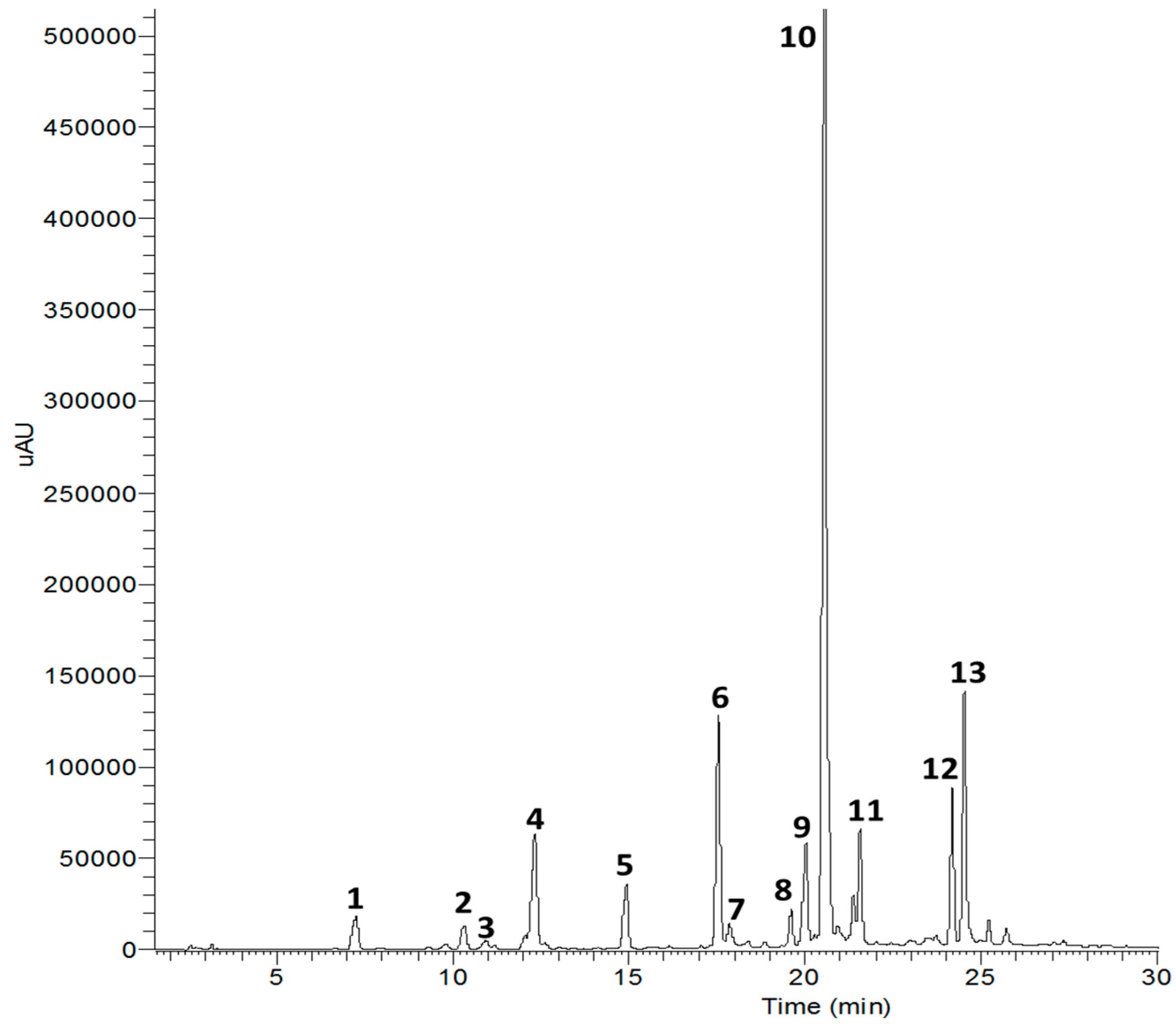
2O fraction (65.7 g). The EtOAc fraction showed strong inhibitory activity on AGEs, so this fraction (3.0 g) was subjected to RP C-18 column chromatography for further with MeOH gradient system (1:4 to 1:1) to yield compounds
1 (25.3 mg),
2 (12.8 mg),
3 (9.7 mg),
4 (12.4 mg),
5 (17.5 mg),
6 (4.8 mg),
7 (8.8 mg),
8 (11.88 mg),
9 (25.4 mg),
10 (5.8 mg),
11 (9.2 mg),
12 (4.2 mg),
13 (13.8 mg) and
14 (8.4 mg) (
Figure 1).
4.4. Assay for the α-Glucosidase Inhibitory Activity
The α-glucosidase inhibitory activity of the extracts and fractions was determined using a modified procedure reported method with a slight modification [
1]. The α-glucosidase activity was measured using the substrate
p-nitrophenyl-α-
d-glucopyranoside (
pNPG), which is hydrolyzed by α-glucosidase to release the product
p-nitrophenol, a colorant that can be monitored at 405 nm. The initial concentration of the enzyme solution was 0.62 unit/mL in 0.1 M phosphate buffer (pH 6.9) and the initial concentration of the substrate solution was 2 mM in the same phosphate buffer. The enzyme solution was mixed with water and the samples or controls in a clear 96-well microplate (flat bottom) and the reaction was initiated by addition of the substrate to the solution. The plates were incubated at 37 °C for 5 min and the reaction was terminated by the addition of 0.1 M Na
2CO
3. Enzyme inhibition was determined by the absorbance of 4-nitrophenol (product) at 405 nm, as measured with a microplate reader. Background absorbance was determined using a non-enzyme control microplate containing the buffer and was subtracted from the absorbance of the samples and controls. The concentration of inhibitors resulting in 50% inhibition of enzyme activity (IC
50) was calculated from the least square regression line of the logarithmic concentrations plotted against the residual activity. Acarbose was used as positive control.
4.5. Assay for the PTP1β Inhibitory Activity
PTP1β tyrosine phosphatase drug discovery kit is a colorimetric, non-radioactive assay designed to measure the phosphatase activity of purified PTP1β. The enzyme activity was measured using IR5 phosphopeptide (insulin receptor B residues 1142–1153, pY-114) as a substrate. To each well of the 96-well microplate (final volume: 125 μL), 75 μM IR5 substrate and PTP1β (2.5 ng/well) in a buffer containing 100 mM MES (pH 6.0), 0.3 M NaCl, 2 mM EDTA, 2 m Mdithiothreitol (DTT) and 0.1% NP-40 were added with or without test compounds. Following incubation at 37 °C for 30 min, the reaction was terminated with the BIOMOL REDTM reagent. The amount of p-nitrophenol produced was estimated by measuring the absorbance at 620 nm. The non-enzymatic hydrolysis of the IR5 substrate was corrected by measuring the increase in absorbance at 620 nm obtained in the absence of PTP1β enzyme.
4.6. Assay for the AGEs Inhibitory Activity
The modified procedure of Lee et al. was followed [
9]. Bovine serum albumin (10 mg/mL) was incubated with 5 mM methylglyoxal in sodium phosphate buffer (100 mM; pH 7.4). Dimethylsulfoxide used for dissolving samples was found to have no effect on the reaction. All of the reagents and samples were dissolved by filtration through 0.2 μm membrane filters and the mixture was incubated at 37 °C for 7 days. The fluorescence intensity was measured at an excitation wavelength of 330 nm and an emission wavelength of 410 nm with a LS50B fluorescence spectrometer Perkin-Elmer Ltd., (Buckinghamshire, FLS, UK). Aminoguanidine was also tested as a known inhibitor.
4.7. Assay for the ABTS+ Inhibitory Activity
The method described by Li et al. was used with slight modifications [
27]. ABTS
+ diammonium salt (2 mM) and potassium persulfate (3.5 mM) were mixed, diluted in distilled water and kept in the dark at room temperature for 24 h before use. After addition of ABTS
+ solution to 10 µL of antioxidant, measurements were recorded at 10 min post reaction. The percentage inhibition of absorbance at 750 nm was calculated and plotted as a function of concentration of antioxidants. Trolox was used as positive control.


