1. Introduction
For centuries honey was not only a popular sweet component of the human diet, but was also one of the most important drugs used in folk medicine [
1]. Regular consumption of this product provides a number of health benefits and its therapeutic potential depends on the botanical origin of the nectar that is used for production of the honey. For instance, in Polish folk medicine, heather (
Calluna vulgaris L.) honey is proposed as a remedy for prostate, liver and biliary system diseases. Honey collected from rapeseed (
Brassica napus L.) is recommended for patients suffering from cardiovascular system diseases. Consumption of buckwheat (
Fagopyrum esculentum Moench) honey is beneficial in relieving the symptoms of hypertension and atherosclerosis [
2]. Moreover, honey is used in treatment of infectious diseases, especially difficult-to-heal infected wounds [
1,
3]. Numerous studies confirm that honey derived from many different botanical and geographical origins exhibit inhibitory effect towards a broad spectrum of Gram-positive and Gram-negative pathogenic bacteria, including antibiotic resistant strains [
4,
5,
6,
7,
8,
9,
10]. The antimicrobial action of honey is based on several mechanisms: the acidity (low pH) [
5,
11,
12], osmotic pressure (high sugar concentration) [
5,
13] and the presence of bacteriostatic and bactericidal factors such as hydrogen peroxide [
14,
15,
16], phenolics [
17], peptide—bee defensin-1 [
5,
18,
19], methylglyoxal [
20,
21] and Maillard reaction products [
22]. The Worobo group also found that honey should be considered as a potential source of microorganisms producing promising antimicrobial compounds, especially bacteriocins of a broad bactericidal spectrum [
23]. Among all these mechanisms, enzymatic production of H
2O
2 is absolutely crucial for antimicrobial activity of the majority of investigated honey types [
5,
8,
12,
20]. Reports of several authors revealed that treatment of honey samples with high temperature and/or catalase completely reduces antimicrobial activity of these products [
5,
9,
14,
24]. In fact, only in the case of New Zealand’s manuka honey, and several Australian and Malaysian honeys, high level of antibacterial activity is mostly caused by a non-peroxide component—methylglyoxal [
21,
25,
26]. The role of phytochemicals in the activity of honeys produced from nectar of other plants remains unclear. However, some important findings in this area have been recently provided by the Brudzynski and coworkers. Within the group of six honeys (excluding manuka honey) they observed an important correlation between high antioxidant and antimicrobial activity of products and lack of activity in the case of products pre-treated with catalase. As an explanation of this observation, they discovered that honeys containing a high concentration of polyphenols of high radical scavenging activity efficiently produce hydroxyl radicals, which are crucial for the antimicrobial potential of the product. The hydroxyl radicals are produced using H
2O
2 (generated by glucose oxidase that is present in honey) as a substrate in the polyphenol-mediated Fenton-like reaction [
7,
24,
27]. As it was shown previously by the groups of Cao [
28] and Sakihama [
29], the efficiency of this reaction increases dramatically in the presence of polyphenols. Thus, polyphenols present in honey emerge as active intermediates that are necessary to confer oxidative action of hydrogen peroxide. The generated OH˙ radicals are powerful but short-lived oxidants that cause proteins and lipid peroxidation and DNA and RNA degradation in bacterial cells [
7,
24,
27]. However, it also should be noted that many reports have not confirmed the correlation between total phenolic content or antioxidant activity and antimicrobial potential of the product [
30,
31]. Thus, in our opinion, the crucial role of polyphenols in the generation of OH˙ radicals is not a complete explanation of the role of phytochemicals in the antimicrobial activity of all hydrogen peroxide dependent honeys. The Brudzynski group also revealed that the two-phase colloidal system, consisting of large, micron-size particles distributed in the concentrated sugar solution, is required for antibacterial activity of honey. The enzyme glucose oxidase efficiently produces the hydrogen peroxide only in the situation when it is involved in the structure of these particles which also contain complexes of other proteins, polyphenols and oligosaccharides. The authors found that dilution of honey allows unpacking and dissociation of large, micron-size, superstructures into smaller nanosize particles. The phase transition (destruction of large particles) ensues at the threshold concentration of molecular crowders (glucose and fructose). The glucose oxidases released from the particles is much less active and the phase transition point is critical for antimicrobial potential of honeys [
32].
The main scope of the current study was: (1) to investigate antibacterial activity of various types of Polish honeys and their mechanism; (2) to investigate anti-staphylococcal activity (against both reference strains and clinical isolates) of honey solutions against planktonic cells and biofilm as well as to compare MIC, MBC and MBEC50; (3) to investigate the role of phytochemicals in H2O2 mediated antibacterial activity of the products.
3. Discussion
The research presented here revealed important differences in the antimicrobial potential of honeys produced by Polish beekeepers. Several products, five in the case of
S. aureus and seven in the case of
S. epidermidis, exhibited high antimicrobial activity with MIC values of only 1.56% (
v/v). On the other hand, within the tested range of concentrations from 0.39 to 12.5% (
v/v), quite a large number of honey samples did not exhibit any activity against the reference strains tested. Similar differentiation of activity was found in our previous research, when honeys from the south of Poland were tested [
9], but other authors also observed important differences in the activities of their honey samples from different geographical regions [
6,
8,
24,
25]. Gram-negative pathogens
E. coli and
P. aeruginosa exhibited lower susceptibility to the honeys’ components. Usually 2 or 4 fold higher concentration was necessary to achieve inhibitory/bactericidal effects in comparison to staphylococci. It is probably a consequence of differences in cell wall construction of these bacteria. Higher susceptibility of staphylococci in comparison to Gram-negative bacteria was also observed by other authors [
6,
26,
27]. However, in many reports the differences in susceptibility were not so evident [
35,
36].
Despite the fact that many reports concerning antibacterial, including anti-staphylococcal, effectiveness of honeys collected in different regions of the world have been published, such a high level of activity (MIC = 1.56% (
v/v)) has been rarely observed. The highest anti-staphylococcal activity of honey has been reported by Nishio and coworkers for products collected by stingless bees
Scaptotrigona bipunctata (MIC = 0.62% or 1.25% depending on the
S. aureus strain tested) and
Scaptotrigona postica (MIC = 1.25% or 2.5% depending on the
S. aureus strain tested) [
37]. High effectiveness (MIC < 2%) against
S. aureus (including MRSA) was confirmed for Scottish heather honey [
38], and chestnut, fir and forest honeys from Slovenia inhibited the growth of
S. aureus at concentration of 2.5% (
v/v) [
8]. MIC values of about 3.0% (
v/v) were observed in the case of e.g., Chilean ulmo tree honey [
39], some Greek and Cypriot honeys [
6], Canadian buckwheat honey [
27] and some Polish honeys investigated in our previous report [
9]. Comparable activity has been also confirmed for honey produced from the manuka bush (
Leptospermum scoparium) indigenous to New Zealand and Australia [
21,
25]. Interesting results have been recently presented by Alvarez-Suarez and coworkers [
40]. The authors found some important differences in properties, including antimicrobial potential, between the Cuban multi-floral honeys produced by two different bee species:
Melipona beecheii and
Apis Mellifera. In the case of
S. aureus the honey collected by
M. beecheii revealed over seven–fold higher activity in comparison to the honey produced by
A. mellifera, with Minimum Active Dilution (MAD) values of 2.0% and 15.0%, respectively. However, a bit different method was used for determination of antimicrobial activity in this study we have no doubts, that the product collected by
M. beecheii belongs to honeys with the highest antimicrobial potential described to date [
40]. Several other
in vitro investigations revealed that honeys produced in many other geographical regions e.g., Malaysia [
26,
41], Thailand [
30], Nordic [
36], Italy [
42] and Africa [
43,
44,
45] are also promising candidates for the treatment of infections caused by staphylococci, including MRSA. The higher values of MBC in comparison to MIC parameter of many products (especially in relation to clinical isolates) identified in our studies, have been also presented by other authors, who compared inhibitory and bactericidal potential of honeys [
26,
30,
36]. This situation is common for many other antimicrobial agents, including some antibiotics.
High potential (MIC ≤ 1.56% (
v/v)) of selected honeys from our collection was also confirmed for
S. aureus strains isolated from animals (bovine mastitis) and humans (infected wounds), which is very optimistic from the point of view of potential application of Polish honeys in clinical practice, e.g., as a component of wound dressings. As it was mentioned above, in folk medicine honey was an essential agent used for treatment of difficult-to-heal infected wounds. Its high potential for this purpose has been recently confirmed and discussed in many scientific reports and publications [
3,
46]. Consequently, several pharmaceutical companies have proposed the dressings containing honey (mostly manuka honey), which are successfully used in treatment of infected wounds. Some authors even suggest the possibility of using honey for treatment of systemic, particularly gastrointestinal infections [
47]. On the basis of papers published up to December 2014, Henatsch and coworkers reviewed the possibilities of application of honey in otorhinolaryngology. The authors concluded that this product can be considered as effective (additional) treatment in mucositis, childhood cough, persistent post-infectious cough and after tonsillectomy [
48]. On the other hand, there are also several reports that revealed lack of positive effects of honey application for infection treatments. In the trial carried out by Kwakman and coworkers, the medical-grade honey did not affect colonization of the skin at central venous catheter insertion sites in intensive care units patients when applied in addition to standard disinfection with 0.5% chlorhexidine in 70% alcohol [
49]. The outcomes of investigation carried out by the mentioned above group of Henatsch revealed that honey eardrops showed a strong in vitro inhibitory activity against all tested strains but did not eradicate
S. aureus infection in vivo [
50]. It also should be emphasized that in vivo infections are often caused by bacteria growing in the form of biofilms. Eradication of this type of infection is especially problematic. Bacterial biofilms are extremely resistant to antibacterial agents including antibiotics and disinfectants. Results of our investigations confirmed much higher resistance of bacterial biofilms in comparison to planktonic cells. The MBEC
50 for most active products was not lower than 25% (
v/v). High resistance of bacterial biofilm was also confirmed in recent studies of Garcia-Tenesaca and coworkers. The dilution containing 20.0% of avocado honey eradicated about 60% of performed staphylococcal biofilm, and activity of eucalyptus and rapeseed honeys were even lower with the level of biofilm eradication below 50% [
51]. Moreover, we observed that a concentration lower than MBEC
50 significantly enhanced the growth of the biofilm—honey probably is used as a source of glucose (easily digestible source of carbon). A similar effect was previously presented by Lu and coworkers [
52]. These observations can be the explanation of failures of treatment of in vivo infections with honey. For successful therapy, the product would have to be used at a concentration not lower than MBEC value. It is not problematic in the case of infected skin wounds (undiluted honey is used for preparation of wound dressing materials), but in the case of systemic infections achievement of such a high concentration would be a challenge, and in most cases is not achievable.
Taking into account botanical sources of the products, especially high activity honeys were derived from buckwheat. It is in agreement with our previous observation [
9] and with results presented by other authors [
27,
36,
53,
54]. High antimicrobial potential exhibited also products qualified as honeydew honey and honey produced from the nectar of a lime tree, which also has been presented in some other reports [
9,
36,
54]. Interestingly high activity was observed in the case of some multi-floral honeys whilst in the previous report presented by Kędzia and coworkers [
54] most of Polish multi-floral honeys were found as products with low antimicrobial potential.
Another important finding of our investigation is the correlation between activity and the source of product (beekeeper, supermarkets and organic grocery stores). The most active honeys were those provided by local beekeepers. High activity was also observed in many honeys coming from abroad. This observation can be (in our opinion) explained by the result of the assay aiming in determination of influence of higher temperature on the activity of the product. We have found that even 10 min treatment the honeys with 60 °C resulted in decrease/complete loss of activity. Honeys delivered by beekeepers have not been processed since harvesting from the hive. Supermarkets and smaller shops usually buy the honeys from big apiaries or from wholesalers. In both cases the product is stored in large-sized containers (usually barrels). During the storage it crystallizes, which is a natural phenomenon (except of honey derived from acacia). Before delivering to the store, the honey is decrystallized and poured into jars. The “liquefaction” of the product should be performed at temperatures not higher than 40 °C, which is safe for honey (its biological value), but is a time consuming process. If the process is performed at higher temperatures, it is much faster, but unfortunately strongly affects stability of some thermal sensitive ingredients including glucose oxidase and other enzymes. The significant correlation between glucose oxidase activity and honey crystals melting time and temperature has been previously revealed by the Kretavicius group [
55]. The presented herein results suggest that some of Polish apiaries/wholesalers perform the decrystallization process at too high temperatures.
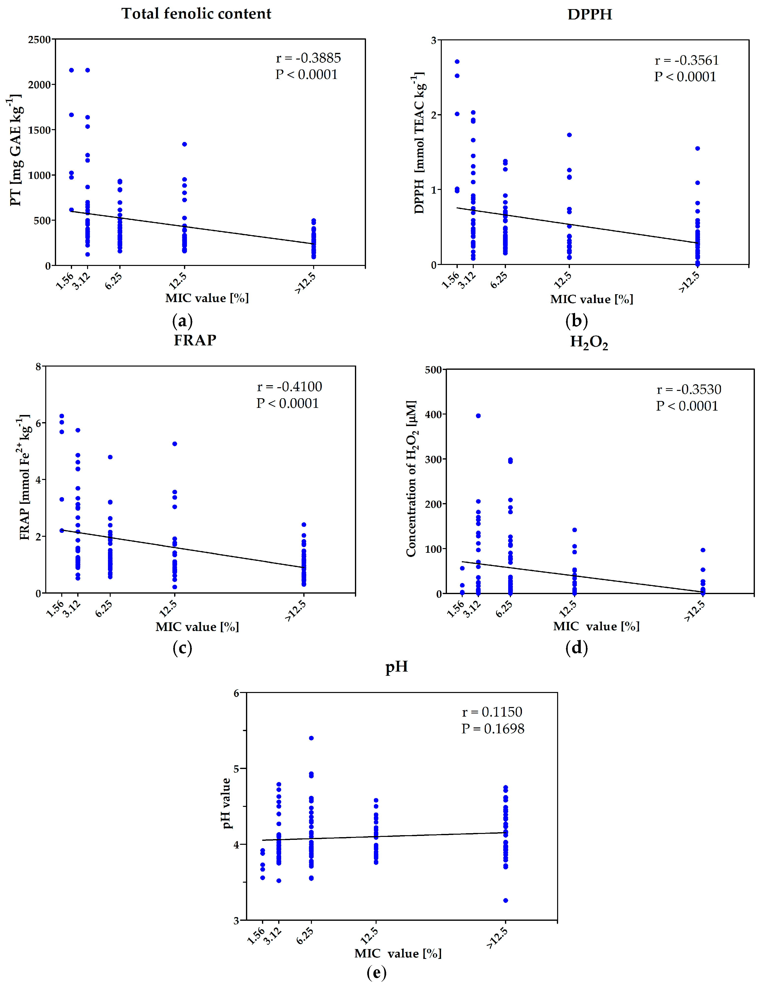
High sensitivity to elevated temperatures and catalase unambiguously confirms that enzymatic generation of hydrogen peroxide is crucial for antimicrobial activity of Polish honeys (
Table 5). On the other hand, the observed strong positive correlations between phenolic content, antioxidant activity, as well as botanical source, and antibacterial activity suggest that components of plant origin (phytochemicals) also importantly affect the products’ activity. The lack of activity with honeys that have inactivated glucose oxidase indicates that the amount of phytochemicals is too low to exert the antibacterial effect alone, or they are present in the honey in less/non-active form. Similar results have been presented by Brudzynski’s group. The authors performed long term research that mostly explained the detailed mechanism of bactericidal potential of hydrogen peroxide dependent honeys and a role of phytochemicals in bacteria elimination. They found that average content of H
2O
2 in honey was over 900-fold lower than that observed in disinfectants and was not sufficient to eliminate the bacteria as the only factor. Thus, they concluded that other components of honey enhance the bactericidal potential of hydrogen peroxide [
56]. Further investigation revealed polyphenols as intermediates that are necessary to confer oxidative action of hydrogen peroxide—generation of hydroxyl and phenoxyl radicals (discussed in more detail in the introduction) that are directly responsible for killing microorganisms [
7,
16,
24,
27,
56]. In their studies, Brudzynski and coworkers mainly used buckwheat honey as a model. Herein, we also investigated the results of the reaction of ingredients of other honeys, namely multi-floral, rape, and lime, with H
2O
2. Adding buckwheat and rape honeys to the 100 μM hydrogen peroxide solution resulted in dramatic decrease of concentration of this agent and the amount of eliminated H
2O
2 correlated with final concentration of the honey. In the light of results presented by Brudzynski and coworkers, the elimination of H
2O
2 in the case of buckwheat honey was rather expected and can be easily explained. Interestingly, a similar effect was observed in the case of, not active, rape honey, which has not been observed to date. Very low or even lack of activity of the product derived from this plant suggests that some ingredients effectively react with H
2O
2 (introduced to the solution or generated by the glucose oxidases) degrade it, and the generated products do not exhibit antimicrobial activity (in contrast to derivatives of reaction with components of buckwheat honey). The results of this experiment also explain the low level of concentration of hydrogen peroxide in the solutions of both products (buckwheat and rape honeys). The low antimicrobial activity of rape honey has been revealed in several independent investigations carried out in Poland [
9,
54,
57]. This could suggest that some essential phytochemicals present in this product are responsible for elimination of hydrogen peroxide without generation bacteriostatic/bactericidal products.
An explanation of the high activities of multi-floral and lime tree honeys seems to be a bit more complicated. It could be the consequence of generation and accumulation of high concentration of hydrogen peroxide in these products. As it was shown in
Figure 4a–d, enzymatically produced H
2O
2 is not strongly affected by phytochemicals present in all four samples of these honeys. In all cases we observed summing up of the hydrogen peroxide introduced to the solution and enzymatically produced one. However, in our opinion the complete understanding of mechanisms of elimination of bacteria by these products, especially the role of phytochemicals requires further research. Thus, the issue of influence of phytochemical components to the antimicrobial activity of honey seems to remain unresolved and quite complicated problem, which cannot be explained by one universal mechanism.
The observed high activity of some Polish honeys with MIC values of 1.56% (
v/v) seems to be also interesting from the point of view of the global structure of honey—the mechanisms stabilizing the structures of micron-size particles, which are crucial for maintaining the glucose oxidase in highly active form. Brudzynski and coworkers proposed concentration of molecular crowders as crucial factors for stabilization of these large-size structures and existence of two-phase coloidal system [
32]. The results presented herein suggest that values of phase transition points of some Polish honeys would have to be lower in comparison to the most active products investigated by the Brudzynski group, with a relatively low concentration of essential sugars (up to 4 fold). In our opinion, this result could suggest that the stability of these large-size particles can be strongly dependent on their chemical composition (presence and concentration of some ingredients). Verification of this thesis and identification of key components will be the issue of our future research.
Because of the climate conditions (cold weather, short season when the bees can collect honey and pollen) and the fact that there are not many places where honey plants (e.g., phacelia, melilot or even rape) are grown the beekeeping is not easy in the region where the samples were collected (north of Poland, near Gdańsk). Moreover, definitely most of local beekeepers do not travel with bees to the places where some honey plants are grown, and only some of them have an access to the fields of buckwheat (
Fagopyrum esculentum Moench), rapeseed (
Brassica napus L.) or parks, alleys or forests with a larger number of black locust (
Robinia pseudoacacia L.) or lime (
Tilia spp.) trees. Consequently, mostly multi-floral honey is produced in this region and the yield of production in local apiaries is very low (according to the last report of Polish Bee Association [
58] it is one of the lowest in Poland, of just about 10–17 kg of honey per hive). Nevertheless, the multi-floral honey produced in this region is characterized by unique features, especially taste. In our opinion, the fact that bees collect the relatively low amount of nectar from different botanical origins in hard weather and climate conditions is also important for high antimicrobial potential of the local honeys. Collecting nectar from many different plant species (mostly weeds and herbs) guarantees the presence of a high concentration of many different phytochemicals in the final product. During the honey-making process, bees add to the nectar enzymes secreted by their salivary glands and remove the water to the final concentration of no more than 20%. The main goal of water removing as well as adding glucose oxidase and bee defensin-1 is antimicrobial protection of honey. The fact that in this region bees convert the relatively low amount of nectar guarantees the high concentration of both antimicrobial agents coming from their bodies, namely glucose oxidase and defensin-1 in the final product. The carried out research has revealed the important correlation between the antimicrobial efficiency of honeys and the content of phytochemicals and enzymatically generated H
2O
2. The high concentration of defensin-1 could be an explanation of high antibacterial efficiency of the products that contain neither a high concentration of polyphenols nor hydrogen peroxide. Valachova and coworkers [
19] have revealed some important differences in the concentration of bee defensin-1 in different honey samples. Comparison of concentration of this peptide in local honeys with the products coming from other geographical regions as well as determination of influence of this agent to the whole antimicrobial efficiency of local honeys will be investigated in our group.
4. Materials and Methods
4.1. Bacterial Strains and Media
In the preliminary studies, antimicrobial activity of all honeys was tested against five reference strains of bacteria:
S. aureus ATCC 25923,
S. aureus ATCC 29213,
S. epidermidis ATCC 12228,
Pseudomonas aeruginosa ATCC 27853, and
Escherichia coli ATCC 25922. The anti-staphylococcal potential of selected—most active honeys, was also investigated against six
S. aureus isolates from patients with skin and soft tissue infections (SSTI)
: Szw35, Szw41, Szw17, Szw48, Szw55, Szw16 [
59] and six
S. aureus strains derived from subclinical bovine mastitis (SCM) milk samples: SA1, SA9, SA70, SA102, SA103, SA105 [
60]. All strains of bacteria were routinely grown on Mueller-Hinton Agar (MHA, Sigma Aldrich, Schnelldorf, Germany) plates. The Minimum Inhibitory Concentration (MIC) was determined using liquid medium—Mueller-Hinton Broth 2 (MHB2, Sigma Aldrich) and for determination of Minimum Bactericidal Concentrations (MBC) the cells were transferred on the Baird Parker Agar plates (Biomaxima, Lublin, Poland).
4.2. Chemicals and Reagents
The standard compounds and reagents (all of analytical grade): MTT (3-(4,5-dimethylthiazol-2-yl)-2,5-diphenyltetrazolium bromide), DMSO (dimethyl sulfoxide), H2O2 (peroxide hydrogen), o-dianisidine dihydrochloride, Peroxidase from horseradish Type VI, ferrous sulphate, gallic acid, Na2CO3, ferric chloride, 1,1-diphenyl-2-picrylhydrazyl radical (DPPH), 2,4,6- tris(2-pyridyl)-1,3,5-triazine (TPTZ), (±)-6-hydroxy- 2,5,7,8-tetramethylchroman-2-carboxylic acid (Trolox), Folin-Ciocalteu’s reagent, PBS were purchased from Merck (Darmstadt, Germany). Methanol and H2SO4 were purchased from (POCH, Gliwice, Poland). Ultrapure H2O (18.0 MΩ) was obtained with Milli-Q Advantage A10 system (Millipore, Billerica, MA, USA). The absorbance of reaction mixture in Folin-Ciocalteu, DPPH and FRAP assays were measured using a Genesys 20 spectrophotometer (Thermo Scientific, Waltham, MA, USA).
4.3. Honey Samples
The studies covered 144 samples of honey (
Table 6.). The primary goal of our research was determination of antimicrobial activity of honeys collected in the northern region of Poland. In 2015 and 2016, beekeepers from this region delivered 95 samples. Most of them (
n = 58; 61%) were classified as multi-floral honeys, 4 (4.2%) were classified as honeydew honeys, 4 (4.2%) honeys were collected in apiaries located in forests, 2 (2.15%) in peatlands and for 27 (28.4 %) samples the beekeepers were able to denominate the leading species of plants that were the source of nectar for bees. Due to some expected differences in activity, honeys bought in organic grocery stores (
n = 22) and supermarkets (
n = 12) were investigated. Moreover the antibacterial activities of Polish north region honeys (PNPH) were compared to the activity of honeys coming from abroad (15), including manuka honey—Man550. The manuka honey was kindly supplied by Propharma (Warszawa, Poland). All the honey samples were stored at 4 °C in the dark.
4.4. Investigation of Antimicrobial Potential of Honey Samples—Determination of MIC (Minimum Inhibitory Concentration) and MBC (Minimum Bactericidal Concentration) Parameters
The assay was performed as described in our previous publications [
9,
61] with slight modifications. Briefly, overnight bacterial cultures grown on MHA plates were suspended in PBS (pH 7.4) to the cell density of OD
600 = 0.132 (equal to 0.5 McFarland turbidity standard)—approximately 1–5 × 10
8 CFU/mL and in the next step diluted in MHB2 medium at a ratio of 1:150
v/v to the final cells concentrations of approximately 1.5 × 10
6 CFU/mL.
The samples of honey were diluted at a volume ratio 1:3 in a concentrated MHB2 medium (CMHB2). The concentrated medium was prepared by dissolution of 22 grams of powdered MHB2 in 750 mL of water instead of 1000 mL as it is required for MHB2 medium. When the honey was added to the concentrated medium in the ratio of 1:3 (v/v) the concentration of solid ingredients were exactly the same as in the case of MHB2 medium prepared according to directions (22 grams per 1 L). The obtained honey solutions were filter sterilized with syringe-driven 0.22 μm PES filters (Merck Milipore, city, Ireland). Subsequent series of two-fold dilution of the honey in range 25.0–0.78% (v/v) were prepared in a 96-well plate using MHB2 broth. Subsequently, the honey solutions in the wells were inoculated with an equal volume of suspension of bacterial cells, prepared as described previously. The final concentrations of inoculated honeys ranged from 0.39% to 12.5% (v/v) (six different concentrations of honeys were tested: 12.5%, 6.25%, 3.12%, 1.56%, 0.78% and 0.39% (v/v)). Additionally, positive control of the growth of tested strain and control of the medium sterility was performed.
The plates were incubated 24 h under static conditions at 37 °C. The lowest concentration of honey with no visible bacterial growth was taken as a MIC value. The MIC assay for each tested strain and honey sample was performed three times.
The minimum bactericidal concentrations (MBCs) were assessed by transferring each dilution used for MIC assay on Baird-Parker agar plates using a sterile 48-well microtiter plate replicator. The plates were incubated for 24 h at 37 °C. Concentrations where no growth of the colonies was observed were assigned as MBC.
4.5. Activity against Staphylococci Growing in the Form of Biofilm
4.5.1. Biofilm Formation Assay
The assay was performed according to the procedure proposed by Walencka and coworkers [
62] with slight modifications. The suspension (OD
600 = 0.1 in PBS buffer) of
S. aureus ATCC 25923 cells was prepared as it was described previously and diluted 1:10 (
v/v) in LB (Luria-Bertani) liquid medium. An aliquot of 200 μL of cells’ suspension were added to the wells of columns 1–7 of vertically set plates. Negative controls were performed with a sterile medium placed in the wells of column 8. The plates were incubated for 24 h at 37 °C in order to allow bacteria to form biofilms.
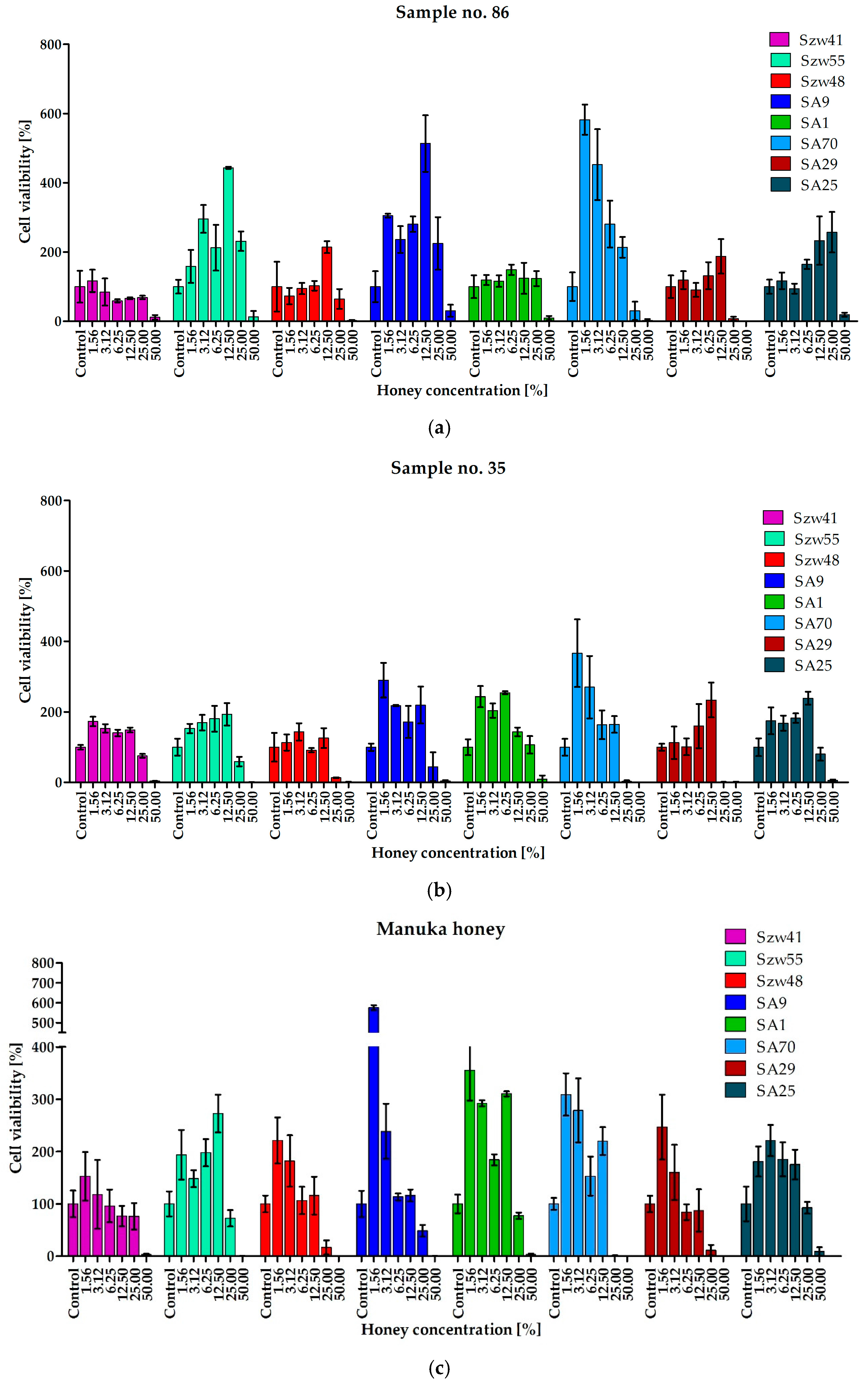
4.5.2. MBEC Assay
The content of the wells was removed and the wells were washed three times with 200 μL of sterile PBS buffer. 50% (
v/v) solutions of honey were prepared by dissolution of honey with an equal volume of 2-fold concentrated LB medium and sterilized by filtration (0.22 μm). The 2-fold serial dilutions of honey ranging from 50% to 1.56% or LB medium as positive/negative controls (determination of biofilm growth in the wells that do not contain any growth inhibitors/control of sterility of media) were added to the wells and incubated for 24 h at 37 °C. The MBEC
50 values of tested honeys were taken as the lowest concentration of honeys that caused at least 50% inhibition of growth of the cells in comparison to the cells growing in the control wells—measured as comparison of ability of living cells to the biotransformation of MTT (3-(4,5-dimethyl-2-thiazolyl)-2,5-diphenyl-2
H-tetrazolium bromide) to insoluble in water violet formazan crystals [
63].
4.5.3. Biofilm MTT-Staining
The MTT assay was performed as described [
62,
63] with minor modifications. Briefly, after the biofilm formation (according to the procedure presented above) the inoculum was removed and the wells of microplate were washed with 200 μL of sterile PBS buffer. Subsequently, 150 μL of PBS and 50 μL of MTT solution (0.3% in PBS) were added to the wells and mixed. Following 2h incubation at 37 °C in dark, the MTT solution was replaced with 200 μL of DMSO for dissolving of formed formazan crystals. The optical density of the obtained solutions was measured at 540 nm using a Victor
3 microtiter reader (Perkin Elmer, Waltham, MA, USA).
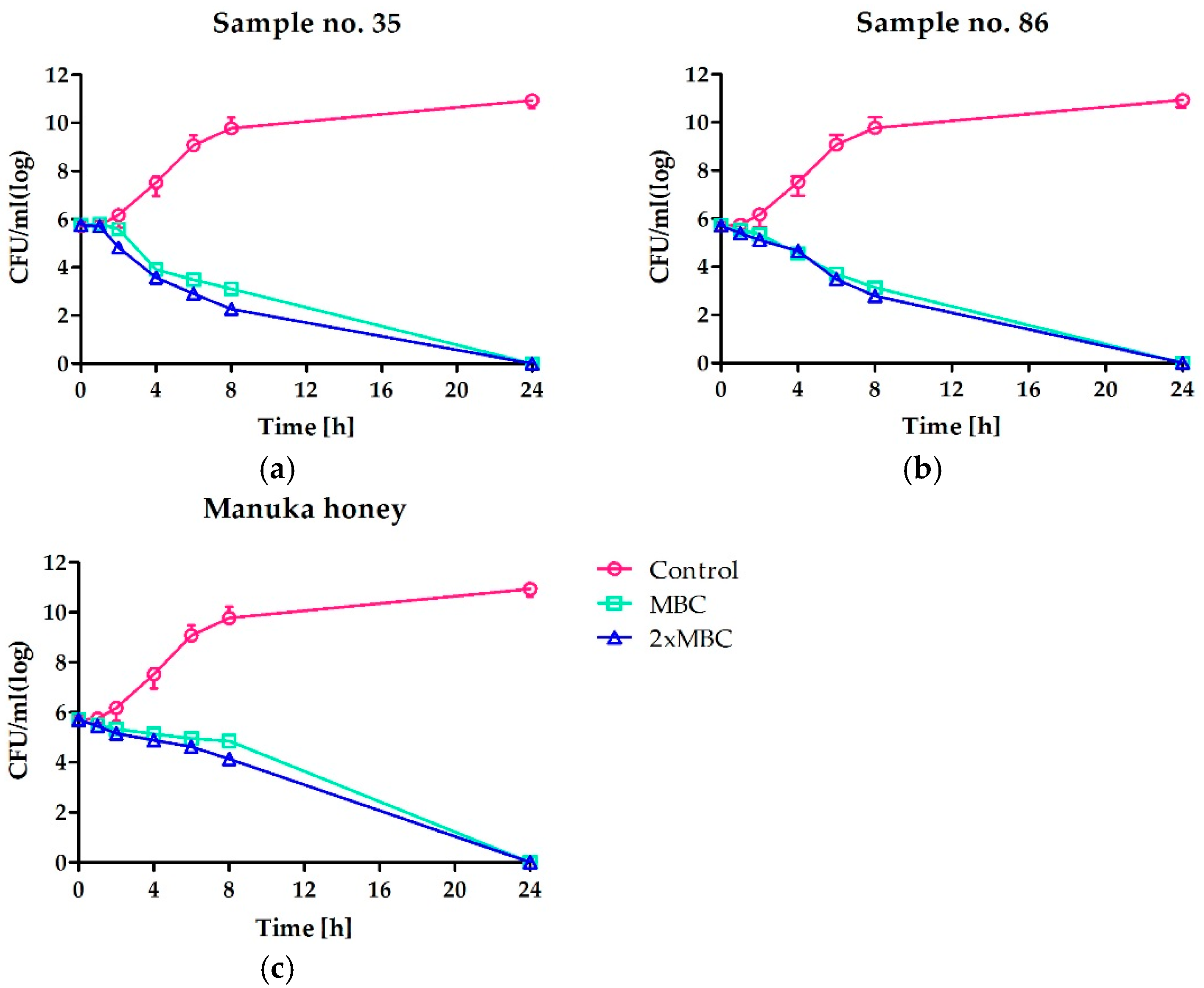
4.6. Time-Kill Assay, Determination of Kinetic of Bactericidal Effect of Honey Against Staphylococci
Time-kill assay was performed for two honeys that revealed the highest anti-staphylococcal activity and manuka honey as a reference. The suspension of approx. cell density 1.5 × 10⁶ CFU/mL of S. aureus ATCC 25923 was prepared in MHB2 broth supplemented with honey to the final concentration equal to MBC or 2 × MBC and incubated at 37 °C with shaking. Viable counts of the cells in the suspensions were obtained from 10-fold serial dilutions at 0, 1, 2, 4, 6, 8 and 24 h by plating 10-fold dilution on Baird-Parker agar plates and incubating at 37 °C for 24 h. The number of the cells in the control suspension, without honey addition, was also determined as a control of growth kinetic of S. aureus ATCC 25923.
4.7. Determination of Hydrogen Peroxide Generation
Hydrogen peroxide concentration in honey was measured according to the modified method described previously by Kwakman and coworkers [
5]. Briefly, 25.0% (
w/v) honey solutions were prepared in deionized water. Samples were incubated at 37 °C for 1 h. 20 μL of diluted honey samples and 67 μL of reagent were added to wells of microtiter plate. The reagent solution consisting of 100 μg/mL of o-dianisidine dihydrochloride and 20 μg/mL of horseradish peroxidase type IV in 10 mM phosphate buffer (pH 6.5). After 5 min of incubation in room temperature, reactions were stopped by addition of 60 μL of 6.0 M H
2SO
4. The Absorbance was measured at 540 nm using a Victor
3 microtiter reader (Perkin Elmer). Concentrations of hydrogen peroxide were calculated using of standard curve the 2-fold serial dilution of H
2O
2 standards (550–2.1 μM).
To determine influence of honey components to H2O2 accumulation 50% (w/v) honey solutions were prepared in deionized water and diluted with H2O2 (200 μM) or water at a ratio 1:1. Samples were incubated at 37 °C for 1 h and concentration of H2O2 (μM) was measured as described above.
4.8. Preliminary Determination of Mechanism of Antimicrobial Activity of Honeys—Heat and Catalase Treatment
Heat treatment and catalase supplementation was performed for preliminary investigation if hydrogen peroxide generation is crucial for antimicrobial activity of tested honeys. To check the influence of elevated temperature to the antimicrobial activity, the 25% (v/v) water solutions of selected, the most active honeys were incubated for 10 or 20 min at 40, 60 or 80 °C. After the incubation, the MIC values against S. aureus ATCC 25923 were determined according to the procedure described above. The determination of MIC value against S. aureus ATCC 25923 was also performed to investigate the influence of catalase on the antibacterial activity of tested honeys samples. It was performed as described above for the procedure of MIC determination, but with the addition of catalase solution to each well in the plates (including positive and negative control) to its final concentration of 250 U/mL.
4.9. Total Phenolics Determination
The total content of phenolic compounds was determined using a modified Folin-Ciocalteu method [
64]. 0.5 mL of Folin-Ciocalteu reagent was mixed with 100 μL of the honey solution (1:5
w/v in ultrapure water) and after 5 min, 3 mL of 100 g/L solution of Na
2CO
3 (
w/v) was added. Following shaking the mixture was made up to a volume of 10 mL with ultrapure water and incubated for 90 min at room temperature. The absorbance at 725 nm was measured against blank in a 10 mm quartz cuvette. Total phenol content was calculated and expressed as milligrams of gallic acid equivalent (GAE) per kilogram, using a calibration curve prepared with fresh gallic acid standard solution (10–500 mg/L). All measurements were performed in triplicate.
4.10. Total Antioxidant Activity (FRAP Assay)
The ferric reducing antioxidant assay (FRAP) was performed according to Kuś et al. [
64]. 30 μL of the aqueous honey solution (1:5,
w/v) were dissolved in 2 mL of ferric complex (10 mmol/L TPTZ and 20 mmol/L ferric chloride in acetate buffer (pH 3.6)). Following the 10 min incubation the absorbance at 593 nm was measured in disposable polystyrene cuvettes and the total antioxidant activity was calculated using a calibration curve prepared with ferrous sulfate (0.1–2 mmol/L) as the standard. The results were expressed as millimoles of Fe
2+ per kilogram of the honey. All measurements were performed in triplicate.
4.11. Antiradical Activity (DPPH Assay)
The DPPH assay was performed according to Kuś et al. [
64]. 50 μL of the watery honey solution (1:5,
w/v) were dissolved in 2 mL of 0.04 mmol/L DPPH in methanol. The mixture was incubated for 15 min in dark, at room temperature. Afterwards, the absorbance was measured at 517 nm using disposable polystyrene cuvettes. The data were expressed as Trolox equivalent antioxidant capacity per kilogram of the honey (TEAC, mmol/kg) using calibration curve prepared with Trolox solution (0.05–1.0 mmol/L). All measurements were performed in triplicate.
4.12. Determination of Water Content
According to the Polish food law the water content for most types of honeys should not be higher than 20%. Honeys containing more than 20% are usually not microbial stable. The content of water was measured using an ATAGO Hand Refractometer (Atago Co. Ltd., Tokyo, Japan) that enables determination in the range 12–26%.
4.13. Determination of pH
The low pH value of honey is also considered as an important factor for microbial potential of this product. It inhibits the growth of endogenous microorganism, but could be also important for antimicrobial activity of honeys used as for example a component of wound dressing materials. The honey samples were dissolved in deionized water to obtain (1:5, w/v) solution. The pH of the samples was measured using Hanna Instruments PH200 pH-meter with automatic temperature compensation (Hanna Instruments, Woonsocket, RI, USA) calibrated with pH standards, 4.01 ± 0.02 and 7.01 ± 0.02.
4.14. Statistical Analysis
Statistical analyses were performed using GraphPad Prism® 5 (Version 5.01, GraphPad Software, Inc., La Jolla, CA, USA). Correlations were obtained by Pearson’s correlation between the investigated parameters and significance was assessed in two-tailed test at the level of significance p < 0.05.


