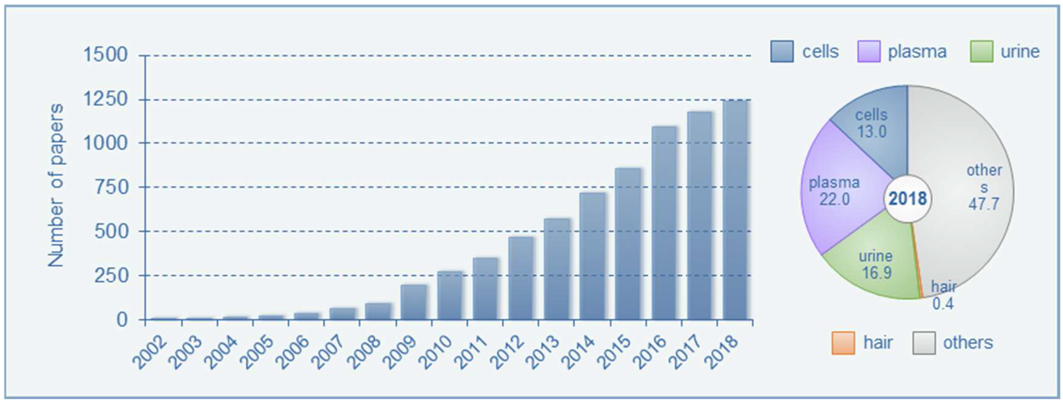
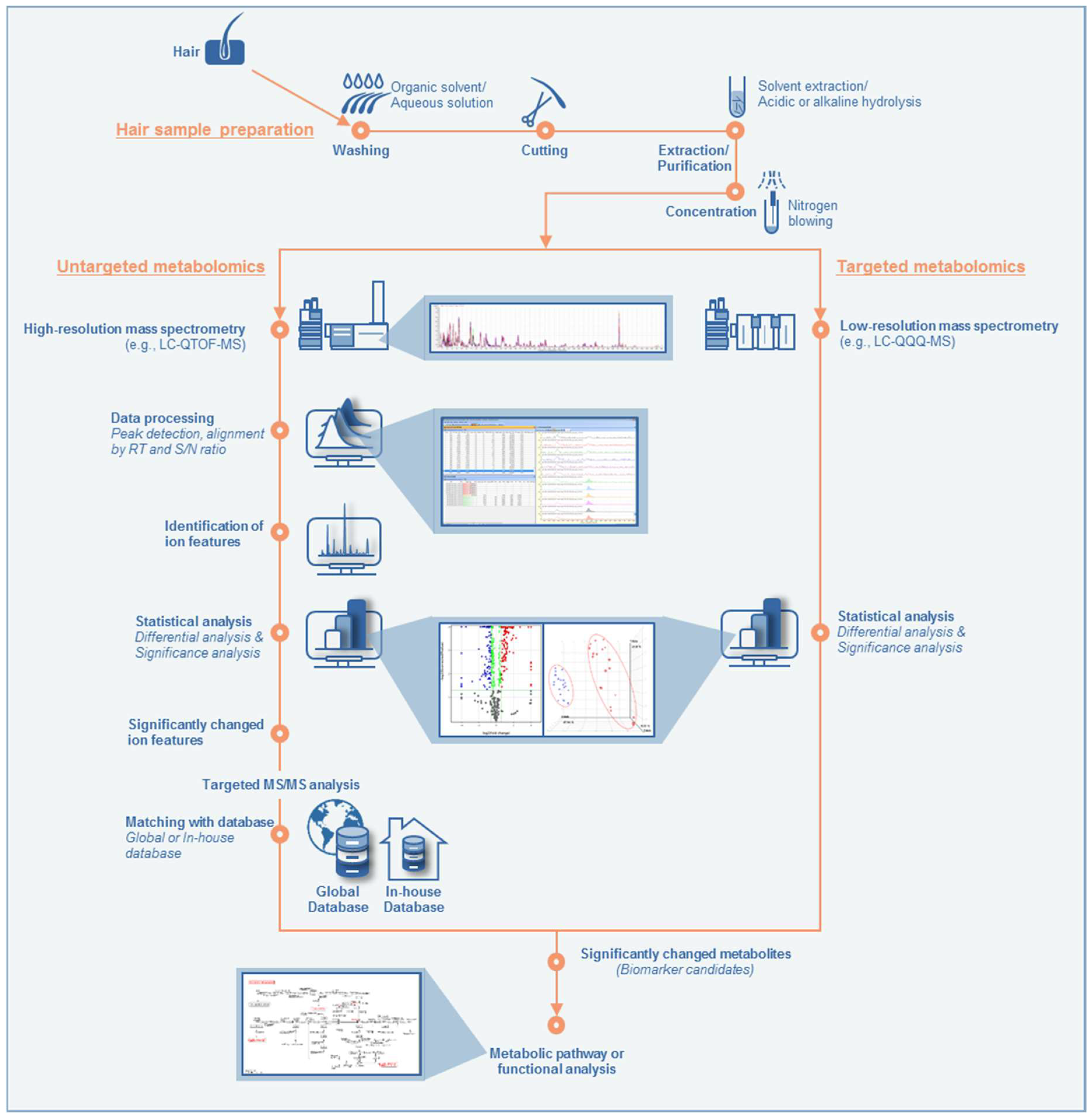
Hair Metabolomics in Animal Studies and Clinical Settings
Abstract
1. Introduction
2. Methods
3. Hair as A Metabolomics Sample
3.1. Hair as An Analytical Sample
3.2. Application of Hair Analysis in Metabolomics
4. Analytical Techniques for Hair Metabolomics
4.1. General Metabolomics Methodology
4.2. Sample Preparation and Instrumental Methods for Hair Metabolomics
5. Use in Animal Studies
6. Use in Clinical Settings
7. Conclusions
Author Contributions
Funding
Conflicts of Interest
References
- Monteiro, M.S.; Carvalho, M.; Bastos, M.L.; Guedes de Pinho, P. Metabolomics analysis for biomarker discovery: Advances and challenges. Curr. Med. Chem. 2013, 20, 257–271. [Google Scholar] [CrossRef] [PubMed]
- Showiheen, S.A.A.; Sun, A.R.; Wu, X.; Crawford, R.; Xiao, Y.; Wellard, R.M.; Prasadam, I. Application of Metabolomics to Osteoarthritis: From Basic Science to the Clinical Approach. Curr. Rheumatol. Rep. 2019, 21, 26. [Google Scholar] [CrossRef] [PubMed]
- Mehrparavar, B.; Minai-Tehrani, A.; Arjmand, B.; Gilany, K. Metabolomics of Male Infertility: A New Tool for Diagnostic Tests. J. Reprod. Infertil. 2019, 20, 64–69. [Google Scholar] [PubMed]
- Voutilainen, T.; Karkkainen, O. Changes in the Human Metabolome Associated With Alcohol Use: A Review. Alcohol Alcohol. 2019, 54, 225–234. [Google Scholar] [CrossRef] [PubMed]
- Jiang, Y.; Zhu, Z.; Shi, J.; An, Y.; Zhang, K.; Wang, Y.; Li, S.; Jin, L.; Ye, W.; Cui, M.; et al. Metabolomics in the Development and Progression of Dementia: A Systematic Review. Front. Neurosci. 2019, 13, 343. [Google Scholar] [CrossRef] [PubMed]
- Dettmer, K.; Aronov, P.A.; Hammock, B.D. Mass spectrometry-based metabolomics. Mass Spectrom. Rev. 2007, 26, 51–78. [Google Scholar] [CrossRef] [PubMed]
- Boudonck, K.J.; Mitchell, M.W.; Nemet, L.; Keresztes, L.; Nyska, A.; Shinar, D.; Rosenstock, M. Discovery of metabolomics biomarkers for early detection of nephrotoxicity. Toxicol. Pathol. 2009, 37, 280–292. [Google Scholar] [CrossRef] [PubMed]
- Li, H.; Bu, Q.; Chen, B.; Shao, X.; Hu, Z.; Deng, P.; Lv, L.; Deng, Y.; Zhu, R.; Li, Y.; et al. Mechanisms of metabonomic for a gateway drug: Nicotine priming enhances behavioral response to cocaine with modification in energy metabolism and neurotransmitter level. PLoS ONE 2014, 9, e87040. [Google Scholar] [CrossRef]
- Bujak, R.; Struck-Lewicka, W.; Markuszewski, M.J.; Kaliszan, R. Metabolomics for laboratory diagnostics. J. Pharm. Biomed. Anal. 2015, 113, 108–120. [Google Scholar] [CrossRef]
- Mastrangelo, A.; Barbas, C. Chronic Diseases and Lifestyle Biomarkers Identification by Metabolomics. Adv Exp. Med. Biol. 2017, 965, 235–263. [Google Scholar] [CrossRef]
- Considine, E.C.; Khashan, A.S.; Kenny, L.C. Screening for Preterm Birth: Potential for a Metabolomics Biomarker Panel. Metabolites 2019, 9, 90. [Google Scholar] [CrossRef] [PubMed]
- Hsu, J.F.; Tien, C.P.; Shih, C.L.; Liao, P.M.; Wong, H.I.; Liao, P.C. Using a high-resolution mass spectrometry-based metabolomics strategy for comprehensively screening and identifying biomarkers of phthalate exposure: Method development and application. Env. Int. 2019, 128, 261–270. [Google Scholar] [CrossRef] [PubMed]
- Yang, L.; Li, Z.; Song, Y.; Liu, Y.; Zhao, H.; Liu, Y.; Zhang, T.; Yuan, Y.; Cai, X.; Wang, S.; et al. Study on urine metabolic profiling and pathogenesis of hyperlipidemia. Clin. Chim. Acta 2019, 495, 365–373. [Google Scholar] [CrossRef] [PubMed]
- Woo, H.M.; Kim, K.M.; Choi, M.H.; Jung, B.H.; Lee, J.; Kong, G.; Nam, S.J.; Kim, S.; Bai, S.W.; Chung, B.C. Mass spectrometry based metabolomic approaches in urinary biomarker study of women’s cancers. Clin. Chim. Acta 2009, 400, 63–69. [Google Scholar] [CrossRef] [PubMed]
- Weljie, A.M.; Bondareva, A.; Zang, P.; Jirik, F.R. (1)H NMR metabolomics identification of markers of hypoxia-induced metabolic shifts in a breast cancer model system. J. Biomol. NMR 2011, 49, 185–193. [Google Scholar] [CrossRef]
- Deja, S.; Barg, E.; Mlynarz, P.; Basiak, A.; Willak-Janc, E. 1H NMR-based metabolomics studies of urine reveal differences between type 1 diabetic patients with high and low HbAc1 values. J. Pharm. Biomed. Anal. 2013, 83, 43–48. [Google Scholar] [CrossRef]
- Garcia-Canaveras, J.C.; Castell, J.V.; Donato, M.T.; Lahoz, A. A metabolomics cell-based approach for anticipating and investigating drug-induced liver injury. Sci. Rep. 2016, 6, 27239. [Google Scholar] [CrossRef]
- Pragst, F.; Balikova, M.A. State of the art in hair analysis for detection of drug and alcohol abuse. Clin. Chim. Acta 2006, 370, 17–49. [Google Scholar] [CrossRef]
- Kempson, I.M.; Lombi, E. Hair analysis as a biomonitor for toxicology, disease and health status. Chem. Soc. Rev. 2011, 40, 3915–3940. [Google Scholar] [CrossRef]
- Barbosa, J.; Faria, J.; Carvalho, F.; Pedro, M.; Queiros, O.; Moreira, R.; Dinis-Oliveira, R.J. Hair as an alternative matrix in bioanalysis. Bioanalysis 2013, 5, 895–914. [Google Scholar] [CrossRef]
- Yu, H.; Jang, W.J.; Jang, J.H.; Park, B.; Seo, Y.H.; Jeong, C.H.; Lee, S. Role of hair pigmentation in drug incorporation into hair. Forensic Sci. Int. 2017, 281, 171–175. [Google Scholar] [CrossRef] [PubMed]
- Behringer, V.; Deschner, T. Non-invasive monitoring of physiological markers in primates. Horm. Behav. 2017, 91, 3–18. [Google Scholar] [CrossRef] [PubMed]
- Ito, S.; Wakamatsu, K. Quantitative analysis of eumelanin and pheomelanin in humans, mice, and other animals: A comparative review. Pigment Cell Res. 2003, 16, 523–531. [Google Scholar] [CrossRef] [PubMed]
- D’Ischia, M.; Wakamatsu, K.; Cicoira, F.; Di Mauro, E.; Garcia-Borron, J.C.; Commo, S.; Galvan, I.; Ghanem, G.; Kenzo, K.; Meredith, P.; et al. Melanins and melanogenesis: From pigment cells to human health and technological applications. Pigment Cell Melanoma Res. 2015, 28, 520–544. [Google Scholar] [CrossRef] [PubMed]
- Wennig, R. Potential problems with the interpretation of hair analysis results. Forensic Sci. Int. 2000, 107, 5–12. [Google Scholar] [CrossRef]
- Cooper, G.A.; Kronstrand, R.; Kintz, P.; Society of Hair, T. Society of Hair Testing guidelines for drug testing in hair. Forensic Sci. Int. 2012, 218, 20–24. [Google Scholar] [CrossRef] [PubMed]
- Jung, H.J.; Kim, S.J.; Lee, W.Y.; Chung, B.C.; Choi, M.H. Gas chromatography/mass spectrometry based hair steroid profiling may reveal pathogenesis in hair follicles of the scalp. Rapid Commun. Mass Spectrom. 2011, 25, 1184–1192. [Google Scholar] [CrossRef]
- Sulek, K.; Han, T.L.; Villas-Boas, S.G.; Wishart, D.S.; Soh, S.E.; Kwek, K.; Gluckman, P.D.; Chong, Y.S.; Kenny, L.C.; Baker, P.N. Hair metabolomics: Identification of fetal compromise provides proof of concept for biomarker discovery. Theranostics 2014, 4, 953–959. [Google Scholar] [CrossRef] [PubMed]
- He, X.; de Seymour, J.V.; Sulek, K.; Qi, H.; Zhang, H.; Han, T.L.; Villas-Boas, S.G.; Baker, P.N. Maternal hair metabolome analysis identifies a potential marker of lipid peroxidation in gestational diabetes mellitus. Acta Diabetol. 2016, 53, 119–122. [Google Scholar] [CrossRef] [PubMed]
- Xie, P.; Wang, T.J.; Yin, G.; Yan, Y.; Xiao, L.H.; Li, Q.; Bi, K.S. Metabonomic Study of Biochemical Changes in Human Hair of Heroin Abusers by Liquid Chromatography Coupled with Ion Trap-Time of Flight Mass Spectrometry. J. Mol. Neurosci. 2016, 58, 93–101. [Google Scholar] [CrossRef]
- Son, H.H.; Lee, D.Y.; Seo, H.S.; Jeong, J.; Moon, J.Y.; Lee, J.E.; Chung, B.C.; Kim, E.; Choi, M.H. Hair sterol signatures coupled to multivariate data analysis reveal an increased 7beta-hydroxycholesterol production in cognitive impairment. J. Steroid Biochem. Mol. Biol. 2016, 155, 9–17. [Google Scholar] [CrossRef] [PubMed]
- Jones, B.; Han, T.L.; Delplancke, T.; McKenzie, E.J.; de Seymour, J.V.; Chua, M.C.; Tan, K.H.; Baker, P.N. Association between maternal exposure to phthalates and lower language ability in offspring derived from hair metabolome analysis. Sci. Rep. 2018, 8, 6745. [Google Scholar] [CrossRef] [PubMed]
- Delplancke, T.D.J.; de Seymour, J.V.; Tong, C.; Sulek, K.; Xia, Y.; Zhang, H.; Han, T.L.; Baker, P.N. Analysis of sequential hair segments reflects changes in the metabolome across the trimesters of pregnancy. Sci. Rep. 2018, 8, 36. [Google Scholar] [CrossRef] [PubMed]
- Chen, X.; de Seymour, J.V.; Han, T.L.; Xia, Y.; Chen, C.; Zhang, T.; Zhang, H.; Baker, P.N. Metabolomic biomarkers and novel dietary factors associated with gestational diabetes in China. Metabolomics 2018, 14, 149. [Google Scholar] [CrossRef] [PubMed]
- De Seymour, J.V.; Tu, S.; He, X.; Zhang, H.; Han, T.L.; Baker, P.N.; Sulek, K. Metabolomic profiling of maternal hair suggests rapid development of intrahepatic cholestasis of pregnancy. Metabolomics 2018, 14, 79. [Google Scholar] [CrossRef] [PubMed]
- Cordero, R.; Lee, S.; Paterson, S. Distribution of concentrations of cocaine and its metabolites in hair collected postmortem from cases with diverse causes/circumstances of death. J. Anal. Toxicol. 2010, 34, 543–548. [Google Scholar] [CrossRef] [PubMed][Green Version]
- Han, E.; Yang, W.; Lee, J.; Park, Y.; Kim, E.; Lim, M.; Chung, H. Correlation of methamphetamine results and concentrations between head, axillary, and pubic hair. Forensic Sci. Int. 2005, 147, 21–24. [Google Scholar] [CrossRef]
- Lee, S.; Cordero, R.; Paterson, S. Distribution of 6-monoacetylmorphine and morphine in head and pubic hair from heroin-related deaths. Forensic Sci. Int. 2009, 183, 74–77. [Google Scholar] [CrossRef]
- Tzatzarakis, M.N.; Alegakis, A.K.; Kavvalakis, M.P.; Vakonaki, E.; Stivaktakis, P.D.; Kanaki, K.; Vardavas, A.I.; Barbounis, E.G.; Tsatsakis, A.M. Comparative Evaluation of Drug Deposition in Hair Samples Collected from Different Anatomical Body Sites. J. Anal. Toxicol. 2017, 41, 214–223. [Google Scholar] [CrossRef][Green Version]
- Lee, S.; Han, E.; In, S.; Choi, H.; Chung, H.; Chung, K.H. Analysis of pubic hair as an alternative specimen to scalp hair: A contamination issue. Forensic Sci. Int. 2011, 206, 19–21. [Google Scholar] [CrossRef]
- Liu, A.Y.; Yang, Q.; Huang, Y.; Bacchetti, P.; Anderson, P.L.; Jin, C.; Goggin, K.; Stojanovski, K.; Grant, R.; Buchbinder, S.P.; et al. Strong relationship between oral dose and tenofovir hair levels in a randomized trial: Hair as a potential adherence measure for pre-exposure prophylaxis (PrEP). PLoS ONE 2014, 9, e83736. [Google Scholar] [CrossRef] [PubMed]
- Han, E.; Lee, S.; In, S.; Park, M.; Park, Y.; Cho, S.; Shin, J.; Lee, H. Relationship between methamphetamine use history and segmental hair analysis findings of MA users. Forensic Sci. Int. 2015, 254, 59–67. [Google Scholar] [CrossRef]
- Gunther, K.N.; Johansen, S.S.; Nielsen, M.K.K.; Wicktor, P.; Banner, J.; Linnet, K. Post-mortem quetiapine concentrations in hair segments of psychiatric patients - Correlation between hair concentration, dose and concentration in blood. Forensic Sci. Int. 2018, 285, 58–64. [Google Scholar] [CrossRef] [PubMed]
- Lee, S.; Han, E.; Park, Y.; Choi, H.; Chung, H. Distribution of methamphetamine and amphetamine in drug abusers’ head hair. Forensic Sci. Int. 2009, 190, 16–18. [Google Scholar] [CrossRef] [PubMed]
- Raul, J.S.; Cirimele, V.; Ludes, B.; Kintz, P. Detection of physiological concentrations of cortisol and cortisone in human hair. Clin. Biochem. 2004, 37, 1105–1111. [Google Scholar] [CrossRef]
- Greff, M.J.E.; Levine, J.M.; Abuzgaia, A.M.; Elzagallaai, A.A.; Rieder, M.J.; van Uum, S.H.M. Hair cortisol analysis: An update on methodological considerations and clinical applications. Clin. Biochem. 2019, 63, 1–9. [Google Scholar] [CrossRef]
- Wester, V.L.; van Rossum, E.F. Clinical applications of cortisol measurements in hair. Eur. J. Endocrinol. 2015, 173, M1-10. [Google Scholar] [CrossRef] [PubMed]
- Veldhorst, M.A.; Noppe, G.; Jongejan, M.H.; Kok, C.B.; Mekic, S.; Koper, J.W.; van Rossum, E.F.; van den Akker, E.L. Increased scalp hair cortisol concentrations in obese children. J. Clin. Endocrinol. Metab. 2014, 99, 285–290. [Google Scholar] [CrossRef][Green Version]
- Yu, H.; Choi, M.; Jang, J.H.; Park, B.; Seo, Y.H.; Jeong, C.H.; Bae, J.W.; Lee, S. Development of a column-switching LC-MS/MS method of tramadol and its metabolites in hair and application to a pharmacogenetic study. Arch. Pharm. Res. 2018, 41, 554–563. [Google Scholar] [CrossRef]
- Cuypers, E.; Flanagan, R.J. The interpretation of hair analysis for drugs and drug metabolites. Clin. Toxicol. (Phila) 2018, 56, 90–100. [Google Scholar] [CrossRef]
- Kim, Y.G.; Hwang, J.; Choi, H.; Lee, S. Development of a Column-Switching HPLC-MS/MS Method and Clinical Application for Determination of Ethyl Glucuronide in Hair in Conjunction with AUDIT for Detecting High-Risk Alcohol Consumption. Pharmaceutics 2018, 10, 84. [Google Scholar] [CrossRef] [PubMed]
- Kim, J.; Yum, H.; Jang, M.; Shin, I.; Yang, W.; Baeck, S.; Suh, J.H.; Lee, S.; Han, S.B. A comprehensive and sensitive method for hair analysis in drug-facilitated crimes and incorporation of zolazepam and tiletamine into hair after a single exposure. Anal. Bioanal. Chem. 2016, 408, 251–263. [Google Scholar] [CrossRef] [PubMed]
- Kuwayama, K.; Nariai, M.; Miyaguchi, H.; Iwata, Y.T.; Kanamori, T.; Tsujikawa, K.; Yamamuro, T.; Segawa, H.; Abe, H.; Iwase, H.; et al. Micro-segmental hair analysis for proving drug-facilitated crimes: Evidence that a victim ingested a sleeping aid, diphenhydramine, on a specific day. Forensic Sci. Int. 2018, 288, 23–28. [Google Scholar] [CrossRef] [PubMed]
- Wang, X.; Johansen, S.S.; Nielsen, M.K.K.; Linnet, K. Hair analysis in toxicological investigation of drug-facilitated crimes in Denmark over a 8-year period. Forensic Sci. Int. 2018, 285, e1–e12. [Google Scholar] [CrossRef] [PubMed]
- Xiang, P.; Shen, M.; Drummer, O.H. Review: Drug concentrations in hair and their relevance in drug facilitated crimes. J. Forensic Leg. Med. 2015, 36, 126–135. [Google Scholar] [CrossRef]
- Gunther, K.N.; Johansen, S.S.; Wicktor, P.; Banner, J.; Linnet, K. Segmental Analysis of Chlorprothixene and Desmethylchlorprothixene in Postmortem Hair. J. Anal. Toxicol. 2018, 42, 642–649. [Google Scholar] [CrossRef]
- Nielsen, M.K.; Johansen, S.S.; Linnet, K. Evaluation of poly-drug use in methadone-related fatalities using segmental hair analysis. Forensic Sci. Int. 2015, 248, 134–139. [Google Scholar] [CrossRef]
- Kintz, P. Issues about axial diffusion during segmental hair analysis. Drug Monit. 2013, 35, 408–410. [Google Scholar] [CrossRef]
- Busardo, F.P.; Jones, A.W. Interpreting gamma-hydroxybutyrate concentrations for clinical and forensic purposes. Clin. Toxicol. (Phila) 2019, 57, 149–163. [Google Scholar] [CrossRef]
- Busardo, F.P.; Vaiano, F.; Mannocchi, G.; Bertol, E.; Zaami, S.; Marinelli, E. Twelve months monitoring of hair GHB decay following a single dose administration in a case of facilitated sexual assault. Drug Test. Anal. 2017, 9, 953–956. [Google Scholar] [CrossRef]
- Noppe, G.; de Rijke, Y.B.; Dorst, K.; van den Akker, E.L.; van Rossum, E.F. LC-MS/MS-based method for long-term steroid profiling in human scalp hair. Clin. Endocrinol. (Oxf) 2015, 83, 162–166. [Google Scholar] [CrossRef] [PubMed]
- Bouhifd, M.; Hartung, T.; Hogberg, H.T.; Kleensang, A.; Zhao, L. Review: Toxicometabolomics. J. Appl. Toxicol. 2013, 33, 1365–1383. [Google Scholar] [CrossRef] [PubMed]
- Gonzalez-Dominguez, R.; Gonzalez-Dominguez, A.; Segundo, C.; Schwarz, M.; Sayago, A.; Mateos, R.M.; Duran-Guerrero, E.; Lechuga-Sancho, A.M.; Fernandez-Recamales, A. High-Throughput Metabolomics Based on Direct Mass Spectrometry Analysis in Biomedical Research. Methods Mol. Biol. 2019, 1978, 27–38. [Google Scholar] [CrossRef] [PubMed]
- Tzoulaki, I.; Castagne, R.; Boulange, C.L.; Karaman, I.; Chekmeneva, E.; Evangelou, E.; Ebbels, T.M.D.; Kaluarachchi, M.R.; Chadeau-Hyam, M.; Mosen, D.; et al. Serum metabolic signatures of coronary and carotid atherosclerosis and subsequent cardiovascular disease. Eur. Heart J. 2019. [Google Scholar] [CrossRef] [PubMed]
- Liu, Z.; Triba, M.N.; Amathieu, R.; Lin, X.; Bouchemal, N.; Hantz, E.; Le Moyec, L.; Savarin, P. Nuclear magnetic resonance-based serum metabolomic analysis reveals different disease evolution profiles between septic shock survivors and non-survivors. Crit. Care 2019, 23, 169. [Google Scholar] [CrossRef] [PubMed]
- Su, F.; Sun, M.; Geng, Y. (1)H-NMR Metabolomics Analysis of the Effects of Sulfated Polysaccharides from Masson Pine Pollen in RAW264.7 Macrophage Cells. Molecules 2019, 24, 1841. [Google Scholar] [CrossRef]
- Lubes, G.; Goodarzi, M. GC-MS based metabolomics used for the identification of cancer volatile organic compounds as biomarkers. J Pharm. Biomed. Anal. 2018, 147, 313–322. [Google Scholar] [CrossRef]
- Pannkuk, E.L.; Laiakis, E.C.; Girgis, M.; Dowd, S.E.; Dhungana, S.; Nishita, D.; Bujold, K.; Bakke, J.; Gahagen, J.; Authier, S.; et al. Temporal Effects on Radiation Responses in Nonhuman Primates: Identification of Biofluid Small Molecule Signatures by Gas Chromatography(-)Mass Spectrometry Metabolomics. Metabolites 2019, 9, 98. [Google Scholar] [CrossRef]
- Zhao, X.; Chen, M.; Zhao, Y.; Zha, L.; Yang, H.; Wu, Y. GC(-)MS-Based Nontargeted and Targeted Metabolic Profiling Identifies Changes in the Lentinula edodes Mycelial Metabolome under High-Temperature Stress. Int. J. Mol. Sci. 2019, 20, 2330. [Google Scholar] [CrossRef]
- Markley, J.L.; Bruschweiler, R.; Edison, A.S.; Eghbalnia, H.R.; Powers, R.; Raftery, D.; Wishart, D.S. The future of NMR-based metabolomics. Curr Opin Biotechnol 2017, 43, 34–40. [Google Scholar] [CrossRef]
- Beale, D.J.; Pinu, F.R.; Kouremenos, K.A.; Poojary, M.M.; Narayana, V.K.; Boughton, B.A.; Kanojia, K.; Dayalan, S.; Jones, O.A.H.; Dias, D.A. Review of recent developments in GC-MS approaches to metabolomics-based research. Metabolomics 2018, 14, 152. [Google Scholar] [CrossRef] [PubMed]
- Alonso, A.; Marsal, S.; Julia, A. Analytical methods in untargeted metabolomics: State of the art in 2015. Front. Bioeng. Biotechnol. 2015, 3, 23. [Google Scholar] [CrossRef] [PubMed]
- Roberts, L.D.; Souza, A.L.; Gerszten, R.E.; Clish, C.B. Targeted metabolomics. Curr. Protoc. Mol. Biol. 2012, 98, 30–32. [Google Scholar] [CrossRef] [PubMed]
- Yin, P.; Xu, G. Current state-of-the-art of nontargeted metabolomics based on liquid chromatography-mass spectrometry with special emphasis in clinical applications. J. Chromatogr. A 2014, 1374, 1–13. [Google Scholar] [CrossRef] [PubMed]
- Lynn, K.S.; Cheng, M.L.; Chen, Y.R.; Hsu, C.; Chen, A.; Lih, T.M.; Chang, H.Y.; Huang, C.J.; Shiao, M.S.; Pan, W.H.; et al. Metabolite identification for mass spectrometry-based metabolomics using multiple types of correlated ion information. Anal. Chem. 2015, 87, 2143–2151. [Google Scholar] [CrossRef] [PubMed]
- Lee, S.; Miyaguchi, H.; Han, E.; Kim, E.; Park, Y.; Choi, H.; Chung, H.; Oh, S.M.; Chung, K.H. Homogeneity and stability of a candidate certified reference material for the determination of methamphetamine and amphetamine in hair. J. Pharm. Biomed. Anal. 2010, 53, 1037–1041. [Google Scholar] [CrossRef]
- Lee, S.; Park, Y.; Kim, J.; In, S.; Choi, H.; Chung, H.; Oh, S.M.; Chung, K.H. Feasibility of rat hair as a quality control material for the determination of methamphetamine and amphetamine in human hair. Arch. Pharm. Res. 2011, 34, 593–598. [Google Scholar] [CrossRef]
- Choi, M.H.; Kim, K.R.; Kim, Y.T.; Chung, B.C. Increased polyamine concentrations in the hair of cancer patients. Clin. Chem. 2001, 47, 143–144. [Google Scholar]
- Joo, K.M.; Kim, A.R.; Kim, S.N.; Kim, B.M.; Lee, H.K.; Bae, S.; Lee, J.H.; Lim, K.M. Metabolomic analysis of amino acids and lipids in human hair altered by dyeing, perming and bleaching. Exp. Derm. 2016, 25, 729–731. [Google Scholar] [CrossRef]
- Khandelwal, P.; Stryker, S.; Chao, H.; Aranibar, N.; Lawrence, R.M.; Madireddi, M.; Zhao, W.; Chen, L.; Reily, M.D. 1H NMR-based lipidomics of rodent fur: Species-specific lipid profiles and SCD1 inhibitor-related dermal toxicity. J. Lipid Res. 2014, 55, 1366–1374. [Google Scholar] [CrossRef]
- Inagaki, S.; Noda, T.; Min, J.Z.; Toyo’oka, T. Metabolic profiling of rat hair and screening biomarkers using ultra performance liquid chromatography with electrospray ionization time-of-flight mass spectrometry. J. Chromatogr. A 2007, 1176, 94–99. [Google Scholar] [CrossRef] [PubMed]
- Tsutsui, H.; Maeda, T.; Min, J.Z.; Inagaki, S.; Higashi, T.; Kagawa, Y.; Toyo’oka, T. Biomarker discovery in biological specimens (plasma, hair, liver and kidney) of diabetic mice based upon metabolite profiling using ultra-performance liquid chromatography with electrospray ionization time-of-flight mass spectrometry. Clin. Chim. Acta. 2011, 412, 861–872. [Google Scholar] [CrossRef] [PubMed]
- Choi, B.; Kim, S.P.; Hwang, S.; Hwang, J.; Yang, C.H.; Lee, S. Metabolic characterization in urine and hair from a rat model of methamphetamine self-administration using LC-QTOF-MS-based metabolomics. Metabolomics 2017, 13. [Google Scholar] [CrossRef]
- Masukawa, Y.; Tsujimura, H.; Imokawa, G. A systematic method for the sensitive and specific determination of hair lipids in combination with chromatography. J. Chromatogr. B Anal. Technol. Biomed. Life Sci. 2005, 823, 131–142. [Google Scholar] [CrossRef] [PubMed]
- Joo, K.M.; Choi, D.; Park, Y.H.; Yi, C.G.; Jeong, H.J.; Cho, J.C.; Lim, K.M. A rapid and highly sensitive UPLC-MS/MS method using pre-column derivatization with 2-picolylamine for intravenous and percutaneous pharmacokinetics of valproic acid in rats. J. Chromatogr. B Anal. Technol. Biomed. Life Sci. 2013, 938, 35–42. [Google Scholar] [CrossRef] [PubMed]
- James, E.L.; Parkinson, E.K. Serum metabolomics in animal models and human disease. Curr. Opin. Clin. Nutr. Metab. Care 2015, 18, 478–483. [Google Scholar] [CrossRef] [PubMed]
- Reed, L.K.; Baer, C.F.; Edison, A.S. Considerations when choosing a genetic model organism for metabolomics studies. Curr. Opin. Chem. Biol. 2017, 36, 7–14. [Google Scholar] [CrossRef] [PubMed]
- Chen, D.; Su, X.; Wang, N.; Li, Y.; Yin, H.; Li, L.; Li, L. Chemical Isotope Labeling LC-MS for Monitoring Disease Progression and Treatment in Animal Models: Plasma Metabolomics Study of Osteoarthritis Rat Model. Sci. Rep. 2017, 7, 40543. [Google Scholar] [CrossRef] [PubMed]
- Koob, G.F.; Le Moal, M. Drug addiction, dysregulation of reward, and allostasis. Neuropsychopharmacology 2001, 24, 97–129. [Google Scholar] [CrossRef]
- Zaitsu, K.; Hayashi, Y.; Kusano, M.; Tsuchihashi, H.; Ishii, A. Application of metabolomics to toxicology of drugs of abuse: A mini review of metabolomics approach to acute and chronic toxicity studies. Drug Metab. Pharm. 2016, 31, 21–26. [Google Scholar] [CrossRef] [PubMed]


| No. | Pathologic Condition | Study Objective | Study Subjects (Animal Species, Animal Model, etc.) | Sample Preparation | Analytical Platform (Untargeted or Targeted) | Key metabolites Changed (Possible Biomarkers) | Consequence of Metabolic Changes | Reference (Year Published) | |
|---|---|---|---|---|---|---|---|---|---|
| Increased | Decreased | ||||||||
| 1 | Stroke | Biomarker discovery | Spontaneously hypertensive rats (SHR/Izm) and stroke-prone SHR rats (SHRSP/Izm) | Acidic solvent sonication | UPLC-ESI-TOF-MS (Untargeted) | m/z 235.40 ion at 2.30 min | - | Potential biomarker of stroke | Inagaki et al., J Chromatogr A (2007) |
| 2 | Diabetes | Biomarker discovery | Spontaneous insulin-resistant mice (ddY-H) | Brief solvent extraction | UPLC-ESI-TOF-MS (Untargeted) | - | N-Acetyl-L-leucine | Potential biomarker of diabetes | Tsutsui et al., Clin Chim Acta (2011) |
| 3 | Dermal toxicity (drug-induced sebaceous gland atrophy) | Metabolic profiling | Rats and hamsters dosed with a stearoyl-CoA desaturase 1 (SCD 1) inhibitor | Solvent incubation for 16 h | NMR (Untargeted) | - | 1,2-distearoyl-3-oleoyl-rac-glycerol, lathosterol-like sterol esters, wax ester, total cholesterol, and cholesterol for rats, and lathosterol-like sterol esters, and wax ester for hamsters | Reduction of lipid levels by an SCD 1 inhibitor | Khandelwal et al., J Lipid Res (2014) |
| 4 | Drug addiction | Metabolic profiling | Methamphetamine self-administering rats | Solvent incubation | UPLC-ESI-QTOF-MS (Untargeted) | Acetylcarnitine, 5-methylcytidine, 1-methyladenosine, palmitoyl-(l)-carnitine | (l)-Norvaline/betaine/5-aminopentanoate/(l)-valine, lumichrome, deoxycorticosterone, oleamide, stearamide, hippurate | Metabolic perturbation in the central nervous system and energy production | Choi et al., Metabolomics (2017) |
| \ | Pathologic Condition | Study Objective | Study Subject (Age, Gender, Number of Subjects, etc.) | Sample Preparation | Analytical Platform (Untargeted or Targeted) | Key Metabolites Changed (Possible Biomarkers) | Consequence of Metabolic Changes | Reference (Year Published) | |
|---|---|---|---|---|---|---|---|---|---|
| Increased | Decreased | ||||||||
| 1 | Cancer | Polyamine measurement for cancer diagnosis | Patients with cervical cancer (34–65 yr, n = 13) or ovarian cancer (37–75 yr, n = 11) | Acidic solution incubation followed by N-ethoxycarbonylation and N-pentafluoropropionylation | GC-MS (Targeted) | Putrescine, spermidine, spermine | - | Deficits in polyamine biosynthesis and accumulation | Choi et al., Clin Chem (2001) |
| 2 | Male pattern baldness | Steroid profiling | Balding men (32.5 yr (mean), n = 19) | Solvent sonication followed by solid phase extraction and trimethylsilation | GC-MS (Targeted) | Dihydrotestosterone, dihydrotestosterone/testosterone ratio, and cortisol/cortisone ratio | - | Increase of 5α-reductase activity | Jung et al., Rapid Commun Mass Spectrom (2011) |
| 3 | Fetal growth restriction | Biomarker discovery | Pregnant women (22–44 yr, 26–28 weeks of gestation, n = 41) | Alkaline hydrolysis followed by chemical derivatization with methyl chloroformate | GC-MS (Untargeted) | Heptadecane, NADPH/NADP, saturated fatty acids (palmitate, 2-methyloctadecanoate, myristate, margarate, stearate, dodecanoate, and octanoate) | Amino acids (lysine, methionine, tyrosine, valine, and threonine), glutathione | Loss of redox control and deficiency of precursors for fetal development and growth | Sulek et al., Theranostics (2014) |
| 4 | Gestational diabetes mellitus (GDM) | Biomarker discovery | Pregnant women (30 yr (median), 26–28 weeks of gestation, n = 94) | Alkaline hydrolysis followed by chemical derivatization with methyl chloroformate | GC-MS (Untargeted) | Adipic acid | - | Lipid peroxidation related to the oxidative stress environment in diabetes | He et al., Acta Diabetol (2016) |
| 5 | Hair damage by dyeing, perming, and bleaching | Metabolic profiling | Treated samples of natural hair of Asians (Beaaulax Co., Saitama, Japan, n = 10) | Acidic or alkaline hydrolysis followed by chemical derivatization with Waters AccQ•Tag reagents for amino acids | UPLC-PDA and | Cysteic acid and cysteine/cysteic acid | Methionine and tryptophan | Quantitative grading of hair damage | Joo et al., Exp Dermatol. (2016) |
| Solvent extraction for extractable lipids and further saponification and solvent extraction followed by chemical derivatization with 2-picolylamine for fatty acids | UPLC-ESI-QQQ-MS (Targeted) | - | Erucic acid, behenic acid, lignoceric acid, nervonic acid, cerotic acid, and 18-methyl eicosanoic acid | ||||||
| 6 | Heroin addiction | Metabolic profiling | Heroin abusers (20–56 yr, male, n = 40, female, n = 18) | Solvent sonication | UFLC-ESI-IT-TOF-MS (Untargeted) | Sorbitol and cortisol | Arachidonic acid, glutathione, linoleic acid, and myristic acid | Deficits in energy metabolism, sorbitol pathway, and immune cell function | Xie et al., J Mol Neurosci (2016) |
| 7 | Cognitive impairment | Sterol profiling | Patients with mild cognitive impairment (MCI, 70.3 yr (mean), female, n = 15) or Alzheimer’s disease (70.8 yr (mean), female, n = 31) | Solvent pulverization followed by trimethylsilation | GC-MS (Targeted) | 7α-Hydroxycholesterol and 7β-hydroxycholesterol | - | Impaired cholesterol metabolism | Son et al., J Steroid Biochem Mol Biol (2016) |
| 8 | Infant lower language ability | Maternal hair metabolic profiling for infant neurodevelopment | Pregnant women (26–28 weeks of gestation, n = 373) | Alkaline hydrolysis followed by chemical derivatization with methyl chloroformate | GC-MS (Untargeted) | Phthalic acid | - | Infant lower language ability caused by high maternal phthalate exposure | Jones et al., Sci Rep 2018 |
| 9 | Small for gestational age infants | Biomarker discovery and metabolic mechanism study | Pregnant women (30.8 yr (mean), 39.1 weeks of gestation, n = 20) | Alkaline hydrolysis followed by chemical derivatization with methyl chloroformate for GC-MS and alkaline hydrolysis followed by solvent extraction for LC-MS | GC-MS and UPLC-ESI-QTOF-MS (Untargeted) | Margaric acid, pentadecanoic acid, and myristic acid | - | Deficits in placental function of fatty acid transfer to the fetus | Delplancke et al., Sci Rep (2018) |
| GDM | Pregnant women (32.7 yr (mean), 38.6 weeks of gestation, n = 11) | 1-Hydroxy-3-nonanone and 22-oxavitamine D3 | Tryptophan, leucine, citric acid, 3,4-oxaolidinercarboxylic acid, 2-oxovaleric acid, 3-pyridinecarboxamide, 2-methylpentan-2-yl trifluoraoacetate, and 2-oxobutyric acid | Deficits in energy metabolism and degradation of amino acids | |||||
| 10 | GDM | Maternal hair metabolic profiling for gestational diabetes mellitus | Pregnant women (32 yr (mean), 24–28 weeks of gestation n = 49) | Alkaline hydrolysis followed by chemical derivatization with methyl chloroformate | GC-MS and UPLC-ESI-QTOF-MS (Untargeted) | Pentachloroethane, 1-hydroxyvitamin D5, (3beta,23E)-3-hydroxy-27-norcycloart-23-en-25-one, (4-methylphenyl)acetaldehyde, linalyl isobutyrate, and 3-phenyl-1-propanol | Proline, 4-methoxy-benzoic acid, 5-methylhexanoic acid, dihydroceramide, 2,2,9,9-tetramethyl-undecan-1,10-diol, palmitoylglycine, benzeneacetic acid, 2-butenoic acid, glutamic acid, but-2-enedioic acid, 2-oxobutyric acid, N,4-diethyl-4-heptanamine, N-methoxycarbonyl-l-proline, pyrrolidine-1,2-dicarboxylic acid, (1-ethyl) ester, NADP_NADPH, malonic acid, 2-methylcyclohexanone, 3-hydroxy-2-octanone, and C17 sphinganine | No correlation between maternal diet in GDM and hair metabolomes | Chen et al., Metabolomics (2018) |
| 11 | Intrahepatic cholestasis of pregnancy (ICP) | Biomarker discovery | Pregnant women (27.9 yr (mean), 17-41 weeks of gestation n = 38) | Alkaline hydrolysis followed by chemical derivatization with methyl chloroformate | GC-MS | - | Adipic acid and succinic acid | No correlation between ICP and hair metabolomes | de Seymour et al., Metabolomics (2018) |
© 2019 by the authors. Licensee MDPI, Basel, Switzerland. This article is an open access article distributed under the terms and conditions of the Creative Commons Attribution (CC BY) license (http://creativecommons.org/licenses/by/4.0/).
Share and Cite
Jang, W.-J.; Choi, J.Y.; Park, B.; Seo, J.H.; Seo, Y.H.; Lee, S.; Jeong, C.-H.; Lee, S. Hair Metabolomics in Animal Studies and Clinical Settings. Molecules 2019, 24, 2195. https://doi.org/10.3390/molecules24122195
Jang W-J, Choi JY, Park B, Seo JH, Seo YH, Lee S, Jeong C-H, Lee S. Hair Metabolomics in Animal Studies and Clinical Settings. Molecules. 2019; 24(12):2195. https://doi.org/10.3390/molecules24122195
Chicago/Turabian StyleJang, Won-Jun, Jae Yoon Choi, Byoungduck Park, Ji Hae Seo, Young Ho Seo, Sangkil Lee, Chul-Ho Jeong, and Sooyeun Lee. 2019. "Hair Metabolomics in Animal Studies and Clinical Settings" Molecules 24, no. 12: 2195. https://doi.org/10.3390/molecules24122195
APA StyleJang, W.-J., Choi, J. Y., Park, B., Seo, J. H., Seo, Y. H., Lee, S., Jeong, C.-H., & Lee, S. (2019). Hair Metabolomics in Animal Studies and Clinical Settings. Molecules, 24(12), 2195. https://doi.org/10.3390/molecules24122195


