Article

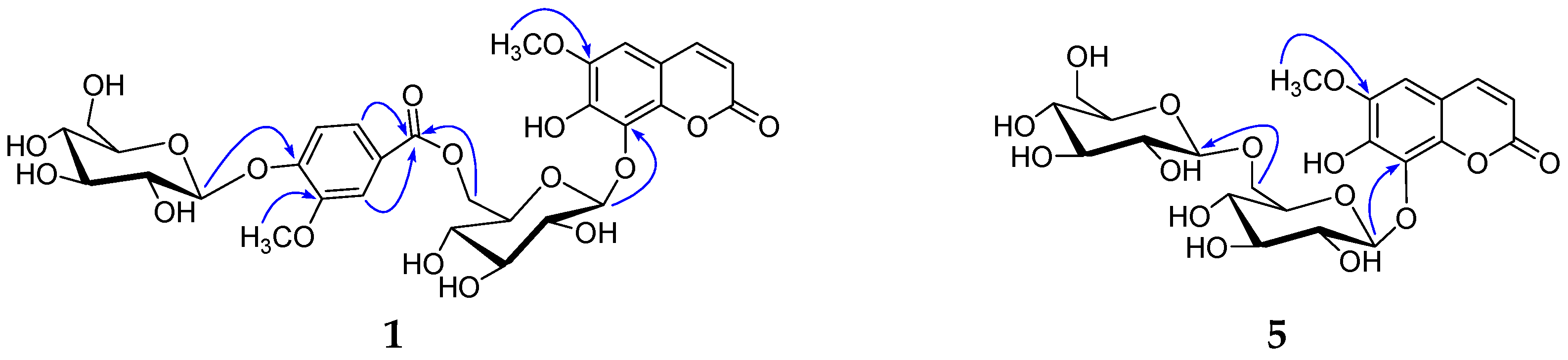
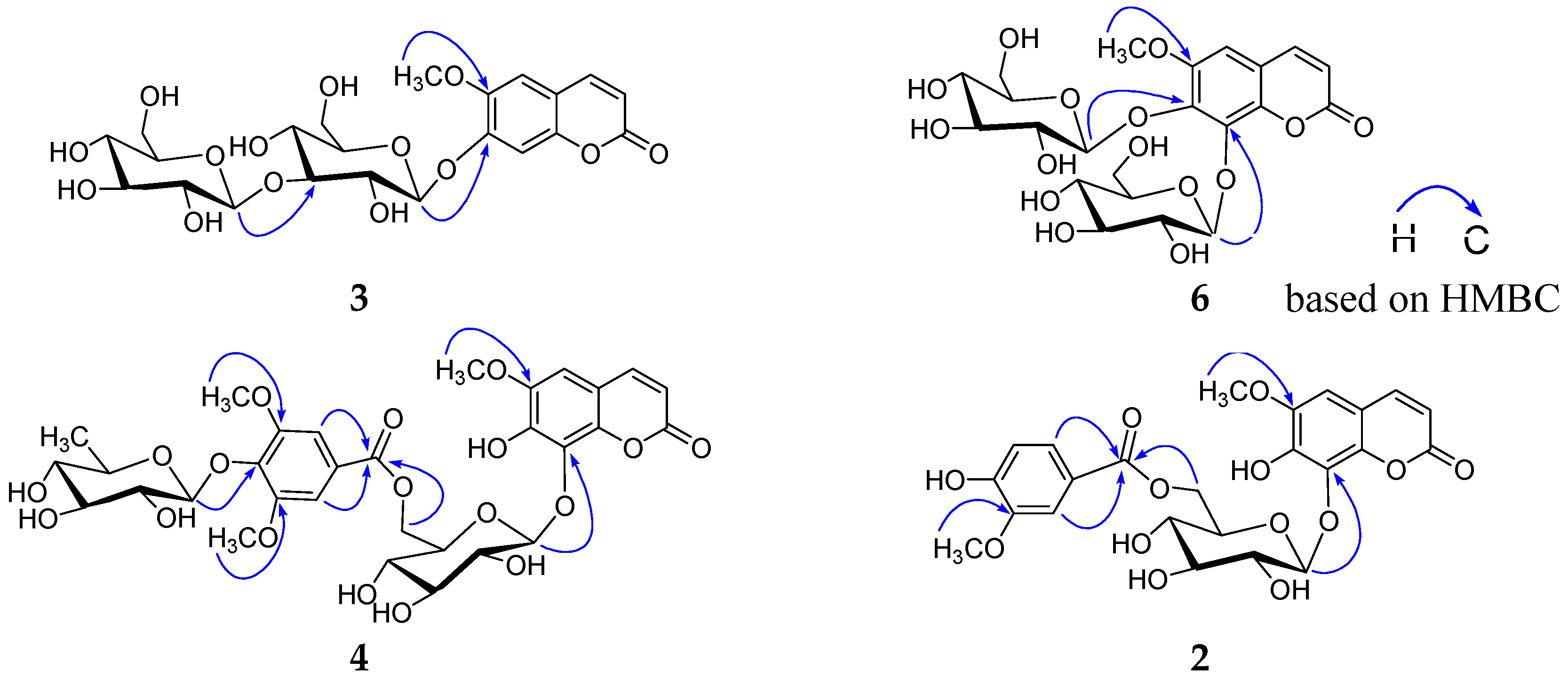
Six New Coumarin Glycosides from the Aerial Parts of Gendarussa vulgaris

1
Collaborative Innovation Center for Respiratory Disease Diagnosis and Treatment & Chinese Medicine Development of Henan Province, Henan University of Chinese Medicine, Zhengzhou 450046, Henan, China
2
School of Pharmacy, Henan University of Chinese Medicine, Zhengzhou 450046, Henan, China
*
Authors to whom correspondence should be addressed.
Molecules 2019, 24(8), 1456; https://doi.org/10.3390/molecules24081456
Submission received: 25 March 2019 / Revised: 9 April 2019 / Accepted: 10 April 2019 / Published: 12 April 2019
(This article belongs to the Section Natural Products Chemistry)

Abstract

:
Six new coumarin glycosides, genglycoside A–F (16), were isolated from the aerial parts of Gendarussa vulgaris, along with ten known analogues (716). Their structures were unambiguously established on the basis of extensive spectroscopic data and HPLC analysis. The cytotoxic activities of all isolated compounds were evaluated by MTT assay. Compound 12 showed the most potent cytotoxicity in Eca-109, MCF-7, and HepG2 cell lines. By the preliminary structure–activity relationships, it was firstly discovered that the glycosylation or esterification at 7,8-dihydroxy or 7-hydroxy drastically reduced the cytotoxic activity of the parent coumarin.

Graphical Abstract

1. Introduction

Gendarussa vulgaris Nees, which belongs to the family of Acanthaceae, is an evergreen dwarf shrub mainly distributed in China, India, Sri Lanka, and the Malay Peninsula [1]. As an important medicinal plant, it has been described in Chinese Pharmacopoeia 2015, Supplement to the Compendium of Materia Medica, Luchuan Materia Medica, Lingnan Medical Records, etc. Its aerial parts (called Xiaobogu in Chinese) are frequently used for the treatment of fascia fracture, traumatic injury, rheumatism, and ostalgia, blood stasis menstrual block, and postpartum abdominal pain [2]. Previous chemical investigations on G. vulgaris revealed the presence of bioactive alkaloids, flavonoids, phenylpropanoids, steroids, and triterpenes [1,2]. Naturally occurring coumarins have exhibited a broad spectrum of pharmacological actions, including as an anticoagulant [3], CNS stimulant [4], antioxidant [5], antiviral [6], hepatoprotective [7], anti-inflammatory [8], anticancer [9], and cyclooxygenase, lipooxygenase, cholinesterase (ChE), and monoamine oxidase (MAO) inhibitory activities [9,10], antimutagenic [10], etc. As potential therapeutic drugs against cancer, coumarins have not only exhibited obvious anti-proliferative activity in malignant melanoma, prostate cancer, and renal cell carcinoma in some clinical trials [11], but also very rare cardiotoxicity, nephrotoxicity, dermal toxicity, and other MDR (multi-drug resistance) side effects [9]. In our search for cytotoxic natural products from the aerial parts of G. vulgaris, six new coumarin glucosides (16) were obtained together with ten known analogues (716). Details of the isolation, structure elucidation, and cytotoxicity of all isolated compounds against Eca-109, MCF-7, and HepG2 cell lines are described here (Figure 1).

2. Results and Discussion

The EtOH extract of the aerial parts of G. vulgaris was partitioned between PE, EtOAc, n-BuOH, and water, respectively. The EtOAc and n-BuOH layers were fractionated and purified by repeated column chromatography, allowing the isolation of sixteen coumarins (116), including six new coumarin glycosides, genglycoside A–F (16), along with ten known analogues. The known metabolites were identified as indidene F (7) [12], isofraxetin 6-O-β-d-glucopyranoside (8) [13], fraxin (9) [14], scopoletin 7-O-β-d-glucopyranoside (10) [15], cleomiscosin A (11) [16], fraxetin (12) [17], scopoletin (13) [18], fraxidin (14) [19], isofraxidin (15) [17], scoparone (16) [20], by comparison of their spectroscopic data with values reported in the literature.
Compound 1 was obtained as a white, amorphous powder. The positive HR-ESI-MS spectrum revealed an [M + K]+ peak at m/z 721.1381 (calcd. 721.1382 for C30H34O18K), suggesting a molecular formula of C30H34O18 with fourteen degrees of unsaturation. Its IR spectrum showed the presence of hydroxyl (3384 cm−1), carbonyl (1712 cm−1), and aromatic ring (1610, 1509 cm−1). The UV spectrum showed the maximum absorptions at 207, 291, and 335 nm. The 1H NMR spectrum (Table 1, see Figure S1 in Supplementary Materials) showed two cis-olefinic protons δ 7.80 (1H, d, J = 9.5 Hz), 6.13 (1H, d, J = 9.5 Hz), and one aromatic proton δ 6.93 (1H, s), suggesting the existence of one 6,7,8-trisubstituted coumarin skeleton. One vanilloyl (4-hydroxy-3-methoxybenzoyl) group was deduced by one ABX system of aromatic protons δ 7.28 (1H, d, J = 1.9 Hz), 7.05 (1H, d, J = 8.5 Hz), 7.18 (1H, dd, J = 8.5, 1.9 Hz), and one aromatic methoxy group δ 3.74 (3H, s). Furthermore, the 1H NMR spectrum also displayed one remaining aromatic methoxy group δ 3.77 (3H, s), two sugar anomeric protons δ 5.06 (1H, d, J = 5.3 Hz), 5.12 (1H, d, J = 5.3 Hz). d-glucose was identified by acid hydrolysis and HPLC analysis. The β-configuration of d-glucose was determined by the large coupling constants (J = 5.3, 5.3 Hz) of the anomeric protons and the chemical shifts (δ 103.2, 99.6) of the anomeric carbons. The 13C-NMR spectrum (Table 2, see Figure S2 in Supplementary Materials) also revealed one coumarin skeleton including one carbonyl group δ 160.1, two olefinic carbons δ 111.9, 144.6, one benzene ring δ 110.0, 104.8, 145.3, 148.4, 131.0, 142.8, besides one methoxyl group δ 56.1, one vanilloyl group δ 122.8, 112.4, 148.4, 150.5, 114.0, 123.6, 165.0, 55.5, and two glucopyranosyl groups δ 103.2, 73.7, 76.2, 70.4, 74.2, 64.0, 99.6, 73.1, 76.8, 69.7, 77.3, 60.8. These spectroscopic data indicated that compound 1 was a coumarin glucoside derivative. The aglycone was identified as fraxetin, by comparison of its NMR data with those reported in the literature [17]. By the HMBC correlations (Figure 2) of the anomeric protons δ 5.06 (1H, d, J = 5.3 Hz, H-1′) and 5.12 (1H, d, J = 5.3 Hz, H-1′′′) with C-8 (δ 131.3) and C-4′′ (δ 150.5), two glucopyranosyl groups were linked to C-7 and C-4′′ respectively. The HMBC correlation of the oxymethylene proton δ 4.19 (1H, dd, J = 11.8, 7.5 Hz, H-6′) with C-7′′ (δ 165.0), indicated that the 6-OH of inner glucopyranosyl group was esterified by vanillic acid. The remaining methoxy group was located at C-6, based on the HMBC correlation between the methoxy group protons δ 3.77 (3H, s) and C-6 (δ 145.3). Thus, compound 1 was established as 8-[6-(4-O-β-d-glucopyranosyloxy-3-methoxybenzoyl)]-O-β-d-glucopyranosyloxy-6-methoxy-7-hydroxycoumarin, and named genglycoside A.
Compound 2 was obtained as a white, amorphous powder. Its IR spectrum showed the presence of hydroxyl (3359 cm−1), carbonyl (1710 cm−1), and aromatic ring (1602, 1505 cm−1). The UV spectrum showed the maximum absorptions at 205, 295, and 345 nm. Its 1H and 13C NMR (Table 1 and Table 2, see Figures S5 and S6 in Supplementary Materials) were quite similar to those of 1, except that one glucopyranosyl group disappeared in 2. This was further supported by its HR-ESI-MS, which gave an [M + Na]+ quasi-molecular ion peak m/z 543.1097 (calcd. for C24H24O13Na, 543.1115), being 162 mass-units less than that of 1. The HMBC correlation (Figure 2) between the sugar anomeric proton δ 4.94 (1H, d, J = 7.7 Hz, H-1′) and C-8 (δ 131.5), indicated that the glucopyranosyl group was also attached to C-8. In addition, the HMBC correlation between the oxymethylene proton δ 4.14 (1H, dd, J = 11.8, 7.5 Hz, H-6′) and C-7′′ (δ 165.3), suggested that the 6-OH of glucopyranosyl group was esterified by vanillic acid. Thus, compound 2 was identified as 8-[6-(3-hydroxy-4-methoxybenzoyl)] -β-d-glucopyranosyloxy-6-methoxy-7-hydroxycoumarin, and named genglycoside B.
Compound 3 was obtained as a white, amorphous powder. The positive HR-ESI-MS spectrum revealed an [M + Na]+ peak at m/z 539.1396 (calcd. 539.1377 for C22H28O14Na), suggesting a molecular formula of C22H28O14 with nine degrees of unsaturation. Its IR spectrum showed the presence of hydroxyl (3333 cm−1), carbonyl (1700 cm−1), and aromatic ring (1612, 1512 cm−1). The UV spectrum showed the maximum absorptions at 204, 290, and 340 nm. The 1H NMR spectrum (Table 1, see Figure S9 in Supplementary Materials) indicated the presence of one 6,7-disubstituted coumarin skeleton δ 7.96 (1H, d, J = 9.6 Hz), 6.33 (1H, d, J = 9.6 Hz), 7.30 (1H, s), 7.17 (1H, s), anomeric protons of two glucopyranosyl groups δ 5.21 (1H, d, J = 7.4 Hz), 4.35 (1H, d, J = 7.8 Hz), one methoxy group δ 3.81 (3H, s). The 13C NMR spectrum (Table 2, see Figure S10 in Supplementary Materials) exhibited one coumarin skeleton including one carbonyl group δ 160.4, two olefinic carbons δ 113.4, 144.1, one benzene ring δ 112.4, 110.0, 146.0, 148.9, 103.1, 148.9, besides two sets of glucopyranosyl groups δ 99.0, 71.8, 87.4, 67.9, 76.5, 61.1, 103.9, 73.8, 75.9, 70.1, 76.9, 60.4, one methoxy group δ 56.1. d-glucose was also identified by the same analytical method as compound 1. The large coupling constants (7.4, 7.8 Hz) of anomeric protons allowed the identification of two β-glucopyranosyl moieties. The HMBC cross peaks (Figure 2) of the anomeric proton δ 5.21 (1H, d, J = 7.4 Hz, H-1′) and 4.35 (1H, d, J = 7.8 Hz, H-1′′) with C-7 (δ 148.9) and C-3′ (δ 87.4), respectively, indicated that one glucopyranosyl group was linked to C-7 of the aglycone and the other was substituted at C-3′ of the inner glucopyranosyl group. The methoxy group was located at C-6, based on the HMBC correlation between methoxy group protons δ 3.81 (3H,s) and C-6 (δ 146.0). Thus, compound 3 was established as 7-[(3-O-β-d-glucopyranosyl-β-d-glucopyranosyl)oxy]-6-methoxycoumarin, and named genglycoside C.
Compound 4 was obtained as a white, amorphous powder. Its IR spectrum showed the presence of hydroxyl (3377 cm−1), carbonyl (1709 cm−1), and an aromatic ring (1603, 1501 cm−1). The UV spectrum showed the maximum absorptions at 208, 292, and 343 nm. Its 1H and 13C NMR (Table 1 and Table 2, see Figures S13 and S14 in Supplementary Materials) were quite similar to those of 1, except for the appearance of one syringoyl group and one rhamnopyranosyl moiety in 4, respectively, instead of one vanilloyl group and one glucopyranosyl moiety found in 1. This was further supported by its HR-ESI-MS, which gave an [M + Na]+ quasi-molecular ion peak m/z 719.1794 (calcd. for C31H36O18Na, 719.1800), being 14 mass-units more than that of 1. The syringoyl (4-hydroxy-3,5-dimethoxybenzoyl) group was deduced by two aromatic protons δ 7.03 (2H, s), two aromatic methoxy groups δ 3.79 (6H, s), combined with the HMBC correlation of the aromatic protons δ 7.03 (2H, s) with the carbonyl group δ 167.0. d-glucose and l-rhamnose were also identified by the same HPLC analysis as compound 1. The large coupling constant (7.8 Hz) of anomeric proton allowed the identification of β-glucopyranosyl moiety. The α configuration for the l-rhamnopyranosyl unit was established by comparison of its NMR data with the literature values [21]. The rhamnopyranosyl group was confirmed by six aliphatic carbons δ 103.4, 72.3, 73.6, 72.2, 72.0, 18.0. By the HMBC correlations (Figure 2) of anomeric protons δ 5.11 (1H, d, J = 7.8 Hz, H-1′) and δ 5.38 (1H, br.s, H-1′′′) with C-8 (δ 132.2) and C-4′′ (δ 139.7), the glucopyranosyl and rhamnopyranosyl groups were linked to C-8 and C-4′′, respectively. The HMBC correlations of the methylene protons δ 4.60 (1H, m, H-6′), 4.41 (1H, m, H-6′) with C-7′′ (δ 167.0), indicated that the 6-OH of inner glucopyranosyl was esterified by syringic acid. The remaining methoxy group was located at C-6, based on the HMBC correlation between methoxy group protons δ 3.80 (3H, s) and C-6 (δ 147.2). Thus, compound 4 was established as 8-[6-(4-O-α-l-rhamnopyranosyloxy-3,5-dimethoxybenzoyl)]-O-β-d-glucopyranosyloxy-6-methoxy-7-hydroxycoumarin, and named genglycoside D.
Compound 5 was obtained as a white, amorphous powder. The positive HR-ESI-MS spectrum revealed an [M + Na]+ peak at m/z 555.1320 (calcd. 555.1326 for C22H28O15Na), suggesting a molecular formula of C22H28O15 with nine degrees of unsaturation. Its IR spectrum showed the presence of hydroxyl (3378 cm−1), carbonyl (1706 cm−1), and aromatic ring (1607, 1509 cm−1). The UV spectrum showed the maximum absorptions at 206, 292, and 339 nm. The 1H NMR spectrum (Table 1, see Figure S17 in Supplementary Materials) indicated the presence of one 6,7,8-trisubstituted coumarin skeleton δ 7.88 (1H, d, J = 9.5 Hz), 6.22 (1H, d, J = 9.5 Hz), 7.02 (1H, s), anomeric protons of two glucopyranosyl groups δ 4.97 (1H, d, J = 7.8 Hz), 4.06 (1H, d, J = 7.8 Hz), one methoxy group δ 3.81 (3H, s). The 13C NMR spectrum (Table 2, see Figure S18 in Supplementary Materials) also exhibited one coumarin skeleton including one carbonyl group δ 160.2, two olefinic carbons δ 111.2, 144.8, one benzene ring δ 110.1, 105.0, 145.3, 143.7, 131.3, 142.7, besides two sets of glucopyranosyl groups δ 103.6, 73.8, 76.4, 69.5, 76.5, 67.7, 103.0, 73.5, 76.2, 69.8, 76.6, 60.8, one methoxy group δ 56.1. d-glucose was also identified by the same analytical method as compound 1. The large coupling constants (7.8, 7.8 Hz) of anomeric protons allowed the identification of two β-glucopyranosyl moieties. The HMBC cross peaks (Figure 2) of the anomeric protons δ 4.97 (1H, d, J = 7.8 Hz, H-1′), 4.06 (1H, d, J = 7.8 Hz, H-1′′) with C-8 (δ 131.3) and C-6′ (δ 67.7), indicated that one glucopyranosyl group was linked to C-8 of the aglycone and the other was substituted at C-6′ of the inner glucopyranosyl group. The methoxy group was located at C-6, based on the HMBC correlation between methoxy group δ 3.81 (3H,s) and C-6 (δ 145.3). Thus, compound 5 was established as 8-[6-(β-d-glucopyranosyloxy)]-O-β-d-glucopyranosyloxy-6-methoxy-7-hydroxycoumarin, and named genglycoside E.
Compound 6 was obtained as a white, amorphous powder. The positive HR-ESI-MS spectrum revealed an [M + Na]+ peak at m/z 555.1320 (calcd. 555.1326 for C22H28O15Na), suggesting a molecular formula of C22H28O15 with nine degrees of unsaturation. Its IR spectrum showed the presence of hydroxyl (3327 cm−1), carbonyl (1682 cm−1), and aromatic ring (1608, 1506 cm−1). The UV spectrum showed the maximum absorptions at 207, 292, and 338 nm. The 1H NMR spectrum (Table 1, see Figure S21 in Supplementary Materials) indicated the presence of one 6,7,8-trisubstituted coumarin skeleton δ 7.94 (1H, d, J = 9.5 Hz), 6.39 (1H, d, J = 9.5 Hz), 7.14 (1H, s), anomeric protons of two glucopyranosyl groups δ 5.26 (1H, d, J = 7.8 Hz), 5.18 (1H, d, J = 7.8 Hz), one methoxy group δ 3.81 (3H, s). The 13C NMR spectrum (Table 2, see Figure S22 in Supplementary Materials) exhibited one coumarin skeleton including one carbonyl group δ 159.9, two olefinic carbons δ 114.9, 144.2, one benzene ring δ 114.4, 105.9, 149.6, 141.3, 136.1, 142.4, besides two sets of glucopyranosyl groups δ 102.6, 74.0, 76.4, 69.9, 77.6, 60.7, 102.5, 74.0, 76.4, 69.9, 77.5, 60.7, one methoxy group δ 56.6. d-glucose was also identified by the same analytical method as compound 1. The large coupling constants (7.8, 7.8 Hz) of two anomeric protons allowed the identification of β-glucopyranosyl moieties. The HMBC cross peaks (Figure 2) of the anomeric protons δ 5.26 (1H, d, J = 7.8 Hz, H-1′) and 5.18 (1H, d, J = 7.8 Hz, H-1′′) with C-7 (δ 141.3) and C-8 (δ 136.1), respectively, indicated that two glucopyranosyl groups were linked to C-7 and C-8 of the aglycone, respectively. The methoxy group was located at C-6, based on the HMBC correlation between methoxy group δ 3.81 (3H,s) and C-6 (δ 149.6). Thus, compound 6 was established as 7,8-bis(β-d-glucopyranosyloxy)-6-methoxycoumarin, and named genglycoside F.
According to the previous procedure [22], all isolated compounds were evaluated for cytotoxic activities against Eca-109, MCF-7, and HepG2 cell lines, as well as a normal human umbilical vein endothelial cell line (HUVEC) (Table 3). Etoposide was used as the positive control. All isolated compounds exerted no cytotoxicity against the normal cell line. Compound 12 showed the highest cytotoxicity against Eca-109, MCF-7, and HepG2 cell lines, with IC50 values of 20.38, 28.61, 30.27 μM, respectively. Compounds 110 and 1516 had no cytotoxicity with IC50 > 100 μM. Furthermore, 7,8-dihydroxy derivative 12 exhibited significantly higher activity compared to corresponding glycosylation and etherification analogues 1, 2, 46, 9, 11, 14, and 15, indicating that 7,8-dihydroxy were structurally required for the cytotoxicity against Eca-109, MCF-7, and HepG2 cells lines. The same effect was found between 7-hydroxy derivative 13 and corresponding analogues 3, 7, 10 and 16. The glycosylation or esterification at 7,8-dihydroxy or 7-dihydroxy drastically reduced the cytotoxic activity of the parent coumarin. With the promising cytotoxicities against three cell lines, compound 12 may be the most valuable lead compound in all tested isolates.

3. Experimental Section

3.1. General Experimental Procedures

The UV spectra were measured on a Shimadzu UV-1700 spectrometer (Shimadzu Corporation, Kyoto, Japan). The IR spectra were measured on a Nicolet 10 Microscope Spectrometer (Thermo Scientific, San Jose, CA, USA). The 1D and 2D NMR spectra were recorded on Bruker-AC (E)-500 spectrometer (Bruker AM 500, Fällanden, Switzerland) using TMS as an internal standard. The HR-ESI-MS was determined on a Bruker microTOF-Q instrument (Bruker BioSpin, Rheinstetten, Germany). Column chromatography was performed with silica gel (200–300 mesh; Qingdao Marine Chemical Inc., Qingdao, China), sephadex LH-20 (GE Healthcare), ODS (50 µm; YMC Co. LTD., Kyoto, Japan.), AB-8 macroporous resin (Qinshi Science and Technology Ltd., Zhengzhou, China). Thin layer chromatography (TLC) was carried out on silica gel GF254 precoated plates (Qingdao Marine Chemical Inc., Qingdao, China), and spots were visualized under UV light. Preparative HPLC separations were performed on a SEP system (Beijing Sepuruisi scientific Co., Ltd., China) equipped with a variable-wavelength UV detector, using a YMC-Pack ODS-A column (250 × 20 mm, 5 μm). Chemical reagents for isolation were of analytical grade and purchased from Tianjin Siyou Co., Ltd., China. Biological reagents were from Sigma Company. Human heptocellular (HepG2), esophageal (Eca-109), and breast (MCF-7) cancer cell lines were from Institute of Materia Medica, Chinese Academy of Medical Sciences and Peking Union Medical College, China.

3.2. Plant Material

The plant materials were collected from Liping, Guizhou province, China, in September 2016, and identified by Cheng-Ming Dong as the aerial parts of G. vulgaris, according to the Chinese Pharmacopoeia 2015. A voucher specimen (GV 20160901) was deposited at the School of Pharmacy, Henan University of Chinese Medicine.

3.3. Extraction and Isolation

The dried aerial parts of G. vulgaris were ground into a power (20 kg) and refluxed with 95% EtOH (60 L × 3). The filtrate was concentrated under reduced pressure to yield a dark-brown residue (1.1 kg). The residue was suspended in water (4.4 L) and partitioned with petroleum ether (PE, 4.4 L × 3), EtOAc (4.4 L × 3), and n-BuOH (4.4 L × 3), successively.
The EtOAc extract (194 g) was fractionated using silica gel column chromatography (CC, 11 × 70 cm) with a gradient of PE (60–90 °C)–acetone. The fractions were combined into ten main fractions E1–10 based on TLC results. Fraction E8 (3.05 g) was chromatographed over open ODS (2.5 × 45 cm) eluted by methanol–H2O (v/v 10:90, 20:80, 30:70, 40:60, 50:50, 60:40, 70:30) to yield sub-fractions E–8–1~E–8–3. Sub-fraction E–8–2 (0.95 g) was further submitted to silica gel CC (1.0 × 20 cm) eluted by CHCl3–MeOH (100:10) to give 15 (3.1 mg). Sub-fraction E–8–3 (1.44 g) was further applied to preparative HPLC, eluted with methanol–H2O (75: 25) at a flow rate of 7 mL/min to give 13 (7.8 mg, tR 15 min), 14 (5.1 mg, tR 18 min), 16 (3.5 mg, tR 26 min). Fraction E9 (2.75 g) was further chromatographed over open ODS (2 × 40 cm) eluted with a gradient of methanol–H2O (v/v 30:70, 60:40, 65:35, 70:30, 80:20, 90:10) to yield sub-fractions E–9–1~E–9–2. Sub-fraction E–9–1 (0.85 g) was purified by preparative HPLC eluted with methanol–H2O (70:30) at a flow rate of 7 mL/min to give 12 (82.6 mg, tR 22 min).
The n-BuOH extract (64 g) was fractionated by AB-8 CC (5 × 90 cm, 900 g) with a gradient system (EtOH-H2O; 0:100, 30:70, 95:5, each 5 L) to give fractions N1 and N2. Fraction N1 (15 g) was separated by silica gel CC (5 × 50 cm, 150 g) with a gradient system of increasing polarity (CH2Cl2–MeOH; 100:3, 100:5, 100:7, 100:10, 100:20, 100:30, 100:50) to afford sub-fraction N-1-1~N-1-3. Sub-Fraction N-1-1 (1.5 g) was subjected to sephadex LH-20 CC (2 × 50 cm) eluted by methanol to give compounds 4 (4.2 mg) and 10 (3.9 mg). Sub-Fraction N-1-3 (1.2 g) was purified by preparative HPLC eluted with MeOH–H2O (40: 60) at 7 mL/min to yield 1 (10.5 mg, tR 12 min), 5 (7.6 mg, tR 16 min), 6 (5.3 mg, tR 18 min), 3 (4.5 mg, tR 21 min), 7 (7.9 mg, tR 25 min). Fraction N2 (6 g) was separated by sephadex LH-20 CC (2.0 × 90 cm), eluted by methanol to yield sub-fraction N-2-1~ N-2-5. Sub-fraction N-2-1 (1.82 g) was purified by silica gel CC (1.5 × 22 cm, 18 g) eluted with CH2Cl2–MeOH (100:3, 100:5, 100:7, 100:10, 100:30) to compounds 2 (3.8 mg), 8 (4.3 mg), 9 (3.1 mg), 11 (3.5 mg).

3.4. Spectroscopic and Physical Data

Genglycoside A (1): white, amorphous powder; UV (MeOH) λmax (log ε ) 207 (0.73), 291 (0.16), 335 (0.14) nm; IR νmax 3384, 2923, 2853, 1702, 1684, 1610, 1509, 1455, 1416, 1271, 1163, 1121 cm−1; HR-ESI-MS (positive): m/z 721.1381 [M + K]+ (calcd. for C30H34O18K, 721.1382); NMR data (DMSO-d6), see Table 1 and Table 2.
Genglycoside B (2): white, amorphous powder; UV (MeOH) λmax (log ε ) 205 (1.08), 295 (0.22), 345 (0.19) nm; IR νmax 3359, 2920, 2851, 1710, 1602, 1505, 1418, 1286, 1217, 1124, 1073 cm−1; HR-ESI-MS (positive): m/z 543.1097 [M + Na]+ (calcd. for C24H24O13Na, 543.1115); NMR data (DMSO-d6), see Table 1 and Table 2.
Genglycoside C (3): white, amorphous powder; UV (MeOH) λmax (log ε ) 204 (1.34), 290 (0.26), 340 (0.27) nm; IR νmax 3333, 2922, 2853, 1700, 1612, 1564, 1512, 1422, 1394, 1281, 1248, 1199, 1076 cm−1; HR-ESI-MS (positive): m/z 539.1396 [M + Na]+ (calcd. for C22H28O14Na, 539.1377); NMR data (DMSO-d6), see Table 1 and Table 2.
Genglycoside D (4): white, amorphous powder; UV (MeOH) λmax (log ε ) 208 (1.25), 292 (0.17), 343 (0.20) nm; IRνmax 3377, 2927, 2854, 1709, 1603, 1577, 1501, 1456, 1416, 1336, 1221, 1187, 1127, 1067 cm−1; HR-ESI-MS (positive): m/z 719.1794 [M + Na]+ (calcd. for C31H36O18Na, 719.1800); NMR data (CD3OD), see Table 1 and Table 2.
Genglycoside E (5): white, amorphous powder; UV (MeOH) λmax (log ε ) 206 (1.09), 292 (0.30), 339 (0.20) nm; IRνmax 3378, 2922, 2854, 1706, 1607, 1566, 1509, 1439, 1412, 1348, 1296, 1197, 1163 cm−1; HR-ESI-MS (positive): m/z 555.1320 [M + Na]+ (calcd. for C22H28O15Na, 555.1326); NMR data (DMSO-d6), see Table 1 and Table 2.
Genglycoside F (6): white, amorphous powder; UV (MeOH) λmax (log ε ) 207 (1.20), 292 (0.31), 338 (0.21) nm; IR νmax 3327, 2924, 2853, 1682, 1608, 1569, 1506, 1487, 1439, 1414, 1294, 1207, 1137, 1072 cm−1; HR-ESI-MS (positive): m/z 555.1320 [M + Na]+ (calcd. for C22H28O15Na, 555.1326); NMR data (DMSO-d6), see Table 1 and Table 2.

3.5. Acid Hydrolysis and Sugar Analysis

According to the literature [23], the absolute configurations of the monosaccharide moieties were determined. Compounds 16 (2 mg) were hydrolyzed with 2 N HCl (5 mL) for 2 h at 90 °C. The HCl was removed by evaporation, and then extracted by EtOAc (7 mL × 3). The aqueous layer was evaporated to dryness under N2 to give a residue. The residue was dissolved in 0.5 mL anhydrous pyridine containing 2 mg l-cysteine methyl ester hydrochloride. The mixture was kept at 60 °C for 1 h, and 20 μL of isothiocyanate was added, followed by heating at 60 °C for another 1 h. Then, the reactant was analyzed by an HPLC system (column: YMC-Triart C18 column (250 × 4.6 mm, 5 μm); eluent: CH3CN/0.1%H3PO4; detection wavelength: 250 nm, injection volume: 10 μL; flow rate: 1.2 mL/min). The derivatives of d-glucose and l-rhamnose in compounds 16 were identified by comparison to the retention times of authentic samples (tR: d-glucose, 17.4 min; l-rhamnose, 26.9 min).

4. Conclusions

With fewer adverse effects, natural products have played an important role in new drug discovery. Extensive research has been focused on natural products with significant cytotoxic activities, such as alkaloids, terpenoids [24], flavonoids [25], and lignans [26]. In contrast, the cytotoxic activities of simple coumarins are rarely reported. Our research revealed that simple coumarins were the major active constituents of G. vulgaris. However, until now, the chemical investigations of G. vulgaris were inadequate, with only two studies [1,27] indicating that it contained seven alkaloids, eight flavonoids, three phenylpropanoids, two steroids, and one triterpene. The phytochemical investigation of G. vulgaris resulted in the isolation of sixteen simple coumarins, including six new glucoside derivatives (16). Among the sixteen isolated coumarins, only compounds 1114 showed certain cytotoxic activities against Eca-109, MCF-7, and HepG2 cell lines. On the basis of the preliminary structure–activity relationship (SAR) studies, 7,8-dihydroxy and 7-hydroxy play a very important role in maintaining cytotoxicity for simple coumarin. The glycosylation and etherification of the 7,8-dihydroxy and 7-hydroxy strongly reduced the cytotoxic activity. This study not only enriches the chemical diversity of coumarin glycosides in Gendarussa plants, but also broadens the application field of G. vulgaris.

Supplementary Materials

The following are available online. Figures S1–S24: NMR spectra of compounds 16.

Author Contributions

Y.J.S. and W.S.F. designed the research; M.L.G., H.J.C., R.J.H., H.C., K.D., Y.L.Z., M.L., and Y.Y.S. performed the research and analyzed the data; Y.J.S. wrote the paper. All authors read and approved the final manuscript.

Funding

This work was supported by the National Natural Science Foundation of China (No. 31300284), Basic Science Foundation of Henan University of Chinese Medicine (No. 2014KYYWF-QN26), Science and Technology Innovation Talent Support Scheme of Henan University of Chinese Medicine (No. 2016XCXRC01), Scientific and Technological Key Project in Henan Province (No. 192102310438), Open Project of Henan Key Laboratory of Zhang Zhong-jing Formulae and Herbs for Immunoregulation (No. kfkt201701).

Acknowledgments

The authors would like to thank Yanbin Guan for the technical assistance in MTT assay.

Conflicts of Interest

The authors declare no conflict of interest.

References

  1. Lu, S.M.; Zhang, G.L. Alkaloids from Gendarussa vulgaris Nees. Nat. Prod. Res. 2008, 22, 1610–1613. [Google Scholar] [CrossRef] [PubMed]
  2. Tang, W.W.; Zeng, J.; Wang, Y.C.; Huang, C. Advances on chemical constituents and pharmacological action of Gendarussa vulgar. Herald Med. 2014, 33, 477–480. [Google Scholar]
  3. Durić, K.; Besovic, E.E.K.; Niksic, H.; Muratovic, S.; Sofic, E. Anticoagulant activity of some Artemisia dracunculus leaf extracts. Bosn. J. Basic Med. Sci. 2015, 15, 9–14. [Google Scholar] [CrossRef]
  4. Mckee, T.C.; Fuller, R.W.; Covington, C.D.; Cardellina, J.H.; Gulakowski, R.J.; Krepps, B.L.; McMahon, J.B.; Boyd, M.R. New pyranocoumarins isolated from Calophyllum lanigerum and Calophyllum teysmannii. J. Nat. Prod. 1996, 59, 754–758. [Google Scholar] [CrossRef] [PubMed]
  5. Borges Bubols, G.; da Rocha Vianna, D.; Medina-Remon, A.; von Poser, G.; Maria Lamuela-Raventos, R.; Lucia Eifler-Lima, V.; Cristina Garcia, S. The antioxidant activity of coumarins and flavonoids. Mini Rev. Med. Chem. 2013, 13, 318–334. [Google Scholar]
  6. Cho, H.J.; Jeong, S.G.; Park, J.E.; Han, J.A.; Kang, H.R.; Lee, D.; Song, M.J. Antiviral activity of angelicin against gammaherpesviruses. Antivir. Res. 2013, 100, 75–83. [Google Scholar] [CrossRef]
  7. Mossa, A.T.; Heikal, T.M.; Belaiba, M.; Raoelison, E.G.; Ferhout, H.; Bouajila, J. Antioxidant activity and hepatoprotective potential of Cedrelopsis grevei on cypermethrin induced oxidative stress and liver damage in male mice. BMC Complem. Altern.Med. 2015, 15, 251–260. [Google Scholar] [CrossRef] [PubMed]
  8. Bansal, Y.; Sethi, P.; Bansal, G. Coumarin: A potential nucleus for anti-inflammatory molecules. Med. Chem. Res. 2013, 22, 3049–3060. [Google Scholar] [CrossRef]
  9. Kumar, M.; Singla, R.; Dandriyal, J.; Jaitak, V. Coumarin derivatives as anticancer agents for lung cancer therapy: A review. Anti-Cancer Agents Med. Chem. 2018, 18, 1–21. [Google Scholar] [CrossRef]
  10. Stefanachi, A.; Leonetti, F.; Pisani, L.; Catto, M.; Carotti, A. Coumarin: A natural, privileged and versatile scaffold for bioactive compounds. Molecules 2018, 23, 250. [Google Scholar] [CrossRef] [PubMed]
  11. Salem, M.A.; Marzouk, M.I.; EI-Kazak, A.M. Synthesis and characterization of some new coumarins with in vitro antitumor and antioxidant activity and high protective effects against DNA damage. Molecules 2016, 21, 249. [Google Scholar] [CrossRef]
  12. He, R.J.; Huang, X.S.; Zhang, Y.J.; Wu, L.D.; Nie, H.; Zhou, D.X.; Liu, B.M.; Deng, S.P.; Yang, R.Y.; Huang, S.; et al. Structural characterization and assessment of the cytotoxicity of 2,3-dihydro-1H-indene derivatives and coumarin glucosides from the bark of Streblus indicus. J. Nat. Prod. 2016, 79, 2472–2478. [Google Scholar] [CrossRef]
  13. Li, Z.L.; Li, X.; Li, D.Y.; Gao, L.S.; Xu, J.; Wang, Y. A new coumarin glycoside from the husks of Xanthoceras sorbifolia. Fitoterapia 2007, 78, 605–606. [Google Scholar] [CrossRef]
  14. Wang, Y.; Pan, Y.; Xing, Y.C.; Tang, Y.Z.; Li, N.; Jiang, S. Isolation and identification of chemical constituents from the testa of Xanthoceras sorbifolia Bunge. Chin. J. Med. Chem. 2013, 23, 397–399. [Google Scholar]
  15. Liu, K.; Hu, H.G.; Wang, J.L.; Jia, Y.J.; Li, H.X.; Li, J. Chemical constituents from Phellinus robustus. Chin. Pharm. J. 2014, 49, 180–183. [Google Scholar]
  16. He, Y.; Zhao, M.; Zong, Y.Y.; Cai, S.X.; Che, Z.T. Chemical constituents from Eurycorymbus cavaleriei. China. Tradit. Herb Drugs 2010, 41, 36–39. [Google Scholar]
  17. Duan, Y.H.; Dai, Y.; Gao, H.; Ye, W.C.; Yao, X.S. Chemical constituents from Sarcandra glabra. China. Tradit. Herb Drugs 2010, 41, 29–32. [Google Scholar]
  18. Wu, Y.B.; Zheng, C.J.; Qin, L.P.; Sun, L.N.; Han, T.; Jiao, L.; Zhang, Q.Y.; Wu, J.Z. Antiosteoporotic activity of anthraquinones from Morinda officinalis on osteoblasts and osteoclasts. Molecules 2009, 14, 573–583. [Google Scholar] [CrossRef]
  19. Liu, W.W.; Zhang, Y.; Hao, X.J.; Wang, Q.; Li, S.L. Chemical constituents from the leaves and twigs of Jatropha podagrica. Nat. Prod. Res. Dev. 2014, 26, 1953–1956. [Google Scholar]
  20. Liu, D.; Shi, N.N.; Wu, Y.H.; Li, W.H.; Zhang, M.L.; Shi, Q.W. Chemical constituents from plant of Artemisia frigida. China. Tradit. Herb Drugs 2017, 48, 5090–5098. [Google Scholar]
  21. Wang, X.Y.; Zhang, W.; Gao, K.; Lu, Y.Y.; Tang, H.F.; Sun, X.L. Oleanane-type saponins from Anemone taipaiensis and their cytotoxic activities. Fitoterapia 2013, 89, 224–230. [Google Scholar] [CrossRef]
  22. Sun, Y.J.; Gao, M.L.; Zhang, Y.L.; Wang, J.M.; Wu, Y.; Wang, Y.; Liu, T. Labdane diterpenes from the fruits of Sinopodophyllum emodi. Molecules 2016, 21, 434. [Google Scholar] [CrossRef]
  23. Tanaka, T.; Nakashima, T.; Ueda, T.; Tomii, K.; Kouno, I. Facile discrimination of aldose enantiomers by reversed-phase HPLC. Chem. Pharm. Bull. 2007, 55, 899–901. [Google Scholar] [CrossRef]
  24. Seca, A.M.L.; Pinto, D.C.G.A. Plant secondary metabolites as anticancer agents: Successes in clinical trials and therapeutic application. Int. J. Mol. Sci. 2018, 19, 263. [Google Scholar] [CrossRef]
  25. Robert, L.; Arvind, S.M. Flavonoids nutraceuticals in prevention and treatment of cancer: A review. Asian. J. Pharm. Clin. Res. 2018, 11, 1–6. [Google Scholar]
  26. Teponno, R.B.; Kusari, S.; Spiteller, M. Recent advances in research on lignans and neolignans. Nat. Prod. Rep. 2016, 33, 1044–1092. [Google Scholar] [CrossRef] [Green Version]
  27. Li, S.H. Chemical constituents of Gendarussa vulgaris. China. Tradit. Herb Drugs 2018, 49, 3998–4002. [Google Scholar]
Sample Availability: Samples of the compounds are not available from the authors.
Figure 1. The chemical structures of compounds 116 from Gendarussa vulgaris.
Molecules 24 01456 g001
Figure 2. Key HMBC correlations of compounds 16.
Molecules 24 01456 g002aMolecules 24 01456 g002b
Table 1. 1H NMR spectroscopic data (500 MHz) of 16.
No.1 a2 a3 a4 b5 a6 a
36.13 d (9.5)6.08 d (9.5)6.33 d (9.6)5.97 d (9.3)6.22 d (9.5)6.39 d (9.5)
47.80 d (9.5)7.79 d (9.5)7.96 d (9.6)7.56 d (9.3)7.88 d (9.5)7.94 d (9.5)
56.93 s6.92 s7.30 s6.65 s7.02 s7.14 s
8 7.17 s
6-OCH33.77 s3.76 s3.81 s3.80 s3.81 s3.81 s
1′5.06 d (5.3)4.94 d (7.7)5.21d (7.4)5.11 d (7.8)4.97 d (7.8)5.26 d (7.8)
2′3.47 m3.44 m3.50 m3.59 m3.37 m3.38 m
3′3.31 m3.31 m3.50 m3.60 m2.97 m3.20 m
4′3.23 m3.23 m3.23 m4.30 m3.24 m3.10 m
5′3.49 m3.47 m3.47 m3.51 m2.89 m3.09 m
6′4.43 dd (11.8, 2.0)
4.19 dd (11.8, 7.5)
4.46 dd (11.8, 1.9)
4.14 dd (11.8, 7.5)
3.61 m
3.48 m
4.60 m
4.41 m
3.87 m
3.60 m
3.58 m
3.41 m
1′′ 4.35 d (7.8) 4.06 d (7.8)5.18 d (7.8)
2′′7.28 d (1.9)7.29 d (1.9)3.10 m7.03 s2.84 m3.38 m
3′′ 3.21 m 3.24 m3.20 m
4′′ 3.02 m 2.99 m3.10 m
5′′7.05 d (8.5)6.79 d (8.2)3.23 m 3.32 m3.12 m
6′′7.18 dd (8.5, 1.9)7.22 dd (8.2, 1.9)3.61 m
3.43 m
7.03 s3.38 m
3.57 m
3.58m
3.40 m
3′′-OCH33.74 s3.75 s 3.79 s
5′′-OCH3 3.79 s
1′′′5.12 d (5.3) 5.38 br.s
2′′′3.25 m 3.89 m
3′′′3.32 m 3.48 m
4′′′3.22 m 3.40 m
5′′′3.47 m 4.15 m
6′′′3.71 m
3.48 m
1.25 d (6.2)
a1H NMR data (δ) were measured in DMSO-d6; b1H NMR data (δ) were measured in CD3OD.
Table 2. 13C NMR Spectroscopic Data (100 MHz) of 16.
No.1a2 a3 a4 b5 a6 aNo.1 a2 a3 a4 b5 a6 a
2160.1 s160.2 s160.4 s163.4 s160.2 s159.9 s1′′122.8 s123.4 s103.9 d126.9 s103.0 s102.5 d
3111.9 d115.0 d113.4 d112.4 d111.2 d114.9 d2′′112.4 d112.4 d73.8 d107.6 d73.5 d74.0 d
4144.6 d144.5 d144.1 d146.0 d144.8 d144.2 d3′′148.4 s147.2 s75.9 d154.3 s76.2 d76.4 d
5104.8 d103.8 d110.0 d105.3 d105.0 d105.9 d4′′150.5 s151.4 s70.1 d139.7 s69.8 d69.9 s
6145.3 s145.8 s146.0 s147.2 s145.3 s149.6 s5′′114.0 d115.0 d76.9 d154.3 s76.6 d77.5 d
7148.4 s147.2 s148.9 s146.0 s143.7 s141.3 s6′′122.6 d120.5 d60.4 t107.6 d60.8 d60.7 t
8131.3 s131.5 s103.1d132.2 s131.3 s136.1 s7′′165.0 s165.3 s 167.0 s
9142.8 s143.2 s148.9 s144.6 s142.7 s142.4 s3′′-OCH355.5 q55.5 q 56.5 q
10110.0 s112.4 s112.4 s110.2 s110.1 s114.4 s5′′-OCH3 56.5 q
6-OCH356.1 q56.0 q56.1 q56.8 q56.1 q56.6 q1′′′99.6 d99.6 d 103.4 d
1′103.2 d104.8 d99.0 d104.4 d103.6 d102.6 d2′′′73.1 d73.1 d 72.3 d
2′73.7 d73.8 d71.8 d75.3 d73.8 d74.0 d3′′′76.8 d76.8 d 73.6 d
3′76.2 d76.3 d87.4 d77.9 d76.4 d76.4 d4′′′69.7 d69.7 d 72.2 d
4′70.4 d70.2 d67.9 d71.4 d69.5 d69.9 d5′′′77.3 d77.3 d 72.0 d
5′74.2 d74.3 d76.5 d75.7 d76.5 d77.6 d6′′′60.8 t60.8 t 18.0 q
6′64.0 t63.7 t61.1 t65.2 t67.7 t60.7 t
a1H NMR data (δ) were measured in DMSO-d6; b1H NMR data (δ) were measured in CD3OD.
Table 3. Cytotoxicities of compounds 1114 against Eca-109, MCF-7, and HepG2 cell lines (IC50, μM).
CompoundEca-109MCF-7HepG2HUVEC
1121.04 ± 1.8535.29 ± 2.6143.72 ± 3.97>100
1220.38 ± 1.9428.61 ± 1.3730.27 ± 1.18>100
1345.72 ± 3.5561.59 ± 5.7053.74 ± 4.09>100
1441.09 ± 3.7859.59 ± 5.24>100>100
etoposide20.48 ± 1.825.82 ± 0.491.15 ± 0.0941. 65 ± 0.32

