Article

Novel South African Rare Actinomycete Kribbella speibonae Strain SK5: A Prolific Producer of Hydroxamate Siderophores Including New Dehydroxylated Congeners

1
Department of Chemistry, University of Cape Town, Rondebosch 7701, South Africa
2
School of Pharmacy, University of the Western Cape, Bellville 7535, South Africa
3
SAMRC/NHLS/UCT Molecular Mycobacteriology Research Unit & DST/NRF Centre of Excellence for Biomedical TB Research, Department of Pathology, Faculty of Health Sciences, University of Cape Town, Observatory 7925, South Africa
4
Institute of Infectious Disease and Molecular Medicine, Faculty of Health Sciences, University of Cape Town, Observatory 7925, South Africa
5
Wellcome Centre for Infectious Diseases Research in Africa, University of Cape Town, Rondebosch 7701, South Africa
6
Department of Molecular and Cell Biology, University of Cape Town, Rondebosch 7701, South Africa
7
College of Science, University of the Philippines Cebu, Lahug, Cebu City 6000, Philippines
8
Marine Biodiscovery Centre, Department of Chemistry, University of Aberdeen, Old Aberdeen AB24 3UE, UK
*
Author to whom correspondence should be addressed.
Molecules 2020, 25(13), 2979; https://doi.org/10.3390/molecules25132979
Submission received: 21 May 2020 / Revised: 4 June 2020 / Accepted: 9 June 2020 / Published: 29 June 2020
(This article belongs to the Section Natural Products Chemistry)

Abstract

:
In this paper, we report on the chemistry of the rare South African Actinomycete Kribbella speibonae strain SK5, a prolific producer of hydroxamate siderophores and their congeners. Two new analogues, dehydroxylated desferrioxamines, speibonoxamine 1 and desoxy-desferrioxamine D1 2, have been isolated, together with four known hydroxamates, desferrioxamine D1 3, desferrioxamine B 4, desoxy-nocardamine 5 and nocardamine 6, and a diketopiperazine (DKP) 7. The structures of 17 were characterized by the analysis of HRESIMS and 1D and 2D NMR data, as well as by comparison with the relevant literature. Three new dehydroxy desferrioxamine derivatives 810 were tentatively identified in the molecular network of K. speibonae strain SK5 extracts, and structures were proposed based on their MS/MS fragmentation patterns. A plausible spb biosynthetic pathway was proposed. To the best of our knowledge, this is the first report of the isolation of desferrioxamines from the actinobacterial genus Kribbella.

Graphical Abstract

1. Introduction

Minerals are essential for the growth, development, and propagation of living organisms. Iron, a crucial element, is involved in various cellular processes including oxygen metabolism, electron transfer, DNA and RNA biosynthesis, and as a catalyst in enzymatic processes where it serves as a cofactor for many enzymes [1]. Microorganisms require iron for biofilm formation, and a lack of this mineral reduces the hydrophobicity of the microbial surface, leading to restriction in biofilm formation [2]. Although the Earth is endowed with copious amounts of iron, it is mostly in an insoluble oxidized form, Fe(OH)3; there are insufficient amounts of the Fe(II) form to meet cellular needs. Plants and microorganisms overcome this limitation by producing Fe(III)-chelating siderophores to scavenge and accumulate iron from soil, fresh and marine water, and sediments for absorption and subsequent reduction to the required Fe(II) form [3].
Siderophores are low molecular weight compounds (200–2000 Da) produced by bacteria, fungi, and graminaceous plants under iron limiting conditions [4]. These compounds form complexes with Fe(III) in the extracellular environment, which are then taken up into the cell via specific high-affinity uptake proteins. Iron, in its soluble Fe(II) form, is subsequently liberated from the siderophore via a redox-mediated process [3]. Siderophores have agricultural, biological control, environmental and medicinal applications. In agriculture, they increase soil fertility by making Fe(II) readily available and aid in nitrogen fixation [3]. Siderophores from nonpathogenic microorganisms compete for iron with those produced by plant and fish fungal pathogens in soil and water habitats, respectively, thereby serving as biological control agents [4]. Since siderophores chelate other metal cations (divalent, trivalent, and actinides) in soil and water, they tend to reduce the level of metal contamination in the environment [5]. The siderophore, desferrioxamine B, isolated from several Streptomyces species, is used to remove excess iron in patients suffering from iron overload [6]. Furthermore, siderophores linked to antibiotics show a higher antibacterial potency compared to normal antibiotics due to an enhanced uptake using the siderophore-mediated iron active transport. Examples of siderophore-enhanced antibiotics are the natural albomycin (thioribosyl pyrimidine antibiotic and tri-hydroxamate siderophore) and the synthetic, FDA-approved cefiderocol (cephalosporin antibiotic and catechol siderophore) [6]. Siderophores are also being explored in the synthesis of sideromycin (siderophore linked to antibiotics) for antibiotic drug discovery and have been explored in a “Trojan horse” approach to a novel anti-tuberculosis agent [7,8].
There are over 500 reported siderophores belonging to a number of different structural classes, about 270 of which have been structurally characterized [3,9]. Siderophores are grouped into four classes, namely hydroxamates, catecholates, carboxylates, and siderophores with mixed ligands, based on characteristic functional groups [10,11]. The hydroxamates are typically made of N-hydroxy-N-succinyl-cadaverine units and normally use three pairs of N-OH and C=O to coordinate with iron in an octahedral geometry [12]. Desferrioxamines and ferrioxamines are good examples, with over 20 known analogues identified to date, which have been mainly characterized by nuclear magnetic resonance (NMR) and extensive mass spectrometric (MS) methods [13].
Species of the genus Kribbella are classified as rare actinomycetes since they are less frequently isolated than species of other actinomycete genera, especially Streptomyces, although they may not be rare in the environment [14]. Kribbella strains are rich sources of novel secondary metabolites; for example, the antifungal alkyl glyceryl ethers, kribelloside A–D, which are produced by Kribbella strain MI481-42F6, which was isolated from a soil sample collected in Japan [15]. Among the Kribbella species isolated, 31 have currently been fully characterized [16]. However, very few Kribbella strains have been investigated for their metabolite profiles and/or isolation of secondary metabolites.
Our research into South African bacterial strains for natural product molecules with antimycobacterial properties has led to the isolation of the rare actinomycete Kribbella speibonae strain SK5, which exhibited an antimycobacterial activity against Mycobacterium aurum strain A+ [17]. Herein, we report the secondary metabolites isolated from K. speibonae strain SK5, including two new desferrioxamines, speibonoxamine 1 and desoxy-desferrioxamine D1 2, four known hydroxamates 26 and a diketopiperazine (DKP) 7. Although siderophores are ubiquitous among metabolites produced by actinomycete strains [3], especially under iron-deficient conditions, this is the first report of the isolation of siderophores from an actinobacterium of the genus Kribbella.

2. Results and Discussion

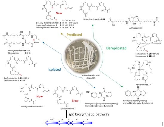
The K. speibonae strain SK5 was isolated from a topsoil sample collected from Stellenbosch in the Western Cape Province of South Africa [17]. The strain was grown in an International Streptomyces Project medium 2 (ISP2) broth and an Amberlite XAD 16N resin was added after 14 days of incubation at 30 °C with constant shaking. Organic solvents, methanol (MeOH), ethyl acetate (EtOAc), and dichloromethane (CH2Cl2), were used sequentially to extract the organics from the combined resin and culture broth. Then, the MeOH, EtOAc, and CH2Cl2 extracts were subjected to a separate high-pressure liquid chromatography-diode array detection, high-resolution electrospray mass spectrometry (HPLC-DAD/HRESIMS) analyses for chemical profiling (Figure S1). Further MS/MS and Global Natural Product Social (GNPS) molecular network analyses of the extracts revealed the presence of several siderophores and DKPs, some of which have not been reported previously (Figure S2). The MeOH, EtOAc, and CH2Cl2 extracts were combined and fractionated using a series of purification steps, including a modified Kupchan method [18], solid phase extraction (SPE), and HPLC to yield two new siderophores, speibonoxamine 1 and desoxy-desferrioxamine D1 2, and four known hydroxamates, 36 and a DKP 7 (Figure 1).

2.1. Structure Elucidation

The structures of the known compounds, desferrioxamine D1 3, desferrioxamine B 4, desoxynocardamine 5, and nocardamine 6 were elucidated by comparison of the HRESIMS and NMR spectra with those reported in the literature (Figures S15–S27) [11,19,20,21,22]. Compounds 36 belong to the hydroxamate class of siderophores [3]. The HRESIMS and NMR spectra (Figures S27–S31) of compound 7 matched the reported diketopiperazine (DKP), hexahydro-3-((4-hydroxyphenyl)methyl)-pyrrolo[1,2-a]pyrazine-1,4-dione [20].
The molecular formula of compound 1, isolated as a colourless amorphous solid, was deduced as C27H50N6O6 by HRESIMS (observed [M + H]+ = 555.3857; calculated [M + H]+ = 555.3870; ∆ = 1.0 ppm), indicating six degrees of unsaturation (Figure S3).
The 1H-NMR of 1 in DMSO-d6 showed only six signals (Figure S4), including four methylenes (δH 3.00, 2.27, 1.36, 1.22), one methyl (δH 1.77), and one NH (δH 7.77) with integrals of 12, 8, 12, 6, 6, and 6, respectively. The number of carbon atoms observed in the HRESIMS was five times higher than the number of signals observed in the 13C-NMR spectrum (δC 171.7, 169.4, 38.9, 31.4, 29.3, 24.3). These results suggested that compound 1 had a symmetrical structure and/or repeating motifs. Analysis of the 1H-1H COSY spectrum together with integrals of the signals in the 1H-NMR spectrum revealed two main spin systems, one of which consisted of three repeating motifs H-3 through H-7, H3′ through H-7′, and H-3” through H-7”, and the other comprised two repeating motifs H-9 through H-10 and H-9′ through H-10′. Careful examination of the 1D NMR data and 2D NMR data suggested that the symmetrical structure of 1 consists of two succinyls, two acyl cadaverine (AC) units, and one cadaverine moiety, characteristic of a hydroxamate siderophore. The heteronuclear multiple bond correlations (HMBC) from H-7 (δH 3.00) to C-8 (δC 171.7) and H-3” (δH 3.00) to C-11′ (δC 171.7) established the connectivity of the AC subunits to the succinyl groups. Furthermore, the cross peaks from H-3′ (δH 3.00) to C-11 (δC 171.7) and H-7′ (δH 3.00) to C-8′ (δC 171.7) established the attachment of the cadaverine moiety to the rest of the molecule.
The final structural analysis of 1 was confirmed by comparison with the spectroscopic data reported for desferrioxamine D1 (3) in the literature [21], which differs from 1 in the presence of N-hydroxy groups in the structure. Therefore, compound 1 represents a new non-hydroxylated desferrioxamine, which was named speibonoxamine after the producing organism, Kribbella speibonae strain SK5. This is the first report of a non-hydroxylated desferrioxamine. Compound 1 may not be efficient in sequestering iron from the environment because of the lack of the hydroxamate moiety.
The molecular formula of compound 2, also isolated as a colourless amorphous solid, was established as C27H50N6O8 by HRESIMS (observed [M + H]+ = 587.3754; calculated [M + H]+ = 587.3768; ∆ = −2.1 ppm), indicating six degrees of unsaturation (Figure S9).
The mass of compound 2 was 32 mass units greater than 1 indicative of the presence of two additional oxygen atoms in the structure. Analysis of the 1H-NMR data of 2 (Figure S10) revealed chemical shifts similar to those of 1, except for signals at δH 1.50 (H-6 and H-6′) and 3.45 (H-7 and H-7′) as the most distinguishable change. The observed proton (δH 3.45) and carbon (δC 47.6) chemical shifts in 2 were more downfield than in 1H 3.00, δC 38.9), indicating that the methylenes C-7 and C-7′ were deshielded by the adjacent N-hydroxy groups in 2. Likewise, the methylene signals at (δH 1.50, δC 26.5) were downfield in 2 compared to that in 1H 1.36, δC 29.3) because of the attachment of C-6 and C-6′ to the deshielded C-7 and C-7′ in 2, respectively. Compound 2 was linked to desoxynocardamine (5) and desferrioxamine D1 (3) in the GNPS molecular network with a mass difference of 2 and 16 Da, respectively (Figure S32) indicating that 2 is an acyclic analogue of 5 or dehydroxylated analogue of 3. The structure of 2 was determined by analyses of the MS/MS fragmentation data together with a comprehensive interpretation of the 1D and 2D NMR data of 2 and comparison with literature data. Compound 2 is a new dehydroxy analogue of compound 3 and hence has been assigned the name desoxy-desferrioxamine D1 [21].

2.2. Molecular Network Analysis

Dereplication of the metabolites produced by the K. speibonae strain SK5 was pivotal in the detection and isolation of the new compounds. Spectrometric data of the metabolites in the crude extract were searched against the natural product database, AntiBase (2017), with subsequent analysis of the data on the GNPS molecular networking platform [23], which grouped the compounds into clusters based on the similarity in MS/MS fragmentation patterns. Several known siderophores and DKPs were identified in the molecular network, including desferrioxamine H 11, ferrioxamine B 12, ferrioxamine E 13, ferrioxamine D1 14, arthrobactin 15 and the DKP, hexahydro-3-(phenylmethyl)-pyrrolo[1,2-a]pyrazine-1,4-dione 16 (Figure S32) [13,19]. Furthermore, three new dehydroxylated siderophores 810 were putatively identified in the molecular network, in addition to the new compounds, speibonoxamine 1 and desoxy-desferrioxamine D1 2, isolated and reported in this study.
Compound 8 was linked to compound 2 when default parameters were used to generate the GNPS molecular network (Figure S33). Compound 8 (m/z 571.3807 [M + H]+, C27H50N6O7) was identified as the dehydroxy analogue of 2 (C27H50N6O8) and the hydroxyl analogue of 1 (C27H50N6O6), as they have a mass difference of 16 Da and the same double bond equivalent (DBE) of six (Figure 2 and Table S1). Inspection of the fragment ions of compounds 1, 2, and 8 was very useful in assigning the position of the hydroxyl group, especially fragment ion 411.2596 (C20H35O5N4), which was present in 1 and 8, but absent in 2 (Figures S34–S36). Although the new compound 8 could not be isolated due to a paucity of material, its structure was proposed based on the MS/MS fragmentation data analysis and was assigned the name didesoxy-desferrioxamine D1 (Figure 2).
Compound 9 (m/z 545.3654 [M + H]+, C25H48N6O7) was linked to desferrioxamine B 4 in the GNPS molecular network and showed a mass difference of 16 Da (Figure 2 and Figure S32). Compound 10 (m/z 529.3702 [M + H]+, C25H48N6O6) was linked to 9 and had a mass difference of 16 Da. The MS/MS fragmentation patterns of 4, 9, and 10 were used to predict the structures of 9 and 10 (Figures S37–S39), and they were named as desoxy-desferrioxamine B and didesoxy-desferrioxamine B, respectively.
The results suggest that the K. speibonae strain SK5 is a prolific producer not only of hydroxamate siderophores, but also dehydroxylated and non-hydroxylated desferrioxamines. Although the presence of siderophores has been previously identified in the genomes of Kribbella species [17,24], to our knowledge this is the first report of the isolation of hydroxamates from this genus.

2.3. Proposed Biosynthetic Pathway

Biosynthesis of the desferrioxamine class of hydroxamate siderophores has been elucidated in Streptomyces and is mediated by nonribosomal peptide synthetase-independent siderophore (NIS) synthetases [9,10,25,26]. Every NIS biosynthetic pathway identified to date contains at least one synthetase with a high sequence similarity to IucA/IucC and such synthetases have thus become the characteristic feature of these pathways [10]. In silico analysis of the annotated genome of the K. speibonae strain SK5 (GenBank accession number: SJJY00000000) identified the biosynthetic gene cluster (BGC) encoding the IucA/IucC synthetase that is likely responsible for producing 16 (Figure 3A, Table 1).
The desferrioxamine (des) BGC in Streptomyces coelicolor strain M145 was proposed to comprise four genes, desABCD [25]. The spbA and spbB genes in the K. speibonae strain SK5 encode enzymes having a high sequence similarity to pyridoxal 5′-phosphate (PLP)-dependent decarboxylase and lysine-6-monooxygenase in Streptomyces strains, respectively [27]. These two enzymes are proposed to catalyze the decarboxylation of lysine 17 to form cadaverine 18 followed by hydroxylation to generate N-hydroxy-cadaverine 19 (Figure 3B). The acyltransferase, SpbC, is likely to catalyze the N-acylation of 19 [9,25], which then undergoes ATP-dependent oligomerization and macrocylization catalyzed by SpbD and SpbE to produce the N-hydroxy-containing hydroxamate desferrioxamines 26 [28].
The isolation of speibonoxamine 1 from K. speibonae strain SK5 suggests that the biosynthetic enzyme, N-acyl-CoA transferase SpbC, may display substrate promiscuity, binding not only to N-hydroxy-cadaverine 19 but also cadaverine 18, the key difference between the biosynthesis of desferrioxamines and speibonoxamine 1. It is proposed that the common decarboxylation intermediate, cadaverine 18, undergoes spontaneous SpbC-catalyzed acylation, followed by SpbD- and SpbE-catalyzed oligomerization of two molecules of succinate, two molecules of N-acetyl-cadaverine, and one molecule of cadaverine to generate 1.

3. Materials and Methods

3.1. General Experimental Procedures

NMR spectra were obtained on a BRUKER Ascend 600 (Bruker, Billerica, MA, USA) Prodigy cryoprobe at 600 and 150 MHz for 1H and 13C nuclei, respectively. DMSO-d6H 2.50, δC 39.7), CD3OD (δH 3.30, δC 49.0), and CDCl3H 7.25, δC 77.00) were used for preparing samples for NMR experiments. High resolution mass spectrometric data were obtained using a Thermo Instruments MS system (LTQ XL/LTQ Orbitrap Discovery, Thermo Scientific, Bremen, Germany) coupled to a Thermo Instruments HPLC system (Accela PDA detector, Accela PDA autosampler, and Accela pump). The MS was run in a positive high-resolution mode (60,000) and MS/MS at a resolution of 7500 and a low-resolution negative mode. The following conditions were used: Capillary voltage 45 V, capillary temperature 260 °C, auxiliary gas flow rate 10–20 arbitrary units, sheath gas flow rate 40–50 arbitrary units, spray voltage 4.5 kV, mass range 100–2000 amu (maximum resolution 30,000). HPLC separations were carried out using a reverse phase C18 ACE 10 µM 10 × 250 mm column connected to an Agilent Technologies 1200 series HPLC system equipped with an Agilent Technologies 1200 series quad pump and Agilent Technologies 1200 series DAD. The amberlite XAD 16N resin was obtained from Sigma-Aldrich, Johannesburg, South Africa. All solvents used throughout were of a HPLC-grade and purchased from both Merck and Sigma-Aldrich (Johannesburg, South Africa).

3.2. Isolation and Characterization of the Strain

The K. speibonae strain SK5 was isolated from a topsoil sample collected from the town of Stellenbosch in the Western Cape Province of South Africa using a newly developed Kribbella-selective medium [17].

3.3. Fermentation

A liquid stock culture of the K. speibonae strain SK5 was inoculated into a 15 mL International Streptomyces Project medium 2 (ISP2) broth (yeast extract 4 g, malt extract 10 g, glucose 4 g, in 1 L H2O, pH 7.3 [29]) in a 250 mL Erlenmeyer flask and incubated for five days at 30 °C with shaking. Then, the entire culture was inoculated into a 50 mL ISP2 broth in a 500 mL Erlenmeyer flask and incubated at 30 °C with shaking for four days. This culture was subsequently split into three parts, which were used as the inocula for 3 × 100 mL ISP2 broths in 1000 mL Erlenmeyer flasks and incubated at 30 °C with shaking for four days. Then, each 100 mL culture was inoculated into a 1000 mL ISP2 broth in a 5000 mL Erlenmeyer flask and incubated at 30 °C with shaking. After 14 days of incubation, the Amberlite XAD 16N resin (50 g/L) was added under sterile conditions to each flask and further incubated for 6 h at 30 °C with shaking. Subsequently, the cultures were harvested and filtered under pressure using a piece of glass wool placed in a Buchner funnel. The filtrate was partitioned in a separating funnel with an equal volume of ethyl acetate. Then, the cell mass mixed with the Amberlite XAD 16N resin (containing the adsorbed organics) was extracted sequentially with MeOH (3×), then EtOAc (3×), and finally, CH2Cl2 (3×). All the organic extracts were concentrated under a reduced pressure to give 3.43 g of the MeOH extract, 0.51 g of the EtOAc extract, and 0.19 g of the CH2Cl2 extract. The extracts were subjected to HPLC-DAD/HRESIMS analyses.

3.4. HPLC-DAD/HRESIMS Analyses

Chemical profiling of the MeOH, EtOAc, and CH2Cl2 extracts was performed by HPLC-DAD/HRESIMS analyses. Each extract (10 µL, 0.1 mg/mL in MeOH) was injected and chromatographically separated on a C18 reverse-phase HPLC column (ACE 10 µM 10 × 250 mm) using a linear gradient from 95% solvent A (0.1% formic acid in water) to 100% solvent B (0.1% formic acid in acetonitrile) for 25 min, followed by 100% B for 5 min at a flow rate of 1.5 mL/min. The DAD of the HPLC was scanned from 200–400 nm. The MS was run in a positive high-resolution mode (60,000) and MS/MS at a resolution of 7500 and a low-resolution negative mode. The Xcalibur software was used to process the raw data. The exact mass and molecular formula of each peak was entered as a single query in the commercially available AntiBase (2017) Natural Compound Identifier (https://www.wiley.com/en-us/AntiBase%3A+The+Natural+Compound+Identifier-p-9783527343591) to ascertain whether the data matched any compound in the database. The HPLC-DAD/HRESIMS profiles of the three extracts showed similar chemical profiles, hence they were combined to obtain 4.13 g.

3.5. Fractionation, Isolation, and Purification of Compounds

The combined crude extract (4.13 g) was suspended in 50 mL of distilled water and extracted with equal volumes of CH2Cl2 (three times). Then, the water layer was partitioned with the same volume of n-butanol (three times). The n-butanol layer (332.2 mg) was concentrated under a reduced pressure and fractionated on a C18 solid phase extraction (SPE) column using a stepwise elution of solvent mixtures of decreasing polarity (8 column volume/solvent mixture): 100% water (SPE1), 12.5% MeOH (SPE2), 25% MeOH (SPE3), 50% MeOH (SPE4), 100% MeOH (SPE5), and 100% MeOH containing 0.05% trifluoroacetic acid (SPE6). All fractions were subjected to the HPLC/HRESIMS analysis.
Fractions SPE2-4 were further purified by the reverse-phase HPLC analysis (C18, linear gradient 100% H2O to 100% MeOH in 45 min, flow rate 1.5 mL/min). SPE2 yielded compounds 6 (19.3 mg) and 7 (0.8 mg), SPE3 yielded compounds 1 (1.2 mg), 2 (0.6 mg), 3 (1.2 mg), and 5 (0.8 mg), and SPE4 yielded 4 (0.5 mg).
Speibonoxamine 1: Colorless amorphous solid; for 1H, 13C NMR data, see Table 2; HRESIMS (positive mode) m/z 555.3857 [M + H]+ and 577.3672 [M + Na]+ Δ 1.004; calcd. for C27H50N6O6.
Desoxy-desferrioxamine D1 2: Colorless amorphous solid; 1H, 13C NMR data, see Table 2; HRESIMS (positive mode) m/z = 587.3754 [M + H]+ and 609.3573 [M + Na]+ Δ −2.092 ppm; calcd. for C27H50N6O8.
Desferrioxamine D1 3: Colorless amorphous solid; 1H NMR data, see Figure S16; HRESIMS (positive mode) m/z = 603.3708 [M + H]+ and 625.3525 [M + Na]+ Δ −0.685 ppm; calcd. for C27H50N6O9.
Desferrioxamine B 4: Colorless amorphous solid; 1H NMR data, see Figure S18; HRESIMS (positive mode) m/z = 561.3602 [M + H]+ and 583.3415 [M + Na]+ Δ −1.049 ppm; calcd. for C25H48N6O8.
Desoxynocardamine 5: Colorless amorphous solid; 1H NMR data, see Figure S20; HRESIMS (positive mode) m/z = 585.3602 [M + H]+ and 607.3418 [M + Na]+ Δ −0.391 ppm; calcd. for C27H48N6O8.
Nocardamine 6: Colorless amorphous solid; 1H, 13C NMR data, see Figures S22–S23; HRESIMS (positive mode) m/z = 601.3552 [M + H]+ and 623.3359 [M + Na]+ Δ −0.161 ppm; calcd. for C27H48N6O9.
Hexahydro-3-((4-hydroxyphenyl)methyl)-pyrrolo[1,2-a]pyrazine-1,4-dione 7: Colorless amorphous solid; 1H NMR data, see Figures S28–S31; HRESIMS (positive mode) m/z = 261.1237 [M + H]+ and 283.1655 [M + Na]+ Δ 1.459 ppm; calcd. for C14H16N2O3.

3.6. Molecular Networking

Raw data obtained from the LC-MS/MS system were converted to a mzXML format using the ProteoWizard tool MSconvert (version 3.0.10051, Vanderbilt University, Nashville, TN, USA) [30]. All mzXML data were uploaded to the Global Natural Products Social (GNPS) molecular networking (MN) webserver3 (http://gnps.ucsd.edu) and analyzed using the MN workflow [23]. The data were filtered by removing all MS/MS fragment ions within +/− 17 Da of the precursor m/z. MS/MS spectra were window filtered by choosing only the top six fragment ions in the +/− 50 Da window throughout the spectrum. The precursor ion mass tolerance was set to 0.02 Da and a MS/MS fragment ion tolerance of 0.02 Da. Then, a network was created where edges were filtered to have a cosine score above 0.7 and more than three matched peaks. Further, edges between two nodes were kept in the network if and only if each of the nodes appeared in each other’s respective top 10 most similar nodes. Finally, the maximum size of a molecular family was set to 100, and the lowest scoring edges were removed from molecular families until the molecular family size was below this threshold. The spectra in the network were then searched against GNPS’ spectral libraries. The library spectra were filtered in the same manner as the input data. All matches kept between the network spectra and library spectra were required to have a score above 0.7 and at least three matched peaks. The output of the molecular network was visualized using the Cytoscape version 3.7.2 (https://cytoscape.org/) [31] and displayed using the settings “preferred layout” with “directed” style. The nodes (compounds) originating from the culture medium and solvent control (MeOH) were excluded from the original network to enable visualization of only the K. speibonae strain SK5 metabolites derived from the MeOH, EtOAC, and CH2Cl2 extracts of the cultures.

4. Conclusions

The K. speibonae strain SK5 is a prolific producer of hydroxamate (desferrioxamine) siderophores and their dehydroxy analogues. Two new desferrioxamines, speibonoxamine and desoxy-desferrioxamine D1, four known hydroxamates, and a DKP were isolated from K. speibonae strain SK5. Furthermore, several new and known siderophores were identified in the K. speibonae strain SK5 extracts, and their plausible structures were determined by the MS/MS fragmentation analysis. This is the first report of siderophores isolated from the genus Kribbella. The proposed speibonoxamine pathway (Figure 3B) suggests a biosynthetic machinery distinct from that reported for desferrioxamine biosynthesis.

Supplementary Materials

The following are available online, Figure S1: MS window of HPLC-DAD/HRESIMS profiles of MeOH (top), EtOAC (middle), and CH2Cl2 (bottom) extracts of the fermentation broth of the Kribbella speibonae strain SK5 cultured in an ISP2 liquid medium; Figure S2: GNPS molecular network of the methanol (red nodes), ethyl acetate (blue nodes), and dichloromethane (green nodes) extracts of the fermentation broth of the K. speibonae strain SK5. The molecular network contains 13 families with the highlighted families (desferrioxamines, ferrioxamines, and diketopiperazines) containing some annotated nodes; Figure S3: HR-ESI-MS spectrum of speibonoxamine (1); Figure S4: 1H-NMR spectrum of speibonoxamine (1) in DMSO-d6 at 600 MHz; Figure S5: 13C-NMR spectrum of speibonoxamine (1) in DMSO-d6 at 600 MHz; Figure S6: Multiplicity edited HSQC NMR spectrum of speibonoxamine (1) in DMSO-d6 at 600 MHz; Figure S7: 1H-1H COSY spectrum of speibonoxamine (1) in DMSO-d6 at 600 MHz; Figure S8: HMBC NMR spectrum of speibonoxamine (1) in DMSO-d6 at 600 MHz; Figure S9: HR-ESI-MS spectrum of desoxy-desferrioxmaine D1 (2); Figure S10: 1H-NMR spectrum of desoxy-desferrioxmaine D1 (2) in DMSO-d6 at 600 MHz; Figure S11: 13C-NMR spectrum of desoxy-desferrioxmaine D1 (2) in DMSO-d6 at 600 MHz; Figure S12: Multiplicity edited HSQC NMR spectrum of desoxy-desferrioxmaine D1 (2) in DMSO-d6 at 600 MHz; Figure S13: 1H-1H COSY spectrum of desoxy-desferrioxmaine D1 (2) in DMSO-d6 at 600 MHz; Figure S14: HMBC NMR spectrum of desoxy-desferrioxmaine D1 (2) in DMSO-d6 at 600 MHz; Figure S15: HR-ESI-MS spectrum of desferrioxamine D1 (3); Figure S16: 1H-NMR spectrum of desferrioxamine D1 (3) in DMSO-d6 at 600 MHz; Figure S17: HR-ESI-MS spectrum of desferrioxamine B (4); Figure S18: 1H-NMR spectrum of desferrioxamine B (4) in DMSO-d6 at 600 MHz; Figure S19: HR-ESI-MS spectrum of desoxynocardamine (5); Figure S20: 1H-NMR spectrum of desoxynocardamine (5) in DMSO-d6 at 600 MHz; Figure S21: HR-ESI-MS spectrum of nocardamine (6); Figure S22: 1H-NMR spectrum of nocardamine (6) in DMSO-d6 at 600 MHz; Figure S23: 13C-NMR spectrum of nocardamine (6) in DMSO-d6 at 600 MHz; Figure S24: Multiplicity edited HSQC NMR spectrum of nocardamine (6) in DMSO-d6 at 600 MHz; Figure S25: 1H-1H COSY spectrum of nocardamine (6) in DMSO-d6 at 600 MHz; Figure S26: HMBC NMR spectrum of nocardamine (6) in DMSO-d6 at 600 MHz; Figure S27: HR-ESI-MS spectrum of hexahydro-3-[(4-hydroxyphenyl)methyl]-Pyrrolo[1,2-a]pyrazine-1,4-dione (7); Figure S28: 1H-NMR spectrum of hexahydro-3-[(4-hydroxyphenyl)methyl]-Pyrrolo[1,2-a]pyrazine-1,4-dione (7) in MeOD at 600 MHz; Figure S29: Multiplicity edited HSQC NMR spectrum of hexahydro-3-[(4-hydroxyphenyl)methyl]-Pyrrolo[1,2-a]pyrazine-1,4-dione (7) in MeOD at 600 MHz; Figure S30: 1H-1H COSY spectrum of hexahydro-3-[(4-hydroxyphenyl)methyl]-Pyrrolo[1,2-a]pyrazine-1,4-dione (7) in MeOD at 600 MHz; Figure S31: HMBC NMR spectrum of hexahydro-3-[(4-hydroxyphenyl)methyl]-Pyrrolo[1,2-a]pyrazine-1,4-dione (7) in MeOD at 600 MHz; Figure S32: Molecular network of the molecular families F1, F2, F6–F8, F11, and the single nodes 477.2555 [M + H]+, 555.3857 [M + H]+, and 571.3807 [M + H]+. Nodes of isolated compounds (16) are shown with solid arrowed lines while dereplicated nodes (816) are indicated by dotted arrowed lines. Nodes were represented as pie charts indicating their intensities or percentages in the methanol (red nodes), ethyl acetate (blue nodes), and dichloromethane (green nodes) extracts of the fermentation broth of K. speibonae strain SK5. Nodes with asterisk (*) indicate dehydroxylated desferrioxamines; Figure S33: A MN showing a link between nodes didesoxy-desferrioxamine D1 8 (571.3807 [M + H]+) and desoxy-desferrioxamine D1 2 (587.3754 [M + H]+) when default parameters were used in generating the MN on the GNPS platform; Figure S34: MS/MS spectrum of desoxy-desferrioxamine D1 2 (587.3754 [M + H]+) with annotation in the structure; Figure S35: MS/MS spectrum of didesoxy-desferrioxamine D1 8 (571.3807 [M + H]+) with annotation in the structure; Figure S36: MS/MS spectrum of speibonoxamine 1 (555.3857 [M + H]+) with annotation in the structure; Figure S37: MS/MS spectrum of desferrioxamine B 4 (561.3606 [M + H]+) with annotation in the structure; Figure S38: MS/MS spectrum of desoxy-desferrioxamine B 9 (545.3654 [M + H]+) with annotation in the structure; Figure S39: MS/MS spectrum of didesoxy-desferrioxamine B 10 (529.3702 [M + H]+) with annotation in the structure; Table S1: Compounds isolated and tentatively identified in the molecular cluster and the MS chromatogram of the fermentation broth of K. speibonae strain SK5 with their corresponding masses, molecular formulae (MF), double bond equivalent (DBE), retention time (Rt), and mass error (ID (Δ ppm)).

Author Contributions

Conceptualization, S.N.S., M.J., P.R.M., D.W.G., and D.R.B.; methodology, S.N.S., M.J., P.R.M., D.R.B., D.W.G., D.F.W., and K.S.A.; investigation, K.S.A.; formal analysis, K.S.A., H.D., and F.M.; resources, S.N.S. and P.R.M.; data curation, K.S.A. and F.M.; writing—original draft preparation, K.S.A.; writing—review and editing, S.N.S., M.J., P.R.M., D.R.B., D.W.G., D.F.W., and H.D.; supervision, S.N.S., D.W.G., and D.R.B.; project administration, S.N.S and D.W.G.; funding acquisition, S.N.S. and D.F.W. All authors have read and agreed to the published version of the manuscript.

Funding

The authors acknowledge funding for this research from the South African Medical Research Council (SAMRC) through the Strategic Health Innovation Partnerships (SHIP) initiative (to D.F.W.), Newton Advanced Fellowship Award (NA160057) (to S.N.S. and M.J.), and the University of Cape Town (to K.S.A.).

Conflicts of Interest

The authors declare no conflict of interest. The funders had no role in the design of the study, in the collection, analyses, interpretation of data, in the writing of the manuscript, or in the decision to publish the results.

References

  1. Butler, A.; Theisen, R.M. Iron(III)–siderophore coordination chemistry: Reactivity of marine siderophores. Coord. Chem Rev. 2011, 254, 288–296. [Google Scholar] [CrossRef] [Green Version]
  2. Weinberg, E.D. Suppression of bacterial biofilm formation by iron limitation. Med. Hypotheses 2004, 63, 863–865. [Google Scholar] [CrossRef] [PubMed]
  3. Hider, R.C.; Kong, X. Chemistry and biology of siderophores. Nat. Prod. Rep. 2010, 27, 637. [Google Scholar] [CrossRef] [PubMed]
  4. Ahmed, E.; Holmström, S.J.M. Siderophores in environmental research: Roles and applications. Microb. Biotechnol. 2014, 7, 196–208. [Google Scholar] [CrossRef]
  5. Rajkumar, M.; Ae, N.; Narasimha, M.; Prasad, V.; Freitas, H. Potential of siderophore-producing bacteria for improving heavy metal phytoextraction. Trends Biotechnol. 2010, 28, 142–149. [Google Scholar] [CrossRef]
  6. Wu, J.Y.; Srinivas, P.; Pogue, J.M. Cefiderocol: A Novel Agent for the Management of Multidrug-Resistant Gram-Negative Organisms. Infect Dis. Ther. 2020, 9, 17–40. [Google Scholar] [CrossRef] [Green Version]
  7. Miller, M.J.; Walz, A.J.; Zhu, H.; Wu, C.; Moraski, G.; Ute, M.; Tristani, E.M.; Crumbliss, A.L.; Ferdig, M.T.; Checkley, L.; et al. Design, Synthesis, and Study of a Mycobactin-Artemisinin Conjugate That Has Selective and Potent Activity against Tuberculosis and Malaria. J. Am. Chem. Soc. 2011, 133, 2076–2079. [Google Scholar] [CrossRef] [PubMed] [Green Version]
  8. Braun, V.; Pramanik, A.; Gwinner, T.; Köberle, M.; Bohn, E. Sideromycins: Tools and antibiotics. BioMetals 2009, 22, 3–13. [Google Scholar] [CrossRef] [Green Version]
  9. Maglangit, F.; Alrashdi, S.; Renault, J.; Trembleau, L.; Victoria, C.; Tong, M.H.; Wang, S.; Kyeremeh, K.; Deng, H. Characterization of the promiscuous N-acyl CoA transferase, LgoC, in legonoxamine biosynthesis. Org. Biomol. Chem. 2020, 18, 2219–2222. [Google Scholar] [CrossRef]
  10. Carroll, C.S.; Moore, M.M. Ironing out siderophore biosynthesis: A review of non-ribosomal peptide synthetase (NRPS)-independent siderophore synthetases. Crit. Rev. Biochem. Mol. Biol. 2018, 53, 356–381. [Google Scholar] [CrossRef]
  11. Maglangit, F.; Tong, M.H.; Jaspars, M.; Kyeremeh, K.; Deng, H. Legonoxamines A-B, two new hydroxamate siderophores from the soil bacterium, Streptomyces sp. MA37. Tetrahedron Lett. 2019, 60, 75–79. [Google Scholar] [CrossRef]
  12. Barry, S.M.; Challis, G.L. Recent advances in siderophore biosynthesis. Curr. Opin. Chem. Biol. 2009, 13, 205–215. [Google Scholar] [CrossRef] [PubMed]
  13. Pluháček, T.; Lemr, K.; Ghosh, D.; Milde, D.; Novák, J.; Havlíček, V. Characterization of microbial siderophores by mass spectrometry. Mass Spectrom. Rev. 2016, 35, 35–47. [Google Scholar] [CrossRef] [PubMed]
  14. Subramani, R.; Sipkema, D. Marine Rare Actinomycetes: A Promising Source of Structurally Diverse and Unique Novel Natural Products. Mar. Drugs 2019, 17, 249. [Google Scholar] [CrossRef] [PubMed] [Green Version]
  15. Igarashi, M.; Sawa, R.; Yamasaki, M.; Hayashi, C.; Umekita, M.; Hatano, M.; Fujiwara, T.; Mizumoto, K.; Nomoto, A. Kribellosides, novel RNA 5′-triphosphatase inhibitors from the rare actinomycete Kribbella sp. MI481-42F6. J. Antibiot. (Tokyo). 2017, 70, 582–589. [Google Scholar] [CrossRef] [PubMed] [Green Version]
  16. Parte, A.C. LPSN—List of prokaryotic names with standing in nomenclature (Bacterio.net), 20 years on. Int. J. Syst. Evol. Microbiol. 2018, 68, 1825–1829. [Google Scholar] [CrossRef] [PubMed]
  17. Curtis, S.M.; Norton, I.; Everest, G.J.; Pelser, J.G.; De Kock, M.C.; Meyers, P.R. Development of a Kribbella-specific isolation medium and description of Kribbella capetownensis sp. nov. and Kribbella speibonae sp. nov., isolated from soil. Antonie van Leeuwenhoek 2020, 113, 617–628. [Google Scholar] [CrossRef]
  18. Zhang, Q.W.; Lin, L.G.; Ye, W.C. Techniques for extraction and isolation of natural products: A comprehensive review. Chinese Med. (UK) 2018, 13, 1–26. [Google Scholar] [CrossRef] [Green Version]
  19. Feistner, G.J.; Stahl, D.C.; Ann, H. Proferrioxamine Siderophores of Erwinia Amylovora. A Capillary Liquid Chromatographic/Electrospray Tandem Mass Spectrometric Study. Org. Mass Spectrom. 1993, 28, 163–175. [Google Scholar] [CrossRef]
  20. Lee, H.S.; Hee, J.S.; Kyoung, H.J.; Tae, S.K.; Oh, K.B.; Shin, J. Cyclic peptides of the nocardamine class from a marine-derived bacterium of the genus Streptomyces. J. Nat. Prod. 2005, 68, 623–625. [Google Scholar] [CrossRef]
  21. Ueki, M.; Suzuki, R.; Takamatsu, S.; Takagi, H.; Uramoto, M.; Ikeda, H.; Osada, H. Nocardamin Production by Streptomyces avermitilis. Actinomycetologica 2009, 23, 34–39. [Google Scholar] [CrossRef] [Green Version]
  22. Fang, Q.; Maglangit, F.; Wu, L.; Ebel, R.; Kyeremeh, K.; Andersen, J.H.; Annang, F.; Pérez-Moreno, G.; Reyes, F.; Deng, H. Signalling and Bioactive Metabolites from Streptomyces sp. RK44. Molecules 2020, 25, 460. [Google Scholar] [CrossRef] [PubMed] [Green Version]
  23. Wang, M.; Carver, J.J.; Phelan, V.V.; Sanchez, L.M.; Garg, N.; Peng, Y.; Nguyen, D.D.; Watrous, J.; Kapono, C.A.; Luzzatto-Knaan, T.; et al. Sharing and community curation of mass spectrometry data with Global Natural Products Social Molecular Networking. Nat. Biotechnol. 2016, 34, 828–837. [Google Scholar] [CrossRef] [PubMed] [Green Version]
  24. Pukall, R.; Lapidus, A.; Del Rio, T.G.; Copeland, A.; Tice, H.; Cheng, J.F.; Lucas, S.; Chen, F.; Nolan, M.; Labutti, K.; et al. Complete genome sequence of Kribbella flavida type strain (IFO 14399 T). Stand. Genomic Sci. 2010, 2, 186–193. [Google Scholar] [PubMed] [Green Version]
  25. Ronan, J.L.; Kadi, N.; Mcmahon, S.A.; Naismith, J.H.; Alkhalaf, L.M.; Challis, G.L.; Challis, G.L. Desferrioxamine biosynthesis: Diverse hydroxamate assembly by substrate- tolerant acyl transferase DesC. R. Soc. Publ. 2018, 373. [Google Scholar] [CrossRef] [PubMed]
  26. Kadi, N.; Song, L.; Challis, G.L. Bisucaberin biosynthesis: An adenylating domain of the BibC multi-enzyme catalyzes cyclodimerization of N-hydroxy-N-succinylcadaverine. Chem. Commun. 2008, 5119–5121. [Google Scholar] [CrossRef]
  27. Barona-Gómez, F.; Wong, U.; Giannakopulos, A.E.; Derrick, P.J.; Challis, G.L. Identification of a cluster of genes that directs desferrioxamine biosynthesis in Streptomyces coelicolor M145. J. Am. Chem. Soc. 2004, 126, 16282–16283. [Google Scholar] [CrossRef]
  28. Rütschlin, S.; Böttcher, T. Dissecting the Mechanism of Oligomerization and Macrocyclization Reactions of NRPS-Independent Siderophore Synthetases. Chem. A Eur. J. 2018, 24, 16044–16051. [Google Scholar] [CrossRef]
  29. Shirling, E.B.; Gottlieb, D. Methods for Characterization of Streptomyces species. Int. J. Syst. Bacteriol. 1966, 16, 313–340. [Google Scholar] [CrossRef] [Green Version]
  30. Holman, J.D.; Tabb, D.L.; Mallick, P. Employing ProteoWizard to Convert Raw Mass Spectrometry Data. Curr Protoc Bioinforma. 2014, 46, 1–7. [Google Scholar] [CrossRef]
  31. Otasek, D.; Morris, J.H.; Bouças, J.; Pico, A.R.; Demchak, B. Cytoscape Automation: Empowering workflow-based network analysis. Genome Biol. 2019, 20, 1–15. [Google Scholar] [CrossRef] [PubMed] [Green Version]
Sample Availability: Samples of the compounds are not available from the authors.
Figure 1. Structures of isolated metabolites 17 from the Kribbella speibonae strain SK5, including depiction of Speibonoxamine 1 with COSY () and key heteronuclear multiple bond correlations (HMBC) (→) correlations.
Molecules 25 02979 g001
Figure 2. (A) Specific subnetwork analysis of the K. speibonae strain SK5; (B) putative dehydroxylated desferrioxamine analogues D1 8 and B 910; (C) structures of plausible desferrioxamines 810 determined by the MS/MS fragmentation pattern.
Molecules 25 02979 g002
Figure 3. (A) Speibonoxamine (spb) biosynthetic gene cluster in the K. speibonae strain SK5; (B) proposed biosynthetic pathway of 14.
Molecules 25 02979 g003
Table 1. Deduced functions of open reading frames (ORFs) in spb biosynthetic gene cluster (BGC).
ProteinAnnotated FunctionStreptomyces Homologue% Identity/% SimilarityAmino Acid Length
SpbDIucA/IucC synthetase74%/83%565
SpbBLysine-6-monooxygenase80%/88%418
SpbCAcyltransferase67%/76%156
SpbESiderophore synthetase78%/84%546
SpbAPLP-dependent decarboxylase86%/93%495
Table 2. 1H and 13C-NMR data of speibonoxamine 1 and desoxy-desferrioxamine D1 2 in DMSO-d6.
Speibonoxamine 1Desoxy-Desferrioxamine D1 2
Position13C1H, Mult. (J, Hz)13C1H, Mult. (J, Hz)
1, 1′23.1, CH31.77, s23.1, CH31.78, s
2, 2′169.4, C-169.4, C-
3, 3′, 3”38.9, CH23.00, t (6.15)38.9, CH23.00, m
4, 4′, 4”29.3, CH21.36, m29.3, CH21.38, dd (7.14, 14.24)
5, 5′, 5”24.3, CH21.22, m24.0, CH21.23, m
6, 6′29.3, CH21.36, m26.5, CH21.50, m
6”29.3, CH21.36, m29.3, CH21.38, dd (7.14, 14.24)
7, 7′38.9, CH23.00, t (6.15)47.6, CH23.45, t (6.96, 6.96)
7”38.9, CH23.00, t (6.15)38.9, CH23.00, m
8171.7, C-172.4, C-
8′171.7, C-171.7, C-
931.4, CH22.27, s28.1, CH22.58, m
9′31.4, CH22.27, s29.8, CH22.40, t (6.89, 6.89)
1031.4, CH22.27, s31.4, CH22.27, m
10′31.4, CH22.27, s30.4, CH22.28, m
11171.7, C-174.4, C-
11′171.7, C-171.2, C-
NH 7.77 7.77