1. Introduction
Carotenoids are one of the essential classes of metabolites in a wide variety of living organisms. More than 750 different carotenoids have been reported to date [
1]. It is known that carotenoids bind to proteins, termed carotenoproteins, to stabilize themselves against light or heat or to manipulate their physical properties, including solubility and color. Crustacyanin, one of the most representative carotenoproteins, binds to a red keto carotenoid, astaxanthin, but exhibits conspicuous blue color because of the bathochromic shift in the visible light region, due to the interaction between the carotenoids and the apoprotein. The structural evidence for specific binding between astaxanthin and the protein was observed by X-ray crystallography [
2]. Although, the blue color of crustacyanin is stable in an ambient milieu, this conspicuous color turns reddish-orange by heating or immersion in organic solvents. The molecular basis of the blue shift is an intriguing subject for biophysical research [
2,
3,
4].
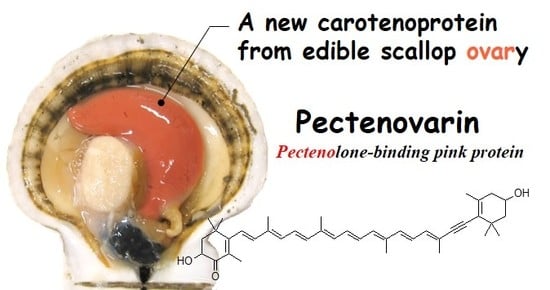
The Japanese scallop
Mizuhopecten yessoensis accumulates carotenoids in the ovary during the gonadal maturation process. These carotenoids are mainly pectenolone, but pectenoxanthin (alloxanthin), diatoxanthin, 3,4,3′-trihydroxy-7′-8′-didehydro-β-carotene, and astaxanthin are known to be minor constituents [
5]. These molecules are accumulated mostly in the ovary, but not in other parts of the body, including the viscera, testes in males, and adductor muscle, and are thought to bear functions such as anti-oxidation and light protection [
6]. These carotenoids originate from food microalgae and are likely subject to structural transformation, then accumulated in the form of carotenoproteins in the ovary. However, their actual functions in the reproduction of shellfish are not precisely known. Although there have been several reports dealing with the characterization and utilization of carotenoids in scallops [
5,
6,
7,
8], isolation and characterization of the carotenoprotein in the ovary have never been reported to the best of our knowledge. We, therefore, conducted isolation and characterization of the carotenoprotein from the ovary of
M. yessoensis. Here, the extraction, purification, and N-terminal and internal amino acid sequences of the new carotenoprotein, named pectenovarin, are described.
3. Discussion
In the present study, a new carotenoprotein, pectenovarin, was isolated and characterized, for the first time, from the mature ovary of the Japanese scallop M. yessoensis as a pink-colored component. We also analyzed the carotenoid components of the protein. Pectenovarin is a sizeable unstable protein with a molecular size of more than 1000 kDa. SDS-PAGE analysis clearly indicated that pectenovarin is composed of subunits with different molecular weights; however, the unusual behavior of these proteins, especially the observation that the bands converge toward lower molecular bands, requires further investigation.
We found that pectenovarin contained three carotenoids—pectenolone, pectenoxanthin, and diatoxanthin—in about a 65:7:11 ratio under AS precipitation, similar to their ratio in the ovary. These carotenoids correspond to the dominant three of the five carotenoids of scallop ovary [
5], which suggests that these carotenoids mostly accumulate in the ovary as pectenovarin in a mature stage.
We also found that the association of carotenoid to the protein resulted in a bathochromic shift of more than 20 nm, leading the compound to be categorized as a “true carotenoprotein” [
13].
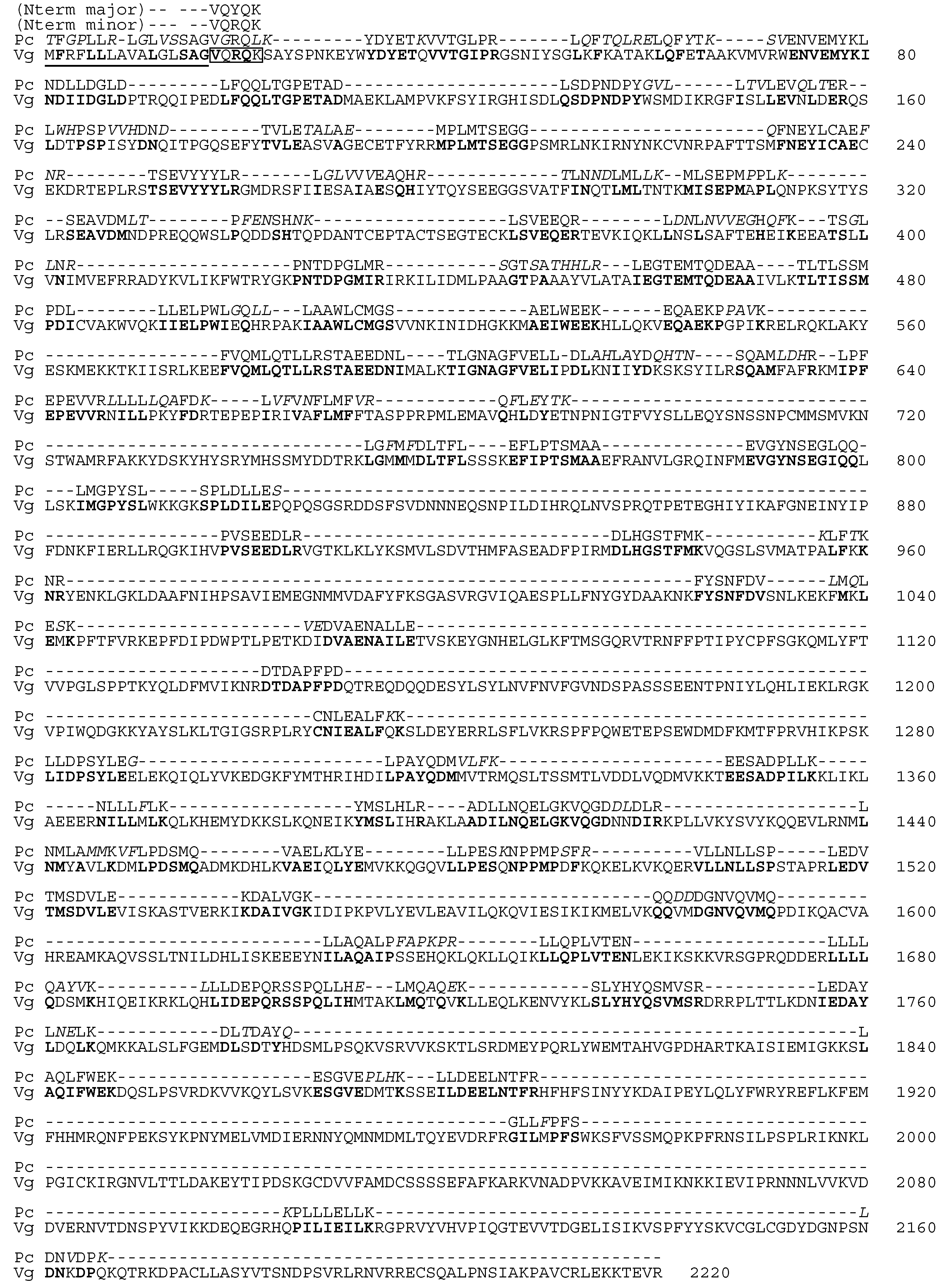
De novo peptide sequencing of the whole pectenovarin revealed that the internal sequences correspond well to those of vitellogenin from
M. yessoensis ovary [
10,
11], with about 80% similarity. Apo-pectenovarin is likely to occur from vitellogenin (Vg) or Vg-like protein. In the noble scallop
Chlamys nobilis, vitellogenin gene expression was shown to be correlated to the total carotenoid contents [
14], which is likely to be directly underpinned by our results.
A short N-terminal amino acid sequence of a pectenovarin subunit was found just adjacent to the putative signal peptide of Vg [
10,
11]. Notably, the N-terminal amino acid sequence VQRQK, however, was found as a minor component corresponding to VQRQK of residues 17–21 in the Vg sequence, while the major N-terminal sequence, VQYQK, had a mutation at residue 3. In the de novo LC-MS/MS sequencing, no peptide fragment corresponding to the N-terminal residues was found in the present study. Although this may be due to experimental limitations, it is worthy to note that some vitellogenins of avian, amphibian, and fish origin are reportedly products of plural gene coding [
15,
16,
17], and pectenovarin could be produced by the processing of plural vitellogenins. Although egg carotenoproteins are widely distributed among gastropods, bivalves, and crustaceans, investigations at the molecular level are limited. The fact that pectenovarin was found to have about 60% similarity with ovorubin, a pink egg carotenoprotein only characterized from the apple snail
P. canaliculata, suggests that egg carotenoproteins originated generally from vitellogenins and have evolved to have certain functions to adapt to different environments [
18,
19]. Interestingly, the Japanese scallop and apple snail differ largely in their reproduction ecology, as the former is a marine broadcast spawner, but the latter is a terrestrial snail depositing its eggmass outside the water. This explains reports that the apple snail carotenoprotein is toxic, as the eggs need to be protected from potential predators [
20]. Pectenovarin, a component of edible scallop ovary, may have functions different from those of ovorubin to protect pelagic eggs during the early stage of development. The function might be related to anti-oxidation and photo protection considering the functions of the carotenoids contained in the protein. Notably, pectenolone, a dominant carotenoid of pectenovarin, was reported to inhibit lipid peroxidation, and its ability was almost as potent as astaxanthin, known to possess high-antioxidant activity [
6]. It is likely that the protein functions as a delivery and storage system of unstable functional small molecules in the ovary.
Structural studies of carotenoproteins at the molecular level were found only for crustacyanin of crustaceans, orange carotenoprotein of cyanobacteria, and, to some extent, ovorubin. The lack of gene and amino acid sequences and structural investigations of carotenoprotein hampers comprehensible discussion as to the physiological role, mode of association with carotenoids, and evolutional aspects of its presence. The present results showing the extremely complex nature of egg carotenoprotein suggest future directions using genetic experiments to deduce the whole amino acid sequences of the subunits and cryo-electron microscopy to reveal its structure. These studies are now in progress and will be reported elsewhere.
4. Materials and Methods
4.1. Materials
Live scallops (Mizuhopecten yessoensis) were purchased at a local grocery store and stored at −30 °C until use. Chemical reagents were purchased from Wako pure chemicals unless otherwise noted.
4.2. Extraction of the Colored Aqueous Components from the Scallop Ovary
Approximately 50 g of the scallop ovary was homogenized with 150 mL of 50 mM Tris-HCl pH 8.0, and then the mixture was centrifuged at 9000 rpm at 4 °C for 30 min. The collected pink-colored supernatant was tested for coloristic stability against the following acidic buffers containing 20 mM NaCl: a buffer of pH 3.5 made of 50 mM citric acid–NaOH, and another buffer of pH 4.0 made of 50 mM AcOH–AcONa. Volumes of 0.5 mL of the specimen and each buffer were mixed, then centrifuged at 12,000 rpm at 4 °C for 15 min.
Another approximately 60 g of ovary specimen was homogenized with 50 mM Tris-HCl (pH 7.2) containing 20 mM NaCl as a neutral condition. The homogenate was centrifuged at 9000 rpm at 4 °C for 30 min, and then the pink-colored precipitate was suspended using the same buffer but at pH 8.0. The above procedure was repeated with buffers containing 150 mM NaCl.
For the optimum extraction buffer, KBr was added to the buffer (50 mM Tris-HCl, pH 8.0) up to 1.12 g/mL so that pink-colored solution was obtained efficiently from the tissue. Twenty grams of scallop ovary were homogenized with the above buffer, and the mixture was centrifuged at 9000 rpm at 4 °C for 30 min; then, the deep-pink-colored supernatant was collected.
4.3. Isolation of Pectenovarin
4.3.1. Extraction of the Colored Aqueous Component
The colored aqueous component was extracted anew from the ovary according to the above procedure. Specifically, 105.8 g of the specimen was homogenized with 50 mM Tris-HCl pH 8.0/150 mM NaCl containing KBr (d = 1.21). The centrifuged supernatant (3 mL) was layered on 10 mL of 45% KBr (saturated) and centrifuged at 15,000 rpm at 4 °C for 3 h. Then, 300 μL of red supernatant was mixed with 600 μL of saturated KBr in water, which was incubated on ice overnight. Then, the sample was centrifuged (15,000 rpm, 16 °C, 3 h) and repeatedly cooled on ice to remove white floating material. Finally, the extract was concentrated using an Amicon Ultra centrifugal filter (100 kDa, Merck KGaA, Darmstadt, Germany, 5000 rpm at 4 °C for 30 min.). The sample solution was filtered using a 0.45 μm syringe filter.
4.3.2. Gel Fractionation Chromatography
The above extract was separated via high-pressure liquid chromatography (HPLC) using a BioSec-5 gel filtration column (300Å, 4.6 × 30 mm, Agilent technology Japan, Tokyo) in several loads and eluted by 50 mM Tris-HCl pH 8.0/150 mM NaCl at 0.35 mL/min. The eluents were monitored using a multiwavelength detector (MD-910, JASCO Corporation, Tokyo, Japan). Portions of the eluents were analyzed by 8% acrylamide gel SDS-PAGE [
21], and then the pale-pink-colored eluents were combined and concentrated by 100 kDa cut-off centrifugal ultrafiltration. The protein quantity of this concentrated specimen was confirmed using a spectrophotometer (NanoDrop2000c, Thermo Fisher Scientific, DE, USA) to 1.667 mg/mL at 20 times dilution.
4.4. Analysis of Carotenoids
4.4.1. Precipitation of Analytical Materials
Ammonium sulfate precipitations of the pink aqueous extracts were prepared to estimate the carotenoid composition of pectenovarin. First, three ovary specimens (23.3, 16.1, and 17.5 g) were homogenized separately with 80 mL of 0.05 M Tris-HCl pH 8.0/150 mM NaCl containing KBr (d = 1.21). After centrifugation (9000 rpm, 4 °C, 30 min), ammonium sulfate was added up to 10% into the reddish supernatants and stirred on ice to dissolve completely. Then, the mixture was cooled on ice for 30 min and centrifuged for 15 min at 12,000 rpm at 4 °C. The concentration of AS was increased until colorless supernatants were observed. The addition of ammonium sulfate up to 25% finally brought the reddish-pink color into the pellet completely. The freeze-dried pellets (13.5, 14.3, and 11.8 mg) were then adjusted to 100 mg/mL with AcCN/MeOH/water at a ratio of 50:50:1. In order to compare carotenoid compositions between the pectenovarin and tissues of the scallop, freeze-dried ovary and midgut gland were also analyzed. First, the freeze-dried specimens of the ovary (131.8, 136.5, and 133.6 mg) and midgut gland (136.8, 133.5, and 133.7 mg) were moistened with water and then extracted three times with 1 mL of acetone. The extracts were partitioned between hexane and water, which separated them into an orange-colored upper layer and a cloudy aqueous layer. The upper layer was collected and evaporated while protecting it from light and then adjusted to 10 mg/mL using the above solvent system.
4.4.2. HPLC Analysis
The above sample (50 μL) was loaded into an HPLC column (Capcell Pack C18-UG80, 4.6 × 30 mm, OSAKA SODA CO., LTD., Osaka, Japan) and then eluted using AcCN/MeOH/water at a ratio of 50:50:1 at 0.35 mL/min. The eluents were monitored using a photodiode array detector (L-7455, Hitachi Ltd., Tokyo, Japan). In order to compare the carotenoid compositions between the AS precipitate, ovary, and midgut gland, the ratios of carotenoid peak area on these chromatograms at 463 nm, which is the maximum absorption of pectenolone, were calculated.
4.4.3. Identification of Carotenoids Using LC-MS/MS
LC-MS/MS was performed in positive mode on a Q-TOF ESI MS/MS (SCIEX TripleTOF, Sciex, MA, USA) equipped with HPLC (Shimadzu Co., Kyoto, Japan) using a BEH C8 column (2.1 × 150, S 2.5 um, Waters, MA, USA) with a gradient of solvent A: 0.1% formate/10 mM ammonium formate containing water, B: 0.1% formate/10 mM ammonium formate containing MeOH/2-propanol (85:15, v/v). The flow rate was set to 0.3 mL/min and the time program was B concentration 75%, 0–2 min; 75%–99%, 2–18 min; 99%, 18–24 min; 99–75%, 24–25 min; 30 min stop. The column oven temperature was 50 degrees, and 5 μL of sample was injected.
4.5. N-terminal Amino Acid Sequence Analysis
Polypeptides on the 6% acrylamide gel after running of SDS-PAGE were transferred to a PVDF membrane using an iBlot Gel Transfer System (Thermo Fisher Scientific Inc., MA, USA). The polypeptides on the PVDF membrane were dyed using acid red 112 (Tokyo Chemical Industry Co., Tokyo, Japan) and then cut out from the membrane and loaded onto a Procise 492HT protein sequencer (PerkinElmer, Inc., MA, USA).
4.6. Internal Amino Acid Sequence Analysis
A 3 μL pectenovarin specimen, with protein concentration 14 μg/μL, was loaded onto 6% polyacrylamide gel for SDS-PAGE, and the separated five bands were respectively cut out and kept in 1.5 mL microtubes. These specimens were reduced using dithiothreitol and then alkylated using iodoacetamide and in-gel. The gel was immersed in 50 mM ammonium hydrogen carbonate; then, the protein was digested in-gel using TPCK trypsin with a molar ratio of 1:50 and incubated for 20 h at 37 °C. The enzyme digests were extracted from the gel using 5% formic acid/50% acetonitrile. The solvents were removed and then dissolved with 80 μL of water and analyzed by LC-MS using a C18 column (Vydac PROTEIN and PEPTIDE C18). The sequences of fragments were extracted using DeNovoGUI 1.16.4 and analyzed and compared using the FASTS program, which finds regions of similarity among proteins with unordered peptides against target sequences [
22].
5. Conclusions
In the present study, we report the isolation and properties of pink carotenoprotein from the ovary of edible Japanese Scallop, Mizuhopecten yessoensis. The carotenoprotein, named pectenovarin, is a large and complex protein composed of more than ten subunits. Extracting the protein from the ovary was a challenge due to the poor stability of the molecule. The extractability of the protein varied from individual to individual, even when using live specimens. The best separation was finally achieved by applying the extract directly to a gel filtration HPLC column. Our success in obtaining reasonably pure protein paved the way for studying the structure of this intriguing molecule. The partial sequence obtained here showed the close relation of the carotenoprotein to vitellogenin. It seems, however, that the protein was highly processed from the vitellogenin of the scallop. Pectenovarin complexes with pectenolone and a few other minor related carotenoids. The mode of complexation is interesting as all other marine carotenoproteins complex solely with astaxanthin. Together, the present results illustrate the interesting utilization of carotenoids in the scallop, in that food-derived carotenoids are converted to pectenolone by the animal and perhaps delivered to the ovary by the formation of a complex with apo-pectenovarin; the complexed protein, pectenovarin, is then stored in the ovary. The structure and behavior of pectenovarin were more complex than expected, but further studies on the unique utilization of carotenoids would provide interesting insights into the physiological and evolutionary aspects of scallops.


