Article

A Total of Eight Novel Steroidal Glycosides Based on Spirostan, Furostan, Pseudofurostan, and Cholestane from the Leaves of Cestrum newellii

School of Pharmacy, Tokyo University of Pharmacy and Life Sciences, 1432-1, Horinouchi, Hachioji, Tokyo 192-0392, Japan
*
Author to whom correspondence should be addressed.
Molecules 2020, 25(19), 4462; https://doi.org/10.3390/molecules25194462
Submission received: 28 August 2020 / Revised: 16 September 2020 / Accepted: 26 September 2020 / Published: 28 September 2020
(This article belongs to the Section Natural Products Chemistry)

Abstract

:
Previously, various steroidal glycosides were reported from plants of Cestrum species. However, phytochemical investigation has not been conducted on Cestrum newellii. A systematic phytochemical investigation of the leaves of C. newellii resulted in the isolation of eight novel steroidal glycosides (18), which were classified into three spirostanol glycosides (13), two furostanol glycosides (4 and 5), two pseudofurostanol glycosides (6 and 7), and one cholestane glycoside (8). In addition, three known cholestane glycosides (911) were isolated and identified. The structures of the new compounds were determined based on spectroscopic data and chemical transformations. Compounds 1 and 2 are spirostanol glycosides having hydroxy groups at C-2, C-3, C-12, and C-24 of the aglycone moiety. Although C. newellii is known to be a poisonous plant, the 3-(4,5-dimethylthiazol-2-yl)-2,5-diphenyl-2H-tetrazolium bromide assay exhibited that none of the isolated compounds were cytotoxic to HL-60 human promyelocytic leukemia cells.

1. Introduction

Plants in the genus Cestrum (Solanaceae) are native to warm subtropical and tropical areas of America, and are now cultivated all over the world for ornamental purposes [1]. Cestrum species are rich sources of steroidal glycosides, and structurally diverse steroidal glycosides have been isolated from C. laevigatum [2,3], C. schlechtendahlii [4], C. ruizteranianum [5], C. parqui [6,7,8], C. diurnum [9,10], C. sendtenerianum [11,12], and C. nocturnum [13,14,15]. C. newellii is reputed to be a poisonous plant. However, a literature survey suggests that no phytochemical investigation has been done on C. newellii. Therefore, a systematic phytochemical analysis of the leaves of C. newellii was conducted with a focus on steroidal glycosides. This paper deals with the structural determination of new compounds (18) on the basis of spectroscopic data and chemical transformations. Furthermore, the cytotoxic activities of the isolated compounds (111) were evaluated.

2. Results and Discussion

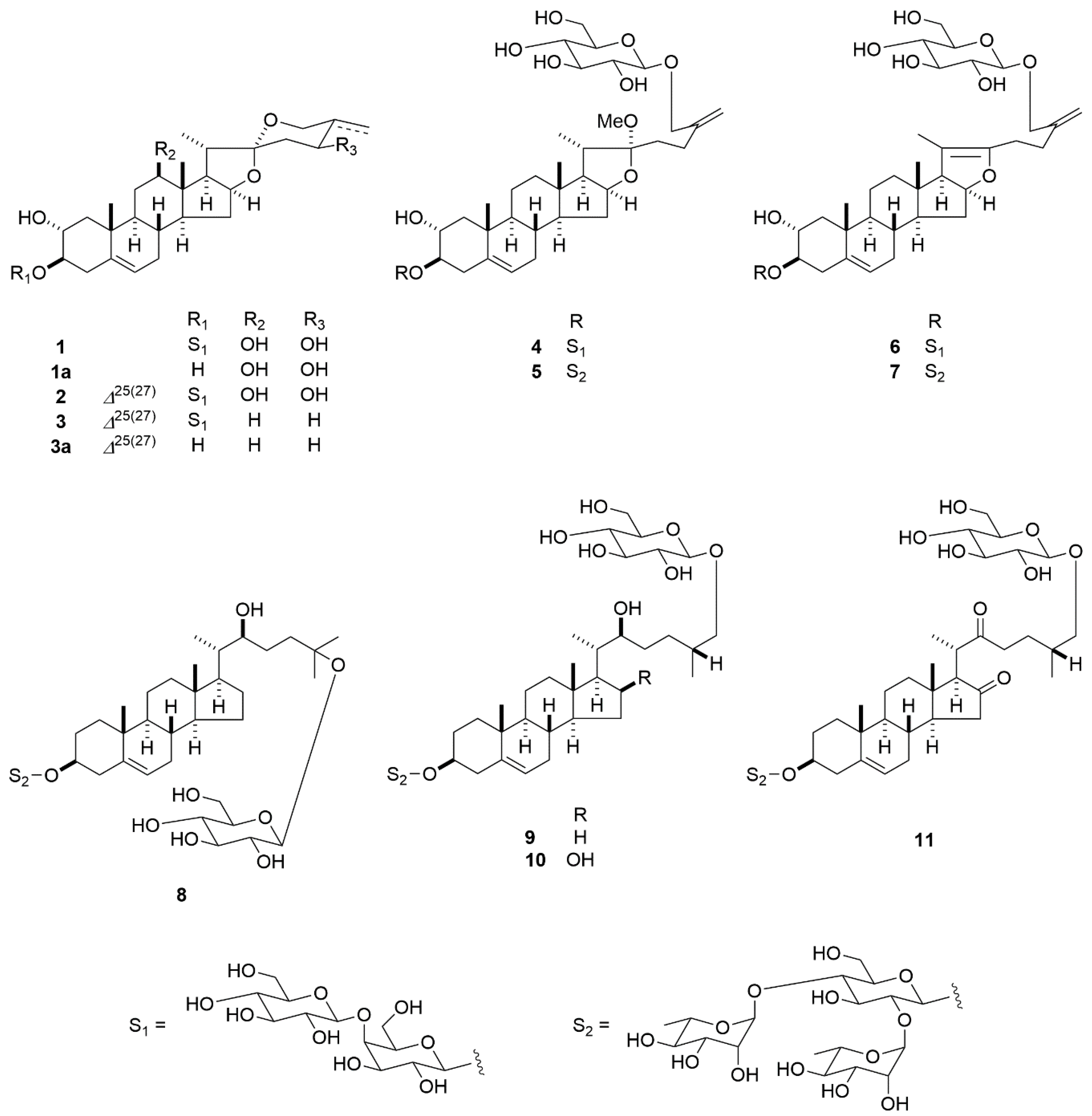
The MeOH extract of the leaves of C. newellii was fractionated by column chromatography (CC) and preparative HPLC to obtain 11 compounds (1-11) (Figure 1). Compounds 911 were identified as (22S,25R)-26-[(β-d-glucopyranosyl)oxy]-22-hydroxycholest-5-en-3β-yl O-α-l-rhamnopyranosyl-(1→2)-O-[α-l-rhamnopyranosyl-(1→4)]-β-d-glucopyranoside (9) [16], (22S,25R)-26-[(β-d-glucopyranosyl)oxy]-16β,22-dihydroxycholest-5-en-3β-yl O-α-l-rhamnopyranosyl-(1→2)-O-[α-l-rhamnopyranosyl-(1→4)]-β-d-glucopyranoside (10) [17], and (25R)-26-[(β-d-glucopyranosyl)oxy]-3β-[(O-α-l-rhamnopyranosyl-(1→2)-O-[α-l-rhamnopyranosyl-(1→4)]-β-d-glucopyranosyl)oxy]-cholest-5-ene-16,22-dione (11) [18], respectively.
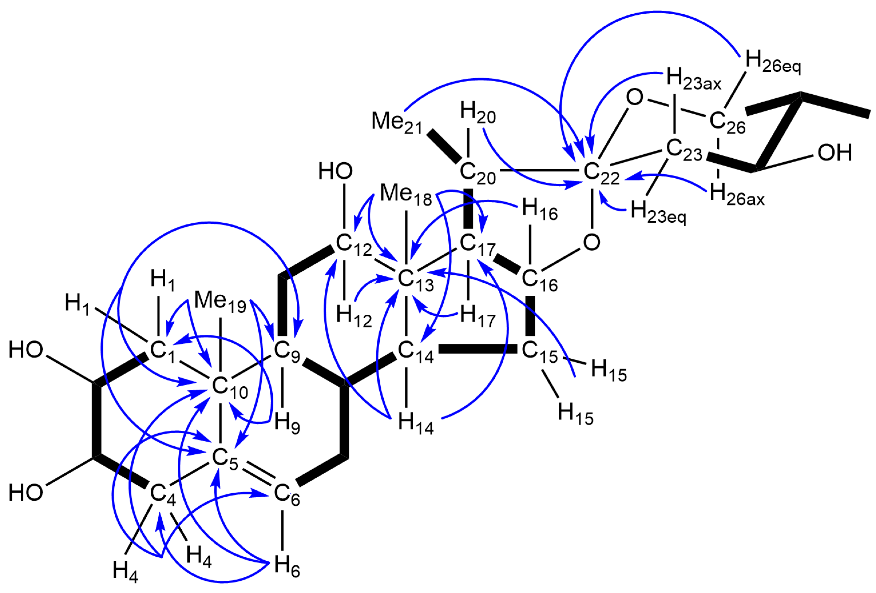
Compound 1 was obtained as an amorphous powder. Its molecular formula was determined to be C39H62O16 based on high-resolution electrospray ionization-time of flight-mass spectroscopy (HRESI-TOF-MS) and 13C-NMR data. The infrared (IR) spectrum of 1 exhibited absorption bands for hydroxy groups at 3381 cm−1. The 1H- and 13C-NMR spectra of 1 displayed signals for two tertiary methyl groups at δH 1.04 (s, Me-18) and 0.94 (s, Me-19); δC 20.3 (C-19) and 10.9 (C-18), two secondary methyl groups at δH 1.42 (d, J = 6.9 Hz, Me-21) and 1.07 (d, J = 6.5 Hz, Me-27); δC 14.2 (C-21) and 13.6 (C-27), an olefinic group at δH 5.30 (br d, J = 3.6 Hz, H-6); δC 140.0 (C-5) and 121.9 (C-6), two quaternary carbons at δC 46.1 (C-13) and 37.9 (C-10), an acetal carbon at δC 112.0 (C-22), and two anomeric protons and carbons at δH 5.22 (d, J = 7.9 Hz) and 4.91 (d, J = 7.8 Hz); δC 106.9 and 103.2. The above spectroscopic data imply that 1 had a spirost-5-ene diglycoside framework. Enzymatic hydrolysis of 1 with naringinase yielded 1a (C27H42O6) as the aglycone, and d-glucose and d-galactose as the carbohydrate moieties. Treatment of 1a with Ac2O in pyridine gave tetraacetate (1b) of 1a, indicating that 1a had four hydroxy groups. In the heteronuclear multiple bond correlation (HMBC) spectrum of 1a, the angular methyl singlet at δH 1.09 showed long-range correlations with C-1 at δC 46.5, C-5 at δC 141.2, C-9 at δC 50.1, and C-10 at δC 38.6, and was assigned to Me-19. The olefinic proton at δH 5.41 attributed to H-6 exhibited HMBC correlations with C-4 at δC 40.7, C-5, and C-10. In the heteronuclear multiple quantum coherence (HMQC) spectrum of 1a, the C-1 and C-4 carbons were correlated to the one-bond coupled protons at δH 2.39 (dd, J = 12.6 and 4.4 Hz, H-1eq) and 1.42 (dd, J = 12.6, 12.3 Hz, H-1ax), and 2.69 (H2-4), respectively. The H-1eq and H-1ax protons showed spin-coupling correlations with the hydroxymethine proton at δH 4.12 (ddd, J = 12.3, 11.2, 4.4 Hz), whereas the H2-4 protons exhibited spin-couplings with the hydroxymethine proton centered at δH 3.82 (m, W1/2 = 18.9 Hz) in the 1H-1H correlation spectroscopy (COSY) spectrum of 1a. A spin-coupling correlation was observed between the two hydroxymethine protons (H-2 and H-3) with a J value of 11.2 Hz. These data are consistent with the presence of a hydroxy group at C-2 and C-3. Another angular methyl singlet at δH 1.08 assigned to Me-18 showed HMBC correlations with C-12 at δC 78.9, C-13 at δC 46.2, C-14 at δC 55.4, and C-17 at δC 62.3. The C-12 carbon was associated with the one-bond coupled proton at δH 3.58 (dd, J = 11.2 and 4.4 Hz) in the HMQC spectrum, from which spin-coupling correlations were observed for the H2-11 methylene protons at δH 2.03 (m, H-11eq) and 1.75 (q-like, J = 11.2 Hz, H-11ax). Thus, a hydroxy group was shown to be present at C-12. The methine proton at δH 1.85 (m) assignable to H-25 displayed spin-coupling correlations with the Me-27 protons at δH 1.10 (d, J = 6.7 Hz), H2-26 methylene protons at δH 3.72 (dd, J = 11.2, 4.9 Hz, H-26eq) and 3.63 (dd, J = 12.3, 11.2 Hz, H-26ax), and H-24 methine proton at δH 4.05 (ddd, J = 10.5, 10.5, 4.8 Hz). These correlations are indicative of the presence of a hydroxy group at C-24 (Figure 2).
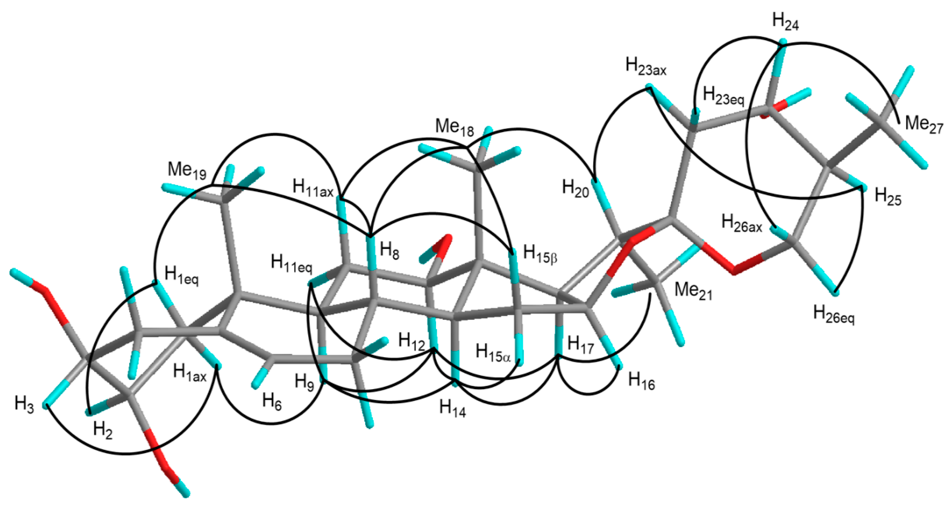
Accordingly, the planar structure of 1a was identified as spirost-5-ene-2,3,12,24-tetrol. NOE correlations in the nuclear Overhauser enhancement spectroscopy (NOESY) and proton spin-coupling constants allowed the stereochemistry of 1a to be determined. The B/C-trans, C/D-trans, and D/E-cis ring junctions, and the configurations of 20α and 22α were confirmed by the following NOE correlations: between H-8 and H-11ax/H-15α/Me-18/Me-19, H-9 and H-11eq/H-12/H-14, H-14 and H-12/H-15α/H-17, H-17 and H-16/Me-21, Me-18 and H-20, and between H-20 and H-23ax (Figure 3). The configurations of the C-2, C-3, and C-12 hydroxy groups were assigned as 2α, 3β, and 12β, respectively, based on the proton spin-coupling constants, 3JH-1ax,H-2 = 12.3 Hz, 3JH-1eq,H-2 = 4.4 Hz, 3JH-11ax,H-12 = 11.2 Hz, and 3JH-11eq,H-12 = 4.4 Hz, and NOE correlations were observed between H-1eq and H-2/Me-19, H-1ax and H-3/H-9, and between H-12 and H-9/H-11eq/H-14/H-17 (Figure 3). The proton spin-coupling constants, 3JH-23ax,H-24 = 10.5 Hz, 3JH-23eq,H-24 = 4.8 Hz, 3JH-24,H-25 = 10.5 Hz, 3JH-25,H-26ax = 12.3 Hz, and 3JH-25,H-26eq = 4.9 Hz, and NOE correlations between H-25 and H-23ax/H-26eq, and between H-24 and H-23eq/H-26ax/Me-27 were consistent with the 24S and 25S configurations (Figure 3). Thus, 1a was identified as (24S,25S)-spirost-5-ene-2α,3β,12β,24-tetrol. The 1H-1H COSY and HMQC spectra of 1 suggest that the sugar moiety of 1 comprised a 4-substituted β-d-galactopyranosyl unit [Gal: δH 4.91 (1H, d, J = 7.8 Hz); δC 103.2, 73.0, 75.0, 79.9, 75.8, and 60.9 (C-1′–6′)] and a terminal β-d-glucopyranosyl unit [Glc: δH 5.22 (1H, d, J = 7.9 Hz); δC 106.9, 75.7, 78.5, 72.0, 78.3, and 62.8 (C-1′′–6′′)]. In the HMBC spectrum of 1, long-range correlations were observed between H-1′′ of Glc (δH 5.22) and C-4′ of Gal (δC 79.9), and between H-1′ of Gal (δH 4.91) and C-3 of the aglycone (δC 84.5). Based on the above data, 1 was identified as (24S,25S)-2α,12β,24-trihydroxyspirost-5-en-3β-yl O-β-d-glucopyranosyl-(1→4)-β-d-galactopyranoside.
The 1H- and 13C-NMR spectral data of 2 (C39H60O16) suggest that 2 is analogous to 1, including the diglycoside moiety attached to C-3 of the aglycone. However, the molecular formula of 2 was smaller than that of 1 by two hydrogen atoms. When the 1H- and 13C-NMR spectra of 2 were compared with those of 1, the Me-27 group was revealed to be displaced by an exomethylene group [δH 5.66 and 5.10 (each br s, H2-27); δC 106.4 (C-27) and 149.3 (C-25)] in 2. Thus, it was speculated that 2 corresponded to the C-25/27 dehydroxy derivative of 1. This was supported by HMBC correlations from H2-27 (δH 5.66 and 5.10) to C-24 (δC 67.0)/C-25 (δC 149.3)/C-26 (δC 64.6). Accordingly, 2 was identified as (24S)-2α,12β,24-trihydroxyspirosta-5,25(27)-dien-3β-yl O-β-d-glucopyranosyl-(1→4)-β-d-galactopyranoside.
Compound 3 (C39H60O14) was obtained as an amorphous solid. The 1H- and 13C-NMR spectral data of 3 were similar to those of 2, including the signals of the diglycoside unit bound to C-3 of the aglycone of 3. However, the molecular formula of 3 was found to be smaller than that of 2 by two oxygen atoms, suggesting that the aglycone of 3 had two less hydroxy groups than 2. Acid hydrolysis of 3 with 1 M HCl (dioxane/H2O, 1:1) gave aglycone (3a), d-galactose, and d-glucose. The 1H- and 13C-NMR spectra of 3a showed signals for two angular methyl groups at δH 1.07 (s, Me-19) and 0.82 (s, Me-18); δC 20.6 (C-19) and 16.3 (C-18), a secondary methyl group at δH 1.08 (d, J = 6.4 Hz, Me-21); δC 14.9 (C-21), an exomethylene group at δH 4.81 and 4.77 (each br s, H2-27); δC 108.7 (C-27) and 144.4 (C-25), an acetal carbon at δC 109.4 (C-22), an olefinic group at δH 5.40 (br d, J = 4.4 Hz, H-6); δC 141.2 (C-5) and 121.2 (C-6), and two vicinal hydroxy groups at δH 4.16 (ddd, J = 12.0, 11.2, 4.2 Hz, H-2) and 3.84 (m, W1/2 = 20.1 Hz, H-3); δC 72.6 (C-2) and 76.7 (C-3). These spectroscopic data imply that 3a was spirosta-5,25(27)-diene-2α,3β-diol. The HMBC spectrum provided evidence that the O-β-d-glucopyranosyl-(1→4)-β-d-galactopyranosyl group was present at C-3 of the aglycone in 3. Therefore, 3 was deduced to be 2α-hydroxyspirosta-5,25(27)-dien-3β-yl O-β-d-glucopyranosyl-(1→4)-β-d-galactopyranoside.
Compound 4 was obtained as an amorphous solid, and its molecular formula was determined to be C46H74O20 based on HRESI-TOF-MS and 13C-NMR data. In the 1H- and 13C-NMR spectra of 4, the following signals were observed: three steroidal methyl groups at δH 1.14 (d, J = 6.9 Hz, Me-21), 0.93 (s, Me-19), and 0.76 (s, Me-18); δC 20.3 (C-19), 16.1 (C-21), and 16.0 (C-18), an exomethylene group at δH 5.34 and 5.04 (each br s, H2-27); δC 146.7 (C-25) and 111.0 (C-27), an olefinic group at δH 5.30 (br d, J = 4.6 Hz, H-6); δC 140.0 (C-5) and 121.8 (C-6), and three anomeric protons and carbons at δH 5.23 (d, J = 7.9 Hz, H-1′′), 4.93 (d, J = 7.8 Hz, H-1′), and 4.90 (d, J = 7.8 Hz, H-1′′′); δC 106.9 (C-1′′), 103.7 (C-1′′′), and 103.3 (C-1′). In addition, an acetal carbon signal at δC 112.3, a methoxy proton and carbon signals at δH 3.23 (s); δC 47.3, and a positive color reaction in Ehrlich’s test suggested that 4 was a 22-methoxyfurostanol glycoside. Compound 4 was treated with β-d-glucosidase to obtain the corresponding spirostanol glycoside (3) and d-glucose. A 3JC,H correlation from H-1′′′ of β-d-glucopyranosyl (δH 4.90) to C-26 of the aglycone (δC 71.9) was observed in the HMBC spectrum of 4. The C-22α configuration was confirmed by the NOE correlation observed between -OMe (δH 3.23) and H-16 (δH 4.41) of the aglycone. Thus, 4 was determined to be 26-[(β-d-glucopyranosyl)oxy]-2α-hydroxy-22α-methoxyfurosta-5,25(27)-dien-3β-yl O-β-d-glucopyranosyl-(1→4)-β-d-galactopyranoside.
The 1H- and 13C-NMR spectroscopic features of 5 (C52H84O23) were similar to those of 4, except for the signals assignable to the sugar moiety attached to C-3 of the aglycone. The molecular formula of 5 was larger than that of 4 by C6H10O3, corresponding to a hexosyl unit. Acid hydrolytic cleavage of 5 with 1 M HCl (dioxane/H2O, 1:1) afforded 3a, d-glucose, and l-rhamnose. Analysis of the 1H-1H COSY and HMQC spectra for the sugar moieties of 5 indicated the presence of a 2,4-disubstituted β- d-glucopyranosyl unit [Glc (I): δH 4.96 (1H, d, J = 7.2 Hz, H-1′); δC 100.9, 77.6, 77.6, 78.5, 76.9, and 61.0 (C-1′–6′)], a terminal β-d-glucopyranosyl unit [Glc (II): δH 4.90 (1H, d, J = 7.8 Hz, H-1′’’’); δC 103.7, 75.0, 78.5, 71.6, 78.4, and 62.7 (C-1′′′′–6′′′′)], and two terminal α-l-rhamnopyranosyl units [Rha (I): δH 6.33 (1H, br s, H-1′’); δC 101.9, 72.2, 72.7, 73.9, 69.4, and 18.5 (C-1′′–6′′); Rha (II): δH 5.79 (1H, br s, H-1′′′); δC 102.7, 72.3, 72.6, 73.8, 70.3, and 18.4 (C-1′′′–6′′′)]. In the HMBC spectrum of 5, long-range correlations were observed between H-1′′ of Rha (I) (δH 6.33) and C-2′ of Glc (I) (δC 77.6), H-1′′′ of Rha (II) (δH 5.79) and C-4′ of Glc (I) (δC 78.5), H-1′ of Glc (I) (δH 4.96) and C-3 of the aglycone (δC 84.9), and between H-1′′′′ of Glc (II) (δH 4.90) and C-26 of the aglycone (δC 71.9). Therefore, 5 was characterized as 26-[(β-d-glucopyranosyl)oxy]-2α-hydroxy-22α-methoxyfurosta-5,25(27)-dien-3β-yl O-α-l-rhamnopyranosyl-(1→2)-O-[α-l-rhamnopyranosyl-(1→4)]-β-d-glucopyranoside.
The 1H- and 13C-NMR spectra of 6 (C45H70O19) and 7 (C51H80O22) were closely related to those of 4 and 5, respectively, except for the signals attributable to the E-ring part of the aglycone. Instead of the secondary methyl signal for Me-21 [4: δH 1.14 (d, J = 6.9 Hz); δC 16.1; 5: δH 1.14 (d, J = 6.9 Hz); δC 16.1] and a methoxy signal [4: δH 3.23 (s); δC 47.3; 5: δH 3.24 (s); δC 47.3], the signals arising from the tertiary methyl groups [6: δH 1.60 (s); δC 11.7; 7: δH 1.59 (s); δC 11.7] and tetrasubstituted olefinic carbons [6: δC 151.6 and 103.9; 7: δC 151.6 and 103.9] were observed in the 1H- and 13C-NMR spectra of 6 and 7. Thus, 6 and 7 were thought to be the corresponding pseudofurostanol glycosides of 4 and 5, respectively. The structures of 6 and 7 were confirmed by the following chemical transformations (Figure 4). Enzymatic hydrolysis of 6 with β-d-glucosidase gave 3 and d-glucose. Furthermore, complete acetylation of 6 with Ac2O in pyridine afforded dodecaacetate (6a), which agreed with the peracetate of the dehydro derivative of 4 prepared by treating 4 with Ac2O in pyridine at 130 °C for 3 h. On the other hand, enzymatic hydrolysis of 7 with β-d-glucosidase gave the spirostanol glycoside (7a), which is in agreement with the glycoside obtained by enzymatic hydrolysis 5. Tridecaacetate (7b) of 7 was consistent with the product prepared from 5 upon treatment of 5 with Ac2O in pyridine at 130 °C for 3 h. Accordingly, 6 and 7 were identified as 26-[(β-d-glucopyranosyl)oxy]-2α-hydroxyfurosta-5,20(22),25(27)-trien-3β-yl O-β-d-glucopyranosyl-(1→4)-β-d-galactopyranoside and 26-[(β-d-glucopyranosyl)oxy]-2α-hydroxyfurosta-5,20(22),25(27)-trien-3β-yl O-α-l-rhamnopyranosyl-(1→2)-O-[α-l-rhamnopyranosyl-(1→4)]-β-d-glucopyranoside, respectively.
Compound 8 (C51H86O21) was obtained as an amorphous solid. The molecular formula of 8 was the same as that of 9, and the 1H- and 13C-NMR spectra of 8 were very similar to those of 9, except for the signals attributable to the side chain of the aglycone units. Compound 8 was enzymatically hydrolyzed with naringinase, yielding (22S)-cholest-5-ene-3β,22,25-triol (8a) [19], d-glucose, and l -rhamnose. The 1H- and 13C-NMR spectra of 8 imply the presence of a 2,4-disubstituted β-d-glucopyranosyl unit [Glc (I)], two terminal α-l-rhamnopyranosyl units [Rha (I) and Rha (II)], and a terminal β-d-glucopyranosyl unit [Glc (II): δH 5.08 (1H, d, J = 7.8 Hz, H-1′′′′); δC 98.6, 75.4, 78.8, 71.8, 78.0, and 62.9 (C-1′′′′–6′′′′)] in this molecule. In the HMBC spectrum of 8, a 3JC,H correlation was observed between H-1′′′′ of Glc (II) (δH 5.08) and C-25 of the aglycone moiety (δC 77.4). Therefore, 8 was determined to be (22S)-25-[(β-d-glucopyranosyl)oxy]-22-hydroxycholest-5-en-3β-yl O-α-l-rhamnopyranosyl-(1→2)-O-[α-l-rhamnopyranosyl-(1→4)]-β-d-glucopyranoside.
The isolated compounds (111) were evaluated for their cytotoxic activity toward HL-60 human promyelocytic leukemia cells using a modified 3-(4,5-dimethylthiazol-2-yl)-2,5-diphenyl-2H-tetrazolium bromide (MTT) assay method. None of 111 exhibited cytotoxicity at a sample concentration of up to 10 μM. Although C. newellii is regarded to be a poisonous plant, the MTT assay exerted that none of the isolated compounds were cytotoxic against HL-60 cells.

3. Materials and Methods

3.1. General

Optical rotations were measured on a JASCO P-1030 and a JASCO DIP-360 (JASCO, Tokyo, Japan) automatic digital polarimeter. IR spectra were obtained using a FT/IR-620 (JASCO) spectrophotometer. NMR spectral data were recorded on a DRX-500 (500 MHz for 1H-NMR, 125 MHz for 13C-NMR) spectrometer using standard Bruker pulse programs at 300 K (Bruker, Karlsruhe, Germany). Chemical shifts are given as δ values with reference to tetramethylsilane (TMS) as an internal standard. HRESI-TOF-MS data were obtained using a Waters Micromass LCT mass spectrometer (Waters, MA, USA). Diaion HP-20 porous polymer polystyrene resin (Mitsubishi-Chemical, Tokyo, Japan), silica gel Chromatrex BW-300 (Fuji-Silysia Chemical, Aichi, Japan), and ODS silica gel COSMOSIL 75C18-OPN (Nacalai Tesque, Kyoto, Japan) were used for CC. Thin-layer chromatography (TLC) analysis was conducted using precoated silica gel 60F254 or RP18 F254S plates (0.25 mm thick; Merck, Darmstadt, Germany), and the spots were made visible by spraying the plates with H2SO4/H2O (1:9), followed by heating. TLC was used to check the progress of the separation of fractions and to confirm the purity of the isolated compounds. A Tosoh CCPM (Tosoh, Tokyo, Japan) or a Tosoh-8020 (Tosoh), Tosoh RI-8020 (Tosoh), or Shodex OR-2 (Showa-Denko, Tokyo, Japan) detector, and a Rheodyne injection port (Rohnert Park, CA, USA) constituted the HPLC system. A Capcell Pak C18 UG120 column (10 mm i.d. × 250 mm, 5 μm, Shiseido, Tokyo, Japan) was used for preparative HPLC. Enzymatic hydrolysis was carried out using β-d-glucosidase (EC 232-589-7; Sigma, St. Louis, MO, USA) or naringinase (EC 232-962-4; Sigma, St. Louis, MO, USA).

3.2. Plant Material

The leaves of C. newellii were purchased from Sakata Seed Corporation (Kanagawa, Japan) and grown in the medicinal botanical garden of Tokyo University of Pharmacy and Life Sciences (TUPLS). A voucher specimen was kept at the herbarium of the TUPLS.

3.3. Extraction and Isolation

C. newellii leaves (dry weight, 3.6 kg) were extracted with MeOH at 60 °C for 2 h, and concentrated under reduced pressure to obtain the MeOH extract (180 g). Then, all MeOH extract was loaded onto a Diaion HP-20 column, and successively eluted with MeOH/H2O (3:7), EtOH, and EtOAc (each 12 L). The EtOH eluted fraction was separated by silica gel CC and eluted with a stepwise gradient mixture of CHCl3/MeOH (9:1, 6:1, 3:1, 1:1) to obtain three fractions (Frs. I–III). Fraction II was further divided by ODS silica gel CC eluted with MeCN/H2O (1:3, 1:1, 3:1, 5:1) to yield seven subfractions (Frs. II-1–II-7). Fraction II-1 was purified by silica gel CC eluted with CHCl3/MeOH/H2O (20:10:1), ODS silica gel CC eluted with MeCN/H2O (1:3), and preparative HPLC using MeCN/H2O (5:11) to obtain 6 (21 mg) and 7 (12 mg). Fraction II-2 was subjected to silica gel CC eluted with CHCl3/MeOH/H2O (20:10:1; 7:4:1), ODS silica gel CC eluted with MeCN/H2O (2:7), and preparative HPLC using MeCN/H2O (1:3) to obtain 4 (1.8 g). Fraction II-3 was separated by silica gel CC eluted with CHCl3/MeOH/H2O (25:10:1; 20:10:1), ODS silica gel CC eluted with MeCN/H2O (5:13; 5:14), and preparative HPLC using MeCN/H2O (10:27) to yield 1 (49 mg) and 2 (8.3 mg). Fraction II-5 was applied to silica gel CC eluted with CHCl3/MeOH/H2O (30:10:1; 25:10:1; 20:10:1; 7:4:1) and ODS silica gel CC eluted with MeCN/H2O (1:1; 1:2; 5:12; 2:5; 10:27; 1:3), and preparative HPLC using MeCN/H2O (5:12) to afford 5 (22 mg), 8 (29 mg), 9 (20 mg), 10 (65 mg), and 11 (71 mg). Fraction II-7 was chromatographed on silica gel and ODS silica gel eluted with CHCl3/MeOH/H2O (30:10:1) and MeCN/H2O (1:1), respectively, to furnish 3 (25 mg).

3.4. Structural Characterization

Compound 1: Amorphous solid; [α]D25 − 62.5 (c = 0.10, MeOH); IR (film) νmax: 3381 (OH), 2907 (CH) cm−1; HRESI-TOF-MS m/z: 787.4146 [M + H]+ (calcd. for C39H63O16: 787.4116). 1H-NMR spectral data (500 MHz, C5D5N): δH 5.30 (1H, br d, J = 3.6 Hz, H-6), 5.22 (1H, d, J = 7.9 Hz, H-1′′), 4.91 (1H, d, J = 7.8 Hz, H-1′), 4.63 (1H, br d, J = 3.5 Hz, H-4′), 4.61 (1H, q-like, J = 8.4 Hz, H-16), 4.56 (1H, dd, J = 10.9, 8.0 Hz, H-6′a), 4.53 (1H, dd, J = 11.2, 2.4 Hz, H-6′′a), 4.41 (1H, dd, J = 9.3, 7.8 Hz, H-2′), 4.25 (1H, dd, J = 9.3, 3.5 Hz, H-3′), 4.23 (1H, dd, J = 9.5, 8.6 Hz, H-3′’), 4.22 (1H, dd, J = 10.9, 4.2 Hz, H-6′b), 4.19 (1H, dd, J = 11.2, 4.7 Hz, H-6′′b), 4.09 (1H, m, H-5′), 4.08 (1H, dd, J = 8.6, 7.9 Hz, H-2′′), 4.07 (1H, dd, J = 9.5, 8.8 Hz, H-4′′), 4.03 (1H, m, H-24), 4.01 (1H, m, H-2), 3.96 (1H, ddd, J = 8.8, 4.7, 2.4 Hz, H-5′′), 3.79 (1H, m, H-3), 3.70 (1H, dd, J = 11.2, 4.8 Hz, H-26eq), 3.60 (1H, dd, J = 12.2, 11.2 Hz, H-26ax), 3.55 (1H, dd, J = 11.0, 4.2 Hz, H-12), 1.42 (3H, d, J = 6.9 Hz, Me-21), 1.07 (3H, d, J = 6.5 Hz, Me-27), 1.04 (3H, s, Me-18), 0.94 (3H, s, Me-19). For 13C-NMR spectral data, see Table 1. For NMR spectral data, see Supplementary Materials.
Compound 1a: Amorphous solid; [α]D25 − 44.2 (c = 0.10, MeOH); IR (film) νmax: 3364 (OH), 2924 and 2872 (CH) cm−1; HRESI-TOF-MS m/z: 463.3084 [M + H]+ (calcd. for C27H43O6: 463.3060). 1H-NMR spectral data (500 MHz, C5D5N): δH 5.41 (1H, br d, J = 4.8 Hz, H-6), 4.63 (1H, q-like, J = 7.5 Hz, H-16), 4.12 (1H, ddd, J = 12.3, 11.2, 4.4 Hz, H-2), 4.05 (1H, ddd, J = 10.5, 10.5, 4.8 Hz, H-24), 3.82 (1H, m, W1/2 = 18.9 Hz, H-3), 3.72 (1H, dd, J = 11.2, 4.9 Hz, H-26eq), 3.63 (1H, dd, J = 12.3, 11.2 Hz, H-26ax), 3.58 (1H, dd, J = 11.2, 4.4 Hz, H-12), 1.45 (3H, d, J = 6.9 Hz, Me-21), 1.10 (3H, d, J = 6.7 Hz, Me-27), 1.09 (3H, s, Me-19), 1.08 (3H, s, Me-18). For 13C-NMR spectral data, see Table 1. For NMR spectral data, see Supplementary Materials.
Tetraacetate of compound 1a (1b): 1H-NMR spectral data (500 MHz, C5D5N): δH 5.34 (1H, br d, J = 4.8 Hz, H-6), 5.04 (1H, m, H-3), 4.84 (1H, dd, J = 11.2, 4.5 Hz, H-12), 1.16 (3H, d, J = 6.8 Hz, Me-21), 1.06 (3H, s, Me-18), 0.92 (3H, s, Me-19), 0.80 (3H, d, J = 6.5 Hz, Me-27), 2.20, 2.10, 2.09 × 2 (each 3H, Ac × 4).
Compound 2: Amorphous solid; [α]D25 − 78.0 (c = 0.10, MeOH); IR (film) νmax: 3381 (OH), 2921 (CH) cm−1; HRESI-TOF-MS m/z: 785.3943 [M + H]+ (calcd. for C39H61O16: 785.3960). 1H-NMR spectral data (500 MHz, C5D5N): δH 5.66 (1H, br s, H-27a), 5.31 (1H, br d, J = 4.8 Hz, H-6), 5.24 (1H, d, J = 7.9 Hz, H-1′′), 5.10 (1H, br s, H-27b), 5.06 (1H, dd, J = 11.3, 6.2 Hz, H-24), 4.92 (1H, d, J = 7.7 Hz, H-1′), 4.60 (1H, q-like, J = 6.9 Hz, H-16), 4.53 (1H, d, J = 12.8 Hz, H-26eq), 4.23 (1H, d, J = 12.8 Hz, H-26ax), 3.98 (1H, m, H-2), 3.80 (1H, m, H-3), 3.55 (1H, dd, J = 11.0, 4.4 Hz, H-12), 1.37 (3H, d, J = 6.9 Hz, Me-21), 1.05 (3H, s, Me-18), 0.95 (3H, s, Me-19). For 13C-NMR spectral data, see Table 1. For NMR spectral data, see Supplementary Materials.
Compound 3: Amorphous solid; [α]D25 − 54.6 (c = 0.10, MeOH); IR (film) νmax: 3369 (OH), 2927 and 2852 (CH) cm−1; HRESI-TOF-MS m/z: 753.4058 [M + H]+ (calcd. for C39H61O14: 753.4061). 1H-NMR spectral data (500 MHz, C5D5N): δH 5.30 (1H, br d, J = 4.7 Hz, H-6), 5.23 (1H, d, J = 7.9 Hz, H-1′′), 4.93 (1H, d, J = 7.8 Hz, H-1′), 4.80 (1H, br s, H-27a), 4.77 (1H, br s, H-27b), 4.51 (1H, q-like, J = 7.9 Hz, H-16), 4.43 (1H, d, J = 12.4 Hz, H-26ax), 4.05 (1H, m, H-2), 4.02 (1H, d, J = 12.4 Hz, H-26eq), 3.82 (1H, m, H-3), 1.06 (3H, d, J = 7.0 Hz, Me-21), 0.94 (3H, s, Me-19), 0.79 (3H, s, Me-18). For 13C-NMR spectral data, see Table 1. For NMR spectral data, see Supplementary Materials.
Compound 3a: Amorphous solid; [α]D25 − 78.6 (c = 0.10, MeOH); IR (film) νmax: 3351 (OH), 2926 and 2852 (CH) cm−1; HRESI-TOF-MS m/z: 429.3010 [M + H]+ (calcd. for C27H41O4: 429.3005). 1H-NMR spectral data (500 MHz, C5D5N): δH 5.40 (1H, br d, J = 4.4 Hz, H-6), 4.81 (1H, br s, H-27a), 4.77 (1H, br s, H-27b), 4.52 (1H, q-like, J = 7.1 Hz, H-16), 4.44 (1H, d, J = 12.1 Hz, H-26ax), 4.16 (1H, ddd, J = 12.0, 11.2, 4.2 Hz, H-2), 4.02 (1H, d, J = 12.1 Hz, H-26eq), 3.84 (1H, m, W1/2 = 20.1 Hz, H-3), 1.08 (3H, d, J = 6.4 Hz, Me-21), 1.07 (3H, s, Me-19), 0.82 (3H, s, Me-18). For 13C-NMR spectral data, see Table 1. For NMR spectral data, see Supplementary Materials.
Compound 4: Amorphous solid; [α]D25 − 53.4 (c = 0.10, MeOH); IR (film) νmax: 3369 (OH), 2936 and 2899 (CH) cm−1; HRESI-TOF-MS m/z: 915.4611 [M + H − MeOH]+ (calcd. for C45H71O19: 915.4590). 1H-NMR spectral data (500 MHz, C5D5N): δH 5.34 (1H, br s, H-27a), 5.30 (1H, br d, J = 4.6 Hz, H-6), 5.23 (1H, d, J = 7.9 Hz, H-1′′), 5.04 (1H, br s, H-27b), 4.93 (1H, d, J = 7.8 Hz, H-1′), 4.90 (1H, d, J = 7.8 Hz, H-1′′′), 4.61 (1H, d, J = 12.8 Hz, H-26a), 4.53 (1H, dd, J = 11.9, 2.4 Hz, H-6′′′a), 4.41 (1H, m, H-16), 4.36 (1H, dd, J = 11.9, 5.6 Hz, H-6′′′b), 4.35 (1H, d, J = 12.8 Hz, H-26b), 4.26 (1H, dd, J = 8.7, 8.7 Hz, H-3′′′), 4.21 (1H, dd, J = 8.7, 8.7 Hz, H-4′′′), 4.06 (1H, dd, J = 8.7, 7.8 Hz, H-2′′′), 4.04 (1H, m, H-2), 3.94 (1H, m, H-5′′′), 3.81 (1H, m, H-3), 3.23 (3H, s, OMe), 1.14 (3H, d, J = 6.9 Hz, Me-21), 0.93 (3H, s, Me-19), 0.76 (3H, s, Me-18). For 13C-NMR spectral data, see Table 1. For NMR spectral data, see Supplementary Materials.
Compound 5: Amorphous solid; [α]D25 − 52.0 (c = 0.10, MeOH); IR (film) νmax: 3382 (OH), 2924 (CH) cm−1; HRESI-TOF-MS m/z: 1045.5264 [M + H − MeOH]+ (calcd. for C51H81O22: 1045.5220). 1H-NMR spectral data (500 MHz, C5D5N): δH 6.33 (1H, br s, H-1′′), 5.79 (1H, br s, H-1′′′), 5.34 (1H, br s, H-27a), 5.32 (1H, br d, J = 4.4 Hz, H-6), 5.05 (1H, br s, H-27b), 4.96 (1H, d, J = 7.2 Hz, H-1′), 4.90 (1H, d, J = 7.8 Hz, H-1′′′′), 4.86 (1H, m, H-5′′), 4.85 (1H, m, H-5′′′), 4.81 (1H, br s, H-2′′), 4.65 (1H, br s, H-2′′′), 4.62 (1H, d, J = 12.7 Hz, H-26a), 4.61 (1H, dd, J = 9.0, 3.7 Hz, H-3′′), 4.53 (1H, dd, J = 12.5, 3.4 Hz, H-6′′′′a), 4.52 (1H, dd, J = 9.3, 3.4 Hz, H-3′′′), 4.42 (1H, q-like, J = 7.3 Hz, H-16), 4.37 (1H, br d, J = 12.5 Hz, H-6′′′′b), 4.36 (1H, d, J = 12.7 Hz, H-26b), 4.35 (1H, dd, J = 9.0, 9.0 Hz, H-4′′), 4.33 (1H, dd, J = 8.9, 8.9 Hz, H-4′), 4.32 (1H, dd, J = 9.3, 9.3 Hz, H-4′′′), 4.25 (1H, dd, J = 8.9, 8.9 Hz, H-4′′′′), 4.24 (1H, dd, J = 8.9, 8.9 Hz, H-3′′′′), 4.21 (1H, br d, J = 12.9 Hz, H-6′a), 4.20 (1H, dd, J = 8.9, 8.4 Hz, H-3′), 4.17 (1H, dd, J = 8.4, 7.2 Hz, H-2′), 4.16 (1H, m, H-2), 4.07 (1H, dd, J = 12.9, 4.6 Hz, H-6′b), 4.06 (1H, dd, J = 8.9, 7.8 Hz, H-2′′′′), 3.93 (1H, m, H-5′′′′), 3.82 (1H, m, H-3), 3.70 (1H, m, H-5′), 3.24 (3H, s, OMe), 1.67 (3H, d, J = 6.2 Hz, Me-6′′), 1.60 (3H, d, J = 6.2 Hz, Me-6′′′), 1.14 (3H, d, J = 6.9 Hz, Me-21), 1.04 (3H, s, Me-19), 0.77 (3H, s, Me-18). For 13C-NMR spectral data, see Table 1. For NMR spectral data, see Supplementary Materials.
Compound 6: Amorphous solid; [α]D25 − 44.7 (c = 0.10, MeOH); IR (film) νmax: 3360 (OH), 2900 (CH) cm−1; HRESI-TOF-MS m/z: 915.4541 [M + H]+ (calcd. for C45H71O19: 915.4590). 1H-NMR spectral data (500 MHz, C5D5N): δH 5.36 (1H, br s, H-27a), 5.31 (1H, br d, J = 4.6 Hz, H-6), 5.25 (1H, d, J = 7.9 Hz, H-1′′), 5.05 (1H, br s, H-27b), 4.93 (1H, d, J = 7.7 Hz, H-1′), 4.90 (1H, d, J = 7.8 Hz, H-1′′′), 4.78 (1H, q-like, J = 7.7 Hz, H-16), 4.59 (1H, d, J = 12.5 Hz, H-26a), 4.35 (1H, d, J = 12.5 Hz, H-26b), 4.11 (1H, m, H-2), 3.82 (1H, m, H-3), 1.60 (3H, s, Me-21), 0.95 (3H, s, Me-19), 0.67 (3H, s, Me-18). For 13C-NMR spectral data, see Table 1. For NMR spectral data, see Supplementary Materials.
Dodecaacetate of Compound 6 (6a): 1H-NMR spectral data (500 MHz, C5D5N): δH 5.41 (1H, br s, H-27a), 5.40 (1H, br s, H-6), 5.06 (1H, br s, H-27b), 4.49 (1H, d, J = 12.5 Hz, H-26a), 4.30 (1H, d, J = 12.5 Hz, H-26b), 1.60 (3H, s, Me-21), 0.94 (3H, s, Me-19), 0.81 (3H, s, Me-18), 2.34, 2.17, 2.14, 2.12 × 2, 2.09, 2.05, 2.03, 2.00 × 3, 1.98 (each 3H, Ac × 12).
Compound 7: Amorphous solid; [α]D25 − 51.0 (c = 0.10, MeOH); IR (film) νmax: 3380 (OH), 2919 (CH) cm−1; HRESI-TOF-MS m/z: 1045.5173 [M + H]+ (calcd. for C51H81O22: 1045.5219). 1H-NMR spectral data (500 MHz, C5D5N): δH 6.34 (1H, br s, H-1′′), 5.80 (1H, br s, H-1′′′), 5.36 (1H, br s, H-27a), 5.34 (1H, br d, J = 5.1 Hz, H-6), 5.04 (1H, br s, H-27b), 4.97 (1H, d, J = 6.6 Hz, H-1′), 4.90 (1H, d, J = 7.8 Hz, H-1′′′′), 4.78 (1H, q-like, J = 7.9 Hz, H-16), 4.59 (1H, d, J = 12.0 Hz, H-26a), 4.35 (1H, d, J = 12.0 Hz, H-26b), 4.16 (1H, m, H-2), 3.82 (1H, m, H-3), 1.67 (3H, d, J = 6.2 Hz, Me-6′′), 1.60 (3H, d, J = 6.1 Hz, Me-6′′′), 1.59 (3H, s, Me-21), 1.06 (3H, s, Me-19), 0.67 (3H, s, Me-18). For 13C-NMR spectral data, see Table 1. For NMR spectral data, see Supplementary Materials.
Compound 7a: Amorphous solid; [α]D25 − 3.9 (c = 0.10, MeOH); IR (film) νmax: 3376 (OH), 2924 and 2852 (CH) cm−1; HRESI-TOF-MS m/z: 883.4738 [M + H]+ (calcd. for C45H71O17: 883.4691). 1H-NMR spectral data (500 MHz, C5D5N): δH 6.36 (1H, br s, H-1′′), 5.81 (1H, br s, H-1′′′), 5.35 (1H, br d, J = 4.6 Hz, H-6), 4.98 (1H, d, J = 7.2 Hz, H-1′), 4.81 (1H, br s, H-27a), 4.78 (1H, br s, H-27b), 4.53 (1H, m, H-16), 4.45 (1H, d, J = 12.1 Hz, H-26a), 4.16 (1H, m, H-2), 4.02 (1H, d, J = 12.1 Hz, H-26b), 3.83 (1H, m, H-3), 1.68 (3H, d, J = 6.2 Hz, Me-6′′), 1.61 (3H, d, J = 6.2 Hz, Me-6′′′), 1.07 (3H, d, J = 6.9 Hz, Me-21), 1.06 (3H, s, Me-19), 0.80 (3H, s, Me-18). 13C-NMR spectral data (125 MHz, C5D5N): δC 45.9, 70.2, 85.0, 37.1, 139.9, 121.9, 32.2, 31.1, 50.1, 37.9, 21.1, 39.6, 40.4, 56.4, 32.1, 81.4, 62.8, 16.2, 20.3, 41.8, 14.9, 109.4, 33.1, 28.9, 144.3, 64.9, 108.7 (C-1-27), 100.9, 77.7, 77.6, 78.6, 77.0, 61.0 (C-1′-6′), 101.9, 72.3, 72.7, 74.0, 69.5, 18.5 (C-1′′-6′′), 102.8, 72.4, 72.6, 73.8, 70.4, 18.4 (C-1′′′-6′′′).
Tridecaacetate of compound 7 (7b): 1H-NMR spectral data (500 MHz, C5D5N): δH 5.49 (1H, br s, H-6), 5.43 (1H, br s, H-27a), 5.34 (1H, br s, H-27b), 4.52 (1H, d, J = 13.1 Hz, H-26a), 4.32 (1H, d, J = 13.1 Hz, H-26b), 1.62 (3H, s, Me-21), 0.94 (3H, s, Me-19), 0.86 (3H, s, Me-18), 2.35, 2.19, 2.18, 2.16, 2.14, 2.09, 2.06, 2.04, 2.03 (×2), 2.01 (×2), 1.96 (each 3H, Ac × 13).
Compound 8: Amorphous solid; [α]D25 − 48.9 (c = 0.10, MeOH); IR (film) νmax: 3381 (OH), 2932 (CH) cm−1; HRESI-TOF-MS m/z: 1035.5762 [M + H]+ (calcd. for C51H87O21: 1035.5740). 1H-NMR spectral data (500 MHz, C5D5N): δH 6.38 (1H, br s, H-1′′), 5.86 (1H, br s, H-1′′′), 5.34 (1H, br d, J = 4.9 Hz, H-6), 5.08 (1H, d, J = 7.8 Hz, H-1′′′′), 4.95 (1H, d, J = 7.7 Hz, H-1′). 3.91 (1H, m, H-22), 3.87 (1H, m, H-3), 1.77 (3H, d, J = 6.2 Hz, H-6′′), 1.62 (3H, d, J = 6.2 Hz, H-6′′′), 1.47 (3H × 2, each s, Me-26 and Me-27), 1.15 (3H, d, J = 6.7 Hz, Me-21), 1.07 (3H, s, Me-18), 0.70 (3H, s, Me-19). For 13C-NMR spectral data, see Table 1. For NMR spectral data, see Supplementary Materials.
Enzymatic hydrolysis of 1 and 48: Compounds 1 (20 mg) and 8 (9.8 mg) were independently treated with naringinase (1: 168 mg, 8: 102 mg) in AcOH/AcOK buffer (pH 4.3, 3.0 mL) at 28 °C for 132 h. Each reaction mixture was purified by silica gel CC eluted with CHCl3/MeOH/H2O (1; 9:1:0, 7:4:1, 8; 9:1:0) to obtain 1a (10.1 mg) from 1, 8a (3.0 mg) from 8, and sugar fractions (6.1 mg from 1, 2.4 mg from 8), respectively. The sugar fraction was analyzed using HPLC under the following conditions: detection, refractive index, and optical rotation; column, Capcell Pak NH2 UG80 (4.6 mm i.d. × 250 mm, 5 μm, Shiseido); solvent, MeCN/H2O (17:3); flow rate, 1.0 mL/min. d-Galactose, d-glucose, and l-rhamnose were identified by comparing their retention times and optical rotations with those of authentic samples: d-galactose (12.13, positive optical rotation), d-glucose (13.62, positive optical rotation), and l-rhamnose (7.48, negative optical rotation). Compounds 4 (29.7 mg), 5 (10.1 mg), 6 (5.6 mg), and 7 (3.1 mg) were independently treated with β-d-glucosidase (4: 25 mg, 5: 15 mg, 6: 8.8 mg, 7: 10 mg) in AcOH/AcONa (pH 5.0, 3.0 mL) at 28 °C for 20 h. Each reaction mixture was chromatographed on silica gel eluted with CHCl3/MeOH/H2O (4, 6, and 7; 20:10:1, 5; 9:1:0) to collect 3 (17.4 mg) from 4, 7a (3.4 mg) from 5, 3 (3.8 mg) from 6, 7a (1.0 mg) from 7, and their sugar fractions. HPLC analysis of the sugar fractions under the same conditions as those of 1 exhibited the presence of d-galactose in 4 and 6, d-glucose in 47, and l-rhamnose in 5 and 7.
Acid hydrolysis of 3 and 5: Compounds 3 (18.2 mg) and 5 (10.1 mg) were independently treated with 1 M HCl (dioxane/H2O, 1:1, 3.0 mL) at 95 °C for 1 h under Ar atmosphere. The reaction solution was neutralized by passing through an Amberlite IRA-93 ZU (Organo, Tokyo, Japan) column and separated using a Sep-Pak C18 cartridge (Waters) eluted with MeOH/H2O (1:4) to yield sugar fractions (3.1 mg from 3 and 1.2 mg from 5) and finally MeOH alone to obtain aglycone fractions. The sugar fractions were analyzed by HPLC under the same conditions as those of 1 showed the presence of d-galactose in 3, d-glucose in 3 and 5, and l-rhamnose in 5. The aglycone fractions were independently subjected to silica gel CC eluted with CHCl3/MeOH (19:1) to furnish 3a (8.3 mg) from 3, and 3a (2.8 mg) from 5.
Acetylation of 47 and 1a: Compounds 4 (100 mg) and 5 (4.5 mg) were independently applied to acetylation with Ac2O (2.0 mL) in pyridine (2.0 mL) at 130 °C for 3 h. The reaction solutions were distributed using Et2O (10 mL × 2). After concentration of the Et2O soluble phases, those were subjected to silica gel CC eluted with hexane hexane/Me2CO (1:1) to obtain 6a (73. 0 mg) from 4, and 7b (3.4 mg) from 5. Compounds 6 (10.2 mg), 7 (8.7 mg), and 1a (8.8 mg) were independently acetylated with Ac2O (1.0 mL) in pyridine (1.0 mL) at 28 °C for 20 h. The reaction solutions were distributed and purified, as well as 4, to afford 6a (4.6 mg) from 6, 7b (4.1 mg) from 7, and 1b (4.0 mg) from 1a.

3.5. Evaluation of Cytotoxic Activity

Cytotoxic activity of 111 against HL-60 cells (JCRB 0085; Human Science Research Resources Bank, Osaka, Japan) was examined by a modified MTT assay method as previously described [20]. In short, HL-60 cells were incubated at 37 °C for 24 h in RPMI-1640 medium with 10% heat-inactivated fetal bovine serum. The cell viability was evaluated using the MTT method.

4. Conclusions

A systematic phytochemical analysis of the leaves of C. newellii was conducted with a focus on steroidal glycosides. As a result, three new spirostanol glycosides (13), two new furostanol glycosides (4 and 5), two new pseudofurostanol glycosides (6 and 7), one new cholestane glycoside (8), and three known cholestane glycosides (911) were isolated. Compounds 1 and 2 are spirostanol glycosides having hydroxy groups at C-2, C-3, C-12, and C-24 of the aglycone moiety. Although C. newellii is known to be a poisonous plant, the MTT assay showed that none of the isolated compounds were cytotoxic toward HL-60 cells.

Supplementary Materials

The following are available online. Figures S1–S52 showed NMR spectral data of 1, 1a, 2, 3, 3a, 411.

Author Contributions

T.I., N.T., and Y.M. conceived and designed the experiments; T.I., N.T., and Y.M. performed the experiments; T.I. and Y.M. wrote the paper. All authors have read and agreed to the published version of the manuscript.

Funding

This research received no external funding.

Conflicts of Interest

The authors declare no conflict of interest.

References

  1. Begum, A.S.; Goyal, Y. Research and medicinal potential of the genus Cestrum (Solanaceae)—A review. Phcog. Rev. 2007, 1, 320–332. [Google Scholar]
  2. Ribeiro, P.R.V.; Araújo, A.J.; Costa-Lotufo, L.V.; Braz-Filho, R.; Nobre Junior, H.V.; Silva, C.R.; Neto, J.B.A.; Silveira, E.R.; Lima, M.A.S. Spirostanol glucosides from the leaves of Cestrum laevigatum L. Steroids 2016, 106, 35–40. [Google Scholar] [CrossRef] [PubMed] [Green Version]
  3. Ribeiro, P.R.V.; Braz-Filho, R.; Araújo, A.J.; Costa-Lotufo, L.V.; Souza, L.G.S.; Nobre Junior, H.V.; Silva, C.R.; Neto, J.B.A.; Silveira, E.R.; Lima, M.A.S. New epimeric spirostanol and furostanol-type steroidal saponins from Cestrum laevigatum L. J. Braz. Chem. Soc. 2016, 27, 2170–2180. [Google Scholar]
  4. Ta, C.A.K.; Guerrero-Analco, J.A.; Roberts, E.; Liu, R.; Mogg, C.D.; Saleem, A.; Otárola-Rojas, M.; Poveda, L.; Sanchez-Vindas, P.; Cal, V.; et al. Antifungal saponins from the maya medicinal plant Cestrum schlechtendahlii G. Don (Solanaceae). Phytother. Res. 2016, 30, 439–446. [Google Scholar] [CrossRef] [PubMed]
  5. Galarraga, E.M.; Mitaine-Offer, A.C.; Amaro-Luis, J.M.; Miyamoto, T.; Tanaka, C.; Pouységu, L.; Quideau, S.; Rojas, L.B.; Lacaille-Dubois, M.A. Steroidal saponins from the fruits of Cestrum ruizteranianum. Nat. Prod. Commun. 2016, 6, 1825–1826. [Google Scholar]
  6. Baqai, F.T.; Ali, A.; Ahmad, V.U. Two new spirostanol glycosides from Cestrum parqui. Helv. Chim. Acta 2001, 84, 3350–3355. [Google Scholar] [CrossRef]
  7. D’abrosca, B.; Dellagreca, M.; Fiorentino, A.; Monaco, P.; Natale, A.; Oriano, P.; Zarrelli, A. Structural characterization of phytotoxic terpenoids from Cestrum parqui. Phytochemistry 2005, 66, 2681–2688. [Google Scholar] [CrossRef] [PubMed]
  8. Mosad, R.R.; Ali, M.H.; Ibrahim, M.T.; Shaaban, H.M.; Emara, M.; Wahba, A.E. New cytotoxic steroidal saponins from Cestrum parqui. Phytochem. Lett. 2017, 22, 167–173. [Google Scholar] [CrossRef]
  9. Ahmad, V.U.; Baqai, F.T.; Ahmad, R. A tigogenin pentasaccharide from Cestrum diurnum. Phytochemistry 1993, 34, 511–515. [Google Scholar] [CrossRef]
  10. Fouad, M.A.; Mohamed, K.M.; Kamel, M.S.; Matsunami, K.; Otsuka, H. Cesdiurins I-III, steroidal saponins from Cestrum diurnum L. J. Nat. Med. 2008, 62, 168–173. [Google Scholar] [CrossRef] [PubMed]
  11. Haraguchi, M.; Motidome, M.; Morita, H.; Takeya, K.; Itokawa, H.; Mimaki, Y.; Sashida, Y. New polyhydroxylated steroidal sapogenin and saponin from the leaves of Cestrum sendtenerianum. Chem. Pharm. Bull. 1999, 47, 582–584. [Google Scholar] [CrossRef] [Green Version]
  12. Haraguchi, M.; Mimaki, Y.; Motidome, M.; Morita, H.; Takeya, K.; Itokawa, H.; Yokosuka, A.; Sashida, Y. Steroidal saponins from the leaves of Cestrum sendtenerianum. Phytochemistry 2000, 55, 715–720. [Google Scholar] [CrossRef]
  13. Ahmad, V.U.; Baqai, F.T.; Fatima, I.; Ahmad, R. A spirostanol glycoside from Cestrum nocturnum. Phytochemistry 1991, 30, 3057–3061. [Google Scholar] [CrossRef]
  14. Mimaki, Y.; Watanabe, K.; Ando, Y.; Sakuma, C.; Sashida, Y.; Furuya, S.; Sakagami, H. Flavonol glycosides and steroidal saponins from the leaves of Cestrum nocturnum and their cytotoxicity. J. Nat. Prod. 2001, 64, 17–22. [Google Scholar] [CrossRef] [PubMed]
  15. Mimaki, Y.; Watanabe, K.; Sakagami, H.; Sashida, Y. Steroidal glycosides from the leaves of Cestrum nocturnum. J. Nat. Prod. 2002, 65, 1863–1868. [Google Scholar] [CrossRef] [PubMed]
  16. Ono, M.; Kakiuchi, T.; Ebisawa, H.; Shiono, Y.; Nakamura, T.; Kai, T.; Ikeda, T.; Miyashita, H.; Yoshimitsu, H.; Nohara, T. Steroidal glycosides from the fruits of Solanum viarum. Chem. Pharm. Bull. 2009, 57, 632–635. [Google Scholar] [CrossRef] [PubMed] [Green Version]
  17. Zhu, X.H.; Tsumagari, H.; Honbu, T.; Ikeda, T.; Ono, M.; Nohara, T. Peculiar steroidal saponins with opened E-ring from Solanum genera plants. Tetrahedron Lett. 2001, 42, 8043–8046. [Google Scholar] [CrossRef]
  18. Honbu, T.; Ikeda, T.; Zhu, X.H.; Yoshihara, O.; Okawa, M.; Nafady, A.M.; Nohara, T. New steroidal glycosides from the fruits of Solanum anguivi. J. Nat. Prod. 2002, 65, 1918–1920. [Google Scholar] [CrossRef] [PubMed]
  19. Kametani, T.; Katoh, T.; Tsubuki, M.; Honda, T. A facile preparation of ecdysone side chain by utilizing furan derivative. Chem. Lett. 1985, 485–488. [Google Scholar] [CrossRef]
  20. Iguchi, T.; Kuroda, M.; Naito, R.; Watanabe, T.; Matsuo, Y.; Yokosuka, A.; Mimaki, Y. Cholestane glycosides from Ornithogalum saundersiae bulbs and the induction of apoptosis in HL-60 cells by OSW-1 through a mitochondrial-independent signaling pathway. J. Nat. Med. 2019, 73, 131–145. [Google Scholar] [CrossRef] [PubMed]
Sample Availability: Samples of the compounds are not available from the authors.
Figure 1. Structures of 1, 1a, 2, 3, 3a, 411.
Molecules 25 04462 g001
Figure 2. HMBC and 1H-1H spin-coupling correlations of 1a. Bold lines indicate the 1H-1H spin couplings traced by 1H-1H COSY spectrum and arrows indicate 1H/13C long-range correlations observed in the HMBC spectrum.
Molecules 25 04462 g002
Figure 3. NOE correlations of 1a.
Molecules 25 04462 g003
Figure 4. Chemical transformations of 47.
Molecules 25 04462 g004
Table 1. 13C-NMR (125MHz, C5D5N) spectral assignments for 1, 1a, 2, 3, 3a, 48.
Positions11a233a45678
145.746.545.745.846.545.845.945.846.037.5
269.972.569.970.072.670.070.170.070.130.2
384.576.784.584.576.784.584.984.684.978.1
437.440.737.537.540.837.537.137.637.139.0
5140.0141.2140.0140.0141.2140.0139.8140.0139.8140.8
6121.9121.3121.9121.8121.2121.8121.9121.8121.9122.0
731.932.031.932.132.232.032.032.232.332.2
830.230.330.231.031.130.931.030.830.832.1
949.850.149.850.150.350.150.150.150.150.4
1037.938.638.037.838.437.837.837.837.836.9
1131.431.531.421.121.221.021.021.321.221.3
1278.778.978.739.639.739.539.539.539.540.1
1346.146.246.240.440.440.340.343.243.242.3
1455.255.455.256.456.556.356.354.754.757.0
1531.731.831.732.032.132.132.134.434.424.5
1681.481.581.881.381.481.381.384.484.428.2
1762.262.362.262.762.863.963.964.464.453.1
1810.911.011.016.216.316.016.114.114.012.0
1920.320.620.320.320.620.320.320.420.319.4
2043.243.243.041.741.840.740.7103.9103.941.9
2114.214.214.114.914.916.116.111.711.712.5
22112.0112.1112.0109.4109.4112.3112.3151.6151.673.2
2341.841.943.533.133.131.531.524.624.630.5
2470.670.667.028.828.928.028.031.031.039.3
2539.839.9149.3144.3144.4146.7146.7146.1146.177.4
2665.265.364.664.964.971.971.971.671.627.1
2713.613.6106.4108.7108.7111.0111.0111.6111.627.1
OMe 47.347.3
Gal GalGal GalGlc (I)GalGlc (I)Glc (I)
1′103.2 103.3103.3 103.3100.9103.4100.9100.2
2′73.0 73.073.0 73.077.673.077.777.8
3′75.0 75.175.1 75.077.675.177.777.9
4′79.9 80.080.0 80.078.580.078.578.5
5′75.8 75.975.9 75.876.975.977.076.9
6′60.9 60.960.9 60.961.060.961.061.2
Glc GlcGlc Glc (I)Rha (I)Glc (I)Rha (I)Rha (I)
1′′106.9 106.9106.9 106.9101.9107.0101.9102.0
2′′75.7 75.775.7 75.772.275.772.372.5
3′′78.5 78.678.6 78.672.778.672.772.8
4′′72.0 72.172.0 72.073.972.174.074.1
5′′78.3 78.478.4 78.369.478.469.569.5
6′′62.8 62.962.9 62.918.563.018.518.6
Glc (II)Rha (II)Glc (II)Rha (II)Rha (II)
1′′′ 103.7102.7103.7102.7102.8
2′′′ 75.072.375.172.472.4
3′′′ 78.572.678.572.672.7
4′′′ 71.673.871.673.873.9
5′′′ 78.470.378.470.370.4
6′′′ 62.718.462.718.418.5
Glc (II) Glc (II)Glc (II)
1′′′′ 103.7 103.798.6
2′′′′ 75.0 75.175.4
3′′′′ 78.5 78.578.8
4′′′′ 71.6 71.671.8
5′′′′ 78.4 78.478.0
6′′′′ 62.7 62.662.9