2.1. Characterization of the Prepared Solid Fuel
Figure 1a showed the X-ray diffraction pattern of the dried gel. Strong reflections were observed at 2θ = 17.98, 22.48, 28.94, 32.96, 36.14, 37.78, 39.86, 40.16, indicating that this structure contains ammonium nitrate in crystalline structure as reported in the JCPDS file No. 47–867. This finding can be explained according to the next reaction:
It means that the dried gel consists of ammonium nitrate and aluminium hydroxide. By comparing with the standard diagrams of boehmite (JCPDS file No. 74–195) and ammonium nitrate (JCPDS file No. 47–867), the peaks of aluminium hydroxide were not observed.
Thermal gravimetric technique is a suitable method for determining percentage of ammonium nitrate in the structure of the prepared gel because ammonium nitrate is completely decomposed with high temperature as shown in the following reaction:
The thermal gravimetric analysis (TGA) of the dried gel was shown in
Figure 1b. The TG curve showed that weight loss of about 77.7 wt.% was observed at 300 °C. It means that the ratio between aluminium nitrate and aluminum hydroxide is 3:1 agreeing with the Equation (1).
2.2. Characterization of the Prepared Nanocomposites
Three nanocomposites were obtained by reaction of the prepared solid fuel of aluminium hydroxide with alcohol as a source of carbon species. X-ray diffraction pattern of the first nanocomposite NC-1, which was produced using methanol as a source of carbon species, is displayed in
Figure 2a. It showed one strong and broad peak at 2θ between 1.8° and 10°, indicating mesoporous amorphous alumina [
10,
11]. This peak was recorded at 1.75 nm (2θ = 5
°). There are no other peaks recorded in the diagram indicating non-crystalline porous structure. Energy-dispersive X-ray spectrometry (EDX) analysis identified the different elements in the outermost parts of the nanocomposite NC-1. Aluminium, carbon, and oxygen were clearly recorded in the spectrum as shown in
Figure 2c. It is known that the signal of the copper belongs to the copper substrate of the sample. The intensity peak of carbon was large indicating the high content of carbon species in the nanocomposite. It means that methanol is working as a source for producing carbon species.
Raman scattering has been widely used to characterize carbon nanotubes (CNTs) [
12,
13]. Raman spectrum exhibited one broad band in the range of 500–2000 cm
−1 as shown in
Figure 2b. CNTs have two bands in this range as reported in the results of Boscarino et al. [
14]. These two bands are labeled as G and D band. They concluded that D and G bands of the ZnO-decorated CNTs were found at 1337 cm
−1 and 1600 cm
−1, respectively. Additionally, our previous study indicated that the LDH-CNTs have two bands at 1320 cm
−1 (D band) and 1581 cm
−1 (G band) [
15]. In the current study, these “D” and “G” bands were observed at 1305 cm
−1 and 1530 cm
−1 by using Gaussian functions. The “D” band indicates disorder induced in the graphitic lattice or defects in carbon nanotubes. The “G” band is due to the tangential stretching modes of carbon nanotubes. The D and G bands were overlapped in a broad band and concentrated at 1305 cm
−1 as shown in
Figure 2b. This overlap indicates that NC-1 has low order of graphitic structure of CNTs. The broadness of the band could be explained by the results of Inbaraj et al. [
16]. The experimental results of Inbaraj et al. indicated that alumina has bands in the Raman spectrum at 1373 cm
−1 and 1403 cm
−1. It means that the alumina bands are adjacent to the bands of CNTs in this small region of the spectrum. Therefore, the four bands were almost merged displaying a broad band. Additionally, this broad band indicates that NC-1 has well dispersed CNTs in the structure of alumina.
TEM images confirmed the well dispersion of CNTs inside the alumina texture as shown in
Figure 3a,b. By calcination at 600 °C,
Figure 3c showed that carbon nano-needles are dispersed inside alumina nanoparticles. While carbon nanotubes were observed in
Figure 3d. It means that the prepared nanocomposite NC-1 is composed of carbon nanotubes and nanorods dispersed inside porous structure of alumina.
The second nanocomposite NC-2 was produced by using ethanol as a source of carbon species. Non-crystalline structure of NC-2 was observed as shown in
Figure 4a. At low 2θ, weak peak was observed at the interval 1.8° and 10° indicating mesoporous amorphous alumina similar to the first nanocomposite. However, the weak peak represents lower porosity at 1.4 nm (2θ = 6.3°). Additionally, there is a weak peak observed at near 2θ = 22.58°. It was indexed as (002) plane indicating hexagonal graphite lattice in association with an existence of large quantity of amorphous carbon.
By energy-dispersive X-ray spectrometry (EDX) analysis, the different elements in the second nanocomposite NC-2 were identified as shown in
Figure 4c.
Figure 4c revealed the high content of carbon species as strong peak in the spectrum. Aluminium oxide was also confirmed by the clear peaks of aluminium and oxygen in
Figure 4c. It means that ethanol can act as a source of carbon species in this technique. Raman spectrum of NC-2 confirmed the presence of carbon species as CNTs. Where,
Figure 4b showed the D and G bands that characterized for graphitic carbon nanomaterials. The main band was observed at 1320 cm
−1 as D-band. It is due to disorder structure of carbon. The G band which indicates the graphitic sheets formation of CNTs was detected at 1595 cm
−1. TEM images of NC-2 showed that the second nanocomposite NC-2 has core-shell structure as shown in
Figure 5a,b. In the other locations, carbon nanotubes were observed in
Figure 5c,d.
These carbon nanotubes became clearer by calcination at 600 °C as shown in
Figure 6. Additionally,
Figure 6 showed network of aluminium oxide nanoparticles. These analyses concluded that the second nanocomposite has two structures. The first structure is aluminium oxide nanoparticles coated by nanolayers of amorphous carbon looking like core-shell structure. The second structure indicated that CNTs are homogeneously distributed through the matrix of aluminium oxide nanoparticles.
By increasing the ratio of carbon source to solid fuel, the third nanocomposite NC-3 was produced through reducing the amount of solid fuel. X-ray diffraction of NC-3 is displayed in
Figure 7a. It is similar to that of the second nanocomposite indicating amorphous structure. Raman spectrum of NC-3 showed the characteristic peaks of graphitic materials.
Figure 7b revealed the D and G bands at 1361 cm
−1 and 1507 cm
−1, respectively. The broad band at the interval 700 cm
−1–2000 cm
−1 confirmed presence of amorphous carbon in addition to aluminium oxide nanoparticles. The similarity of X-ray patterns and Raman spectra between both the second and the third nanocomposites indicated that both of them have the same structure.
TEM images and Raman spectra indicated that the carbon source in the explosive reactions played an important role for building the structure of nanocomposites. In case of the first nanocomposite, NC-1, the using of methanol as a carbon source led to producing carbon nanotubes homogeneously dispersed in the matrix of aluminium oxides. While, by using ethanol as a source of carbon, two kinds of carbon species were produced and combined with aluminium oxides through two structures for building both nanocomposites NC-2 and NC-3. The first structure is composed of CNTs dispersed in the matrix of aluminium oxides and the second structure comprised of aluminium oxides nanoparticles coated with a thin film of carbon looking like core-shell structure.
2.3. Optical Properties
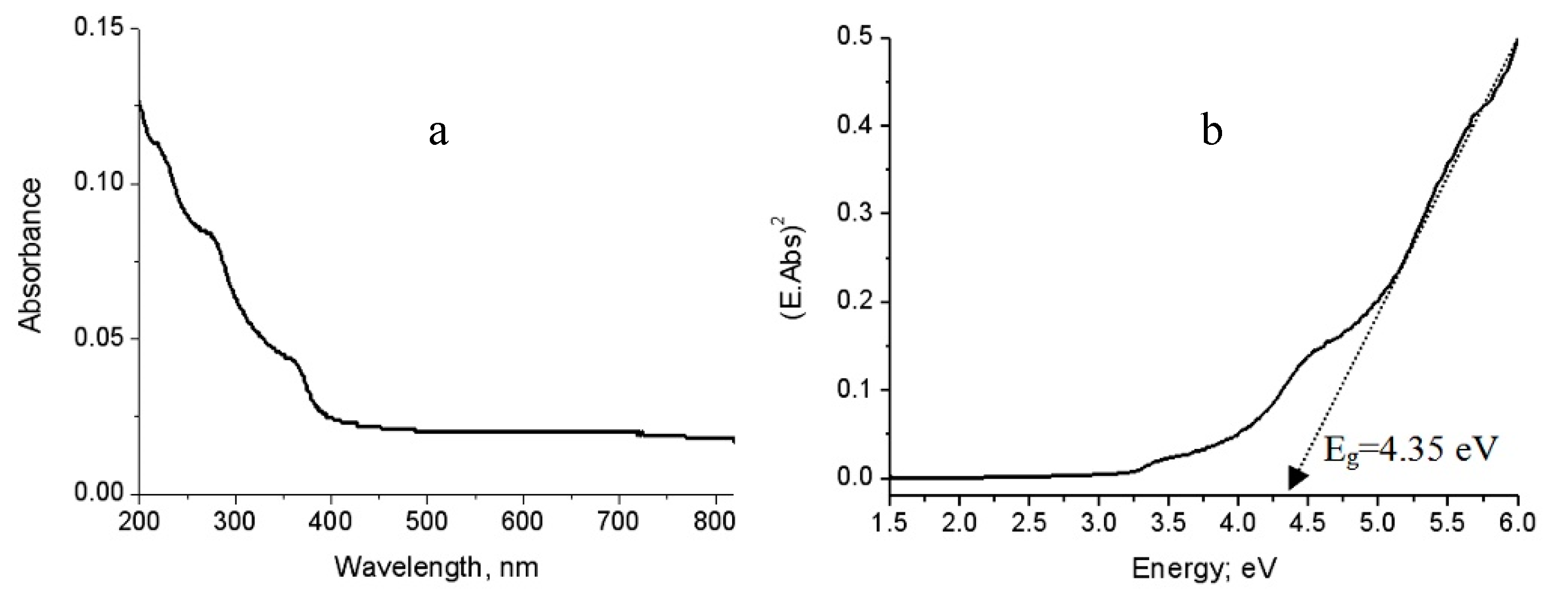
It is known that the bulk phase of alumina has a wide band gap energy in the range of 8.7–9.4 eV and is considered as an insulator. There have been many attempts to modify alumina structures to improve their optical properties. To investigate the optical behavior of the alumina nanoparticles and their nanocomposites, the UV-Vis absorption spectra have used as a powerful tool for providing important details about their absorbance and band gaps.
Figure 8a is displayed the UV-Vis absorbance of the pure aluminium oxide nanoparticles that was prepared without carbon species. It showed absorption edge near to 400 nm with three maxima at 220 nm, 290 nm and 360 nm. By comparing with the bulk aluminium oxide in which no absorptions are recorded within the spectral region 200–400 nm, the nano size plays positive role for improving the absorbance of alumina. These peaks were attributed to the intrinsic defect centers that have absorption bands in these regions [
17].
By using absorbance data, the energy band gap (E
g) could be calculated through the scientific relation between the absorbance coefficient of the materials (Abs) and the incident photon energy (E) according to the following equation [
18]:
Where E is calculated by multiplying the Planck’s constant (h) with speed of light (c) and divided by the wavelength (λ). The absorption coefficient (Abs) describes the optical absorption process. The value (2) is used for the allowed direct transitions.
Therefore, the band gap energy of the prepared alumina could be determined by plotting both (E.Abs)
2 and (E). By extending the straight line to the (E) axis, the optical band gap energy can be obtained when the value (E.Abs)
2 equal zero as shown in
Figure 8b.
Figure 8b showed that the band gap energy of the prepared aluminium oxide nanoparticles is 4.34 eV. Comparing with the bulk aluminium oxide, the band gap energy of alumina decreased from 8.7 eV to 4.34 eV because of the positive effect of the nano size for producing defect states in the band gap region agreeing with the results of Amirsalari and Shayesteh [
1].
By growing both carbon species and alumina nanoparticles through the explosive reactions of aluminium hydroxide, the optical properties of the prepared nanocomposites based on alumina are strongly modified as shown in
Figure 9,
Figure 10 and
Figure 11.
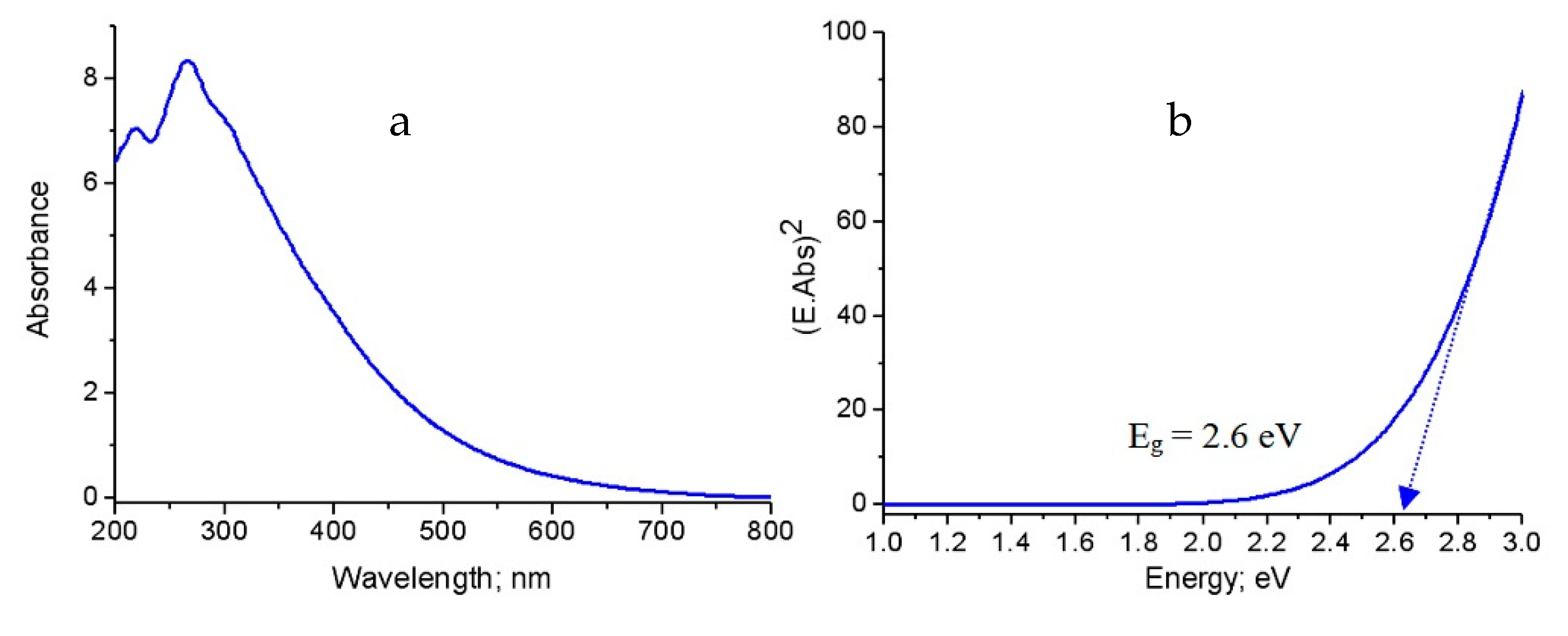
Figure 9a displayed the absorbance of the first nanocomposite NC-1. It showed that the absorption edge shifted to 700 nm with the maximum intensity at 280 nm. Additionally, the absorbance spectrum became wider as shown in
Figure 9a. It means that the material became active in the visible region. This finding was confirmed by determining the band gap energy. The band gap energy of NC-1 was 2.62 eV as seen in
Figure 9b. This band gap energy was quite different from those of aluminium oxides bulk (E
g = 8.7 eV) [
1] and nanoparticles (E
g = 4.35 eV). This large narrowing of the band gap energy may be explained according to the presence of new levels in the band gap region because of defect states of CNTs. This shrinkage is due to a consequence of many body effects on the conduction and the valence bands of aluminium oxides such as exchange energy due to electron–electron and electron-impurity interactions of CNTs. These effects lead to a narrowing of the band gap (red shift) [
17].
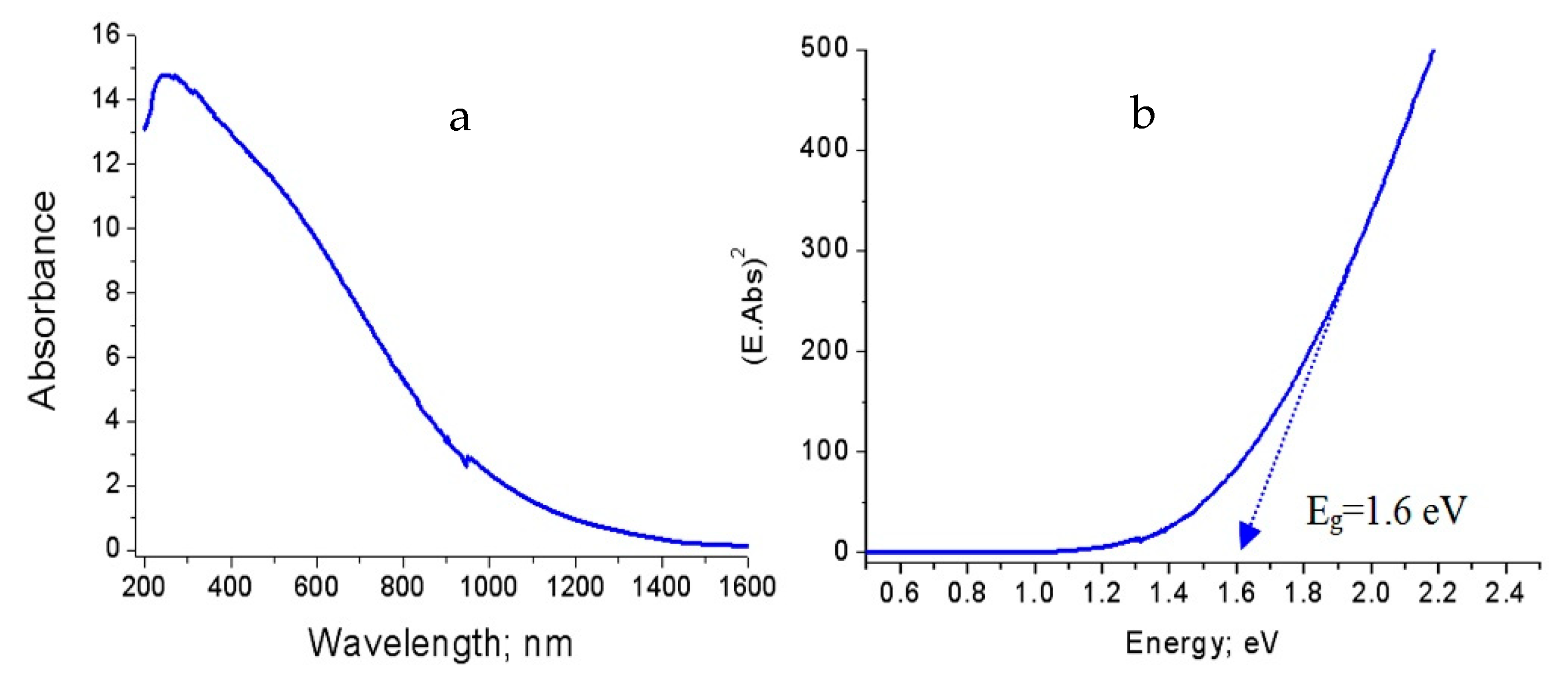
In case of the second nanocomposite NC-2,
Figure 10a reveals the absorbance spectrum in the wavelength range of 200–1500 nm. This broad absorbance indicates that the material became very sensitive for visible light. The calculated band gap energy of NC-2 confirmed this observation as shown in
Figure 10b. It revealed 1.6 eV for the second nanocomposite. Compared to the band gap energy of both the first nanocomposite and alumina nanoparticles, more narrowing of band gap of alumina happened after changing methanol to ethanol as a source of carbon.
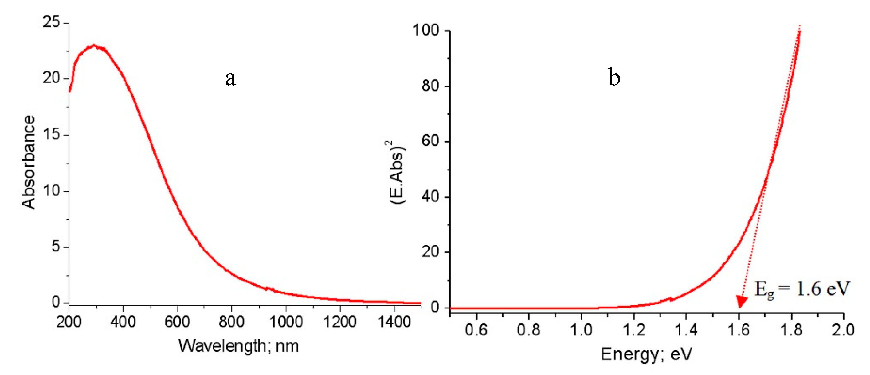
These observations were also noticed in the third nanocomposite NC-3 as shown in
Figure 11.
Figure 11a showed a broad absorbance of NC-3 in the wavelength range 200–1100 nm. It means that the third nanocomposite became optically active in the visible region. The band gap energy of NC-3 was 1.6 eV as shown in
Figure 11b.
Comparing with the band gap energy of the second nanocomposite, the third nanocomposite has similar effect in the visible region. This large narrowing of the band gap energy of the prepared nanocomposites is probably due to chemical interaction between CNTs and aluminium oxide nanoparticles, generating a new energy level to reduce the band gap energy [
19,
20]. The narrowing of the band gap energy of the second and third nanocomposites may be due to the growth of another type of carbon species in which the core-shell structure formed. The link between the core (aluminium oxide) and the shell (carbon nanolayers) generates other energy levels, decreasing the band gap energy. These experimental results are summarized in
Figure 12 to indicate the conversion of bulk alumina from non-optical behavior to photo-active products.
2.4. Fast Removal of Pollutants
In order to test the solar activity of the prepared nanocomposites, the photocatalytic removal of the green dyes (naphthol green B; NGB) from water were studied using NC-1, NC-2, and NC-3 in the presence of sunlight. At the same time, to evaluate the effect of explosion reactions on the optical properties of alumina, the solar activity of the prepared nanoparticles of alumina was measured by the same process and compared with the solar activity of the nanocomposites.
The concentration of the green pollutants was determined by the intensity of the absorption of naphthol green B at λmax= 720 nm. A blank experiment indicated that NGB has high stability in sunlight because no variation in the concentration was noticed in sunlight.
A green mixture with one of the prepared materials was illuminated for ten minutes in sunlight. After that the concentration of the green dyes was determined through measuring the absorbance of the green solution. These results are displayed in
Figure 13 and
Figure 14. The percentage of naphthol green B in presence of NC-1 sharply decreased after illuminating 10 min in sunlight as shown in
Figure 13.
Figure 14 showed 80.9% removal of the green pollutant. In case of using NC-2, the removal increased to be 92.3% in the same period, while complete decolorization and removal of the green dyes were nearly achieved after using NC-3.
Figure 13 showed approximately complete disappearance for the absorbance of the green dyes. Additionally,
Figure 14 revealed that 99% of the green dyes was removed by NC-3 after irradiating 10 min in sunlight. In case of using the nanoparticles of alumina in sunlight, slight variation was noticed for the intensity of the absorbance of the green dyes after 10 min as shown in
Figure 13. Additionally,
Figure 14 showed low removal for the green dyes 2.1%. It means that the prepared nanoparticles of alumina are ineffective in sunlight. The comparison between the results of alumina nanoparticles and their nanocomposites indicated that the explosive technique is very effective for converting non-optical alumina to optical nanomaterials. By comparing with our previous results of zinc oxide nanoparticles, the prepared nanocomposites were more effective in sunlight than the most familiar optical materials because the Al doped ZnO nanoparticles caused reduction for the band gap energy from 3.29 to 3.23 eV leading to complete removal of NGB in 6 h under sunlight [
21]. Recently, the complete decolorization and decomposition of green dye were achieved after 1.25–1.3 h of irradiation of UV light using the aluminium zinc oxide nanocomposite-coated CNTs and nanofibers [
21,
22].


