
Scaffold Hopping in Discovery of HIV-1 Non-Nucleoside Reverse Transcriptase Inhibitors: From CH(CN)-DABOs to CH(CN)-DAPYs

 , and
, and
Abstract
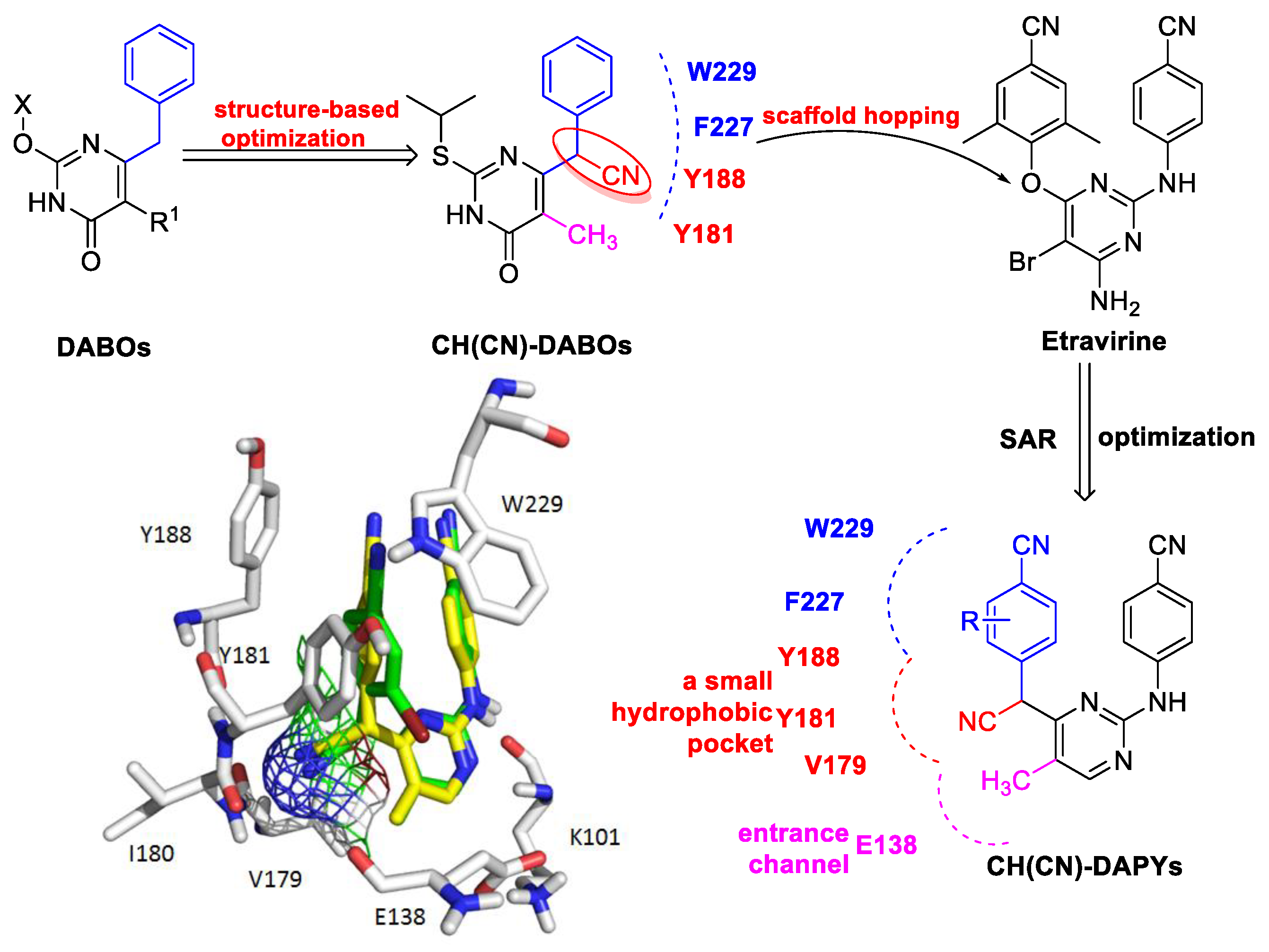
:1. Introduction
2. Results and Discussion
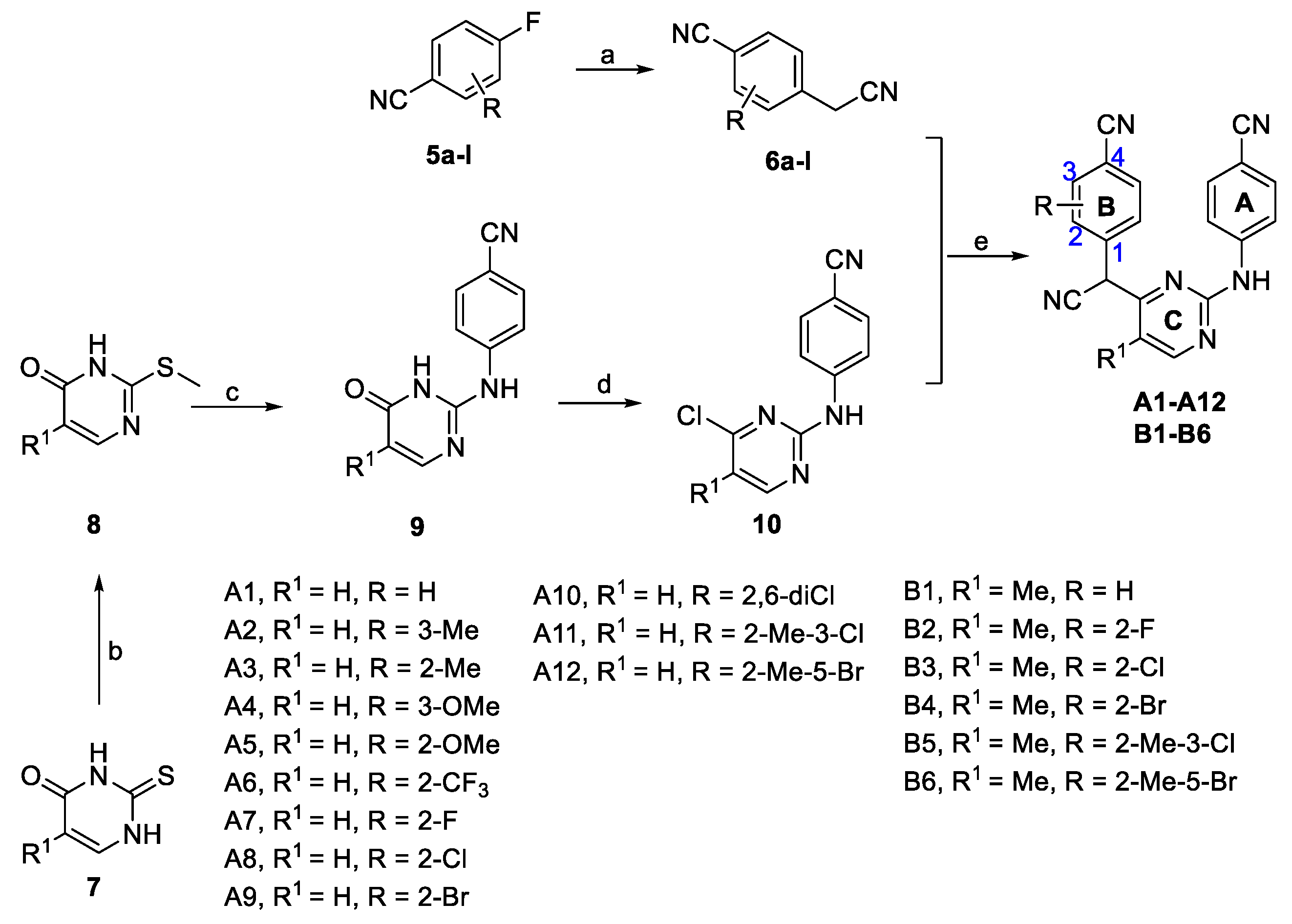
2.1. Chemistry
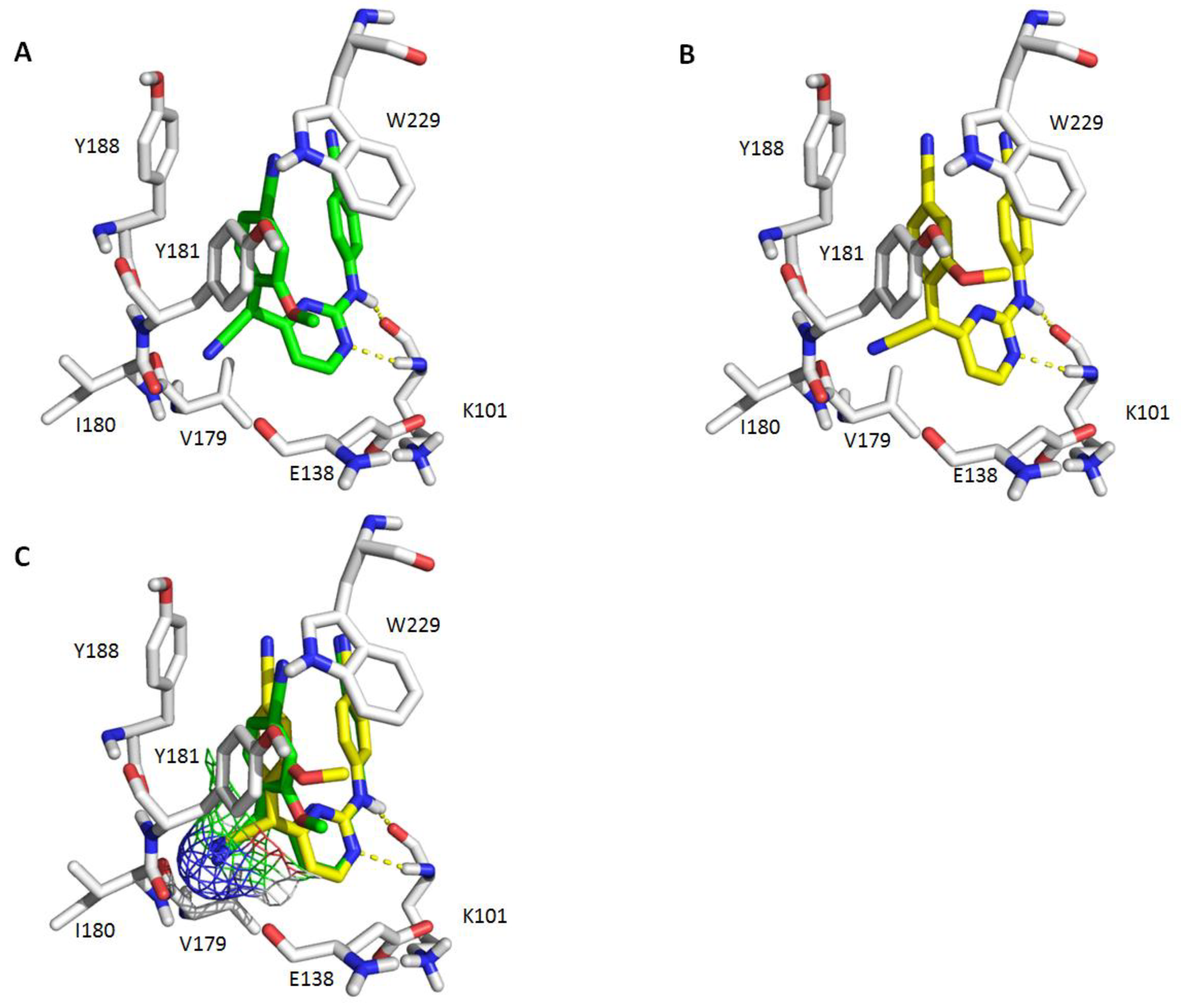
2.2. Antiviral Activity of Compounds A1–A12 and Binding Conformation Analysis
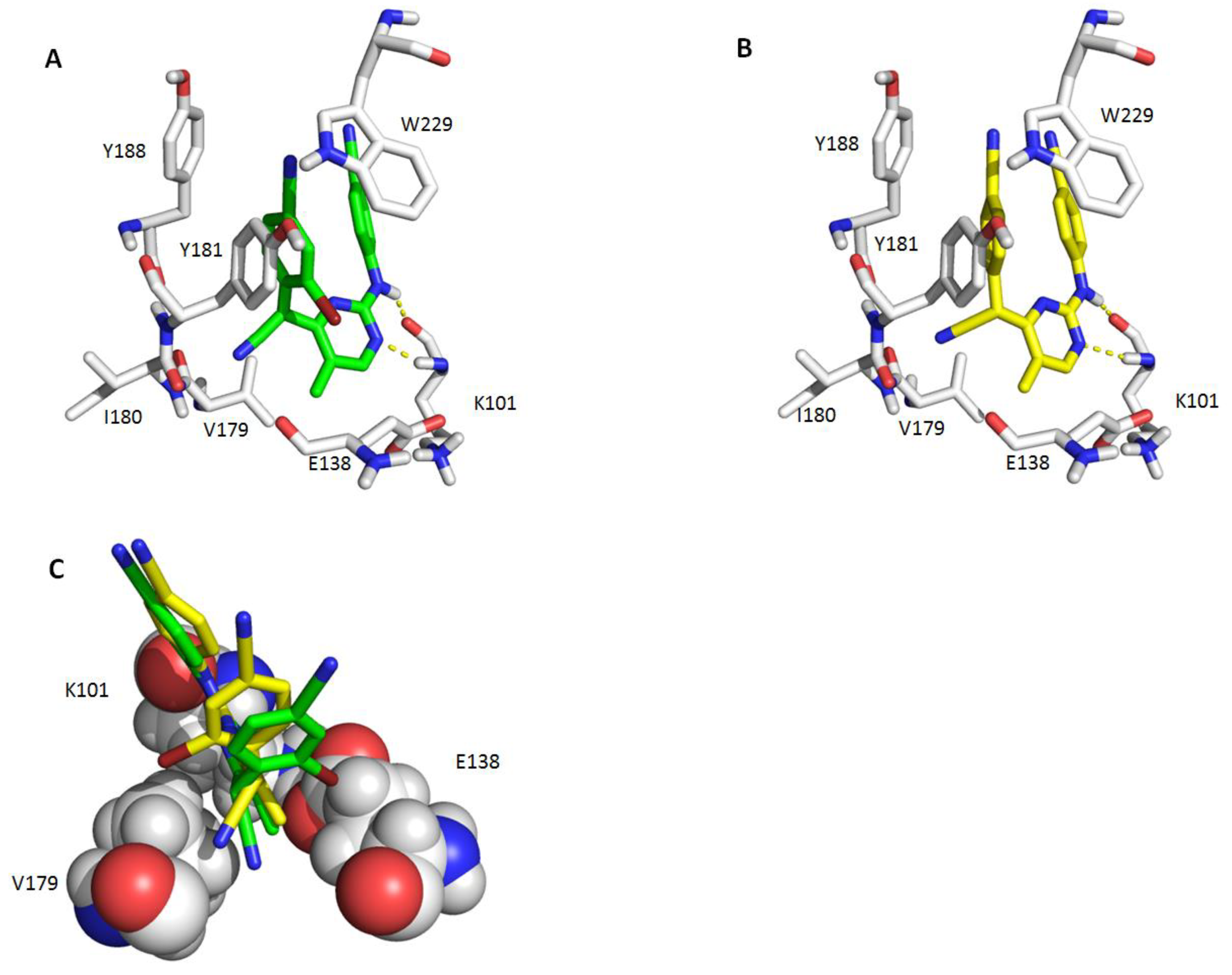
2.3. Antiviral Activity of Compounds B1–B6 and Binding Conformation Analysis
2.4. Antiviral Activity against Mutants of Compounds A1–A12 and B1–B6 and Enzymatic Assay of Compounds B1–B6
3. Materials and Methods
3.1. Synthesis
3.1.1. General Procedure for the Preparation of Compounds 6a-l
3.1.2. General Procedure for the Preparation of Compounds A1–A12 and B1–B6
3.2. In Vitro Anti-HIV Assay
3.3. Reverse Transcriptase Assay
3.4. Molecular Modeling
4. Conclusions
Supplementary Materials
Author Contributions
Funding
Acknowledgments
Conflicts of Interest
References
- Ren, J.; Stammers, D.K. Structural basis for drug resistance mechanisms for non-nucleoside inhibitors of HIV reverse transcriptase. Virus Res. 2008, 134, 157–170. [Google Scholar] [CrossRef] [PubMed]
- Namasivayam, V.; Vanangamudi, M.; Kramer, V.G.; Kurup, S.; Zhan, P.; Liu, X.; Kongsted, J.; Byrareddy, S.N. The Journey of HIV-1 Non-Nucleoside Reverse Transcriptase Inhibitors (NNRTIs) from Lab to Clinic. J. Med. Chem. 2019, 62, 4851–4883. [Google Scholar] [CrossRef] [PubMed]
- Zhan, P.; Pannecouque, C.; De Clercq, E.; Liu, X. Anti-HIV Drug Discovery and Development: Current Innovations and Future Trends. J. Med. Chem. 2016, 59, 2849–2878. [Google Scholar] [CrossRef] [PubMed]
- Zhang, H.; Tian, Y.; Kang, D.; Huo, Z.; Zhou, Z.; Liu, H.; De Clercq, E.; Pannecouque, C.; Zhan, P.; Liu, X. Discovery of uracil-bearing DAPYs derivatives as novel HIV-1 NNRTIs via crystallographic overlay-based molecular hybridization. Eur. J. Med. Chem. 2017, 130, 209–222. [Google Scholar] [CrossRef]
- Qin, B.; Jiang, X.; Lu, H.; Tian, X.; Barbault, F.; Huang, L.; Qian, K.; Chen, C.H.; Huang, R.; Jiang, S.; et al. Diarylaniline derivatives as a distinct class of HIV-1 non-nucleoside reverse transcriptase inhibitors. J. Med. Chem. 2010, 53, 4906–4916. [Google Scholar] [CrossRef] [Green Version]
- Sergeyev, S.; Yadav, A.K.; Franck, P.; Michiels, J.; Lewi, P.; Heeres, J.; Vanham, G.; Arien, K.K.; Vande Velde, C.M.; De Winter, H.; et al. 2,6-Di(arylamino)-3-fluoropyridine Derivatives as HIV Non-Nucleoside Reverse Transcriptase Inhibitors. J. Med. Chem. 2016, 59, 1854–1868. [Google Scholar] [CrossRef]
- Sang, Y.; Han, S.; Han, S.; Pannecouque, C.; De Clercq, E.; Zhuang, C.; Chen, F. Follow on-based optimization of the biphenyl-DAPYs as HIV-1 nonnucleoside reverse transcriptase inhibitors against the wild-type and mutant strains. Bioorg. Chem. 2019, 89, 102974. [Google Scholar] [CrossRef]
- Casado, J.L. Liver toxicity in HIV-infected patients receiving novel second-generation nonnucleoside reverse transcriptase inhibitors etravirine and rilpivirine. AIDS Rev. 2013, 15, 139–145. [Google Scholar]
- Casado, J.L.; Banon, S. Recent advances in rilpivirine: New data and promising treatment option. AIDS Rev. 2014, 16, 172–181. [Google Scholar]
- Sang, Y.; Han, S.; Pannecouque, C.; De Clercq, E.; Zhuang, C.; Chen, F. Ligand-Based Design of Nondimethylphenyl-Diarylpyrimidines with Improved Metabolic Stability, Safety, and Oral Pharmacokinetic Profiles. J. Med. Chem. 2019, 62, 11430–11436. [Google Scholar] [CrossRef]
- Zhuang, C.; Pannecouque, C.; De Clercq, E.; Chen, F. Development of non-nucleoside reverse transcriptase inhibitors (NNRTIs): Our past twenty years. Acta Pharm. Sin. B 2019. [Google Scholar] [CrossRef]
- Gu, S.X.; Yang, S.Q.; He, Q.Q.; Ma, X.D.; Chen, F.E.; Dai, H.F.; Clercq, E.D.; Balzarini, J.; Pannecouque, C. Design, synthesis and biological evaluation of cycloalkyl arylpyrimidines (CAPYs) as HIV-1 NNRTIs. Bioorg. Med. Chem. 2011, 19, 7093–7099. [Google Scholar] [CrossRef] [PubMed]
- Jin, K.; Yin, H.; De Clercq, E.; Pannecouque, C.; Meng, G.; Chen, F. Discovery of biphenyl-substituted diarylpyrimidines as non-nucleoside reverse transcriptase inhibitors with high potency against wild-type and mutant HIV-1. Eur. J. Med. Chem. 2018, 145, 726–734. [Google Scholar] [CrossRef]
- Sang, Y.; Han, S.; Pannecouque, C.; De Clercq, E.; Zhuang, C.; Chen, F. Conformational restriction design of thiophene-biphenyl-DAPY HIV-1 non-nucleoside reverse transcriptase inhibitors. Eur. J. Med. Chem. 2019, 182, 111603. [Google Scholar] [CrossRef]
- Han, S.; Sang, Y.; Wu, Y.; Tao, Y.; Pannecouque, C.; De Clercq, E.; Zhuang, C.; Chen, F.E. Molecular Hybridization-Inspired Optimization of Diarylbenzopyrimidines as HIV-1 Nonnucleoside Reverse Transcriptase Inhibitors with Improved Activity against K103N and E138K Mutants and Pharmacokinetic Profiles. ACS Infect. Dis. 2019. [Google Scholar] [CrossRef]
- Wan, Z.Y.; Tao, Y.; Wang, Y.F.; Mao, T.Q.; Yin, H.; Chen, F.E.; Piao, H.R.; De Clercq, E.; Daelemans, D.; Pannecouque, C. Hybrid chemistry. Part 4: Discovery of etravirine-VRX-480773 hybrids as potent HIV-1 non-nucleoside reverse transcriptase inhibitors. Bioorg. Med. Chem. 2015, 23, 4248–4255. [Google Scholar] [CrossRef]
- Han, S.; Sang, Y.; Wu, Y.; Tao, Y.; Pannecouque, C.; De Clercq, E.; Zhuang, C.; Chen, F.E. Fragment hopping-based discovery of novel sulfinylacetamide-diarylpyrimidines (DAPYs) as HIV-1 nonnucleoside reverse transcriptase inhibitors. Eur. J. Med. Chem. 2020, 185, 111874. [Google Scholar] [CrossRef]
- Han, S.; Lei, Y.; Pannecouque, C.; De Clercq, E.; Zhuang, C.; Chen, F. Fragment-based discovery of sulfur-containing diarylbenzopyrimidines as novel nonnucleoside reverse transcriptase inhibitors. Chin. Chem. Lett. 2020, 31, 764–768. [Google Scholar] [CrossRef]
- Ji, L.; Chen, F.E.; De Clercq, E.; Balzarini, J.; Pannecouque, C. Synthesis and anti-HIV-1 activity evaluation of 5-alkyl-2-alkylthio-6-(arylcarbonyl or alpha-cyanoarylmethyl)-3,4-dihydropyrimidin-4(3H)-ones as novel non-nucleoside HIV-1 reverse transcriptase inhibitors. J. Med. Chem. 2007, 50, 1778–1786. [Google Scholar] [CrossRef]
- Wang, Y.; Du, Y.; Huang, N. A survey of the role of nitrile groups in protein-ligand interactions. Future Med. Chem. 2018, 10, 2713–2728. [Google Scholar] [CrossRef]
- Kang, D.; Fang, Z.; Li, Z.; Huang, B.; Zhang, H.; Lu, X.; Xu, H.; Zhou, Z.; Ding, X.; Daelemans, D.; et al. Design, Synthesis, and Evaluation of Thiophene [3,2-d]pyrimidine Derivatives as HIV-1 Non-nucleoside Reverse Transcriptase Inhibitors with Significantly Improved Drug Resistance Profiles. J. Med. Chem. 2016, 59, 7991–8007. [Google Scholar] [CrossRef] [PubMed]
- Yang, Y.; Kang, D.; Nguyen, L.A.; Smithline, Z.B.; Pannecouque, C.; Zhan, P.; Liu, X.; Steitz, T.A. Structural basis for potent and broad inhibition of HIV-1 RT by thiophene [3,2-d]pyrimidine non-nucleoside inhibitors. Elife 2018, 7, e36340. [Google Scholar] [CrossRef] [PubMed]
- Zeng, Z.S.; Liang, Y.H.; Feng, X.Q.; Chen, F.E.; Pannecouque, C.; Balzarini, J.; De Clercq, E. Lead optimization of diarylpyrimidines as non-nucleoside inhibitors of HIV-1 reverse transcriptase. ChemMedChem 2010, 5, 837–840. [Google Scholar] [CrossRef] [PubMed]
- Pannecouque, C.; Daelemans, D.; De Clercq, E. Tetrazolium-based colorimetric assay for the detection of HIV replication inhibitors: Revisited 20 years later. Nat. Protoc. 2008, 3, 427–434. [Google Scholar] [CrossRef] [PubMed]
- Pauwels, R.; Balzarini, J.; Baba, M.; Snoeck, R.; Schols, D.; Herdewijn, P.; Desmyter, J.; De Clercq, E. Rapid and automated tetrazolium-based colorimetric assay for the detection of anti-HIV compounds. J. Virol. Methods 1988, 20, 309–321. [Google Scholar] [CrossRef]
- Balzarini, J.; Karlsson, A.; Perez-Perez, M.J.; Camarasa, M.J.; Tarpley, W.G.; De Clercq, E. Treatment of human immunodeficiency virus type 1 (HIV-1)-infected cells with combinations of HIV-1-specific inhibitors results in a different resistance pattern than does treatment with single-drug therapy. J. Virol. 1993, 67, 5353–5359. [Google Scholar] [CrossRef] [Green Version]
- Balzarini, J.; Karlsson, A.; Perez-Perez, M.J.; Vrang, L.; Walbers, J.; Zhang, H.; Oberg, B.; Vandamme, A.M.; Camarasa, M.J.; De Clercq, E. HIV-1-specific reverse transcriptase inhibitors show differential activity against HIV-1 mutant strains containing different amino acid substitutions in the reverse transcriptase. Virology 1993, 192, 246–253. [Google Scholar] [CrossRef]
- Balzarini, J.; Karlsson, A.; Vandamme, A.M.; Perez-Perez, M.J.; Zhang, H.; Vrang, L.; Oberg, B.; Backbro, K.; Unge, T.; San-Felix, A.; et al. Human immunodeficiency virus type 1 (HIV-1) strains selected for resistance against the HIV-1-specific [2’,5’-bis-O-(tert-butyldimethylsilyl)-3’-spiro- 5”-(4”-amino-1”,2”-oxathiole-2”,2”-dioxide)]-beta-D-pentofurano syl (TSAO) nucleoside analogues retain sensitivity to HIV-1-specific nonnucleoside inhibitors. Proc. Natl. Acad. Sci. USA 1993, 90, 6952–6956. [Google Scholar] [CrossRef] [Green Version]
- Auwerx, J.; North, T.W.; Preston, B.D.; Klarmann, G.J.; De Clercq, E.; Balzarini, J. Chimeric human immunodeficiency virus type 1 and feline immunodeficiency virus reverse transcriptases: Role of the subunits in resistance/sensitivity to non-nucleoside reverse transcriptase inhibitors. Mol. Pharmacol. 2002, 61, 400–406. [Google Scholar] [CrossRef] [Green Version]
- Singer, V.L.; Jones, L.J.; Yue, S.T.; Haugland, R.P. Characterization of PicoGreen reagent and development of a fluorescence-based solution assay for double-stranded DNA quantitation. Anal. Biochem. 1997, 249, 228–238. [Google Scholar] [CrossRef] [Green Version]
Sample Availability: Samples are available from the authors. |


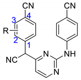
| Compounds | R | EC50 (μM) 2 | CC50 (μM) 3 | SI 4 |
|---|---|---|---|---|
| HIV-1 IIIB 1 | ||||
| A1 | H | 3.27 ± 2.25 | 32.7 ± 9.9 | 10 |
| A2 | 3-Me | 1.17 ± 0.34 | 16.1 ± 3.6 | 14 |
| A3 | 2-Me | 0.069 ± 0.008 | 10.9 ± 3.6 | 157 |
| A4 | 3-OMe | 11.74 ± 12.13 | 78.4 ± 10.4 | 7 |
| A5 | 2-OMe | 0.059 ± 0.011 | 25.8 ± 8.7 | 434 |
| A6 | 2-CF3 | 0.49 ± 0.30 | 6.2 ± 1.4 | 13 |
| A7 | 2-F | 0.24 ± 0.16 | 10.3 ± 4.5 | 43 |
| A8 | 2-Cl | 0.063 ± 0.009 | 4.6 ± 2.7 | 72 |
| A9 | 2-Br | 0.079 ± 0.02 | 3.4 ± 0.3 | 42 |
| A10 | 2,6-diCl | 0.082 ± 0.02 | 4.5 ± 1.3 | 55 |
| A11 | 2-Me-3-Cl | 0.30 ± 0.03 | 6.1 ± 0.5 | 20 |
| A12 | 2-Me-5-Br | 0.24 ± 0.04 | 13.2 ± 1.6 | 54 |
| Nevirapine | 0.20 ± 0.10 | >15.0 | >76 | |
| Etravirine | 0.005 ± 0.001 | >4.6 | >1012 | |
| Efavirenz | 0.003 ± 0.001 | >6.3 | >3939 | |
| Rilpivirine | 0.0016 ± 0.0003 | 5.9 ± 0.3 | 3747 |

| Compounds | R | EC50 (μM) 2 | CC50 (μM) 3 | SI 4 |
|---|---|---|---|---|
| HIV-1 IIIB 1 | ||||
| B1 | H | 0.30 ± 0.11 | 108.6 ± 80.0 | 356 |
| B2 | 2-F | 0.04 ± 0.02 | 20.6 ± 3.6 | 586 |
| B3 | 2-Cl | 0.01 ± 0.01 | 9.6 ± 2.7 | 920 |
| B4 | 2-Br | 0.006 ± 0.001 | 6.6 ± 1.3 | 1086 |
| B5 | 2-Me-3-Cl | 0.02 ± 0.008 | 8.6 ± 2.2 | 370 |
| B6 | 2-Me-5-Br | 0.008 ± 0.004 | 9.8 ± 3.5 | 1215 |
| Nevirapine | 0.20 ± 0.10 | >15.0 | >76 | |
| Etravirine | 0.005 ± 0.001 | >4.6 | >1012 | |
| Efavirenz | 0.003 ± 0.001 | >6.3 | >3939 | |
| Rilpivirine | 0.0016 ± 0.0003 | 5.9 ± 0.3 | 3747 |
| Compounds | EC50(μM) 1 | IC50 (μM) 2 | |||||
|---|---|---|---|---|---|---|---|
| L100I | K103N | Y181C | E138K | F227L + V106A | K103N + Y181C | ||
| A1 | ≥12.31 | >32.70 | 11.26 ± 1.51 | >32.70 | >32.70 | >32.70 | ND 3 |
| A2 | >16.07 | >16.07 | >16.07 | >16.07 | >16.07 | >16.07 | ND |
| A3 | 5.03 ± 0.55 | 1.96 ± 0.45 | 1.71 ± 0.13 | 3.04 ± 0.39 | >10.93 | >10.93 | ND |
| A4 | ≥61.36 | ≥62.18 | ≥48.12 | ≥56.55 | ≥61.00 | ≥65.72 | ND |
| A5 | 4.72 ± 0.79 | 0.68 ± 0.16 | 1.25 ± 0.58 | 1.63 ± 0.32 | ≥12.42 | >25.85 | ND |
| A6 | >6.23 | >6.23 | 2.29 ± 0.02 | >6.23 | >6.23 | >6.23 | ND |
| A7 | >10.36 | >10.36 | >10.36 | >10.36 | >10.36 | >10.36 | ND |
| A8 | ≥2.37 | 2.13 ± 0.22 | 1.87 ± 0.21 | 2.20 ± 0.35 | >4.59 | >4.59 | ND |
| A9 | >3.35 | ≥1.88 | 1.88 ± 0.42 | 2.31 ± 0.02 | >3.35 | >3.35 | ND |
| A10 | 2.32 ± 0.48 | 1.78 ± 0.12 | 2.20 ± 0.27 | 2.19 ± 0.22 | >4.49 | >4.49 | ND |
| A11 | 2.32 ± 0.48 | >6.11 | >6.11 | >6.11 | >6.11 | >6.11 | ND |
| A12 | >13.23 | 2.52 ± 0.42 | 2.78 ± 0.36 | 3.73 ± 0.59 | >13.23 | >13.23 | ND |
| B1 | 8.03 ± 4.43 | 3.95 ± 0.22 | 7.05 ± 0.96 | 4.92 ± 0.3 | ≥58.97 | >108.57 | 1.46 ± 0.09 |
| B2 | 3.04 ± 1.13 | 1.07 ± 0.37 | 1.59 ± 0.18 | 0.97 ± 0.14 | >20.63 | >20.63 | 0.22 ± 0.05 |
| B3 | 0.82 ± 0.15 | 0.08 ± 0.02 | 0.26 ± 0.02 | 0.14 ± 0.03 | 4.04 ± 0.70 | >9.56 | 0.05 ± 0.03 |
| B4 | 0.51 ± 0.19 | 0.08 ± 0.03 | 0.16 ± 0.17 | 0.11 ± 0.04 | 2.35 ± 0.18 | >6.62 | 0.06 ± 0.03 |
| B5 | 1.82 ± 0.4 | 0.39 ± 0.07 | 1.02 ± 0.2 | 0.60 ± 0.10 | 1.66 ± 0.27 | >8.58 | 0.07 ± 0.02 |
| B6 | 0.86 ± 0.19 | 0.06 ± 0.01 | 0.33 ± 0.10 | 0.17 ± 0.06 | 1.52 ± 0.15 | >9.79 | 0.06 ± 0.02 |
| Nevirapine | 0.98 ± 0.39 | 4.70 ± 0.63 | 6.07 ± 1.55 | 0.20 ± 0.15 | ≥4.28 | >15.02 | 0.45 ± 0.25 |
| Etravirine | 0.008 ± 0.002 | 0.003 ± 0.001 | 0.017 ± 0.003 | 0.008 ± 0.004 | 0.014 ± 0.005 | 0.04 ± 0.01 | 0.01 ± 0.009 |
| Efavirenz | 0.04 ± 0.008 | 0.08 ± 0.01 | 0.006 ± 0.001 | 0.005 ± 0.002 | 0.28 ± 0.07 | 0.24 ± 0.06 | ND |
| Rilpivirine | 0.002 ± 0.001 | 0.001 ± 0.0002 | 0.003 ± 0.001 | 0.003 ± 0.001 | 0.04 ± 0.01 | 0.009 ± 0.002 | ND |
© 2020 by the authors. Licensee MDPI, Basel, Switzerland. This article is an open access article distributed under the terms and conditions of the Creative Commons Attribution (CC BY) license (http://creativecommons.org/licenses/by/4.0/).
Share and Cite
Li, T.-T.; Pannecouque, C.; De Clercq, E.; Zhuang, C.-L.; Chen, F.-E. Scaffold Hopping in Discovery of HIV-1 Non-Nucleoside Reverse Transcriptase Inhibitors: From CH(CN)-DABOs to CH(CN)-DAPYs. Molecules 2020, 25, 1581. https://doi.org/10.3390/molecules25071581
Li T-T, Pannecouque C, De Clercq E, Zhuang C-L, Chen F-E. Scaffold Hopping in Discovery of HIV-1 Non-Nucleoside Reverse Transcriptase Inhibitors: From CH(CN)-DABOs to CH(CN)-DAPYs. Molecules. 2020; 25(7):1581. https://doi.org/10.3390/molecules25071581
Chicago/Turabian StyleLi, Ting-Ting, Christophe Pannecouque, Erik De Clercq, Chun-Lin Zhuang, and Fen-Er Chen. 2020. "Scaffold Hopping in Discovery of HIV-1 Non-Nucleoside Reverse Transcriptase Inhibitors: From CH(CN)-DABOs to CH(CN)-DAPYs" Molecules 25, no. 7: 1581. https://doi.org/10.3390/molecules25071581