Article

Antibacterial Activity of Fluorobenzoylthiosemicarbazides and Their Cyclic Analogues with 1,2,4-Triazole Scaffold

1
Department of Pharmaceutical Microbiology, Faculty of Pharmacy, Medical University of Lublin, Chodźki 1, 20-093 Lublin, Poland
2
Department of Organic Chemistry, Faculty of Pharmacy, Medical University of Lublin, Chodźki 4a, 20-093 Lublin, Poland
3
FQS-Fujitsu Poland, Parkowa 11, 33-332 Kraków, Poland
4
Institute of Applied Radiation Chemistry, Faculty of Chemistry, Lodz University of Technology, Żeromskiego 116, 90-924 Lodz, Poland
*
Author to whom correspondence should be addressed.
Molecules 2021, 26(1), 170; https://doi.org/10.3390/molecules26010170
Submission received: 2 November 2020 / Revised: 18 December 2020 / Accepted: 28 December 2020 / Published: 31 December 2020

Abstract

:
The development of drug-resistant bacteria is currently one of the major challenges in medicine. Therefore, the discovery of novel lead structures for the design of antibacterial drugs is urgently needed. In this structure–activity relationship study, a library of ortho-, meta-, and para-fluorobenzoylthiosemicarbazides, and their cyclic analogues with 1,2,4-triazole scaffold, was created and tested for antibacterial activity against Gram-positive bacteria strains. While all tested 1,2,4-triazoles were devoid of potent activity, the antibacterial response of the thiosemicarbazides was highly dependent on substitution pattern at the N4 aryl position. The optimum activity for these compounds was found for trifluoromethyl derivatives such as 15a, 15b, and 16b, which were active against both the reference strains panel, and pathogenic methicillin-sensitive and methicillin-resistant Staphylococcus aureus clinical isolates at minimal inhibitory concentrations (MICs) ranging from 7.82 to 31.25 μg/mL. Based on the binding affinities obtained from docking, the conclusion can be reached that fluorobenzoylthiosemicarbazides can be considered as potential allosteric d-alanyl-d-alanine ligase inhibitors.

1. Introduction

A huge number of drugs and clinical candidates for development contain halogen atoms. For a long time, predominantly only the steric and hydrophobic factors of halogens were considered when trying to exploit their role in ligand recognition and target–ligand binding complex stabilization. Today, the ability of halogens to form favorable intermolecular interactions, such as halogen bonds, hydrogen bonds, and multipolar interactions that significantly contribute to the stability of target–ligand complexes has been well recognized, and widely utilized in the rational design of new therapeutics against clinically significant targets [1,2,3,4]. According to an analysis by Hernandes and co-workers [5], the majority of halogenated drugs are fluorine drugs (57%), followed by chlorine ones (38%), while those with bromine are rare (4%), and only a few iodinated drugs are known (1%) due to their relatively instability, caused by the high polarizability of the C–I bond.
Regarding the fluorinated drugs and drug candidates, fluorine substitutions have been extensively investigated in drug research and there are some important reasons that justify their predominance. For instance, several studies have documented that selective introduction of a trifluoromethyl group or even a single fluorine atom into a in a key position of a therapeutic molecule significantly enhances its pharmacokinetic and physicochemical properties, such as improved metabolic stability and membrane permeability [6]. The increased binding affinity of fluorine-containing drug candidates to molecular targets has also been proved in several cases [7,8,9,10,11,12,13,14,15,16,17,18,19]. Currently, several fluorinated pharmaceuticals are widely used in clinical practice, and nearly one-third of the 100 top-selling drugs are organofluorine compounds. The most common among these are fluoroquinolone antibiotics, antimalarials, antidepressants, antivirals, anesthetics, anticancers, cholesterol-lowering drugs, anti-inflammatory drugs, and asthma drugs, and several reviews on potential therapeutic applications of fluorinated molecules have been published in recent years [4,20,21,22]. For these reasons, we explored the antibacterial potential within the structure–activity relationship of a library of 1-fluorobenzoyl-4-aryl(alkyl)thiosemicarbazides. Previously, we had searched for the N1 substituents which would improve the antibacterial potency of thiosemicarbazide-based compounds and had found that the fluorobenzoyl group might constitute a particularly useful series [23]. Other research groups also confirmed the thiosemicarbazide structure as a novel scaffold for potential antibacterial agents [24,25,26,27,28,29,30,31,32,33], although a systematic survey of the effects of the N1 and N4 terminal substituents on antibacterial potency has not been presented thus far. Thus, in an attempt to more closely define the factors controlling the potency of these compounds, we prepared three series, with ortho-, meta-, and para-fluorobenzoyl groups, and determined their antibacterial activity in vitro with the structure–activity relationship. In carrying out this study of the structure–activity relationship, our strategy was four-stage. First, we sought to investigate the effect of the aromatic fluorine substitution (ortho, meta, and para) and the N4 alkyl and aryl groups. Second, we sought to extend the antibacterial assay on methicillin-sensitive and methicillin-resistant Staphylococcus aureus clinical isolates. Third, we set out to alter the linear structure of the thiosemicarbazide by the synthesis of cyclic analogues with a 1,2,4-triazole scaffold, and to compare these with the corresponding thiosemicarbazide counterparts. Finally, our results of docking our analogues to the allosteric site of d-alanyl-d-alanine ligase concurred with the pioneering reports made by Ameryckx et al. [24,25] on the molecular basis of the antibacterial activity of benzoylthiosemicarbazides.

2. Results and Discussion

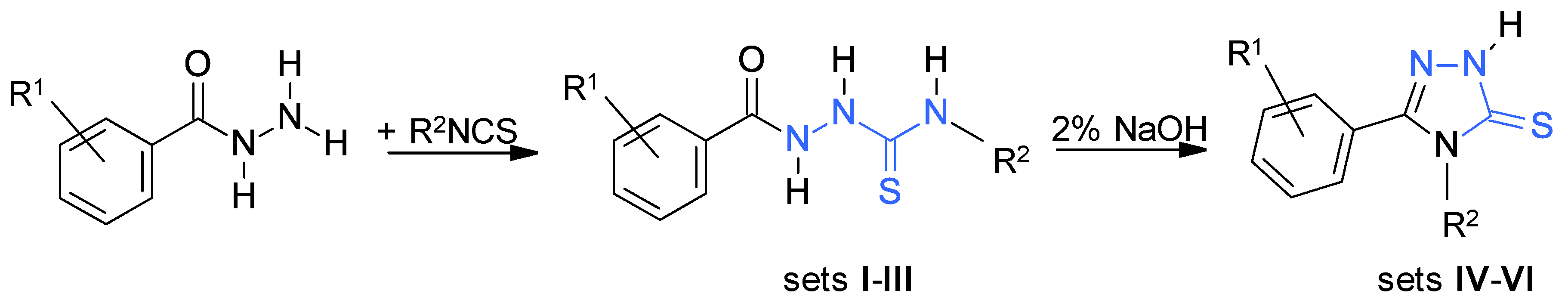
2.1. Synthetic Protocols

The synthesis of ortho-, meta-, and para-fluorobenzoylthiosemicarbazides (sets IIII) and their cyclic analogues with a 1,2,4-triazole-5-thione scaffold (sets IVVI) was performed according to a well-established synthetic route, described elsewhere [23,34] (Scheme 1). The reaction of the corresponding fluorobenzoylhydrazide with related alkyl/aryl isothiocyanate in boiling ethanol gave 1-fluorobenzoyl-4-aryl/(alkyl)thiosemicarbazides (sets IIII) in 40–90% yields. Resulting compounds were cyclized in boiling aqueous sodium hydroxide to give corresponding 1,2,4-triazole-3-thiones (sets IVVI), which were obtained in 48–90% yields.

2.2. Antibacterial Screening

The thiosemicarbazides (sets IIII) were evaluated for their antibacterial activity against a panel of the reference Gram-positive bacteria using the standard two-fold serial microdilution assay described by the Clinical and Laboratory Standards Institute [35]. The results for the minimum inhibitory concentrations (MICs) are reported in Table 1. Commercially available antibiotic cefuroxime was used as the reference control.
A few general conclusions arose from the antibacterial assay, which can be summarized as follows. The most important is that within set I of ortho-fluorobenzoylthiosemicarbazides 1a16a, the compounds that bear substituents at the N4 aryl position that are electron acceptors were more active than those that are either unsubstituted or bear electron donors. Electron-withdrawing substituents in the meta position (7a, 9a, 11a, 13a, 15a) generally increased the antibacterial potency, and within meta electron-withdrawing compounds the activity follows the trend: meta-CF3 15a (MIC 7.82–15.63 µg/mL; 0.02–0.04 mM) > meta-Cl 9a (MIC 7.82–62.5 µg/mL; 0.02–0.19 mM) ~ meta-Br 11a (MIC 7.82–62.5 µg/mL; 0.02–0.17 mM) > meta-F 7a = meta-I 13a (n.a.; MICs at 2000 µg/mL; 6.51 and 4.82 mM, respectively). The assessment of the results of the meta electron-withdrawing compounds was made in three terms: (i) an electronic one, expressed by the Hammett substituent constant σm [37,38,39] and inductive substituent constant σI; (ii) a steric, expressed by the molar refractivity MR, the Taft’s constant Es [39,40,41,42], and the Charton steric parameter σν [39,43,44,45,46]; and (iii) a hydrophobic, expressed by cLogP and π, the Hansch substituent constant [39,47], and led to the conclusion that the single most important physicochemical property that could explain the variance in the MICs of these compounds is the Hammett substituent constant σm. Indeed, as presented in Figure 1, a good correlation exists between the MICs of the meta-substituted compounds 7a, 9a, 11a, 13a, 15a, and the σm constants, whereas no correlation could be found between their MICs and steric or hydrophobic parameters. Comparative σm, σI, σν, MR, Es, π, and cLogP values are listed in Table S1. The fact that the improvement in activity of these compounds is generally correlated with the Hammett substituent constant σm (F < I < Cl < Br < CF3) suggests that the electronic properties of the meta electron withdrawing substituents control the antibacterial activity. While it remains unclear what role, if any, the electronegativity of the meta substituents plays in antibacterial activity, possible interpretations of the correlation between the antibacterial activity of 7a, 9a, 11a, 13a, 15a, and σm are therefore that: (i) antibacterial activity increases with decreasing electron density on the thiosemicarbazide scaffold because enhancement in binding of the thiosemicarbazide to the molecular target is created through the increased positive charge character of the N4 aryl ring or whole thiosemicarbazide scaffold; or (ii) antibacterial activity increases with decreasing pKa of the thiosemicarbazide core (NH-NH-C(=S)-NH), because the compound must be deprotonated to be active (deprotonation hypothesis) [48]. A combination of both effects is possible. A second important observation is that although the steric substituent constants, such as the Taft Es constant or Charton σν parameter, do not directly explain the trend in antibacterial activity of meta compounds 7a, 9a, 11a, 13a, and 15a, the most potent antibacterial meta-CF3 15a proved to possess the lowest Taft Es constant and the highest Charton σν parameter, thus suggesting that the bulkiness effect of the CF3 substituent induced an improvement in antibacterial activity. The last but not least observation that arose from the antibacterial assay of the thiosemicarbazides of set I, is that substitutions in the para position of the N4 phenyl ring abolished activity (Table 1, compounds 6a, 8a, 10a, 12a, and 14a). The only exceptions were para-CF3 16a and para-Cl 10a, which inhibited the growth of Bacillus cereus at a concentration similar to cefuroxime, and Micrococcus luteus at the MIC of 7.82 µg/mL (0.02 mM). The fact that 10a and 16a still possess antibacterial activity suggest that although the protein binding pocket provides sufficient space for additional groups at the para position, the combination of both steric and specific, i.e., hydrophobic and electrostatic or intermolecular interactions might be the most prominent factor regulating the activity of the para substituted thiosemicarbazides of set I.
For the meta-fluorobenzoylthiosemicarbazides of set II (1b16b), a reverse trend of antibacterial activity was observed compared to ortho set I. Clearly, within set II, para-Cl 10b with MICs in the range of 7.82–15.63 µg/mL (0.02–0.19 mM) against Staphylococcus spp., B. subtilis, and M. luteus showed a clear preference for the potency whereas the antibacterial activity of para-CF3 16b was comparable to that of meta-CF3 15b. Furthermore, meta-I 13b, in contrast to inactive meta-I 13a, was able to inhibit the growth of S. aureus, B. subtilis, B. cereus, and M. luteus at the concentration of 15.63 µg/mL (0.04 mM), whereas the antibacterial activity of meta-Cl 9b and meta-Br 11b against S. aureus strains was two to four times higher compared to their isomers, 9a and 11a, respectively. Clearly, within set II, meta-Cl and para-Cl compounds (9b, 10b), and meta-CF3 and para-CF3 compounds (15b, 16b), proved to be the most effective. No important contribution(s), however, of electronic, steric, and/or hydrophobic factors on antibacterial trend within set II was found.
Finally, the antibacterial activity of the para-fluorobenzoylthiosemicarbazides of set III (1c16c) was tested. Although these compounds were generally less effective than their ortho (set I) and meta (set II) isomers, the efficiency of the compounds with chloro (9c, 10c), bromo (11c, 12c), and iodo (13c, 14c) substitutions against B. cereus was still close to cefuroxime. In addition, these compounds with MICs at 15.63 µg/mL (0.05 mM for 9c, 10c, and 0.04 mM for 11c, 12c, 13c, 14c) showed good inhibitory action against M. luteus. The presence of other substituents, both an electron withdrawing (CF3, F) and an electron donating (CH3), reduced potency, whereas the presence of a propyl, butyl, phenyl, or naphthyl group abolished activity; similarly to the antibacterial trends observed for sets I and II.
To confirm antibacterial potential of the most potent thiosemicarbazides, 15a, 16a, 9b, 10b, 11b, 15b, 16b, these compounds were subsequently assayed against a panel of pathogenic methicillin-sensitive and methicillin-resistant Staphylococcus aureus clinical isolates. As indicated from the data collected in Table 2, the best antibacterial response, with MICs in the range of 3.91–15.63 µg/mL, was noted for trifluoromethyl derivatives; meta 15a (0.02–0.04 mM) and para 16b (0.04–0.09 mM). Meta trifluoromethyl derivative 15b showed an inhibitory action at a slightly higher concentration (MICs 7.82–31.25 µg/mL; 0.04–0.08 mM), while with the remaining compounds activity was lost.
Since SAR analysis of the fluorobenzoylthiosemicarbazides (sets IIII) seemed to be more complicated than we expected, as mentioned above, we also prepared their cyclic analogues with a 1,2,4-triazole-3-thione scaffold (sets IVVI), and submitted them to antimicrobial assay to determine the role, if any, of the thiosemicarbazide core NH-NH-C(=S)-NH on antibacterial activity. As shown in Table 3, all tested triazoles showed much lower antibacterial potency compared to their acyclic precursors (e.g., 15a vs. 15at) or were even inactive (e.g., 13b vs. 13bt). It is important to note, that none of them showed potent antibacterial activity. Thus, these results confirmed that, although the linear NH-NH-C(=S)-NH core is a key structural element for antibacterial activity, the electronic effect of the N1 and N4 substituents on the whole thiosemicarbazide skeleton is equally important. It seems equally possible, however, that the N1 and N4 substituents could lead to structural benefits, either by providing favorable conformation of the thiosemicarbazide in the molecular target binding site, or by providing favorable intermolecular contacts. Of course, a combination of both electronic and steric effects is also possible. Another important observation is that the hydrophobic factor cLogP, alone, had a negligible influence on the antibacterial response of the tested thiosemicarbazides and s-triazoles.

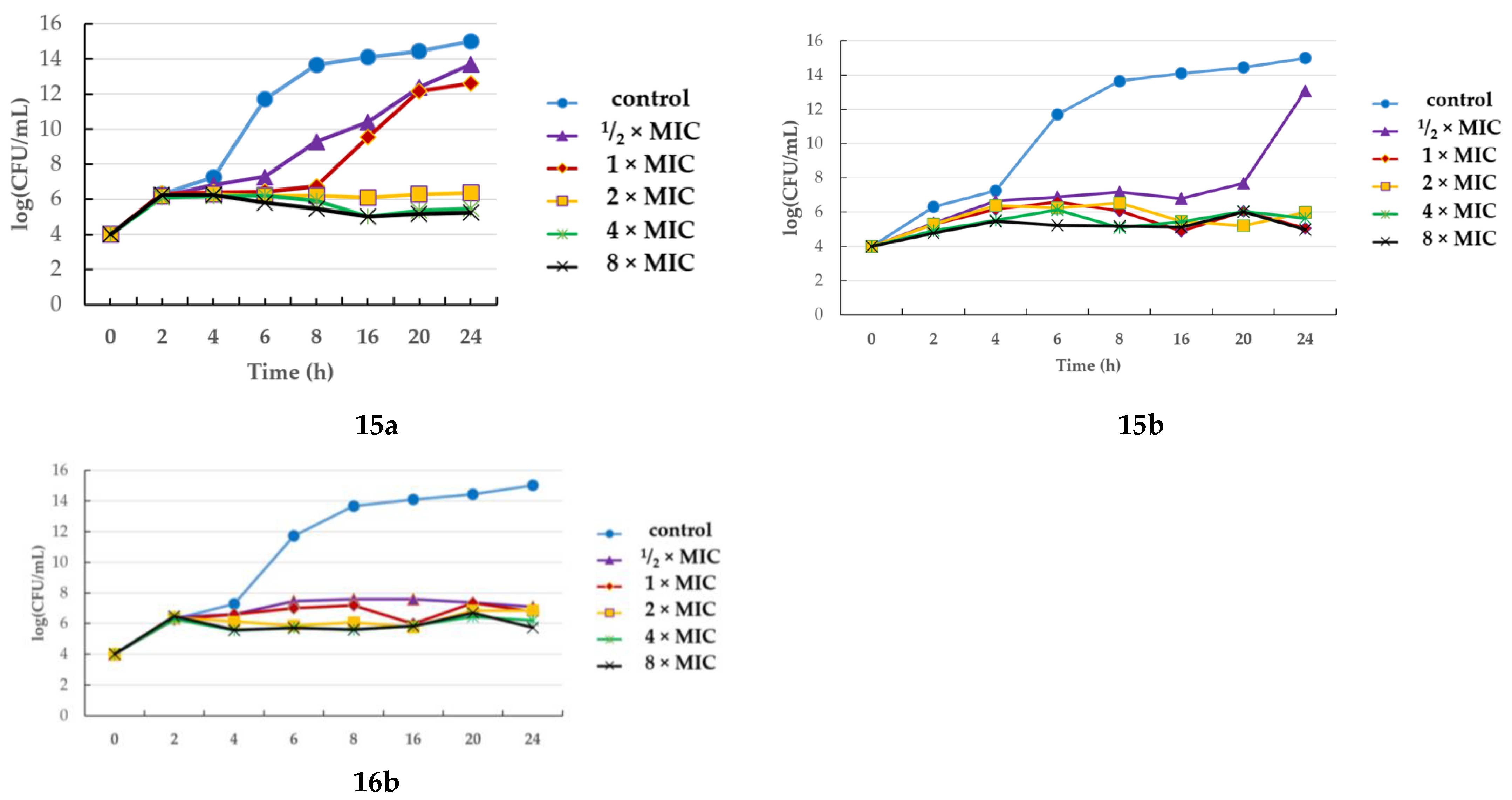
2.3. Assessing Bactericidal/Bacteriostatic Characteristics

Although the molecular mechanism of the antibacterial activity of thiosemicarbazides is still unknown, their inhibitory action towards bacterial topoisomerases has been proposed as one possible explanation [49,50,51]. Experiments aimed at identifying possible additional targets of these compounds are currently ongoing, but so far the only conclusive results have been yielded by Ameryckx et al., for ortho-hydroxybenzoylthiosemicarbazides [24,25]. According to these results, d-alanyl-d-alanine ligase (Ddl) might be considered as a potential bacterial target for the thiosemicarbazides, with the N1 ortho-hydroxybenzoyl substitution and subsequent time kill assay for representative analogue, with 2,3-dichlorophenyl group at the N4 position (PR), proving that it acts as a bactericidal agent. Generally, when considering the molecular mechanism by which antibacterial agents control microorganisms, the mode of their action can also be classified according to whether they lead to the death of the microbe (bactericidal action), or inhibit its growth (bacteriostatic action). Thus, to distinguish whether our thiosemicarbazides were bactericidal or bacteriostatic in nature, we next submitted them to a minimal bactericidal concentration (MBC) assay. Upon analysis, all compounds were found to be bacteriostatic (MBC/MIC >4) toward all bacteria strains tested (data not shown). Subsequent results from the concentration dependent time-kill assay, for the most potent antibacterial agents 15a, 15b, 16b against S. aureus ATCC 25923 over a period of 24 h, confirmed this observation (Figure 2). A concentration-dependent and time-dependent bacteriostatic activity was found for all the compounds tested, even at a high concentration of 4 × MIC or 8 × MIC. Bactericidal activity, defined as a reduction of at least 99.9% of the total count of CFU/mL in the original inoculum within a period of 24 h, was not observed.
As a general rule, antibacterial agents that disrupt the cell wall or cell membrane, or interfere with essential enzymes are often bactericidal, whereas those that inhibit protein synthesis, e.g., bacterial topoisomerases, or interfere with metabolic processes tend to be bacteriostatic [52,53,54]. Taking this fact into account, the results of the MBC assay are not surprising. In fact, in our previous studies [23], on the thiosemicarbazides 9a and 10a, we showed that their inhibitory action against S. aureus DNA gyrase was very weak; compound 10a was able to inhibit ~50% of S. aureus DNA gyrase activity at as high a concentration as 100 µg/mL, whereas the inhibitory activity of 9a at this concentration was even lower (~33%). Collectively, the results of the MBC assay further confirm the assumption that, for our bacteriostatic thiosemicarbazides of sets IIII, bacterial topoisomerases are not primary factors contributing to their antibacterial activity.

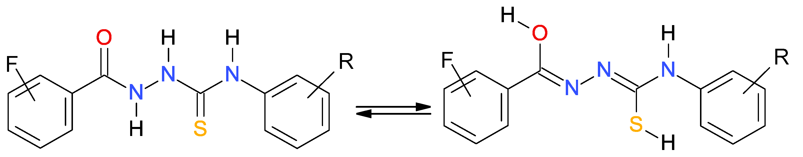
2.4. Thiol−ThioneTautomerismintheThiosemicarbazides
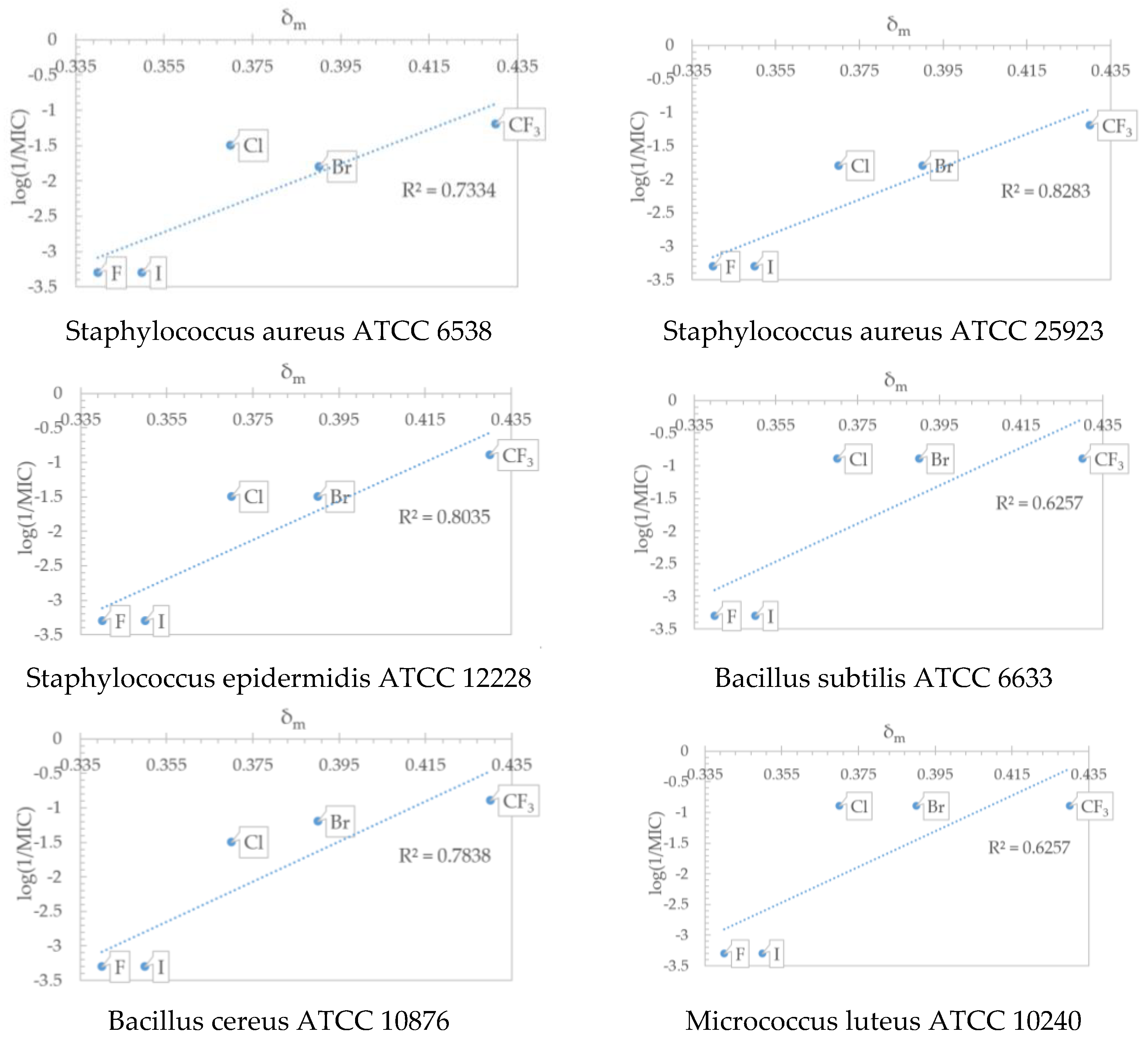
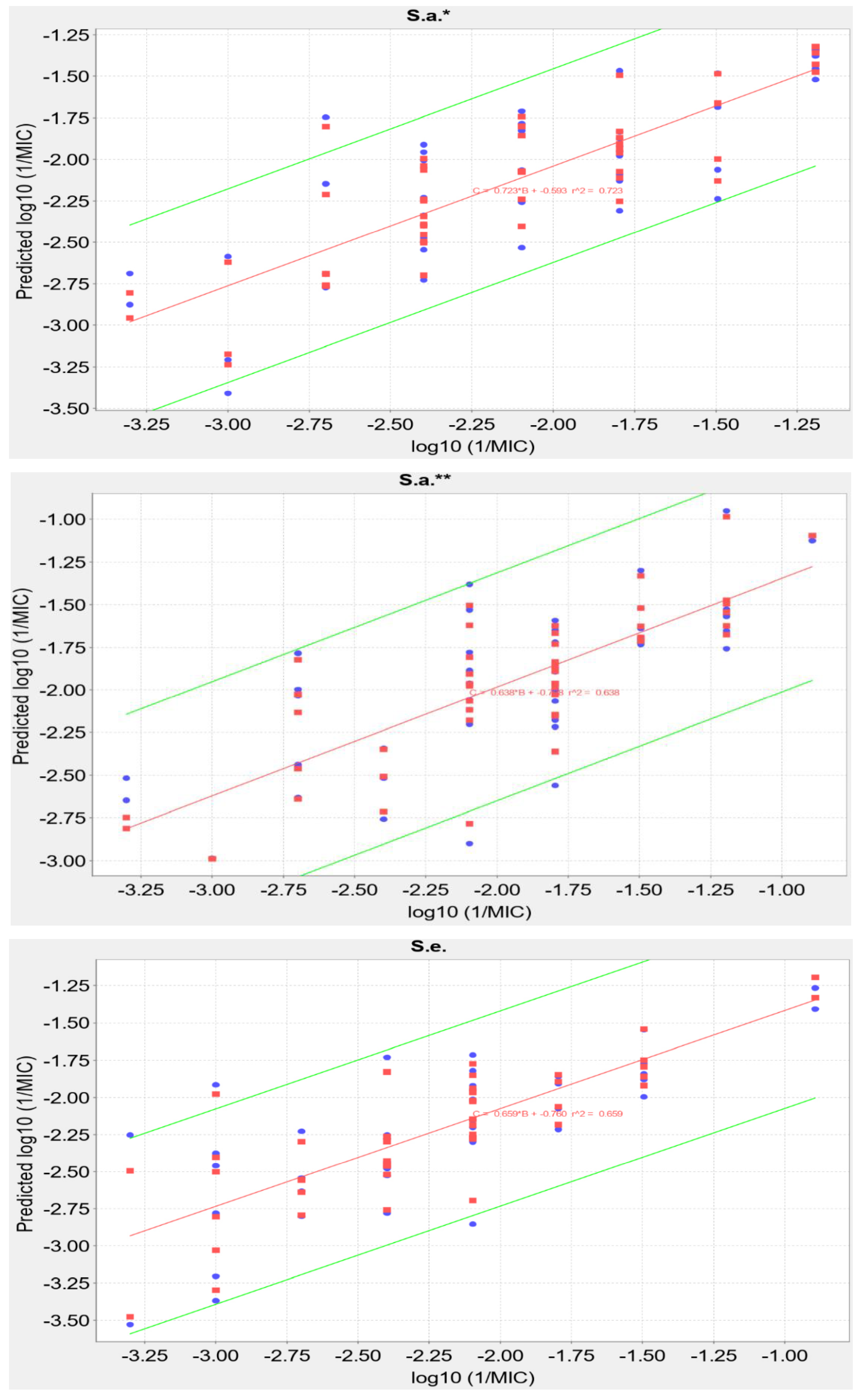
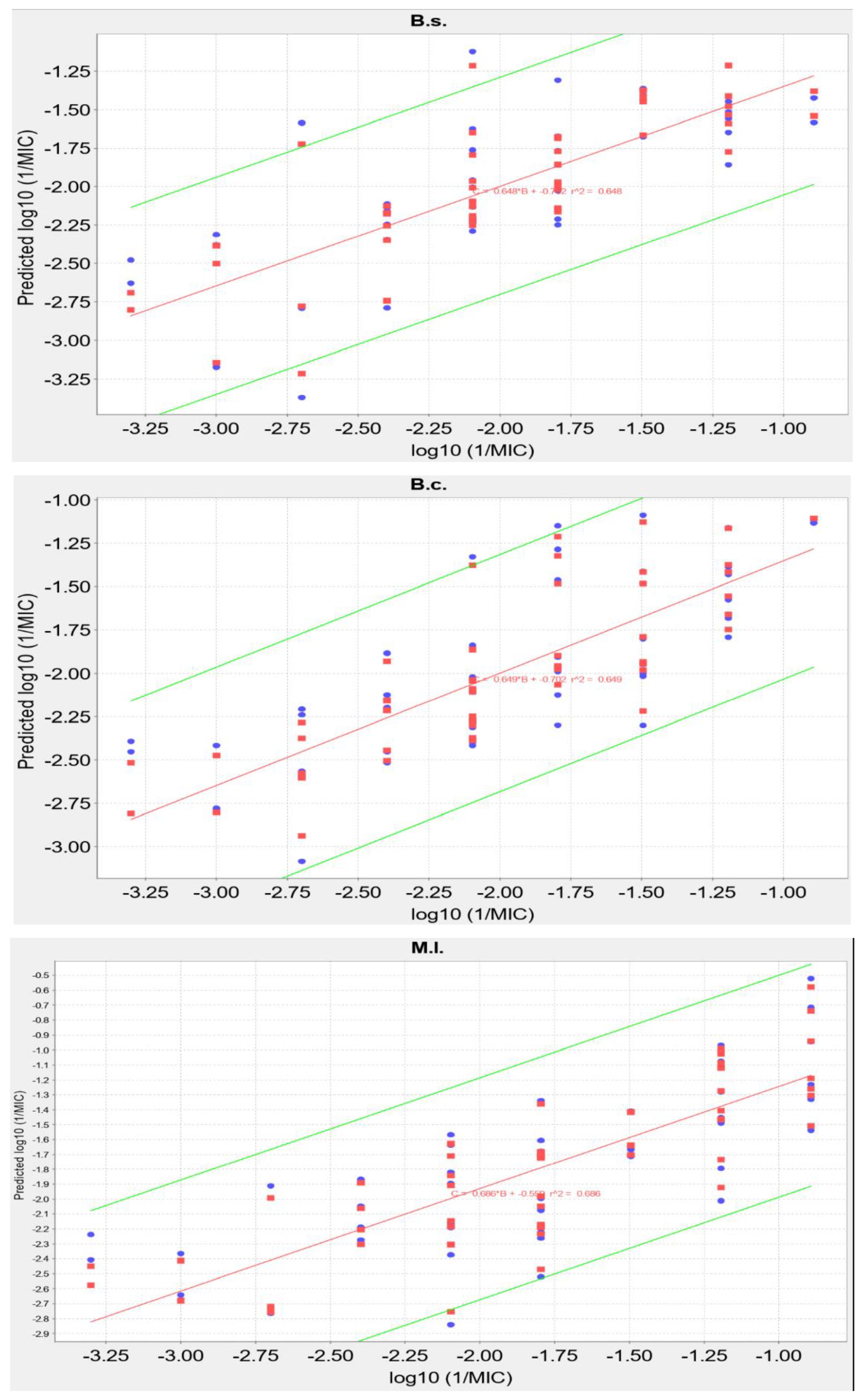
In an attempt to extract a general background of the relationship between the antibacterial activity and physicochemical properties of the tested thiosemicarbazides and s-triazoles, we also performed a QSAR analysis. Before experiments, however, the relative stabilities of the thiosemicarbazide tautomeric forms were studied, based on computational analysis.
As is known, the proton transfers influence conformational and electronical changes in a molecule, and thus induce changes in its flexibility, polarity, lipophilicity, and acceptor–donor capacity. All these factors have a significant impact on biological activity, including cell membrane permeability, metal complexation, and inter- and intra-molecular interactions. Since the thiosemicarbazides studied here formally can exist in tautomeric thione (C=S) and thiole (C–S) forms [24,25,55,56] (Figure 3), we evaluated theoretically their relative Gibbs free energies. For this purpose, geometries of the thione and thiole forms of the thiosemicarbazides were optimized at the DFT level of theory, using the ωB97X-D functional [57] expressed in the def2-TZVP basis set [58], as implemented in the Gaussian16 program [59]. Vibrational analysis was used to ensure that the optimized geometry corresponded to a stationary point representing a minimum on the potential energy surface (3n − 6 real vibrations). A SMD continuum solvent model [60] was used to model the aqueous solution. Our results indicated that the thiosemicarbazides in the thiole tautomeric form were less stable by about 21.8 kcal/mol, and thus their presence in the aqueous solution can be disregarded. The relative stability in the tautomers of s-triazole was studied previously, and it was established that thione tautomer is also the energetically most stable form [61].

2.5. QSAR Analysis

Having determined the relative stabilities of the thiosemicarbazides and s-triazoles, QSAR analysis was performed using SCIGRESS software (SCIGRESS v 3.4.4 www.scigress.com), to find correlations between the structure and the minimal inhibitory concentration for each microbial species separately. For these studies, only the thiosemicarbazides and s-triazoles that were active in the antibacterial assay were included. For the purpose of the analysis the values of the MICs were converted to Log10(1/MIC). The 466 descriptors used included both topological and quantum descriptors, calculated by the PM6/COSMO method (see Supplementary Materials). A fully automated procedure of descriptor selection by the enhanced replacement method (ERM) was applied to select five descriptors for best fitting of the linear regression equation (Table 4). The obtained values of R2 are in the range of 0.64–0.72, so, while some correlation was observed, it is not high enough for predicting unknown compounds. It is worth noticing, however, that the presence of a chlorine atom in the molecule appears to be beneficial for activity, while a bromine atom apparently decreases activity against S. aureus ATCC 25923. The resulting equations and fit (R2 and leave-one-out cross validation R2) are shown in Table 4. The graphs of linear regressions and cross validations are shown in Figure 4.

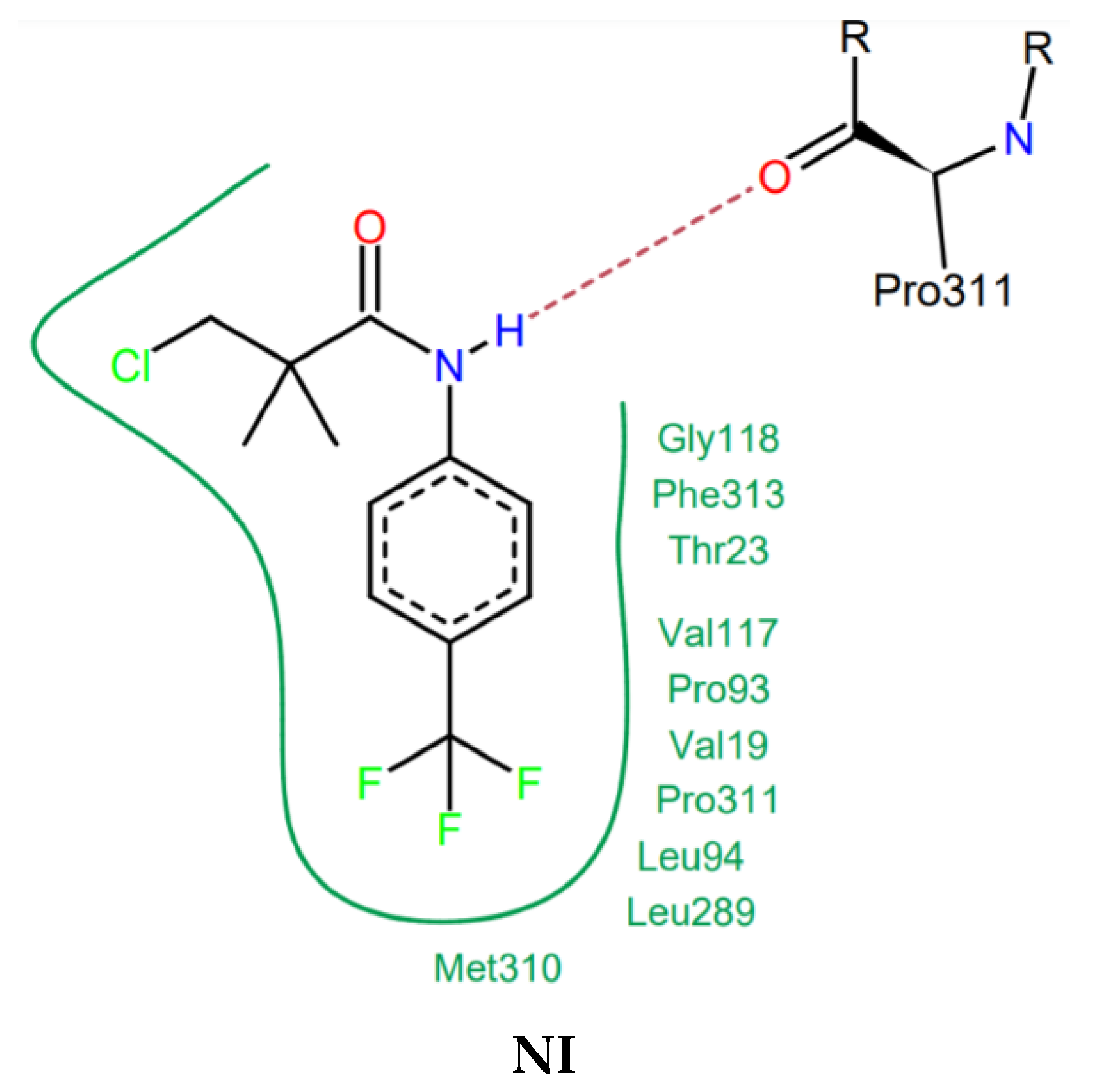
2.6. Docking Studies

As mentioned above, recently Ameryckx et al. [24,25] reported the discovery of a series of 4-aryl-1-(2-hydroxybenzoyl)thiosemicarbazides as promising allosteric inhibitors of d-alanyl-d-alanine ligase (Ddl); an essential enzyme in bacterial cell wall biosynthesis, and an important target for developing new antibacterial agents. This enzyme catalyzes the formation of d-alanile:d-alanine dipeptide, the crucial precursor of bacterial cell wall peptidoglycan, by two half-reactions. In the first half-reaction, Ddl uses one d-alanine and one ATP as substrates to produce a phosphorylated d-alanine intermediate. In the second half-reaction, Ddl uses a second d-alanine substrate to complete the reaction to the normal d-alanine:d-alanine dipeptide product [62]. According to previously reported results, the 4-(3,4-dichlorophenyl)-1-(2-hydroxybenzoyl)thiosemicarbazide (PR, Figure 5) was identified as one of the most potent Ddl inhibitors, with a bactericidal activity against Gram-positive bacteria, including multidrug resistant strains and, at the same time, very low cytotoxicity on a THP-1 human monocytic cell line. Although the authors concluded their extensive experiments with the judgment that the ortho hydroxy group is critical for the inhibitory action of benzoylthiosemicarbazides against Ddl, none of the tested compounds that were inactive on ligase exhibited an antibacterial activity at the same time. This fact supports the hypothesis that the inhibition of Ddl activity could explain, at least in part, the antibacterial effect observed for the benzoylthiosemicarbazides.
Given the above, we decided to dock the best of our benzoylthiosemicarbazides, 9a, 11a, 15a, 16a, 9b, 10b, 11b, 13b, 15b, and 16b, to the allosteric site of S. aureus Ddl (PDB code: 2I80). The best docking scores for the thiosemicarbazides, and previously reported inhibitor PR, are listed in Table 5. In Figure 5 the best binding pose for representative model compound 9a is presented. The best binding poses of the remaining thiosemicarbazides at the allosteric site of S. aureus Ddl are available in the Supplementary Materials.
According to the docking results presented in Table 5, all the thiosemicarbazides are identified as binding at an allosteric site of Ddl, with much higher affinity than the native inhibitor. Their predicted binding sites overlap well with the binding site of the native inhibitor NI, and in all cases at least one hydrogen bond is observed between the NH group of the thiosemicarbazide skeleton and Pro311, Met 310, or Pro93 (see Supporting Material). The binding of the thiosemicarbazides 9a, 11a, 15a, 16a, 9b, 10b, 11b, 13b, 15b, and 16b is further stabilized by numerous hydrophobic interactions with surrounding residues, and most of these interactions are identical to those in the crystal structure of native amide NI in complex with S. aureus Ddl [62]. Notably, the binding mode of our thiosemicarbazides in the allosteric site of Ddl is stabilized by similar intermolecular interactions as predicted for previously discovered inhibitor PR (see Figure 5). Thus, the docking results indicate that our fluorobenzoylthiosemicarbazides can be at least theoretically considered as potential d-alanyl-d-alanine allosteric ligase inhibitors. Enzymatic experiments, however, are needed to confirm this assumption.

3. Materials and Methods

3.1. Chemistry

All commercial reactants and solvents were purchased from either Sigma-Aldrich (St. Louis, MO, USA) or Alfa Aesar (Karlsruhe, Germany), with the highest purity and used without further purification. The melting points were determined on a Fischer-Johns block (Fischer Scientific, Schwerte, Germany) and were uncorrected. Elemental analyses were determined by a AMZ-CHX elemental analyzer (PG, Gdańsk, Poland) and were within ±0.4% of the theoretical values. 1H-NMR spectra were recorded on a Bruker Avance (300 MHz) spectrometer (Bruker BioSpin GmbH, Rheinstetten, Germany). Physicochemical characterizations of the compounds 3a, 6a, 8a, 9a, 10a, 12a, 15a, 2b, 3b, 8b, 10b, 12b, 1c, 3c, 6c, 8c, 9c, 10c, 12c, 3at, 6at, 8at, 10at, 12at, 2bt, 3bt, 8bt, 10bt, 12bt, 1ct, 3ct, 4ct, 6ct, 8ct, 9ct, 10ct, 12ct, were reported elsewhere [51,63,64,65,66,67,68,69,70]. These compounds were prepared in our lab for the purpose of this work, and their characteristics (melting points, 1H-NMR spectra) compared well with the characteristics already reported. The structures of the compounds 1a, 2a, 5a, 7a, 1b, 4b, 5b, 6b, 7b, 9b, 15b, 2c, 4c, 5c, 7c, 15c, 1at, 2at, 4at, 5at, 9at, 1bt, 2ct, and 5ct are known (CAS numbers); however, there are no references reporting their preparation and physicochemical characterization; therefore, these data have been included into the manuscript, and are presented in the Supplementary Materials file.

3.1.1. General Procedure for Synthesis of the Thiosemicarbazides

A solution of appropriate fluorobenzoic hydrazide (0.01 mol) and an equimolar amount of aryl or alkyl isothiocyanate (0.01 mol) in anhydrous ethanol (25 mL) was heated under reflux for 10–40 min. After cooling, the solid formed was filtered off, dried, and crystallized from ethanol.

3.1.2. General Procedure for Synthesis of the 1,2,4-Triazole-3-thiones

A solution of appropriate thiosemicarbazide (0.01 mol) in 2% NaOH (20 mL) was heated under reflux for 2 h. After cooling, the solution was acidified with 3M HCl whereupon a solid separated out. The solid formed was filtered off, dried, and crystallized from ethanol.

3.2. Antibacterial Screening

The antimicrobial activity of the title compounds was tested on six reference Gram-positive strains: S. aureus ATCC 25923, S. aureus ATCC 6538, Staphylococcus epidermidis ATCC 12228, Bacillus subtilis ATCC 6633, B. cereus ATCC 10876, Micrococcus luteus ATCC 10240, eight MSSA clinical isolates (MSSA 1–MSSA 8), and eight MRSA clinical isolates (MRSA 11–MRSA 18). For this purpose, microbial suspensions were prepared in sterile saline (0.85% NaCl) with an optical density of 0.5 McFarland standard; 150 × 106 CFU/mL (CFU, colony-forming units). All the stock solutions of the tested compounds were dissolved in DMSO. Mueller-Hinton broth medium was used with a series of 2-fold dilutions of the tested substances in the range of final concentrations from 3.91 to 1000 µg/mL. For SAR analysis, compounds 7a and 13a were also tested at the concentration of 2000 µg/mL. Cefuroxime was used as positive control.
The in vitro antibacterial activity of the tested compounds was screened on the basis of MIC (minimal inhibitory concentration), defined as the lowest concentration of the compound at which there was no visible growth of the tested microorganisms. Determination of the MIC values was achieved by a broth microdilution method, according to CLSI recommendation [71]. MBC (minimal bactericidal concentration), defined as the lowest concentration of compound that resulted in a > 99.9% reduction in CFU of the initial inoculum, was determined by plating out the contents of wells (20 µL), that showed no visible growth of bacteria, onto Mueller-Hinton agar plates, and incubating at 35 °C for 18 h. Both MIC and MBC values are given in µg/mL, according to the CLSI reference [35].

Time-Kill Assay

The time-kill curve analyses were performed by culturing Staphylococcus aureus ATCC 25923 in Mueller-Hinton broth medium (bioMerieux, France), in the presence of seven antimicrobial-concentrations, in doubling dilutions ranging from 3.9 to 250 µg/mL (⅟2 × MIC to 8 × MIC) of compounds 15a, 15b, and 16b. Subsequently, 0.5 McFarland Standard (1.5 × 10⁸ CFU/mL, colony-forming units per milliliter) was prepared in MHB medium from culture grown in MHB medium overnight at 35 °C. Next, 1 µL of inoculum was refreshed into 10 mL MHB medium in tubes with antimicrobial-free (control) and with various concentrations of compounds 15a, 15b, and 16b (multiples MICs—⅟2 × MIC; 1 × MIC, 2 × MIC, 4 × MIC and 8 × MIC), and incubated at 35 °C and shaking at 150 rpm. Aliquots (1 µL) of the cultures were removed at 0, 2, 4, 6, 8, 16, 20, and 24 h, and serial 10-fold dilution were prepared in PBS buffer before planting on a Mueller-Hinton agar (bioMerieux, France, Grenoble) plate. For samples obtained from each time-point, the number of viable colonies was determined on a compound-free MHA plate following incubation at 24 h at 35 °C (number of colonies as reciprocal of the dilution factor). The lower limit for the colony counts was 5 to 300 CFU on the plate with proper dilution. Antimicrobial compounds were considered to the bactericidal at the lowest concentration that reduced the original inoculum by 99.9% for each of the indicated time-points [72].

3.3. Docking Studies

The docking simulations were performed using the FlexX docking module of the LeadIT environment as implemented in the LeadIT 2.1.9 program (BioSolveIT GmbH, Augustin, Germany) using the crystal structure of Staphylococcus aureus d-alanine:d-alanine ligase in complex with 3-chloro-2,2-dimethyl-N-[4-(trifluoromethyl)phenyl]propanamide (PDB code 2I80). The active sites were defined to include all of the atoms within 6.5 Å radius of the native ligand. To validate the docking protocol, ligands co-crystallized with the proteins were initially docked into the crystal structure of Ddl; the best conformations obtained were practically identical with the experimental ones. Subsequently, the studied thiosemicarbazides were docked using the same docking parameters. The first 100 top ranked docking poses were saved for each docking run.

4. Conclusions

We have shown that for a range of fluorobenzoylthiosemicarbazides, alteration of the electronic or structural nature of the thiosemicarbazide, by incorporation of the N4 alkyl or electron-donating aryl system or by the preparation of cyclic derivatives with the 1,2,4-triazole scaffold, offers no advantages over the N4 electron-withdrawing aryl system. The activity of the N4 electron-withdrawing aryl thiosemicarbazides is highly dependent on the steric effects of the substituents. The optimum activity lies with trifluoromethyl derivatives such as 15a, 15b, and 16b. The docking results support the idea presented by Ameryckx et al. [24,25] that these compounds are able to act as allosteric inhibitors of d-alanyl-d-alanine ligase. Enzymatic experiments, however, are needed to provide evidence for this assumption.

Supplementary Materials

The following are available online: steric and electronic parameters for F, Cl, Br, I, and CF3 substituents, physicochemical characterization of the thiosemicarbazides and s-triazoles, list of descriptors used in QSAR modeling, docking binding poses obtained for all studied compounds.

Author Contributions

Conceptualization, A.P., M.W; methodology, U.K., M.W., N.T., W.P., P.P.; validation, U.K.; formal analysis, A.P.; investigation, U.K., A.P; resources, U.K., M.W.; data curation, M.W.; writing—original draft preparation, A.P.; writing—review and editing, A.P.; visualization, A.P., N.T., W.P.; supervision, A.P.; project administration, M.W., A.P; funding acquisition, U.K., M.W.; QSAR calculations, W.P.; computational studies, P.P. All authors have read and agreed to the published version of the manuscript.

Funding

This research was funded by Medical University of Lublin, Poland, grant number DS 15.

Institutional Review Board Statement

Not applicable.

Informed Consent Statement

Not applicable.

Data Availability Statement

Not applicable.

Acknowledgments

We thank Edyta Stefaniszyn and Dr Szymon Kosiek for the synthesis of title compounds.

Conflicts of Interest

The authors declare no conflict of interest.

Sample Availability

Samples of title compounds are available from the authors.

References

  1. Lu, Y.; Liu, Y.; Xu, Z.; Li, H.; Liu, H.; Zhu, W. Halogen bonding for rational drug design and new drug discovery. Expert Opin. Drug Discov. 2012, 7, 375–383. [Google Scholar] [CrossRef] [PubMed]
  2. Hunter, C.A. Quantifying intermolecular interactions: Guidelines for the molecular recognition toolbox. Angew. Chemie Int. Ed. 2004, 43, 5310–5324. [Google Scholar] [CrossRef] [PubMed]
  3. Richter, F.; Leaver-Fay, A.; Khare, S.D.; Bjelic, S.; Baker, D. De novo enzyme design using Rosetta3. PLoS ONE 2011, 6, e19230. [Google Scholar] [CrossRef] [PubMed]
  4. Seeman, N.C. Structural DNA nanotechnology: An overview. Methods Mol. Biol. 2005, 303, 143–166. [Google Scholar]
  5. Hernandes, M.Z.; Cavalcanti, S.M.T.; Moreira, D.R.M.; de Azevedo Junior, W.F.; Leite, A.C.L. Halogen atoms in the modern medicinal chemistry: Hints for the drug design. Curr. Drug Targets 2010, 11, 303–314. [Google Scholar] [CrossRef]
  6. Shah, P.; Westwell, A.D. The role of fluorine in medicinal chemistry. J. Enzyme Inhib. Med. Chem. 2007, 22, 527–540. [Google Scholar] [CrossRef] [Green Version]
  7. Böhm, H.; Banner, D.; Bendels, S.; Kansy, M.; Kuhn, B.; Müller, K.; Obst-Sander, U.; Stahl, M. Fluorine in medicinal chemistry. ChemBioChem 2004, 5, 637–643. [Google Scholar]
  8. Dugar, S.; Yumibe, N.; Clader, J.W.; Vizziano, M.; Huie, K.; Heek, M.V.; Compton, D.S.; Davis, H.R. Metabolism and structure activity data based drug design: Discovery of (2)SCH 53079 an analog of the potent cholesterol absorption inhibitor(2)SCH 48461. Bioorg. Med. Chem. Lett. 1996, 6, 1271–1274. [Google Scholar] [CrossRef]
  9. Hutchinson, I.; Chua, M.-S.; Browne, H.L.; Trapani, V.; Bradshaw, T.D.; Westwell, A.D.; Stevens, M.F.G. Antitumor benzothiazoles. 14. Synthesis and in vitro biological properties of fluorinated 2-(4-aminophenyl)benzothiazoles. J. Med. Chem. 2001, 44, 1446–1455. [Google Scholar] [CrossRef]
  10. Brantley, E.; Trapani, V.; Alley, M.C.; Hose, C.D.; Bradshaw, T.D.; Stevens, M.F.G.; Sausville, E.A.; Stinson, S.F. Fluorinated 2-(4-amino-3-methylphenyl)benzothiazoles induce CYP1A1 expression, become metabolised, and bind to macromolecules in sensitive human cancer cells. Drug Metab. Dispos. 2004, 32, 1392–1401. [Google Scholar] [CrossRef] [Green Version]
  11. Akama, T.; Ishida, H.; Shida, Y.; Kimura, U.; Gomi, K.; Saito, H.; Fuse, E.; Kobayashi, S.; Yoda, N.; Kasai, M. Design and synthesis of potent antitumor 5,40-diaminoflavone derivatives based on metabolic considerations. J. Med. Chem. 1997, 40, 1894–1900. [Google Scholar] [CrossRef] [PubMed]
  12. Mortimer, C.G.; Wells, G.; Crochard, J.-P.; Stone, E.L.; Bradshaw, T.D.; Stevens, M.F.G.; Westwell, A.D. Antitumor benzothiazoles. 26. 2-(3,4-Dimethoxyphenyl)-5-fluorobenzothiazole (GW610, NSC 721648), a simple fluorinated 2-arylbenzothiazole, shows potent and selective inhibitory activity against lung, colon, and breast cancer cell lines. J. Med. Chem. 2006, 49, 179–185. [Google Scholar] [CrossRef] [PubMed]
  13. Kim, C.-Y.; Chang, J.S.; Doyon, J.B.; Baird, T.T.; Fierke, C.A.; Jain, A.; Christianson, D.W. Contribution of fluorine to protein-ligand affinity in the binding of fluoroaromatic inhibitors to carbonic anhydrase II. J. Am. Chem. Soc. 2000, 122, 12125–12134. [Google Scholar] [CrossRef]
  14. Maren, T.H.; Conroy, C.W. A new class of carbonic anhydrase inhibitors. J. Biol. Chem. 1993, 268, 26233–26239. [Google Scholar]
  15. Riley, K.E.; Merz, K.M. Effects of fluorine substitution on edge-to-face interaction of the benzene dimer. J. Phys. Chem. 2005, 109, 17752–17756. [Google Scholar] [CrossRef]
  16. Hof, F.; Scofield, D.M.; Schweizer, W.B.; Diederich, F. A weak attractive interaction between organic fluorine and an amide group. Ang. Chem. Int. Ed. 2004, 43, 5056–5059. [Google Scholar] [CrossRef]
  17. Olsen, J.A.; Banner, D.W.; Seiler, P.; Sander, U.O.; D’Arcy, A.; Stihle, M.; Müller, K.; Diederich, F. A fluorine scan of thrombin inhibitors to map the fluorophilicity/fluorophobicity of an enzyme active site: Evidence for C-F···CvO interactions. Ang. Chem. Int. Ed. 2003, 42, 2507–2511. [Google Scholar] [CrossRef]
  18. Domagala, J.M.; Hanna, L.D.; Heifetz, C.L.; Hutt, M.P.; Mich, T.F.; Sanchez, J.P.; Solomon, M. New structure-activity relationships of the quinolone antibacterials using the target enzyme. The development and application of a DNA gyrase assay. J. Med. Chem. 1986, 29, 394–404. [Google Scholar] [CrossRef]
  19. Wright, D.H.; Brown, G.H.; Peterson, M.L.; Rotschafer, J.C. Application of fluoroquinolone pharmacodynamics. J. Antimicrob. Chemother. 2000, 46, 669–683. [Google Scholar] [CrossRef]
  20. Purser, S.; Moore, P.R.; Swallow, S.; Gouverneur, V. Fluorine in medicinal chemistry. Chem. Soc. Rev. 2008, 37, 320–330. [Google Scholar] [CrossRef]
  21. Ojima, I. Exploration of fluorine chemistry at the multidisciplinary interface of chemistry and biology. J. Org. Chem. 2013, 78, 6358–6383. [Google Scholar] [CrossRef] [PubMed] [Green Version]
  22. Gillis, E.P.; Eastman, K.J.; Hill, M.D.; Donnelly, D.J.; Meanwell, N.A. Applications of fluorine in medicinal chemistry. J. Med. Chem. 2015, 58, 8315–8359. [Google Scholar] [CrossRef] [PubMed]
  23. Chudzik-Rzad, B.; Malm, A.; Trotsko, N.; Wujec, M.; Plech, T.; Paneth, A. Synergistic effects of thiosemicarbazides with clinical drugs against S. aureus. Molecules 2020, 25, 2302. [Google Scholar] [CrossRef] [PubMed]
  24. Ameryckx, A.; Thabault, L.; Pochet, L.; Leimanis, S.; Poupaert, J.H.; Wouters, J.; Joris, B.; Van Bambeke, F.; Frédérick, R. 1-(2-Hydroxybenzoyl)-thiosemicarbazides are promising antimicrobial agents targeting D-alanine-D-alanine ligase in bacterio. Eur. J. Med. Chem. 2018, 159, 324–338. [Google Scholar] [CrossRef] [PubMed]
  25. Ameryckx, A.; Pochet, L.; Wang, G.; Yildiz, E.; Saadi, B.E.; Wouters, J.; Van Bambeke, F.; Frédérick, R. Pharmacomodulations of the benzoyl-thiosemicarbazide scaffold reveal antimicrobial agents targeting D-alanyl-D-alanine ligase in bacterio. Eur. J. Med. Chem. 2020, 200, 112444. [Google Scholar] [CrossRef]
  26. Al-Mutairi, A.A.; Al-Alshaikh, M.A.; Al-Omary, F.A.M.; Hassan, H.M.; El-Mahdy, A.M.; El-Emam, A.A. Synthesis, antimicrobial, and anti-proliferative activities of novel 4-(adamantan-1-yl)-1-arylidene-3-thiosemicarbazides, 4-arylmethyl N′-(Adamantan-1-yl)piperidine-1-carbothioimidates, and related derivatives. Molecules 2019, 24, 4308. [Google Scholar] [CrossRef] [Green Version]
  27. Chen, R.; Huo, L.; Jaiswal, Y.; Huang, J.; Zhong, Z.; Zhong, J.; Williams, L.; Xia, X.; Liang, Y.; Yan, Z. Design, synthesis, antimicrobial, and anticancer activities of acridine thiosemicarbazides derivatives. Molecules 2019, 24, 2065. [Google Scholar] [CrossRef] [Green Version]
  28. El-Sharief, M.A.M.S.; Abbas, S.Y.; El-Bayouki, K.A.M.; El-Gammal, E.W. Synthesis of thiosemicarbazones derived from N-(4-hippuric acid)thiosemicarbazide and different carbonyl compounds as antimicrobial agents. Eur. J. Med. Chem. 2013, 67, 263–268. [Google Scholar] [CrossRef]
  29. Wang, Y.; Dang, Q.; Liu, C.; Yu, D.; Pu, X.; Wang, Q.; Gao, H.; Zhang, B.; Cha, D. Selective adsorption toward Hg(II) and inhibitory effect on bacterial growth occurring on thiosemicarbazide-functionalized chitosan microsphere surface. ACS Appl. Mater. Interfaces 2018, 10, 40302–40316. [Google Scholar] [CrossRef]
  30. Chen, Y.-L.; Fang, K.-C.; Sheu, J.-Y.; Hsu, S.-L.; Tzeng, C.-C. Synthesis and antibacterial evaluation of certain quinolone derivatives. J. Med. Chem. 2001, 44, 2374–2377. [Google Scholar] [CrossRef]
  31. Devar, S.B.; Swamy, B.H.M.; Rao, B.N.; Shivkumar, H.; Shivkumar, B. Synthesis of new tetrazoloquinoline thiocarbohydrazides as potential antimicrobial agents. Indian J. Heterocycl. Chem. 2011, 21, 37–40. [Google Scholar]
  32. Lalezari, I.; Rezvani, N.; Malekzaseh, F. Synthesis and antimicrobial activity of thiocarbohydrazide-1,5-dicarboxylic acid diesters. J. Pharm. Sci. 1972, 61, 1486–1487. [Google Scholar] [CrossRef] [PubMed]
  33. Zabin, S.A.; Jejurkar, C.R. Fluorescence, antibacterial and pigmentation studies of some binuclear Schiff base complexes. Asian J. Chem. 1995, 7, 542–550. [Google Scholar]
  34. Kapron, B.; Luszczki, J.J.; Siwek, A.; Karcz, T.; Nowak, G.; Zagaja, M.; Andres-Mach, M.; Stasilowicz, A.; Cielecka-Piontek, J.; Kocki, J.; et al. Preclinical evaluation of 1,2,4-triazole-based compounds targeting voltage-gated sodium channels (VGSCs) as promising anticonvulsant drug candidates. Bioorg. Chem. 2020, 94, 103355. [Google Scholar] [CrossRef]
  35. CLSI. Performance Standards for Antimicrobial Susceptibility Testing, 27th ed.; Clinical and Laboratory Standards Institute: Wayne, PA, USA, 2017. [Google Scholar]
  36. Molinspiration. Available online: https://www.molinspiration.com/ (accessed on 1 October 2020).
  37. Hansch, C.; Leo, A.; Taft, R.W. A survey of Hammett substituent constants and resonance and field parameters. Chem. Rev. 1991, 91, 165–195. [Google Scholar] [CrossRef]
  38. Hammett, L.P. The effect of structure upon the reactions of organic compounds. Benzene derivatives. Am. Chem. Soc. 1937, 59, 96–103. [Google Scholar] [CrossRef]
  39. Hansch, C.H.; Leo, A. Substituent Constants for Correlation Analysis in Chemistry and Biology; Wiley: New York, NY, USA, 1979. [Google Scholar]
  40. Taft, R.W., Jr. Linear free energy relationships from rates of esterification and hydrolysis of aliphatic and ortho-substituted benzoate esters. J. Am. Chem. Soc. 1952, 74, 2729–2732. [Google Scholar] [CrossRef]
  41. Taft, R.W., Jr. Linear steric energy relationships. J. Am. Chem. Soc. 1953, 75, 4538–4539. [Google Scholar] [CrossRef]
  42. Taft, R.W., Jr. Separation of polar, steric, and resonance effects in reactivity. In Steric Effects in Organic Chemistry; Newman, M.S., Ed.; Wiley: New York, NY, USA, 1969; pp. 556–675. [Google Scholar]
  43. Charton, M. Nature of the ortho effect. II. Composition of the Taft steric parameters. J. Am. Chem. Soc. 1969, 91, 615–618. [Google Scholar] [CrossRef]
  44. Charton, M. Steric effects. I. Esterification and acid-catalyzed hydrolysis of esters. J. Am. Chem. Soc. 1975, 97, 1552–1556. [Google Scholar] [CrossRef]
  45. Charton, M. Steric effects. II. Base-catalyzed ester hydrolysis. J. Am. Chem. Soc. 1975, 97, 3691–3693. [Google Scholar] [CrossRef]
  46. Charton, M. Steric effects. III. Bimolecular nucleophilic substitution. J. Am. Chem. Soc. 1975, 97, 3694–3697. [Google Scholar] [CrossRef]
  47. Fujita, T.; Iwasa, J.; Hansch, C. A new substituent constant, π, derived from partition coefficients. J. Am. Chem. Soc. 1964, 86, 5175–5180. [Google Scholar] [CrossRef]
  48. Röhrig, U.F.; Majjigapu, S.R.; Grosdidier, A.; Bron, S.; Stroobant, V.; Pilotte, L.; Colau, D.; Vogel, P.; Van den Eynde, B.J.; Zoete, V.; et al. Rational design of 4-aryl-1,2,3-triazoles for indoleamine 2,3-dioxygenase 1. Inhibition J. Med. Chem. 2012, 55, 5270–5290. [Google Scholar] [CrossRef]
  49. Paneth, A.; Stączek, P.; Plech, T.; Strzelczyk, A.; Dzitko, K.; Wujec, M.; Kuśmierz, E.; Kosikowska, U.; Grzegorczyk, A.; Paneth, P. Biological evaluation and molecular modelling study of thiosemicarbazide derivatives as bacterial type IIA topoisomerases inhibitors. J. Enzym. Inhib. Med. Chem. 2016, 31, 14–22. [Google Scholar] [CrossRef] [Green Version]
  50. Siwek, A.; Stączek, P.; Wujec, M.; Stefańska, J.; Kosikowska, U.; Malm, A.; Jankowski, S.; Paneth, P. Biological and docking studies of topoisomerase IV inhibition by thiosemicarbazides. J. Mol. Model. 2011, 17, 2297–2303. [Google Scholar] [CrossRef]
  51. Siwek, A.; Stączek, P.; Stefańska, J. Synthesis and structure-activity relationship studies of 4-arylthiosemicarbazides as topoisomerase IV inhibitors with Gram-positive antibacterial activity. Search for molecular basis of antibacterial activity of thiosemicarbazides. Eur. J. Med. Chem. 2011, 46, 5717–5726. [Google Scholar] [CrossRef]
  52. Finberg, R.W.; Moellering, R.C.; Tally, F.P.; Craig, W.A.; Pankey, G.A.; Dellinger, E.P.; West, M.A.; Joshi, M.; Linden, P.K.; Rolston, K.V.; et al. The importance of bactericidal drugs: Future directions in infectious disease. Clin. Infect. Dis. 2004, 39, 1314–1320. [Google Scholar] [CrossRef]
  53. French, G.L. Bactericidal agents in the treatment of MRSA infections—The potential role of daptomycin. J. Antimicrob. Chemother. 2006, 58, 1107–1117. [Google Scholar] [CrossRef] [Green Version]
  54. Kohanski, M.A.; Dwyer, D.J.; Collins, J.J. How antibiotics kill bacteria: From targets to networks. Nat. Rev. Microbiol. 2010, 8, 423–435. [Google Scholar] [CrossRef] [Green Version]
  55. Hall, I.H.; Chen, S.; Barnes, B.J.; West, D.X. The hypolipidemic activity of heterocyclic thiosemicarbazones, thioureas and their metal complexes in Sprague dawley male rats. Met-Based Drugs 1999, 6, 143–147. [Google Scholar] [CrossRef] [PubMed] [Green Version]
  56. Abu-Melha, S. Pyridyl thiosemicarbazide: Synthesis, crystal structure, DFT/B3LYP, molecular docking studies and its biological investigations. Chem. Cent. J. 2018, 12, 101. [Google Scholar] [CrossRef]
  57. Chai, J.-D.; Head-Gordon, M. Long-range corrected hybrid density functionals with damped atom-atom dispersion corrections. Phys. Chem. Chem. Phys. 2008, 10, 6615–6620. [Google Scholar] [CrossRef] [PubMed] [Green Version]
  58. Weigend, F.; Ahlrichs, R. Balanced basis sets of split valence, triple zeta valence and quadruple zeta valence quality for H to Rn: Design and assessment of accuracy. Phys. Chem. Chem. Phys. 2005, 7, 3297–3305. [Google Scholar] [CrossRef] [PubMed]
  59. Frisch, M.J.; Trucks, G.W.; Schlegel, H.B. Gaussian, Version 16; Gaussian, Inc.: Wallingford, CT, USA, 2016. [Google Scholar]
  60. Marenich, A.V.; Cramer, C.J.; Truhlar, D.G. Universal solvation model based on solute electron density and a continuum model of the solvent defined by the bulk dielectric constant and atomic surface tensions. J. Phys. Chem. B 2009, 113, 6378–6396. [Google Scholar] [CrossRef]
  61. Siwek, A.; Wujec, M.; Wawrzycka-Gorczyca, I.; Dobosz, M.; Paneth, P. Thiol-thione tautomeric forms recognition on the example of 4-[3-(2-methyl-furan-3-yl)-5-thioxo-1,2,4-triazolin-4-yl]acetic acid. Heteroatom Chem. 2008, 19, 337–344. [Google Scholar] [CrossRef]
  62. Liu, S.; Chang, J.S.; Herberg, J.T.; Horng, M.-M.; Tomich, P.K.; Lin, A.H.; Marotti, K.R. Allosteric inhibition of Staphylococcus aureus D-alanine:D-alanine ligase revealed by crystallographic studies. Proc. Nat. Acad. Sci. USA 2006, 103, 15178–15183. [Google Scholar] [CrossRef] [Green Version]
  63. Zhang, Y.; Wang, Y.; Lin, Q.; Wei, T. Compound N-Fluorobenzamido-N’-Phenylthiocarbamide as Well as Preparation and Application Thereof. Patent No CN 101514176, 21 November 2012. [Google Scholar]
  64. Sarac, K. Synthesis, spectroscopic properties, quantum chemical calculations, and biological activities of 2-{[5-(2-fluorophenyl)-4-(4-methylphenyl)-4H-1,2,4-triazol-3-yl]sulfanyl}-1-[3-methyl-3-(2,4,6-trimethylphenyl)-cyclobutyl]ethan-1-one. Russ. J. Org. Chem. 2020, 56, 119–128. [Google Scholar] [CrossRef]
  65. Joshi, K.C.; Mehta, D.S. Synthesis of some 3-(fluorinated aryl)-4-alkyl-/aryl-5-mercapto-1,2,4-triazoles and related compounds as possible CNS [central nervous system] depressants. J. Indian Chem. Soc. 1974, 51, 613–615. [Google Scholar]
  66. Carter, D.I.; Cheeseright, T.J.; Vinter, J.G. Preparation of N-Thiadiazolyl Acetamides for Treating and Preventing Diseases. Patent No WO 2008107677, 30 October 2008. [Google Scholar]
  67. Aouad, M. Synthesis, characterization and antimicrobial evaluation of some new Schiff, Mannich and acetylenic Mannich bases incorporating a 1,2,4-triazole nucleus. Molecules 2014, 19, 18897–18910. [Google Scholar] [CrossRef] [Green Version]
  68. Gulerman, N.; Rollas, S.; Kiraz, M.; Ekinci, A.C.; Vidin, A. Evaluation of antimycobacterial and anticonvulsant activities of new 1-(4-fluorobenzoyl)-4-substituted-thiosemicarbazide and 5-(4-fluorophenyl)-4-substituted-2,4-dihydro-3H-1,2,4-triazole-3-thione derivatives. Farmaco 1997, 52, 691–695. [Google Scholar] [CrossRef] [PubMed]
  69. Paneth, A.; Trotsko, N.; Popiolek, L.; Grzegorczyk, A.; Krzanowski, T.; Janowska, S.; Malm, A.; Wujec, M. Synthesis and antibacterial evaluation of mannich bases derived from 1,2,4-triazole. Chem. Biodivers. 2019, 16, e1900377. [Google Scholar] [CrossRef] [PubMed]
  70. Liu, X.; Tong, J.; Li, B.; Shi, Y.; Cao, Y. (Fluorophenyl)Triazole Derivatives as Fungicides and Their Preparation, Agrochemical Compositions and Use in the Treatment of Plant Fungal Infection. Patent No. CN 102766102, 3 December 2014. [Google Scholar]
  71. CLSI. Performance Standards for Antimicrobial Susceptibility Testing, 30th ed.; CLSI Document M100; Clinical Laboratory Standards Institute: Wayne, PA, USA, 2020. [Google Scholar]
  72. Mun, S.-H.; Kang, O.-H.; Joung, D.-K.; Kim, S.-B.; Choi, J.-G.; Shin, D.-W.; Kwon, D.-Y. In vitro anti-MRSA activity of carvone with gentamicin. Exp. Ther. Med. 2014, 7, 891–896. [Google Scholar] [CrossRef] [PubMed]
Scheme 1. Synthetic route for 1-fluorobenzoyl-4-aryl(alkyl)thiosemicarbazides (sets IIII) and 1,2,4-triazole-3-thiones (sets IVVI). R1 = o-F (set I-a, set IV-at), m-F (set II-b, set V-bt), p-F (set III-c, set VI-ct). R2 = Pr (1), Bu (2), Ph (3), 1-naph (4), m-tol (5), p-tol (6), m-F (7), p-F (8), m-Cl (9), p-Cl (10), m-Br (11), p-Br (12), m-I (13), p-I (14), m-CF3 (15), p-CF3 (16).
Molecules 26 00170 sch001
Figure 1. Plot of values of log(1/MIC) for bacterial strains determined experimentally for the compounds of set I with meta electron-withdrawing substitution (7a, 9a, 11a, 13a, 15a), versus the appropriate values of the σm Hammett electronic substituent constant.
Molecules 26 00170 g001
Figure 2. Time-kill kinetics of 15a, 15b, and 16b against S. aureus ATCC 25923 over a period of 24 h. Control—S. aureus ATCC 25923 growth without tested compounds.
Molecules 26 00170 g002
Figure 3. Thione-thiole tautomeric forms of the thiosemicarbazides.
Molecules 26 00170 g003
Figure 4. Predicted vs. experimental log10(1/MIC) values for the respective species for all active compounds (red squares) and leave-one-out model validation results (blue circles). Note: S. a.*—S. aureus ATCC 6538, S. a.**—S. aureus ATCC 25923, S. e.—S. epidermidis ATCC 12228, B. s.—B. subtilis ATCC 6633, B. c.—B. cereus ATCC 10876, M. l.—M. luteus ATCC 10240.
Molecules 26 00170 g004aMolecules 26 00170 g004b
Figure 5. Interactions of previously reported Ddl inhibitor PR, our representative model thiosemicarbazide (9a), and native ligand NI in the allosteric binding site of Ddl.
Molecules 26 00170 g005aMolecules 26 00170 g005b
Table 1. In vitro antibacterial activities of the fluorobenzoylthiosemicarbazides of the ortho set I (1a16a), meta set II (1b16b), and para set III (1c16c) expressed as minimum inhibitory concentrations (MICs) (μg/mL, [mM]).
R1R2cLogPS. a.*S. a.**S. e.B. s.B. c.M. l.
3a2FPh2.26500
[1.73]
500
[1.73]
500
[1.73]
250
[0.87]
250
[0.87]
250
[0.87]
4a2F1-naph3.42250
[0.74]
125
[0.37]
250
[0.74]
125
[0.37]
125
[0.37]
125
[0.37]
8a2Fpara-FPh2.42250
[0.81]
250
[0.81]
250
[0.81]
125
[0.41]
125
[0.41]
62.5
[0.20]
9a2Fmeta-ClPh2.9131.25
[0.10]
62.5
[0.19]
31.25
[0.10]
15.63
[0.05]
31.25
[0.10]
7.82
[0.02]
10a2Fpara-ClPh2.94250
[0.77]
500
[1.54]
250
[0.77]
500
[1.54]
31.25
[0.10]
7.82
[0.02]
11a2Fmeta-BrPh3.0462.5
[0.17]
62.5
[0.17]
31.25
[0.09]
15.63
[0.04]
15.63
[0.04]
7.82
[0.02]
15a2Fmeta-CF3Ph3.1315.63
[0.04]
15.63
[0.04]
7.82
[0.02]
7.82
[0.02]
7.82
[0.02]
7.82
[0.02]
16a2Fpara-CF3Ph3.15500
[1.40]
500
[1.40]
125
[0.35]
7.82
[0.02]
15.63
[0.04]
7.82
[0.02]
3b3FPh2.28250
[0.87]
250
[0.87]
1000
[3.46]
250
[0.87]
250
[0.87]
125
[0.44]
4b3F1-naph3.44250
[0.74]
125
[0.37]
250
[0.74]
125
[0.37]
125
[0.37]
125
[0.37]
5b3Fmeta-tol2.71n.a.125
[0.41]
n.a.n.a.n.a.250
[0.83]
6b3Fpara-tol2.731000
[3.30]
1000
[3.30]
1000
[3.30]
1000
[3.30]
1000
[3.30]
1000
[3.30]
7b3Fmeta-FPh2.4262.5
[0.20]
62.5
[0.20]
125
[0.41]
62.5
[0.20]
62.5
[0.20]
31.25
[0.10]
8b3Fpara-FPh2.44250
[0.81]
125
[0.41]
500
[1.62]
125
[0.41]
250
[0.81]
250
[0.81]
9b3Fmeta-ClPh2.9415.63
[0.05]
15.63
[0.05]
31.25
[0.10]
31.25
[0.10]
15.63
[0.05]
7.82
[0.02]
10b3Fpara-ClPh2.9615.63
[0.05]
7.82
[0.02]
7.82
[0.02]
15.63
[0.05]
62.5
[0.19]
7.82
[0.02]
11b3Fmeta-BrPh3.0731.25
[0.09]
15.63
[0.04]
62.5
[0.17]
15.63
[0.04]
15.63
[0.04]
15.63
[0.04]
12b3Fpara-BrPh3.0962.5
[0.17]
31.25
[0.09]
1000
[2.72]
125
[0.34]
62.5
[0.17]
15.63
[0.04]
13b3Fmeta-IPh3.3431.25
[0.08]
15.63
[0.04]
62.5
[0.15]
15.63
[0.04]
15.63
[0.04]
15.63
[0.04]
15b3Fmeta-CF3Ph3.1515.63
[0.04]
31.25
[0.09]
31.25
[0.09]
31.25
[0.09]
31.25
[0.09]
15.63
[0.04]
16b3Fpara-CF3Ph3.1815.63
[0.04]
15.63
[0.04]
31.25
[0.09]
31.25
[0.09]
15.63
[0.04]
15.63
[0.04]
8c4Fpara-FPh2.47250
[0.81]
62.5
[0.20]
250
[0.81]
125
[0.41]
125
[0.41]
62.5
[0.20]
9c4Fmeta-ClPh2.96250
[0.77]
n.a.n.a.62.5
[0.19]
62.5
[0.19]
15.63
[0.05]
10c4Fpara-ClPh2.9862.5
[0.19]
31.25
[0.10]
125
[0.39]
62.5
[0.19]
31.25
[0.10]
15.63
[0.05]
11c4Fmeta-BrPh3.0962.5
[0.17]
125
[0.34]
125
[0.34]
31.25
[0.09]
31.25
[0.09]
15.63
[0.04]
12c4Fpara-BrPh3.1262.5
[0.17]
31.25
[0.09]
62.5
[0.17]
31.25
[0.09]
31.25
[0.09]
15.63
[0.04]
13c4Fmeta-IPh3.3631.25
[0.08]
15.63
[0.04]
125
[0.30]
62.5
[0.15]
31.25
[0.08]
15.63
[0.04]
14c4Fpara-IPh3.39125
[0.30]
62.5
[0.15]
125
[0.30]
62.5
[0.15]
62.5
[0.15]
15.63
[0.04]
15c4Fmeta-CF3Ph3.18125
[0.35]
125
[0.35]
125
[0.35]
125
[0.35]
125
[0.35]
62.5
[0.18]
16c4Fpara-CF3Ph3.20125
[0.35]
62.5
[0.18]
125
[0.35]
62.5
[0.18]
62.5
[0.18]
31.25
[0.09]
Cef. 0.98
[0.002]
0.49
[0.001]
0.24
[0.0006]
15.63
[0.04]
31.25
[0.07]
0.98
[0.002]
Note: S. a.*—S. aureus ATCC 6538, S. a.**—S. aureus ATCC 25923, S. e.—S. epidermidis ATCC 12228, B. s.—B. subtilis ATCC 6633, B. c.—B. cereus ATCC 10876, M. l.—M. luteus ATCC 10240. Cef.—cefuroxime. n.a.—no active. The remaining thiosemicarbazides (1a, 2a, 5a7a, 12a14a, 1b, 2b, 14b, 1c7c) showed no antibacterial activity. The cLogP values were obtained through the online data server, Molinspiration [36].
Table 2. In vitro antibacterial activities of 15a, 15b, and 16b against clinical isolates of methicillin-sensitive and methicillin-resistant Staphylococcus aureus, expressed as MICs (μg/mL, [mM]).
Strain15a15b16bVancomycin
MSSA-115.63 [0.04]31.25 [0.09]7.82 [0.02]0.39 [0.0003]
MSSA-215.63 [0.04]31.25 [0.09]15.63 [0.04]0.78 [0.0005]
MSSA-37.82 [0.02]15.63 [0.04]7.82 [0.02]0.39 [0.0003]
MSSA-415.63 [0.04]31.25 [0.09]15.63 [0.04]0.78 [0.0005]
MSSA-57.82 [0.02]15.63 [0.04]15.63 [0.04]0.78 [0.0005]
MSSA-67.82 [0.02]15.63 [0.04]15.63 [0.04]0.78 [0.0005]
MSSA-77.82 [0.02]15.63 [0.04]7.82 [0.02]0.78 [0.0005]
MSSA-87.82 [0.02]15.63 [0.04]15.63 [0.04]1.56 [0.001]
MRSA-1115.63 [0.04]31.25 [0.09]7.82 [0.02]0.78 [0.0005]
MRSA-127.82 [0.02]31.25 [0.09]3.92 [0.01]0.39 [0.0003]
MRSA-137.82 [0.02]31.25 [0.09]7.82 [0.02]0.78 [0.0005]
MRSA-1415.63 [0.04]15.63 [0.04]7.82 [0.02]0.78 [0.0005]
MRSA-1515.63 [0.04]31.25 [0.09]7.82 [0.02]0.78 [0.0005]
MRSA-167.82 [0.02]15.63 [0.04]7.82 [0.02]0.78 [0.0005]
MRSA-1715.63 [0.04]15.63 [0.04]3.92 [0.01]0.78 [0.0005]
MRSA-1815.63 [0.04]15.63 [0.04]7.82 [0.02]0.78 [0.0005]
Note: multidrug-sensitive clinical isolates of S. aureus (MSSA1–MSSA8) and multidrug-resistant clinical isolates of S. aureus (MRSA11–MRSA18) from hospital sources were obtained from wound swabs.
Table 3. In vitro antibacterial activities of the s-triazoles of ortho set IV (1at16at), meta set V (1bt16bt), and para set VI (1ct16ct) expressed as MICs (μg/mL, [mM]).
R1R2cLogPS. a.*S. a.**S. e.B. s.B. c.M. l.
1at2FPr2.561000
[4.21]
125
[0.53]
1000
[4.21]
1000
[4.21]
500
[2.11]
500
[2.11]
8at2Fpara-FPh3.12500
[1.73]
250
[0.86]
n.a.250
[0.86]
500
[1.73]
125
[0.43]
11at2Fmeta-BrPh3.95500
[1.43]
125
[0.36]
125
[0.36]
125
[0.36]
500
[1.43]
500
[1.43]
12at2Fpara-BrPh3.761000
[2.86]
500
[1.43]
1000
[2.86]
1000
[2.86]
1000
[2.86]
1000
[2.86]
13at2Fmeta-IPh4.22125
[0.31]
62.5
[0.16]
125
[0.31]
15.63
[0.04]
125
[0.31]
125
[0.31]
14at2Fpara-IPh4.0462.5
[0.16]
125
[0.31]
62.5
[0.16]
62.5
[0.16]
250
[0.63]
62.5
[0.16]
15at2Fmeta-CF3Ph4.0362.5
[0.18]
62.5
[0.18]
125
[0.37]
62.5
[0.18]
31.25
[0.09]
62.5
[0.18]
16at2Fpara-CF3Ph3.85250
[0.74]
62.5
[0.18]
250
[0.74]
250
[0.74]
125
[0.37]
62.5
[0.18]
9bt3Fmeta-ClPh3.84125
[0.41]
62.5
[0.20]
250
[0.82]
250
[0.82]
125
[0.41]
62.5
[0.20]
10bt3Fpara-ClPh3.65250
[0.82]
31.25
[0.10]
500
[1.64]
125
[0.41]
250
[0.82]
31.25
[0.10]
11bt3Fmeta-BrPh3.9762.5
[0.18]
62.5
[0.18]
125
[0.36]
125
[0.36]
62.5
[0.18]
125
[0.36]
16bt3Fpara-CF3Ph3.87250
[0.74]
125
[0.37]
500
[1.47]
250
[0.74]
250
[0.74]
125
[0.37]
8ct4Fpara-FPh3.16500
[1.73]
500
[1.73]
1000
[3.46]
500
[1.73]
500
[1.73]
500
[1.73]
10ct4Fpara-ClPh3.68125
[0.41]
31.25
[0.10]
125
[0.41]
125
[0.41]
125
[0.41]
125
[0.41]
12ct4Fpara-BrPh3.81125
[0.36]
62.5
[0.18]
125
[0.36]
62.5
[0.18]
125
[0.36]
62.5
[0.18]
13ct4Fmeta-IPh4.27125
[0.31]
62.5
[0.16]
125
[0.31]
62.5
[0.16]
62.5
[0.16]
62.5
[0.16]
15ct4Fmeta-CF3Ph4.08250125
[0.37]
250
[0.74]
125
[0.37]
125
[0.37]
62.5
[0.18]
16ct4Fpara-CF3Ph3.90n.a.n.a.n.a.n.a.125
[0.37]
62.5
[0.18]
Cef. 0.98
[0.002]
0.49
[0.001]
0.24
[0.0006]
15.63
[0.04]
31.25
[0.07]
0.98
[0.002]
Note: S. a.*—S. aureus ATCC 6538, S. a.**—S. aureus ATCC 25923, S. e.—S. epidermidis ATCC 12228, B. s.—B. subtilis ATCC 6633, B. c.—B. cereus ATCC 10876, M. l.—M. luteus ATCC 10240. Cef.—cefuroxime. n.a.—no active. Remaining triazoles (2at7at, 9at, 10at, 1bt8bt, 12bt-15bt, 1ct7ct, 9ct, 11ct, 14ct) showed no antibacterial activity. The cLogP values were obtained through the online data server, Molinspiration [36].
Table 4. Parameters of the five descriptor multiple linear regression QSAR models of antibacterial activity.
SpeciesR2CV r2Equation
S. a.*0.7229 0.6346 Log10(1/MIC) = 0.4570 × Chlorine count + 5851.7193 × highest electrophilic susceptibility on O − 275.6544 × ring count all aromatic/MW − 1600.7357 × highest nucleophilic susceptibility/MW − 0.7411 × ln(highest nucleophilic susceptibility on H) − 3.6088
S. a.**0.6376 0.5042 Log10(1/MIC) = −0.8910 × Bromine count + 68.6203 × highest electrophilic susceptibility on C − 10.8611 × highest radical susceptibility on C + 3544.5345 × highest electrophilic susceptibility on O + 0.0205 × hydrophobicity weighted positive area − 6.2195
S. e.0.6589 0.5465 Log10(1/MIC) = 0.6767 × Chlorine count + 23.1042 × highest radical susceptibility on C + 0.0282 × atomic charge weighted positive area-atomic charge weighted negative area − 5541.1048 × highest nucleophilic susceptibility on C/MW − 1.7821 × ln(Csp2 bonded to 2 C) + 4.8737
B. s.0.6477 0.5163 Log10(1/MIC) = 0.3005 × Chlorine count − 40.9730 × all count/MW − 5.3291 × highest nucleophilic susceptibility2 + 1.3923 × ln(rotatable bond count) + 2.26699e-03 × 1.0/highest nucleophilic susceptibility on H − 0.0468
B. c.0.6493 0.5250 Log10(1/MIC) = 8728.8101 × highest electrophilic susceptibility on O − 16.0298 × highest radical susceptibility on C2 − 16.8597 × 1.0/log P + 1.64514e-03 × 1.0/highest nucleophilic susceptibility on H − 2.7091 × sqrt(ring count all aromatic) + 4.5796
M. l.0.6858 0.5990 Log10(1/MIC) = 0.7892 × Chlorine count + 5623.4960 × highest electrophilic susceptibility on O − 344.9316 × ring count all aromatic/MW − 1613.2362 × highest nucleophilic susceptibility/MW + 1.84660e-03 × 1.0/highest radical susceptibility on H + 0.3114
Note: S. a.*S. aureus ATCC 6538, S. a.**S. aureus ATCC 25923, S. e.—S. epidermidis ATCC 12228, B. s.B. subtilis ATCC 6633, B. c.—B. cereus ATCC 10876, M. l.—M. luteus ATCC 10240.
Table 5. FleXx docking scores of the thiosemicarbazides 9a, 11a, 15a, 16a, 9b, 10b, 11b, 13b, 15b, and 16b in relation to S. aureus d-alanyl-d-alanine ligase (Ddl).
9a11a15a16a9b10b11b13b15b16bPRNI
−25.9−23.4−28.4−25.4−27.9−28.3−28.5−26.0−27.9−28.2−27.7−14.3
Note: PR—4-(3,4-dichlorophenyl)-1-(2-hydroxybenzoyl)thiosemicarbazide; previously reported Ddl inhibitor [24]; NI—native inhibitor.
Publisher’s Note: MDPI stays neutral with regard to jurisdictional claims in published maps and institutional affiliations.

