Abstract
Herbs and spices have been used since antiquity for their nutritional and health properties, as well as in traditional remedies for the prevention and treatment of many diseases. Therefore, this study aims to perform a chemical analysis of both essential oils (EOs) from the seeds of Carum carvi (C. carvi) and Coriandrum sativum (C. sativum) and evaluate their antioxidant, antimicrobial, anti-acetylcholinesterase, and antidiabetic activities alone and in combination. Results showed that the EOs mainly constitute monoterpenes with γ-terpinene (31.03%), β-pinene (18.77%), p-cymene (17.16%), and carvone (12.20%) being the major components present in C. carvi EO and linalool (76.41%), γ-terpinene (5.35%), and α-pinene (4.44%) in C. sativum EO. In comparison to standards, statistical analysis revealed that C. carvi EO showed high and significantly different (p < 0.05) antioxidant activity than C. sativum EO, but lower than the mixture. Moreover, the mixture exhibited two-times greater ferric ion reducing antioxidant power (FRAP) (IC50 = 11.33 ± 1.53 mg/mL) and equipotent chelating power (IC50 = 31.33 ± 0.47 mg/mL) than the corresponding references, and also potent activity against 2,2-diphenyl-1-picrylhydrazyl (DPPH) (IC50 = 19.00 ± 1.00 mg/mL), β-carotene (IC50 = 11.16 ± 0.84 mg/mL), and superoxide anion (IC50 = 10.33 ± 0.58 mg/mL) assays. Antimicrobial data revealed that single and mixture EOs were active against a panel of pathogenic microorganisms, and the mixture had the ability to kill more bacterial strains than each EO alone. Additionally, the anti-acetylcholinesterase and α-glucosidase inhibitory effect have been studied for the first time, highlighting the high inhibition effect of AChE by C. carvi (IC50 = 0.82 ± 0.05 mg/mL), and especially by C. sativum (IC50 = 0.68 ± 0.03 mg/mL), as well as the mixture (IC50 = 0.63 ± 0.02 mg/mL) compared to the reference drug, which are insignificantly different (p > 0.05). A high and equipotent antidiabetic activity was observed for the mixture (IC50 = 0.75 ± 0.15 mg/mL) when compared to the standard drug, acarbose, which is about nine times higher than each EO alone. Furthermore, pharmacokinetic analysis provides some useful insights into designing new drugs with favorable drug likeness and safety profiles based on a C. carvi and C. sativum EO mixture. In summary, the results of this study revealed that the combination of these EOs may be recommended for further food, therapeutic, and pharmaceutical applications, and can be utilized as medicine to inhibit several diseases.
1. Introduction
Plant essential oils (EOs) and their bioactive components extracted from herbs and spices have received increasing interest as new natural alternatives in many different areas such as the pharmaceutical, cosmetic, and food industries [1,2]. Their apparently safe nature, along with their potential effectiveness and well-acknowledged antioxidant capacity, help to provide remedies to mankind against various diseases [3,4]. The exposure of the human body to different types of reactive species such as free radicals (ROS/RNS) induces oxidative stress that leads to lipid peroxidation, protein glycation/oxidation and nitration, enzyme inactivation, and DNA damage, which leads to the development of various pathological conditions such as diabetes mellitus (DM) and neurodegenerative diseases, which in turn can be neutralized by the presence of endogenous or exogenous antioxidant systems [5,6,7]. In addition, emerging infectious and chronic diseases due to rising antibiotic resistance are responsible for several epidemics and pandemics with disastrous consequences, and therefore pose a serious threat to public health [8,9]. Along with the drastically increased interest from consumers in the use of natural agents to treat a variety of ailments, there have been numerous reports that have described the beneficial effects of using EOs from culinary herbs that are usually considered a crucial pillar of healthcare with high safety and no side effects [10]. EOs and their molecules can modulate different signaling pathways that are over activated or down regulated during acute or chronic inflammatory responses [11].
Coriander (Coriandrum sativum L.) is an herbaceous and glabrous medicinal and aromatic plant, belonging to the family of Umbelliferae (Apiaceae), typically found in several parts of the world such as the Mediterranean and Middle Eastern regions, with India being the biggest producer, consumer, and exporter [12]. This herb is widely used in the Mediterranean region for the treatment of gastrointestinal disorders such as anorexia and diarrhea, as well as to alleviate spasms, gastric complaints, bronchitis, and gout [13]. Tunisian people widely use this condiment as a flavoring and adjuvant agent and/or as traditional remedies for the treatment of different diseases such as insufficient milk, postpartum, and eczema. It has been reported that coriander possess a broad spectrum of therapeutic effects, including antioxidant, antimutagenic, antidiabetic, sedativehypnotic, antihelmintic, anticonvulsant, diuretic, antifungal, anxiolytic, anticancer, hepatoprotective, etc. [14]. The EO of coriander is composed of many bioactive molecules mainly used in the food and cosmetic industries, because of its perfume and antioxidant potential [15]. Caraway (Carum carvi L.) is an aromatic, biennial umbelliferous plant belonging to the Apiaceae family. This plant has been served traditionally for a long time for various medical prescriptions such as digestive unrest including gas, loss of appetite, bloating, heartburn, as well as mild spasms of the stomach and intestines [16]. Traditionally, caraway oil has been used to help people to avoid phlegm, relieve constipation, and provide urination control. Nursing mothers use caraway to increase the flow of breast milk [17]. Many pharmacological potencies of caraway seeds have been demonstrated such as anti-convulsant, antimicrobial, analgesic, anti-inflammatory, anti-anxiety, anti-hyperglycemic, and anti-spasmodic properties, and it has been used as a cure for dyspepsia, flatulent indigestion, diarrhea, and hysteria [18]. Caraway fruits are used in various traditional systems as curative plants for the management of different ailments, such as digestive disorders, functional dyspepsia, thyroid hormones, as well as a remedy to cure indigestion, pneumonia, carminative, appetizer, and galactagogue [19,20,21]. Also, it has been used as an important ingredient in anti-obesity drugs in Unani traditional medicine [22], largely employed to reduce the plasma triglycerides and cholesterol levels in normal and streptozotocin rats [23].
These two spices have been widely exploited as remedies in traditional folk medicine for many diseases globally. A combination of their EOs is a novel alternative to create better medication and create a synergistic blend creating even more benefits.
In continuation of our research to discover potential natural therapeutic agents [24,25,26,27,28,29,30,31,32,33], this study aimed to investigate the composition of C. carvi and C. sativum EOs by GC-MS analysis. To elucidate their pharmacotherapeutic virtues, we assessed their antioxidant and antimicrobial activities, and we reported for the first time their anti-acetylcholinesterase and antidiabetic inhibitory effect alone and in combination. As a part of our endeavor to increase the potential and exploration of these activities, a pharmacokinetics study has been performed.
2. Results
2.1. Chemical Composition of C. carvi and C. sativum EOs
In this study, the C. carvi and C. sativum EOs prepared by the hydro-distillation method have a yield of 1.32% and 0.83% (v/w) based on dry weight, respectively. EOs were characterized by gas chromatography coupled with flame ionization detectors (GC-FID) and mass spectrometry (GC-MS), resulting in the identification of 28 volatile compounds for C. carvi EO, representing 99.1% of the total oil composition, with the major compounds being γ-terpinene (31.03%), β-pinene (18.77%), p-cymene (17.16%), and carvone (12.20%). Meanwhile, C. sativum EO highlighted 27 components representing 99.46% with high levels of linalool (76.41%), followed respectively by γ-terpinene (5.35%) and α-pinene (4.44%) (Table 1). Our results were different to what has been previously reported in other provenances of C. carvi EO, demonstrating that it was essentially dominated by γ-carvone (45–95%), followed by γ-limonene (1.5–51%) [19,34]. It has been reported that EOs from three Tunisian ecotypes were dominated by carvone (76.8–80.5%) and limonene (13.1–20.3%) [35]. Chinese caraway seed EO contain limonene (43.5%), carvone (32.6%), and apiole (15.1%) as major components [36], however only carvone (65.77 to 78.8%) and limonene (19.38 to 31.64%) are very abundant in other EOs [37]. EO cultivated from C. carvi in the high hills of the Uttarakhand Himalayas mainly contains carvone (44.5–95.9%), limonene (1.5–51.3%), β-myrcene (0–0.4%), trans-dihydrocarvone (0–0.5%), and trans-carveole (0–0.2%) [38]. Based on the results of the chemical composition of C. carvi EO from different ecotypes, it has been found that carvone (61.6–77.4%) and limonene (16.2–29.1%) were the major components of caraway oils. German caraway EO mainly contains carvone (77.3%) and limonene (16.2%), while the corresponding values were 76.3% and 19.5% for other Tunisian chemotypes. Carvone (61.6%), limonene (29.1%), β-myrcene (3.9%) and α-selinene (10.9%) were the main components of Egyptian chemotype [39].
The EO composition of C. sativum from the same organ has been widely studied and the literature survey showed a great variability in essential oil composition, where the main detected compounds were linalool (55.59%), γ-terpinene (7.47%), α-pinene (7.14%), and camphor (5.59%) [40], similar to that obtained in our study. Gil et al. [41] reported that the main components found in Argentinean and European coriander EO are linalool (72.3% and 77.7%), followed by α-pinene (5.9% and 4.4%), γ-terpinene (4.7% and 5.6%), camphor (4.6% and 2.4%), and limonene (2.0% and 0.9%, respectively). Also, linalool (65.8%), α-pinene (6.8%), γ-terpinene (6.1%), and camphor (5.1%), were found in the EO from New Zealand [42]. Similarly, Russian coriander seed EO was dominated by linalool (68.0%) [43]. Consequently, we can conclude the predominant presence of linalool in a large number of wild coriander seed EO all over the world.

Table 1.
Chemical composition (%) of the EOs isolated from the seeds of two Tunisian spices C. carvi and C. sativum.
Table 1.
Chemical composition (%) of the EOs isolated from the seeds of two Tunisian spices C. carvi and C. sativum.
| No | Compounds Identified | RI a | RI b | (%) C. carvi | (%) C. sativum | Identification |
|---|---|---|---|---|---|---|
| 1 | α-thujene | 921 | 927 | 0.34 | tr | RI,MS |
| 2 | α-pinene | 932 | 935 | 0.62 | 4.44 | RI,MS,CS |
| 3 | Camphene | 946 | 950 | - | 0.40 | RI,MS |
| 4 | Sabinene | 969 | 974 | - | 0.21 | RI,MS |
| 5 | β-pinene | 979 | 981 | 18.77 | 0.66 | RI,MS |
| 6 | Myrcene | 992 | 991 | 1.00 | 0.56 | RI,MS |
| 7 | Octanal | 1004 | 1002 | - | tr | RI,MS |
| 8 | α-phellendrene | 1002 | 1006 | 0.22 | - | RI,MS |
| 9 | δ-3-carene | 1011 | 1011 | 0.06 | - | RI,MS |
| 10 | α-terpinene | 1014 | 1018 | 0.16 | tr | RI,MS,CS |
| 11 | p-cymene | 1027 | 1026 | 17.16 | 1.74 | RI,MS |
| 12 | Limonene | 1032 | 1034 | 0.42 | 1.23 | RI,MS |
| 13 | γ-terpinene | 10,599 | 1064 | 31.03 | 5.35 | RI,MS,CS |
| 14 | Cis-linalool oxide | 1072 | 1076 | - | 0.5 | RI,MS |
| 15 | Octanol | 1070 | 1085 | - | 0.30 | RI,MS |
| 16 | Linalool | 1095 | 1089 | tr | 76.41 | RI,MS,CS |
| 17 | Cis-sabinene hydrate | 1098 | 1099 | 0.25 | - | RI,MS |
| 18 | Trans-allo-ocimene | 1128 | 1141 | tr | - | RI,MS |
| 19 | Camphor | 1146 | 1151 | - | 2.20 | RI,MS |
| 20 | Citronellal | 1148 | 1153 | - | 0.49 | RI,MS |
| 21 | p-mentha-1,5-dien-8-ol | 1166 | 1158 | tr | - | RI,MS |
| 22 | Trans-pinocarveol | 1145 | 1161 | 0.19 | - | RI,MS |
| 23 | Borneol | 1169 | 1179 | 0.18 | 0.23 | RI,MS |
| 24 | Terpinen-4-ol | 1177 | 1193 | - | 0.23 | RI,MS |
| 25 | α-terpeniol | 1195 | 1204 | - | .026 | RI,MS |
| 26 | Cryptone | 1193 | 1195 | 0.41 | - | RI,MS |
| 27 | Myrtenal | 1196 | 1204 | tr | - | RI,MS |
| 28 | Decanal | 1205 | 1227 | 0.12 | - | RI,MS |
| 29 | Citronellol | 1230 | 1243 | - | 0.58 | RI,MS |
| 30 | Carvone | 1242 | 1247 | 12.20 | - | RI,MS |
| 31 | Cuminaldehyde | 1247 | 1254 | - | 0.90 | RI,MS |
| 32 | Geraniole | 1265 | 1261 | - | 0.15 | RI,MS,CS |
| 33 | 1-phenylbutanol | - | 1289 | 3.29 | - | RI,MS |
| 34 | Decanol | 1277 | 1287 | - | 0.10 | RI,MS |
| 35 | Safrole | 1285 | 1293 | - | 0.27 | RI,MS |
| 36 | Bornyl acetate | 1292 | 1298 | 12.84 | - | RI,MS,CS |
| 37 | (E)-anethole | 1282 | 1302 | tr | - | RI,MS |
| 38 | Undecanal | 1308 | 1305 | - | tr | RI,MS |
| 39 | δ-elemene | 1334 | 1330 | tr | - | RI,MS |
| 40 | α-terpenyl acetate | 1346 | 1350 | - | 0.14 | RI,MS |
| 41 | Geranyl acetate | 1379 | 1382 | 0.14 | 1.81 | RI,MS |
| 42 | α-copaene | 1385 | 1383 | 0.15 | - | RI,MS |
| 43 | Trans-caryophyllene | 1417 | 1425 | 0.10 | - | RI,MS |
| 44 | γ-selinene | - | 1437 | tr | - | RI,MS |
| Total identification | 99.10 | 99.46 | ||||
a RI = literature retention indices on HP-5MS column, according to Adams, 2007 [44]; b RI = experimental retention index calculated against a C6–C28 n-alkanes mixture on the HP-5MS column; % = percentage peak area of essential oil components; - = not detected; tr = traces (<0.1%); MS = mass spectra; CS = co-injection with authentic standard.
2.2. Antioxidant Activity Evaluation
The antioxidant activity was evaluated following five assays: DPPH, superoxide anion, reducing power, chelating power, and β-carotenes. The results (Table 2) revealed that C. carvi EO (IC50 = 34.00 ± 3.46 mg/mL) and C. sativum EO (IC50 = 38.83 ± 0.70 mg/mL) displayed moderate DPPH scavenging activity and were significantly different (p < 0.05) when compared to the mixture EO (IC50 = 19.00 ± 1.00 mg/mL), which exhibited the strongest antioxidant activity but was about two times weaker than the reference drug. Superoxide anion and β-carotene assays followed the same trend as DPPH assays but with less effectiveness. In contrast, potent and significantly different (p < 0.05) antioxidant activity was recorded for the mixture (EC50 = 11.33 ± 1.53 mg/mL) in the FRAP test, which is two times higher than the tested standard (EC50 = 23.00 ± 1.00 mg/mL), showcasing the high effect of synergism between the EO components. However, C. sativum EO exhibited equipotent and insignificantly different (p > 0.05) reducing power to the control and was less potent than the C. carvi EO, but statistically different (p < 0.05). The chelating activity of C. sativum was found to be significantly different (p < 0.05) to C. carvi, mixture, and EDTA. Our results justified the finding that the addition of caraway and coriander to food increased the antioxidant content of the food due to the presence of antioxidant and anti-inflammatory compounds [45,46,47,48,49,50].
Table 2.
DPPH test, superoxide anion radical-scavenging activity, reducing power, chelating power, and β-carotene of two EOs (C. carvi and C. sativum) and their mixture compared to authentic standards (BHT and EDTA).
From the above data, the high antioxidant potential of C. carvi and C. sativum EOs and their mixture is due to their high terpene contents (Table 1). The non-phenolic compound, γ-terpinene, as a pre-aromatic monoterpene hydrocarbon, has demonstrated antioxidant activity and is also able to inhibit lipid peroxidation [51]. The addition of EO rich in γ-terpinene to edible lipids may provide an alternative or supplementary strategy for obtaining large increases in their oxidative stability and shelf life, as well as the addition of vitamin E [52]. Similar to previous studies, the high free radical scavenging ability of C. sativum may be ascribed also to the presence of carvone at a high level, because carvone has conjugated double bonding, and, hence, high antioxidant activity [53]. Moreover, carvone has been demonstrated as a protective agent against lipid peroxidation [54]. The alcohol linalool, which is the predominant component in C. sativum EO, plays an important role in the activity of EOs as a synergy component rather than a single antioxidant [53]. p-cymene, as a monoterpene, has been considered a high antioxidant activity constituent of complex natural mixtures [53]. The potent antioxidant power of p-cymene has been proven through its interactions with other monoterpenes, leading to synergistic interactions, which explains in part the strong antioxidant effect of the EO mixture in our study.
2.3. Antimicrobial Activity Evaluation
The results of antimicrobial potential are reported via the determination of IZ (inhibition zone), MIC, MBC, and MFC values of C. carvi and C. sativum EOs alone and in combination. Furthermore, antibiotics against eighteen human pathogenic strains were also used as standards in the present study, and are outlined in Table 3 and Table 4. Table 3 showed that IZDs are in the range of 11.00 ± 00 mm to 25.00 ± 0.00 mm for C. carvi EO, 8.33 ± 0.57 mm to 21.66 ± 1.15 mm for C. sativum EO, 12.00 ± 1.00 mm to 25.66 ± 0.57 mm for the mixture, and 17.00 ± 1.00 mm to 32.67 ± 0.58 mm for the standards (tetracycline and gentamycin).
Table 3.
Inhibition zones of growth (IZ mm ± SD), showing the qualitative antibacterial activity of two EOs (C. carvi and C. sativum) and their mixture against human pathogenic bacteria compared to standard antibiotics (gentamycin and tetracycline).
Table 4.
Inhibition zones of growth (IZ mm ± SD), showing the qualitative antifungal activity of two EOs (C. carvi and C. sativum) and their mixture against human pathogenic fungal strains compared to a standard antifungal (Amphotericin B).
In addition, C. carvi exhibited potent antifungal activity (Table 4) when compared to C. sativum against all strains (except equipotent activity with C. glabrata), however the mixture displayed more potent activity as compared to C. carvi and C. sativum alone for all strains. The combined EOs displayed the same IZDs as the reference drug except for C. parapsilosis and S. cerevisae strains. IZDs, meaning a comparison of all tested microorganisms (bacteria and yeast), showed that the mixture has the highest activity (p < 0.05) compared to the two oils alone. Moreover, mixture IZDs for yeast strains were similar to amphotericin B except against C. parapsilosis and S. cerevisiae.
The results of MIC and MBC values against all strains ranged from 0.059 mg/mL (M. luteus, E. feacalis) to 1.875 mg/mL (P. aeruginosa, V. natrigens) and 0.234 mg/mL (M. luteus) to 3.750 mg/mL (P. aeruginosa, V. parahaemolyticus, V. furnisii, V. mimicus, V. natrigens) for C. carvi EO, and from 0.234 mg/mL (S. aureus, M. luteus, V. parahaemolyticus) to 1.875 mg/mL (P. aeruginosa, V. alginolyticus, V. proteolyticus, V. furnisii, V. mimicus, V. natrigens, V. carhiaccae) and 0.938 mg/mL (S. aureus, M. luteus, E. feacalis) to 7.500 mg/mL (V. alginolyticus, V. furnisii, V. mimicus, V. carhiaccae) for C. sativum EO. Interestingly, in the case of the EO mixture, MIC and MBC values have been reduced and were found to be in the range of 0.029 mg/mL (S. epidermidis) to 0.469 mg/mL (P. aeruginosa, V. natrigens) and 0.059 mg/mL (M. luteus) to 0.938 mg/mL (P. aeruginosa, V. mimicus, V. natrigens), which are very close to the used antibiotics.
To better underline the capability of EOs in destroying bacterial and fungal cells (bactericidal and fungicidal) or simple growth inhibition effects (bacteriostatic and fungistatic), the MBC/MIC and MFC/MIC ratios have been determined for each strain (Table 5 and Table 6). As shown, C. carvi EO was found to be bactericidal against all strains, except E. faecalis, V. alginolyticus and V. furnisii, and fungicidal against only C. glabrata and C. parapsilosis. Meanwhile, C. sativum was only bacteriostatic against V. parahaemolyticus and V. fluvialis and fungistatic against C. krusei. Additionally, the mixture was found to be more effective at killing some bacterial strains than each EO alone and even more so than standard drugs, especially against B. cereus.
Table 5.
Minimal inhibition concentration (MIC), minimal bactericidal concentration (MBC), and MBC/MIC ratio showing quantitative antibacterial activity of two EOs (C. carvi and C. sativum) and their mixture against human pathogenic bacteria compared to standard antibiotics (gentamycin and tetracycline).
Table 6.
Minimal inhibition concentration (MIC), minimal fungicidal concentration (MFC) and ratio MFC/MIC showing antifungal activity for two EOs (C. carvi and C. sativum) and their mixture against human pathogenic fungi compared to standard antifungals (Amphotericin B). MIC and MBC values are expressed in mg/mL.
According to statistical analysis, means comparison of IZDs of all strains showed that Gram positive (Gram+) bacteria were more sensitive than Gram negative (Gram−). Moreover, the HCA (Figure 1) of these bacterial strains (Gram+ and Gram−), according to their responses to all EOs, based on the MIC and MBC values, allowed us to classify these stains on three clusters. The first one (C1) regroups all Gram+ bacteria, specifically S. typhimurium and L. monocytogenes, that reveal a similarity in their response against the assessed EO. Gram-strains, meanwhile, were clustered in two groups, C2 and C3, and according to the Ward clustering method C2 was found to be more related to C1 than C3.
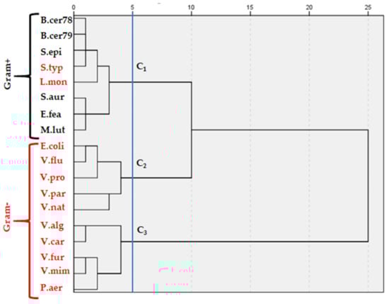
Figure 1.
Hierarchical cluster analysis (HCA) of bacterial strains, according to their responses to all EOs, based on MIC and MBC values. Clustering was established according to ward method.
As shown, the high resistance of Gram-negative bacteria when compared to the Gram-positive ones, may be due to their cell walls having a thick layer of peptidoglycan, making it difficult for antimicrobial agents to pass through, and thus imparting rigidity to their cells [55]. It has been mentioned previously that 75% of antibacterial drugs were terpenes [9]. The antimicrobial activity of EOs can vary with the type of the RO and the used microorganism. In this study, potent antimicrobial activity was especially observed for the mixture, mainly due to the richness of monoterpenes. Among them, carvone has been reported for its potential in inhibiting the growth of bacteria and fungi [56]. Also, carvone is known to possess potent inhibitory activity against E. coli and S. typhimurium [9]. Linaloole extracted from lavender EO by membrane disruption has demonstrated antimicrobial activity against resistant K. pneumoniae [57]. A previous study conducted by Giweli et al. [58] showed the strong antimicrobial activity of γ-terpinene that was obtained in high proportion in C. carvi.
2.4. Enzymes Inhibitory Activity Evaluation
2.4.1. Cholinesterase Inhibition
Acetylcholinesterase ends the effect of this neurotransmitter at cholinergic synapses by hydrolyzing acetylcholine to choline and acetate. Therefore, the inhibition of cholinesterase enzymes (anti-AChE) is considered promising in the management of neurological and neurodegenerative disorders such as AD. In this study, we evaluated for the first time the anti-cholinesterase activity of EOs alone and in combination. Results (Table 7) showed that C. carvi (IC50 = 0.82 ± 0.05 mg/mL) and C. sativum (IC50 = 0.68 ± 0.03 mg/mL) EOs alone, as well as in combination (IC50 = 0.63 ± 0.02 mg/mL), significantly inhibited the acetylcholinesterase enzyme when compared to the reference drug galanthamine (IC50 = 1.05 ± 0.05 mg/mL). Statistical analysis indicates the similarly strong inhibition potency of the mixture and C. sativum, which are not significantly different (p > 0.05). However, the inhibitory effect of C. carvi was significantly different (p < 0.05) from those of C. sativum and the standard galanthamine. Our finding has been supported by Öztürk [59], who reported that EO rich in terpenes shows high AChE inhibitory activity, especially γ-terpinene (IC50 = 181 μg/mL). Tnidis et al. [60], in a similar work, reported the ability of terpenes from Stachys lavandulifolia Vahl (Lamiaceae) to inhibit AChE. Menichini et al. [61] indicated that the Pimpinella anisoides plant, which is rich in terpenes, exhibited enzyme inhibition towards AChE. The anti-AChE activity of Salvia lavandulaefolia Vahl EOs containing β-pinene and linalool, which are obtained in high proportion in our oils, have also been confirmed [62]. The study confirmed the potency of combined terpenes. In another study, linalool’s high AChE inhibition was confirmed [63].
Table 7.
Inhibitory activities of acetylcholinesterase and α-glucosidase of two EOs (C. carvi and C. sativum) and their mixture compared to authentic standards (Galanthamine, Acarbose).
2.4.2. Antidiabetic Inhibition
T2DM is a metabolic disorder characterized by persistent hyperglycemia with serious complications. α-glucosidase is one of the enzymes involved in the deconstruction of long chain carbohydrates by breaking down starch and disaccharides to glucose. To reduce the high levels of glucose in the blood, enzyme inhibitors are a key. An anti-diabetic bioassay was carried out for the first time to test C. carvi, C. sativum, and a mixture of both EOs as α-glucosidase inhibitors. It was obvious that C. carvi (IC50 = 6.83 ± 0.76 mg/mL) and C. sativum (IC50 = 6.24 ± 0.86 mg/mL) EOs demonstrated similar antidiabetic activity, which is about nine-fold lower than the standard drug, acarbose (IC50 = 0.73 ± 0.10 mg/mL); however, the EO mixture (IC50 = 0.75 ± 0.15 mg/mL) displayed equipotent activity to acarbose. Our finding justifies the increased dietary intake of coriander seeds in decreasing the oxidative burden in DM. Terpenes have been provided for their antidiabetic potential. Studying the EO from Harita cheirifolia L., Majouli et al. [64] have reported that among the active monoterpenes, p-cymene and γ-terpinene revealed potent inhibitory effects. The strongest α-glucosidase inhibition effect was also displayed by Sideritis galactic EO containing high levels of β-pinene (32.2%), and the activity was ascribed to the high level of monoterpene hydrocarbons.
2.5. Pharmacokinetics Profiling of the Major Identified Components from C. carvi and C. sativum EOs
The pharmacokinetic profile of the major identified compounds defines their absorption, distribution, metabolism, excretion, and toxicity (ADMET) properties, which will be carefully considered in the early stages of drug development, leading to a significant reduction in the number of compounds that failed in clinical trials. The data depicted in Table 8 outlined that log BB values were in the range of 0.818 to 0.478 for all components, indicating their ability to penetrate the blood-brain barrier moderately and the fact that they will be highly distributed. They also manifested high Caco-2 permeability expressed by log Papp values > 0.90 cm/s. The volume of distribution (VDss) gives an indication about the circulation of a medicine at an equal level of blood plasma. Results showed that amongst the tested compounds, only α-pinene, β-pinene and linalool exhibited VDss values >0.45, suggesting that they may be more distributed in tissue rather than plasma. All tested compounds were predicted not to be a substrate or inhibitor of the human cytochrome P450 (CYP) isoforms (CYP2D6, CYP3A4, CYP1A2, CYP2C19, CYP2C9, CYP2D6 and CYP3A4), which are involved in drug metabolism. The transfer of a candidate compound by OCT2 gives useful information concerning its clearance, as well its potential contraindications. None of them are OCT2 substrates. The toxicity profile including AMES test, the human ether-a-go-go related gene (hERG), hepatotoxicity, and skin sensitization revealed that the selected identified compounds do not have any toxicity (Table 8).
Table 8.
Pharmacokinetics profiles of the major identified components.
Water solubility ≤ −4 is soluble; Intestinal absorption below 30% indicates poor absorbance; Blood brain barrier permeability ≤ −1 is considered poorly distributed to the brain; Central nervous system (CNS) permeability ≥ −2 is considered to penetrate the CNS; Low total clearance (logCLtot) value indicates high drug half lifetime.
The BOILED-Egg graph (WLOGP vs. TPSA) prediction of GI absorption and BBB permeation, which helps in the computation of polarity and lipophilicity, revealed that all molecules fall in the yellow region with red points indicating that they possess a high probability of brain penetration and are non-substrate of P-gp (PGP-) (Figure 2).
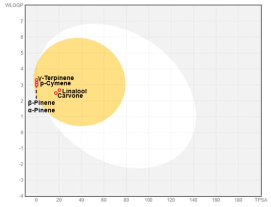
Figure 2.
Boiled egg plot of WLogP vs. TPSA. Yellow indicates BBB-permeant, white indicates gastrointestinal permeant, blue indicates P-glycoprotein, and red indicates P-glycoprotein.
3. Materials and Methods
3.1. Plant Material and Extraction of EOs
Our work focused on the seeds (achenes) of two spices: caraway and coriander. These two species were collected from a farmer in the Kelibia region, North-East of Tunisia (latitude 36,965918; longitude 11,037080) and identified according to the flora of Tunisia. The seeds were separated and then dried at room temperature for about ten days. Once dried, the plant material was extracted for EOs. An amount of 100 g of aerial part was transferred to hydro-distillation for 3 h with 500 mL distilled water using a Clevenger-type apparatus. The distilled EO was dried over anhydrous sodium sulfate, filtered, and stored at 4 °C. The yield was calculated based on the dried weight of the sample.
3.2. Essential Oils Analysis
3.2.1. Gas Chromatography (GC)
A Hewlett-Packard 5890 series II gas chromatograph equipped with HP-5MS capillary column (30 m × 0.25 mm i.d., film thickness 0.25 µm; Hewlett-Packard, Palo Alto, CA, USA) and connected to a flame ionization detector (FID) was applied using the following conditions. The column temperature was programmed at 50 °C for 1 min, then increased by 7 °C/min to 250 °C, and then left at 250 °C for 5 min. The injection port temperature was 240 °C, and the detector temperature was 250 °C (split ratio: 1/60). The carrier gas was helium with a flow rate of 1.2 mL/min, and the analyzed sample volume was 2 µL. The percentages of the constituents were calculated by the electronic integration of FID peak areas, without the use of response factor correction. The mean percentage of compounds in the EO represents the average calculated from three individual experiments. Retention indices (RI) were calculated for separate compounds relative to C6-C28 n-alkanes mixture.
3.2.2. Gas Chromatography-Mass Spectrometry (GC-MS)
The isolated volatile compounds were analyzed by GC-MS, using a Hewlett-Packard 5890 series II gas chromatograph. The fused HP-5MS capillary column (the same as that used in the GC analysis) was coupled to a HP 5972A masse-selective detector (Hewlett-Packard, Palo Alto, CA, USA). The oven temperature was programmed at 50 °C for 1 min, then increased by 7 °C/min to 250 °C, and then left at 250 °C for 5 min. The temperature of the injection port was set to 250 °C and that of the detector to 280 °C (split ratio: 1/100). The carrier gas was helium with a flow rate of 1.2 mL/min, and the analyzed sample volume was 2 µL. The mass spectrometer conditions were as follows: ionization voltage, 70 eV; ion source temperature, 150 °C; electron ionization mass spectra were acquired over the mass range 50–550 m/z. The components of these oils were identified following the same protocol as Hajlaoui et al. [27].
3.3. Antioxidant Activity
3.3.1. Scavenging Ability on DPPH Radical
The DPPH quenching ability of the EO was measured according to the same experiment as described by Felhi et al. [65,66]. Anti-radical activity was expressed as IC50 (µg/mL) values, reflecting the extract doses required to cause a 50% inhibition. A lower IC50 value corresponded to a higher antioxidant activity of plant extract.
3.3.2. Superoxide Anion Radical-Scavenging Activity
Superoxide anion scavenging activity was assessed using the method described by Saini et al. [67] with slight modifications. The reaction mixture contained 0.2 mL of EO assayed at different concentrations, 0.2 mL of 60 mM PMS stock solution, 0.2 mL of 677 mM NADH, and 0.2 mL of 144 mM NBT, all in phosphate buffer (0.1 mol/L, pH 7.4). After incubation at ambient temperature for 5 min, absorbance was read at 560 nm against a blank. Antioxidant activity was evaluated based on IC50 values, which was defined as the amount of antioxidant needed to reduce the generation of superoxide radical anions by 50% and expressed as μg/mL (as determined from three replicates per treatment). The inhibition percentage of superoxide anion generation was calculated using the following formula:
where, A0 and A1 had the same references presented in the above Equation.
Superoxide quenching (%) = [(A0 − A1) × 100]/A0
3.3.3. Reducing Power
The ability of the EO to reduce Fe3+ was assayed using the method described by Bakari et al. [68] and Kadri et al. [69] Briefly, 1 mL of the EO was mixed with 2.5 mL of phosphate buffer (0.2 M, pH 6.6) and 2.5 mL of 1% K3Fe(CN)6. Absorbance was measured at 700 nm. The mean of absorbance values was plotted against concentration values, and a linear regression analysis was performed. Increased absorbance of the reaction mixture indicated increased reducing power. The EC50 value (µg/mL) is the effective concentration at which absorbance was 0.5 for reducing power. BHT and ascorbic acid were used as positive control.
3.3.4. Chelating Effect on Ferrous Ions
The use of the ferrozine method was assessed to evaluate in vitro chelating power as reported by Ballester-Costa et al. [70]. The completion of the kinetics of this activity to determine the concentration that is 50% of chelating of ferric iron, the value of EC50 (µg.mL−1) corresponds to the lower efficiency of the oil highest.
3.3.5. β-Carotene-Linoleic Acid Model System (β-CLAMS)
The β-CLAMS method is based on the discoloration of β-carotene by the peroxides generated during the oxidation of linoleic acid at an elevated temperature, and was performed based on the protocol done by Kadri et al. [69]. In this study the β-CLAMS was modified for the 96-well micro-plate reader. In brief, the β-carotene was dissolved in 2 mL of CHCl3, to which 20 mg of linoleic acid and 200 mg of tween 40 were added. The results are expressed as IC50 values (µg/mL). All samples were prepared and analyzed in triplicate.
3.4. Antimicrobial Activity
3.4.1. Microorganisms
The microorganisms tested in this study belonged to 18 reference bacterial strains and 5 fungal strains, which are presented in Table 3 and Table 4, respectively. The bacterial species consisted of 6 Gram-positive and 12 Gram-negative bacterial strains. The fungal species belonged to four ATCC Candida strains and one Saccharomyces strain. A sterile cotton swab (Nippon Menbo, Tokyo, Japan) was immediately cultured into Sabouraud Chloramphenicol agar (Bio-Rad®, Mions, France) to obtain isolated colonies.
3.4.2. Disc-Diffusion Assay
Antimicrobial activity testing was performed according to the protocol described by Vuddhakul et al. [71] and slightly modified by Hajlaoui et al. [72] and Snoussi et al. [73] for Vibrio spp. strains. For the experiments, a loopful of the microorganisms working stocks were enriched on a tube containing 9 mL of Mueller-Hinton broth (for bacteria) and Sabouraud Chloramphenical broth (for Yeast strains), then incubated at 37 °C for 18 to 24 h. The overnight cultures were used for the antimicrobial activity of the EO used in this study, and optical density was adjusted at 0.5 McFarland turbidity standards with a DENSIMAT (BioMérieux®, Marcy l’Etoile, France). The inoculums of the respective bacteria and fungi were streaked onto MH or SB agar plates using a sterile swab. For Vibrio strains, the MH medium was supplemented with 1% NaCl.
Sterile filter discs (diameter 6 mm, Whatman paper No. 3) were impregnated with 10 μL of EO placed on the appropriate agar media (SB, MH and MH + 1%NaCl). Gentamycin (10 μg/disc) and Amphotericin B (20 μg/disc) were used as positive reference standards to determine the sensitivity of one strain/isolate to each of the tested microbial species. Antibiotic susceptibility was determined using the Kirby–Bauer method on Mueller Hinton agar plates supplemented with 1% NaCl. After incubation at 37 °C for 18 to 24 h, the diameter of inhibition zone was measured with 1 mm flat rule, and diameters were interpreted according to the Committee of the French Society of the Antibiogram [74]. The dishes were incubated at 37 °C for 18–24 h for microbial strains. The diameter of inhibition zones around each of the discs was taken as a measure of antimicrobial activity. Each experiment was carried out in triplicate, and the mean diameter of the inhibition zone was recorded.
3.4.3. Micro-Well Determination of MIC, MBC and MFC
Minimal inhibition concentration (MIC), minimal bactericidal concentration (MBC) and minimal fungicidal concentration (MFC) values were determined for all bacterial and fungal strains used in this study as described by Hajlaoui et al. [72]. A 100 μL aliquot from the stock solutions of EO was added into the first wells. Then, 100 μL from the serial dilutions were transferred into eleven consecutive wells. The last well containing 195 μL of nutrient broth without EO and 5 μL of the inoculum on each strip was used as the negative control. The final volume in each well was 200 μL. The plates were incubated at 37 °C for 18–24 h. The EO tested in this study was screened two times against each organism. The MIC value was defined as the lowest concentration of the compounds to inhibit the growth of the microorganisms. The MBC and MFC values were interpreted as the highest dilution (lowest concentration) of the sample, which showed clear fluid with no development of turbidity and without visible growth. MBC/MIC and MFC/MIC ratios were also calculated. All tests were performed once.
3.5. Enzyme Inhibition Assays
3.5.1. Anti-Acetylcholinesterase Inhibitory Assay
Acetylcholinesterase enzymatic activity was measured using a slightly modified version of the method described by Ingkaninan et al. [75]. In brief, 98 μL (50 mM) Tris–HCl buffer pH 8, 30 μL sample, and 7.5 μL acetylcholinesterase solution containing 0.26 U/mL were mixed in an ELISA well plate and left to incubate for 15 min. Subsequently, 22.5 μL of AChI (acetylthiocholine iodide) 0.023 mg/mL and 142 μL of (3 mM) DTNB were added. The absorbance at 405 nm was read when the reaction reached the equilibrium. A control reaction was carried out using water instead of extract. Galanthamine was used as a positive control. The absorbance value obtained was considered 100% activity. Inhibition (%) was calculated using the following equation:
where, Asample refers to the absorbance of the reaction containing the extract and Acontrol to the absorbance of the reaction control. Tests were carried out in triplicate, and a blank with Tris–HCl buffer instead of enzyme solution was performed. An extract concentration providing 50% inhibition (IC50) was obtained by plotting the inhibition percentage against extract solution concentrations.
I% = 100 − (Asample/Acontrol) × 100
3.5.2. α-Glucosidase Inhibitory Assay
The α-glucosidase assay of the tested EOs was conducted according to the standard method with slight modifications [75]. Inside the 96-well plate, 50 µL of phosphate buffer (100 mM, pH = 6.8), 10 µL α-glucosidase (1 U/mL), 20 µL of samples, and standard acarbose of different concentration were incubated for 15 min at 37 °C. Briefly, 20 µL of 5 mM substrate (4-nitrophenyl β-d-glucopyranoside) was added to each well and left to incubate for 20 min at 37 °C. The reacting mixture was stopped after incubation by adding 0.1 M sodium carbonate (50 µL). The release of p-nitrophenol of the reacting mixture relating to the activity of the enzyme was read at a wavelength of 405 nm using a multiplate reader (Multiskan, Thermo Scientific, Waltham, MA, USA). The enzyme inhibition rate, expressed as percentage of inhibition, was calculated using the following formula:
where, A is the absorbance in the presence of test substance, and B is the absorbance in the presence of phosphate buffer (control). The results are expressed as IC50 values (µg/mL). All samples were prepared and analyzed in triplicate.
Percentage inhibitory activity (%) = (1 − A/B) × 100
3.6. Pharmacokinetics Study
The pharmacokinetic and drug-likeness properties of the selected compounds were estimated using ADME (absorption, distribution, metabolism and excretion) descriptors by a SwissADME online server (http://www.swissadme.ch/, accessed on 20 April 2021) and pkCSM (http://biosig.unimelb.edu.au/pkcsm/prediction, accessed on 22 April 2021) online tools [76,77,78].
3.7. Statistical Analysis
All experiments were performed in triplicates, and average values were calculated using the SPSS 25.0 statistical package (version 25, Chicago, IL, USA) for Windows. Differences in means were calculated using the Duncan’s multiple-range tests for means with a 95% confidence interval (p ≤ 0.05).
4. Conclusions
As mentioned above, combined C. carvi and C. sativum seed EOs exhibited potent antioxidant effects and a broad spectrum of antimicrobial activity against the tested pathogenic strains, even higher than each EO alone. Also, the strongest inhibiting power of AChE and α-glucosidase enzymes have been outlined for the first time demonstrating the highest activity of the mixture, especially the antidiabetic one. The biological properties of this EO mixture, along with its pharmacokinetic profile, support the efficiency and traditional applications of these species in food, show that mixed EOs might provide an alternative way to fight microbial contamination, and indicate that EOs may be potential health-promoting antidiabetic and anti-Alzheimer agents.
Author Contributions
Conceptualization, H.H., A.K., and S.A.; methodology, H.H., E.N., and S.A.; software, M.S., S.A., M.A., and A.K.; investigation, A.K., E.N., K.A., and M.A.K.; writing—original draft preparation, K.A., H.H., A.K., M.A.K., and M.S.; writing—review and editing, M.A.K., M.A., and M.S.; supervision, A.K. and M.S.; project administration, A.K. All authors have read and agreed to the published version of the manuscript.
Funding
This research has been funded by the United Arab Emirates University start up grant- 12M003.
Data Availability Statement
All data generated or analyzed during this study are included in this article.
Conflicts of Interest
The authors declare no conflict of interest. The funders had no role in the design of the study; in the collection, analyses, or interpretation of data; in the writing of the manuscript; or in the decision to publish the results.
Sample Availability
The essential oils of Carum carvi L. and Coriandrum sativum L. are available from the authors.
References
- Elshafie, H.S.; Camele, I. An Overview of the Biological Effects of Some Mediterranean Essential Oils on Human Health. Biomed. Res. Int. 2017, 2017, 9268468. [Google Scholar] [CrossRef] [PubMed]
- Liang, J.Y.; Xu, J.; Yang, Y.Y.; Shao, Y.Z.; Zhou, F.; Wang, J.L. Toxicity and Synergistic Effect of Elsholtzia ciliata Essential Oil and Its Main Components against the Adult and Larval Stages of Tribolium castaneum. Foods 2020, 9, 345. [Google Scholar] [CrossRef]
- Pellegrini, M.; Ricci, A.; Serio, A.; Chaves-López, C.; Mazzarrino, G.; D’Amato, S.; Lo Sterzo, C.; Paparella, A. Characterization of Essential Oils Obtained from Abruzzo Autochthonous Plants: Antioxidant and Antimicrobial Activities Assessment for Food Application. Foods 2018, 7, 19. [Google Scholar] [CrossRef] [PubMed]
- Kwiatkowski, P.; Mnichowska-Polanowska, M.; Pruss, A.; Masiuk, H.; Dzięcioł, M.; Giedrys-Kalemba, S.; Sienkiewicz, M. The effect of fennel essential oil in combination with antibiotics on Staphylococcus aureus strains isolated from carriers. Burns 2017, 43, 1544–1551. [Google Scholar] [CrossRef] [PubMed]
- Alminderej, F.; Bakari, S.; Almundarij, T.I.; Snoussi, M.; Aouadi, K.; Kadri, A. Antioxidant Activities of a New Chemotype of Piper cubeba L. Fruit Essential Oil (Methyleugenol/Eugenol): In Silico Molecular Docking and ADMET Studies. Plants 2020, 9, 1534. [Google Scholar] [CrossRef] [PubMed]
- Abrahamse, H.; George, S. Redox Potential of Antioxidants in Cancer Progression and Prevention. Antioxidants 2020, 9, 1156. [Google Scholar] [CrossRef]
- Alfei, S.; Marengo, B.; Zuccari, G. Oxidative Stress, Antioxidant Capabilities, and Bioavailability: Ellagic Acid or Urolithins? Antioxidants 2020, 9, 707. [Google Scholar] [CrossRef] [PubMed]
- Alminderej, F.; Bakari, S.; Almundarij, T.I.; Snoussi, M.; Aouadi, K.; Kadri, A. Antimicrobial and Wound Healing Potential of a New Chemotype from Piper cubeba L. Essential Oil and In Silico Study on S. aureus tyrosyl-tRNA Synthetase Protein. Plants 2021, 10, 205. [Google Scholar] [CrossRef]
- Mahizan, N.A.; Yang, S.K.; Moo, C.L.; Song, A.A.L.; Chong, C.M.; Chong, C.W.; Abushelaibi, A.; Lim, S.H.E.; Lai, K.S. Terpene Derivatives as a Potential Agent against Antimicrobial Resistance (AMR) Pathogens. Molecules 2019, 24, 2631. [Google Scholar] [CrossRef]
- Taiwo, M.O.; Adebayo, O.S. Plant Essential Oil: An Alternative to Emerging Multidrug Resistant Pathogens. J. Microbiol. Exp. 2017, 5, 00163. [Google Scholar] [CrossRef]
- Spisni, E.; Petrocelli, G.; Imbesi, V.; Spigarelli, R.; Azzinnari, D.; Sarti, M.D.; Campieri, M.; Valerii, M.C. Antioxidant, Anti-Inflammatory, and Microbial-Modulating Activities of Essential Oils: Implications in Colonic Pathophysiology. Int. J. Mol. Sci. 2020, 21, 4152. [Google Scholar] [CrossRef]
- Sahib, N.G.; Anwar, F.; Gilani, A.H.; Hamid, A.A.; Saari, N. Coriander (Coriandrum sativum L.): A Potential Source of High-Value Components for Functional Foods and Nutraceuticals—A Review. Phytother. Res. 2013, 27, 1439–1456. [Google Scholar] [CrossRef]
- Sayed-Ahmed, B.; Talou, T.; Saad, Z.; Hijazi, A.; Merah, O. The Apiaceae: Ethnomedicinal Family as source for industrial uses. Ind. Crop. Prod. 2017, 109, 661–671. [Google Scholar] [CrossRef]
- Momin, A.H.; Acharya, S.S.; Gajjar, A.V. Coriandrum sativum-review of advances in phytopharmacology. Int. J. Pharma. Sci. Res. 2012, 3, 1233. [Google Scholar]
- Al-Snaf, A.E. A review on chemical constituents and pharmacological activities of Coriandrum sativum. IOSR J. Pharm. 2016, 6, 17–42. [Google Scholar] [CrossRef]
- Keshavarz, A.; Minaiyan, M.; Ghannadi, A.; Mahzouni, P. Effects of Carum carvi L. (Caraway) extract and essential oil on TNBS-induced colitis in rats. Res. Pharm. Sci. 2013, 8, 1–8. [Google Scholar]
- Kabiri, M.; Kamalinejad, M.; Sohrabvand, F.; Bioos, S.; Babaeian, M. Management of breast milk oversupply in traditional Persian medicine. J. Evid. Based Complement Altern. Med. 2017, 22, 1044–1050. [Google Scholar] [CrossRef]
- Miraj, S.; Kiani, S. Pharmacological activities of Carum carvi L. Der Pharm. Lett. 2016, 8, 135–138. [Google Scholar]
- Rasooli, I.; Allameh, A. Chapter 32—Caraway (Carum carvi L.) essential oils. In Essential Oils in Food Preservation, Flavor and Safety; Preedy, V.R., Ed.; Academic Press: San Diego, CA, USA, 2016; pp. 287–293. [Google Scholar]
- Malhotra, S. (Ed.) Caraway: Handbook of Herbs and Spices; Elsevier: Amsterdam, The Netherlands, 2006; Volume 3, pp. 270–298. [Google Scholar]
- Mahboubi, M. Caraway as Important Medicinal Plants in Management of Diseases. Nat. Prod. Bioprospect. 2019, 9, 1–11. [Google Scholar] [CrossRef]
- Meena, A.; Brijendra, S.; Yadav, A.; Uttam, S.; Ramanjeet, K.; Ayushy, S.; Vertika, G.; Bhavana, P. Review on medicinal properties and bioactive constituents of herbal spices commonly used in India. J. Pharm. Res. 2010, 3, 866–868. [Google Scholar]
- Lemhadri, A.; Hajji, L.; Michel, J.B.; Eddouks, M. Cholesterol and triglycerides lowering activities of caraway fruits in normal and streptozotocin diabetic rats. J. Ethnopharmacol. 2006, 106, 321–326. [Google Scholar] [CrossRef] [PubMed]
- Mseddi, K.; Alimi, F.; Noumi, E.; Veettil, V.N.; Deshpande, S.; Adnan, M.; Hamdi, A.; Elkahoui, S.; Alghamdi, A.; Kadri, A.; et al. Thymus musilii Velen. as a promising source of potent bioactive compounds with its pharmacological properties: In vitro and in silico analysis. Arab. J. Chem. 2020, 13, 6782–6801. [Google Scholar] [CrossRef]
- Gad-Elkareem, M.A.M.; Abdelgadir, E.H.; Badawy, O.M.; Kadri, A. Potential antidiabetic effect of ethanolic and aqueous-ethanolic extractsof Ricinus communis leaves on streptozotocin-induced diabetes in rats. Peer J. 2019, 7, e6441. [Google Scholar] [CrossRef] [PubMed]
- Ben Mefteh, F.; Daoud, A.; Bouket, A.C.; Thissera, B.; Kadri, Y.; Cherif-Silini, H.; Eshelli, M.; Alenezi, F.N.; Vallat, A.; Oszako, T.; et al. Date Palm Trees Root-Derived Endophytes as Fungal Cell Factories for Diverse Bioactive Metabolites. Int. J. Mol. Sci. 2018, 19, 1986. [Google Scholar] [CrossRef]
- Hajlaoui, H.; Mighri, H.; Aouni, M.; Gharsallah, N.; Kadri, A. Chemical composition and in vitro evaluation of antioxidant. antimicrobial. cytotoxicity and anti-acetylcholinesterase properties of Tunisian Origanum majorana L. essential oil. Microb. Pathog. 2016, 95, 86–94. [Google Scholar] [CrossRef]
- Hajlaoui, H.; Arraouadi, S.; Mighri, H.; Chaaibia, M.; Gharsallah, N.; Ros, G.; Nieto, G.; Kadri, A. Phytochemical Constituents and Antioxidant Activity of Oudneya Africana L. Leaves Extracts: Evaluation Effects on Fatty Acids and Proteins Oxidation of Beef Burger during Refrigerated Storage. Antioxidants 2019, 8, 442. [Google Scholar] [CrossRef]
- Snoussi, M.; Noumi, E.; Hajlaoui, H.; Usai, D.; Sechi, L.; Zanetti, S.; Bakhrouf, A. High potential of adhesion to abiotic and biotic materials in fish aquaculture facility by Vibrio alginolyticus strains. J. Appl. Microbiol. 2009, 106, 1591–1599. [Google Scholar] [CrossRef]
- Znati, M.; Jabrane, A.; Hajlaoui, H.; Harzallah-Skhiri, F.; Bouajila, J.; Casanova, J.; Ben Jannet, H. Chemical Composition and in vitro Evaluation of Antimicrobial and Anti-acetylcholinesterase Properties of the Flower Oil of Ferula lutea. Nat. Prod. Commun. 2012, 7, 947–950. [Google Scholar] [CrossRef]
- Bakari, S.; Hajlaoui, H.; Daoud, A.; Mighri, H.; Ross-Garcia, J.M.; Gharsallah, N.; Kadri, A. Phytochemicals, antioxidant and antimicrobial potentials and LC-MS analysis of hydroalcoholic extracts of leaves and flowers of Erodium glaucophyllum collected from Tunisian Sahara. Food Sci. Technol. 2018, 38, 310–317. [Google Scholar] [CrossRef]
- Akrout, A.; Hajlaoui, H.; Mighri, H.; Najjaa, H.; Jani, H.E.; Zaidi, S.; Neffati, M. Chemical and Biological Characteristics of Essential Oil of Rosmarinus officinalis Cultivated in Djerba. J. Essent. Oil-Bear. Plants 2013, 13, 398–411. [Google Scholar] [CrossRef]
- Bnina, E.B.; Hajlaoui, H.; Chaieb, I.; Said, M.B.; Jannet, H.B.; Daami-Remadi, M. Chemical composition, antimicrobial and insecticidal activities of the tunisian Citrus aurantium essential oils. Czech J. Food Sci. 2017, 37, 81–92. [Google Scholar] [CrossRef]
- Khan, R.M.; Ahmad, W.; Ahmad, M.; Hasan, A. Phytochemical and pharmacological properties of Carum carvi. Eur. J. Pharm. Med. Res. 2016, 3, 231–236. [Google Scholar]
- Laribi, B.; Kouki, K.; Mougou, A.; Marzouk, B. Current awareness in flavour and fragrance. J. Sci. Food Agric. 2010, 90, 391–396. [Google Scholar] [CrossRef]
- Jiang, Z.-T.; Sun, M.-L.; Li, R.; Wang, Y. Essential oil Composition of Chinese Caraway (Carum carvi L.). J. Essent. Oil Bear. Plants 2011, 14, 379–382. [Google Scholar] [CrossRef]
- Gwari, G.; Bhandari, U.; Andola, H.C.; Lohani, H.; Chauhan, N. Aroma profile of seeds of Carum carvi Linn. cultivated in higher hills of Uttarakhand Himalaya. Indian J. Nat. Prod. Res. 2012, 3, 411–413. [Google Scholar]
- Raal, A.; Arak, E.; Orav, A. The content and composition of the essential oil Found in Carum carvi L. commercial fruits obtained from different countries. J. Essent. Oil Res. 2012, 24, 53–59. [Google Scholar] [CrossRef]
- Laribi, B.; Kouki, K.; Bettaieb, T.; Mougou, A.; Marzouk, B. Essential oils and fatty acids composition of Tunisian, German and Egyptian caraway (Carum carvi L.) seed ecotypes: A comparative study. Ind. Crops Prod. 2013, 41, 312–318. [Google Scholar] [CrossRef]
- Shahwar, M.K.; El-Ghorab, A.H.; Anjum, F.M.; Butt, M.S.; Hussain, S.; Nadeem, M. Characterization of Coriander (Coriandrum sativum L.) Seeds and Leaves: Volatile and Non Volatile Extracts. Int. J. Food. Prop. 2012, 15, 736–747. [Google Scholar] [CrossRef]
- Gil, A.; Fuente, E.B.; Lenardis, A.E.; Pereira, M.L.; Suárez, S.A.; Bandoni, A.; Baren, C.V.; Lira, P.D.L.; Ghersa, C.M. Coriander Essential Oil Composition from Two Genotypes Grown in Different Environmental Conditions. J. Agric. Food Chem. 2002, 50, 2870–2877. [Google Scholar] [CrossRef]
- Smallfield, B.; John, W.; Nigel, B.P.; Kenneth, G.D. Coriander spice oil: Effects of fruit crushing and distillation time on yield and composition. J. Agric. Food Chem. 2001, 49, 118–123. [Google Scholar] [CrossRef]
- Misharina, T.A. Effect of conditions and duration of storage on composition of essential oil from coriander seeds. Prikl. Biokhimiia Mikrobiol. 2001, 37, 726–732. [Google Scholar]
- Adams, R.P. Identification of Essential Oil Components by Gas Chromatography/Mass Spectrometry; Allured Publishing: Carol Stream, IL, USA, 2007. [Google Scholar]
- Peana, A.T.; D’Aquila, P.S.; Serra, F.P.G.; Pippia, P.; Moretti, M.D.L. Anti-inflammatory activity of linalool and linalyl acetate constituents of essential oils. Phytomedicine 2002, 9, 721–726. [Google Scholar] [CrossRef]
- Ramalho, T.R.; Oliveira, M.T.; Lima, A.L.; Bezerra-Santos, C.R.; Piuvezam, M.R. Gamma-Terpinene modulates acute inflammatory response in mice. Planta Med. 2015, 81, 1248–1254. [Google Scholar] [CrossRef]
- Salehi, B.; Upadhyay, S.; Orhan, I.E.; Jugran, A.K.; Jayaweera, S.L.D.; Dias, D.A.; Sharopov, F.; Taheri, Y.; Martins, N.; Baghalpour, N.; et al. Therapeutic Potential of α- and β-Pinene: A Miracle Gift of Nature. Biomolecules 2019, 9, 738. [Google Scholar] [CrossRef]
- De Santana, M.F.; Guimarães, A.G.; Chaves, D.O.; Silva, G.C.; Bonjardim, L.R.; Lucca Júnior, W.D.; de Souza Ferro, J.N.; de Oliveira Barreto, E.; dos Santos, F.E.; Soares, M.B.P.; et al. The anti-hyperalgesic and anti-inflammatory profiles of p-cymene: Evidence for the involvement of opioid system and cytokines. Pharm. Biol. 2015, 53, 1583–1590. [Google Scholar] [CrossRef]
- Zhao, M.; Du, J. Anti-inflammatory and protective effects of D-carvone on lipopolysaccharide (LPS)-induced acute lung injury in mice. J. King Saud Univ. Sci. 2020, 32, 1592–1596. [Google Scholar] [CrossRef]
- Samojlik, I.; Lakic, N.; Mimica-Dukić, N.; Daković-Svajcer, K.; Bozin, B. Antioxidant and Hepatoprotective Potential of Essential Oils of Coriander (Coriandrum sativum L.) and Caraway (Carum carvi L.) (Apiaceae). J. Agric. Food Chem. 2010, 11, 8848–8853. [Google Scholar] [CrossRef]
- De Oliveira, T.M.; de Carvalho, R.B.F.; da Costa, I.H.F.; de Oliveira, G.A.L.; de Souza, A.A.; de Lima, S.G.; de Freitas, R.M. Evaluation of p-cymene, a natural antioxidant. Pharm. Biol. 2015, 53, 423–428. [Google Scholar] [CrossRef]
- Foti, M.C.; Ingold, K.U. Mechanism of Inhibition of Lipid Peroxidation by γ-Terpinene, an Unusual and Potentially Useful Hydrocarbon Antioxidant. J. Agric. Food Chem. 2003, 51, 2758–2765. [Google Scholar] [CrossRef]
- Wojtunik, K.A.; Ciesla, L.M.; Waksmundzka-Hajnos, M. Model studies on the antioxidant activity of common terpenoid constituents of essential oils by means of the 2.2-diphenyl-1-picrylhydrazyl method. J. Agric. Food Chem. 2014, 63, 9088–9094. [Google Scholar] [CrossRef]
- Vinothkumar, R.; Sudha, P.; Viswanathan, M.; Kabalimoorthy, J.; Balasubramanian, T.; Nalini, N. Modulating effect of d-carvone on 1.2-dimethylhydrazine-induced pre-neoplastic lesions, oxidative stress and biotransforming enzymes, in an experimental model of rat colon carcinogenesis. Cell Prolif. 2013, 46, 705–720. [Google Scholar] [CrossRef] [PubMed]
- Guimarães, A.C.; Meireles, L.M.; Lemos, M.F.; Guimarães, M.C.C.; Endringer, D.C.; Fronza, M.; Scherer, R. Antibacterial Activity of Terpenes and Terpenoids Present in Essential Oils. Molecules 2019, 24, 2471. [Google Scholar] [CrossRef] [PubMed]
- Znini, M.; Bouklah, M.; Majidi, L.; Kharchouf, S.; Aouniti, A.; Bouyanzer, A.; Hammouti, B.; Costa, J.; Al Deyab, S.S. Chemical composition and inhibitory effect of Mentha spicata essential oil on the corrosion of steel in molar hydrochloric acid. Int. J. Electrochem. Sci. 2011, 6, 691–704. [Google Scholar]
- Yang, S.K.; Yusoff, K.; Thomas, W.; Akseer, R.; Alhosani, M.S.; Abushelaibi, A.; Lai, K.S. Lavender essential oil induces oxidative stress which modifies the bacterial membrane permeability of carbapenemase producing Klebsiella pneumoniae. Sci. Rep. 2020, 10, 819. [Google Scholar] [CrossRef]
- Giweli, A.; Džamić, A.M.; Soković, M.; Ristić, M.S.; Marin, P.D. Antimicrobial and Antioxidant Activities of Essential Oils of Satureja thymbra Growing Wild in Libya. Molecules 2012, 17, 4836–4850. [Google Scholar] [CrossRef] [PubMed]
- Öztürk, M. Anticholinesterase and antioxidant activities of Savoury (Satureja thymbra L.) with identified major terpenes of the essential oil. Food Chem. 2012, 134, 48–54. [Google Scholar] [CrossRef]
- Tundis, R.; Bonesi, M.; Pugliese, A.; Nadjafi, F.; Menichini, F.; Loizzo, M.R. Tyrosinase, acetyl- and butyryl-cholinesterase inhibitory activity of Stachys lavandulifolia Vahl (Lamiaceae) and its major constituents. Rec. Nat. Prod. 2015, 9, 81–93. [Google Scholar]
- Menichini, F.; Tundis, R.; Loizzo, M.R.; Bonesi, M.; Marrelli, M.; Statti, G.A.; Menichini, F.; Conforti, F. Acetylcholinesterase and butyrylcholinesterase inhibition of ethanolic extract and monoterpenes from Pimpinella anisoides V Brig. (Apiaceae). Fitoterapia 2009, 80, 297–300. [Google Scholar] [CrossRef]
- Savelev, S.; Okelloa, E.; Perry, N.S.L.; Wilkinsa, R.M.; Perry, E.K. Synergistic and antagonistic interactions of anticholinesterase terpenoids in Salvia lavandulaefolia essential oil. Pharmacol. Biochem. Behav. 2003, 75, 661–668. [Google Scholar] [CrossRef]
- López, M.D.; Pascual-Villalobos, M.J. Mode of inhibition of acetylcholinesterase by monoterpenoids and implications for pest control. Ind. Crop. Prod. 2010, 31, 284–288. [Google Scholar] [CrossRef]
- Majouli, K.; Hlila, M.B.; Hamdi, A.; Flamini, G.; Ben Jannet, H.; Kenani, A. Antioxidantactivity and α-glucosidase inhibition by essential oils from Hertia cheirifolia (L.). Ind. Crop. Prod. 2016, 82, 23–28. [Google Scholar] [CrossRef]
- Felhi, S.; Saoudi, M.; Daoud, A.; Hajlaoui, H.; Ncir, M.; Chaabane, R.; El Feki, A.; Gharsallah, N.; Kadri, A. Investigation of phytochemical contents, in vitro antioxidant and antibacterial behavior and in vivo anti-inflammatory potential of Ecballium elaterium methanol fruits extract. Food Sci. Technol. 2017, 37, 558–563. [Google Scholar] [CrossRef]
- Felhi, S.; Hajlaoui, H.; Ncir, M.; Bakari, S.; Ktari, N.; Saoudi, M.; Gharsallah, N.; Kadri, A. Nutritional, phytochemical and antioxidant evaluation and FT-IR analysis of freeze-dried extracts of Ecballium elaterium fruit juice from three localities. Food Sci. Technol. 2016, 36, 646–655. [Google Scholar] [CrossRef]
- Saini, A.; Pandey, A.; Sharma, S.; Suradkar, U.S.; Ambedkar, Y.R.; Meena, P.; Raman, R.; Gurjar, A.S. Assessment of antioxidant activity of rosemary (Rosmarinus officinalis) leaves extract. J. Pharmacogn. Phytochem. 2020, 9, 14–17. [Google Scholar]
- Bakari, S.; Daoud, A.; Felhi, S.; Smaoui, S.; Gharsallah, N.; Kadri, A. Proximate analysis, mineral composition, phytochemical contents, antioxidant and antimicrobial activities and GC-MS investigation of various solvent extracts of Cactus cladode. Food Sci. Technol. 2017, 27, 286–293. [Google Scholar] [CrossRef]
- Kadri, A.; Zarai, Z.; Chobba, I.B.; Gharsallah, N.; Damak, M.; Békir, A. Chemical composition and in vitro antioxidant activities of Thymelaea hirsuta L: Essential oil from Tunisia. Afr. J. Biotechnol. 2011, 10, 2930–2935. [Google Scholar]
- Ballester-Costa, C.; Sendra, E.; Fernández-López, J.; Pérez-Álvarez, J.A.; Viuda-Martos, M. Assessment of Antioxidant and Antibacterial Properties on Meat Homogenates of Essential Oils Obtained from Four Thymus Species Achieved from Organic Growth. Foods 2017, 6, 59. [Google Scholar] [CrossRef] [PubMed]
- Vuddhakul, V.; Bhooponga, P.; Hayeebilana, F.; Subhadhirasakul, S. Inhibitory activity of Thai condiments on pandemic strain of Vibrio parahaemolyticus. Food Microbiol. 2007, 24, 413–418. [Google Scholar] [CrossRef]
- Hajlaoui, H.; Snoussi, M.; Noumi, E.; Zanetti, S.; Ksouri, R.; Bakhrouf, A. Chemical composition, antioxidant and antibacterial activities of the essential oils of five Tunisian aromatic plants. Ital. J. Food Sci. 2010, 3, 323–332. [Google Scholar]
- Snoussi, M.; Hajlaoui, H.; Noumi, E.; Usai, D.; Sechi, L.A.; Zanetti, S.; Bakhrouf, A. In vitro anti-Vibrio spp. activity and chemical composition of some Tunisian aromatic plants. World J. Microbiol. Biotechnol. 2008, 24, 3071–3076. [Google Scholar] [CrossRef]
- Ingkaninan, K.; Temkittawon, P.; Chuenchon, K.; Yuyaem, T.; Thongnoi, W. Screening for acetylcholinesterase inhibito activity in plants used in Thai traditional rejuvenating and neurotonic remedies. J. Ethnopharmacol. 2003, 89, 261–264. [Google Scholar] [CrossRef]
- Asghari, B.; Salehi, P.; Sonboli, A.; Ebrahimi, S.N. Flavonoids from Salvia chloroleuca with alpha-amylsae and alpha-glucosidase inhibitory effect. Iran. J. Pharm. Res. 2015, 14, 609. [Google Scholar]
- Kadri, A.; Aouadi, K. In vitro antimicrobial and α-glucosidase inhibitory potential of enantiopure cycloalkylglycine derivatives: Insights into their in silico pharmacokinetic, druglikeness, and medicinal chemistry properties. J. Appl. Pharm. Sci. 2020, 10, 107–115. [Google Scholar]
- Othman, I.M.M.; Gad-Elkareem, M.A.M.; Anouar, E.H.; Aouadi, K.; Kadri, A.; Snoussi, M. Design, synthesis ADMET and molecular docking of new imidazo[4.5-b]pyridine-5-thione derivatives as potential tyrosyl-tRNA synthetase inhibitors. Bioorg. Chem. 2020, 102, 104105. [Google Scholar] [CrossRef]
- Ghannay, S.; Kadri, A.; Aouadi, K. Synthesis, in vitro antimicrobial assessment, and computational investigation of pharmacokinetic and bioactivity properties of novel trifluoromethylated compounds using in silico ADME and toxicity prediction tools. Monatsh. Chem. 2020, 151, 267–280. [Google Scholar] [CrossRef]


Publisher’s Note: MDPI stays neutral with regard to jurisdictional claims in published maps and institutional affiliations. |
© 2021 by the authors. Licensee MDPI, Basel, Switzerland. This article is an open access article distributed under the terms and conditions of the Creative Commons Attribution (CC BY) license (https://creativecommons.org/licenses/by/4.0/).