Article

Spectrophotometric Study of Bridging N-Donor Ligand-Induced Supramolecular Assembly of Conjugated Zn-Trisporphyrin with a Triphenylamine Core

Department of Applied Chemistry, Kumoh National Institute of Technology, Gumi 39177, Korea
*
Author to whom correspondence should be addressed.
Molecules 2021, 26(16), 4771; https://doi.org/10.3390/molecules26164771
Submission received: 29 June 2021 / Revised: 3 August 2021 / Accepted: 4 August 2021 / Published: 6 August 2021

Abstract

:
This article studies the supramolecular assembly behavior of a Zn-trisporphyrin conjugate containing a triphenylamine core (1) with bridging N-donor ligands using the UV-vis spectrophotometric titration method at micromolar concentrations. Our results show that pyridine, a non-bridging ligand, formed a 3:1 open complex with 1. The corresponding binding constant was estimated to be (2.7 ± 0.15) × 1014 M−3. In contrast, bridging ligands, 4,4-bipyridine (BIPY) and 1,3-di(4-pyridyl)propane (DPYP), formed stable 3:2 double-decker complexes with 1 in solution, which collapsed to yield a 3:1 open complex when excess BIPY or DPYP was added. The binding constants for forming BIPY and DPYP double-decker complexes were estimated to be (9.26 ± 0.07) × 1027 M−4 and (3.62 ± 0.16) × 1027 M−4, respectively. The UV-vis titration profiles supported the conclusion that the degradation of the 3:2 double-decker 1∙BIPY complex is less favorable compared to that of 1∙DPYP. Consequently, the formation of the 3:1 1∙DPYP open complex proceeded more readily than that of 1∙BIPY.

1. Introduction

The construction of well-defined nano- and microscopic architectures provides access to outstanding materials [1,2,3] for use in artificial electron transfer [4], photocatalysis [5], sensing [6], molecular recognition [7], and medicinal applications [8]. Porphyrin materials are attractive building blocks for forming self-assembled, highly ordered supramolecular structures [9,10,11]. Porphyrin compounds are important because of their symmetric aromatic structures and excellent electronic and photophysical properties, and are used for catalysis [12,13,14]. Porphyrin molecules self-assemble to form large-scale aggregates with well-defined nanostructures of definite sizes, shapes, and dimensions. Various intermolecular non-covalent forces or interactions, such as hydrogen bonding, hydrophobic or hydrophilic interactions, π-π stacking, ligand coordination, electrostatic interactions, and van der Waals interactions, are responsible for porphyrin self-assembly [15,16,17,18,19]. Various strategies, such as ionic self-assembly [20], re-precipitation methods [21], ligand coordination [22], coordination polymerization [23], and surfactant assistance by either surfactants or amphipathic molecules [24], have been utilized to form porphyrin aggregates. However, covalent metalloporphyrins, such as dendrimers, double-stranded conjugated ladder porphyrins, porphyrin arrays, double-decker porphyrins, cyclic porphyrins, and tetra-porphyrins, readily self-assemble without any external stimuli and form a larger aggregation of porphyrin arrays in solution [25,26,27,28,29,30]. Among the various supramolecular architectures, tris-metalloporphyrins assembled with N-donor ligands, such as 1,4-diazabicyclo[2.2.2]octane (DABCO), form sandwich-type coordination cages that are attractive for supramolecular catalysis applications and artificial molecular receptor synthesis [31,32,33].
We have previously reported the strong two-photon absorption of octupolar trisporphyrin conjugates containing a triphenylamine core (1) (Scheme 1) [34]. The optimized geometry obtained using semi-empirical AM1 calculations revealed that the trisporphyrin molecule adopts a propeller-shaped structure. Incorporating the unique C3-symmetrical structure of conjugated porphyrins, we expected to construct an interesting architecture via supramolecular assembly within this system. In this report, we describe the spectrophotometric study of the supramolecular assembly of Zn-trisporphyrin induced by coordination with bridging N-donor ligands. We chose 4,4-bipyridine (BIPY, a rigid ligand) and 1,3-di(4-pyridyl)propane (DPYP, a flexible ligand) as the bridging N-donor ligands for the present study.

2. Results and Discussion

UV-vis spectroscopy was applied to evaluate the binding properties of Zn-trisporphyrin 1 (1 × 10⁻⁶ M) with bridging N-donor ligands in toluene at room temperature. For a comparative study, we used monomeric Zn-porphyrin 2 ([5,10,15-tri-(p-tolyl)-20-trimethylsilylethynylporphyrinato]Zn(II), see Scheme 1) as a reference model and performed supramolecular assembly studies under similar conditions.
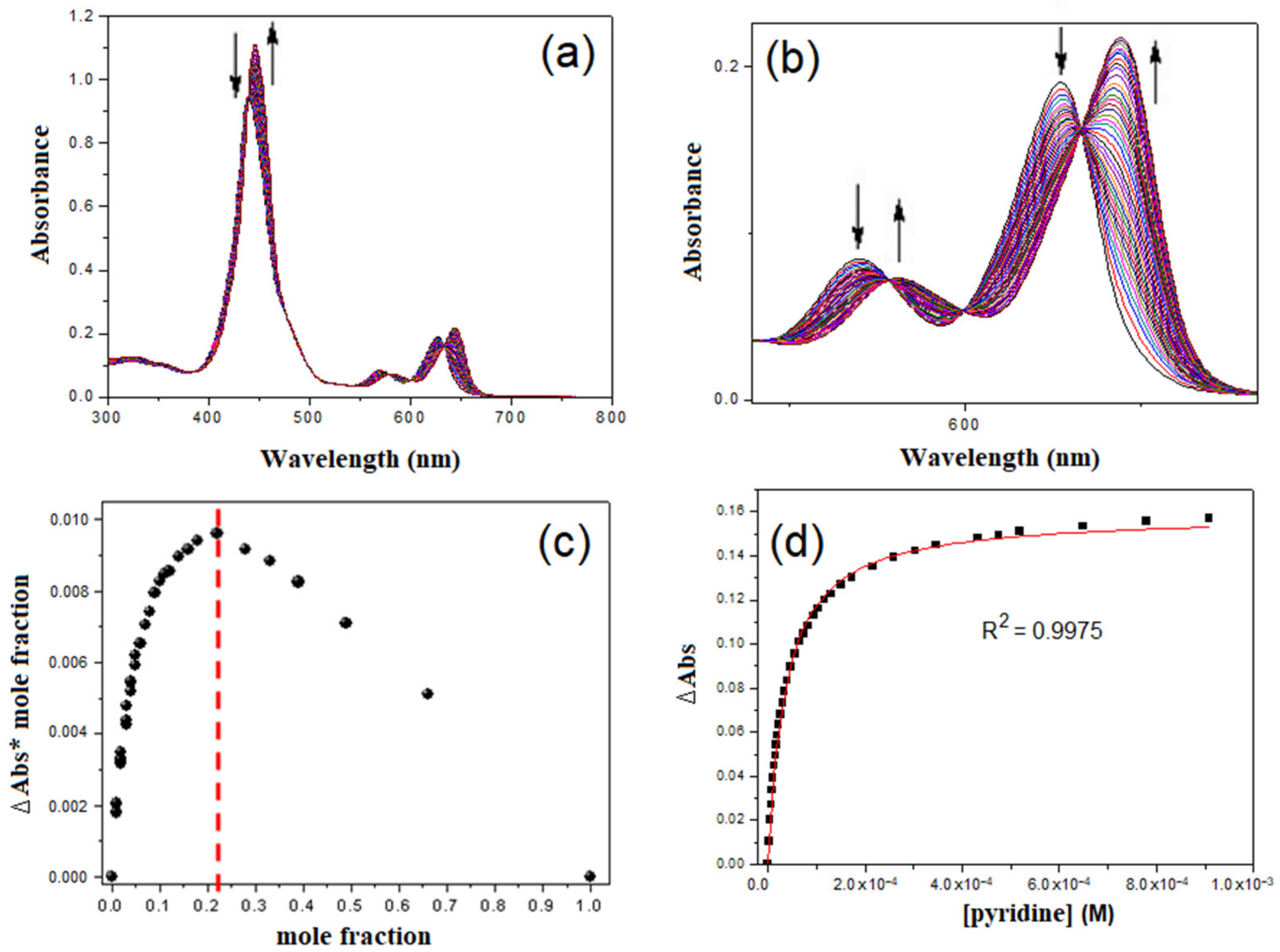
The stability constant and the related species resulting from the coordination of pyridine with 1 are shown in Scheme 2. The UV-vis spectrum of 1 in toluene contained an intense, broad Soret band (439 nm) and weak Q-bands (570 nm and 627 nm) (Figure 1 and Supplementary Materials Figure S1) [34].
Titrimetric addition of pyridine to a toluene solution of 1 created a series of isosbestic points (439, 578, 599, and 632 nm) along with a shift in the Soret band (439 to 446 nm) and in the Q-bands (570 to 581 nm and 627 to 644 nm). The associated 7 nm redshift in the Soret band indicated pyridine binding to 1. No further changes in the absorbance were observed after the addition of 3 equiv. of pyridine. Job’s plot for the titration data (Figure 1c) clearly supports forming a 3:1 complex, as shown in Scheme 2. Equilibrium constant K31 for 1∙pyridine was calculated as follows:
K31 = [(PY)31]/[PY]3[1]
The binding constant, K31 for the 1∙pyridine complex, was estimated to be (2.7 ± 0.15) × 1014 M−3. On the other hand, monomeric Zn-porphyrin 2 exhibits typical electronic absorption features in toluene, including an intense Soret band at 434 nm and weak Q bands at 563 nm and 605 nm (Figure S2). The addition of pyridine to a toluene solution of monomeric 2 resulted in a series of isosbestic points (436, 570, 592, and 611 nm) along with a shift in the Soret band (434 to 438 nm) and in the Q-bands (563 to 575 nm and 605 to 622 nm). The associated 4 nm redshift in the Soret band is characteristic of the formation of a 2∙pyridine complex in a 1:1 ratio. The binding constant, K11 (= [PY∙2]/[PY][2]) for the 2∙pyridine complex, was calculated to be (1.60 ± 0.08) × 104 M−1 (Figure S2).
The addition of 4,4-bipyridine (BIPY) to a toluene solution of conjugated Zn-trisporphyrin 1 led to forming a series of coordination complexes. The overall stability constants and associated species are depicted in Scheme 3. The general equilibrium constants, shown in Scheme 3, were determined as follows:
K 31 = [ L 3 · 1 / L ] 3 1
K 32 = [ L 3 · 1 2 / L ] 3 [ 1 ] 2
K 32 31 = K 31 2 / K 32 = L 3 · 1 2 / L 3 · 1 2 [ L ] 3
where, L = BIPY or DPYP.
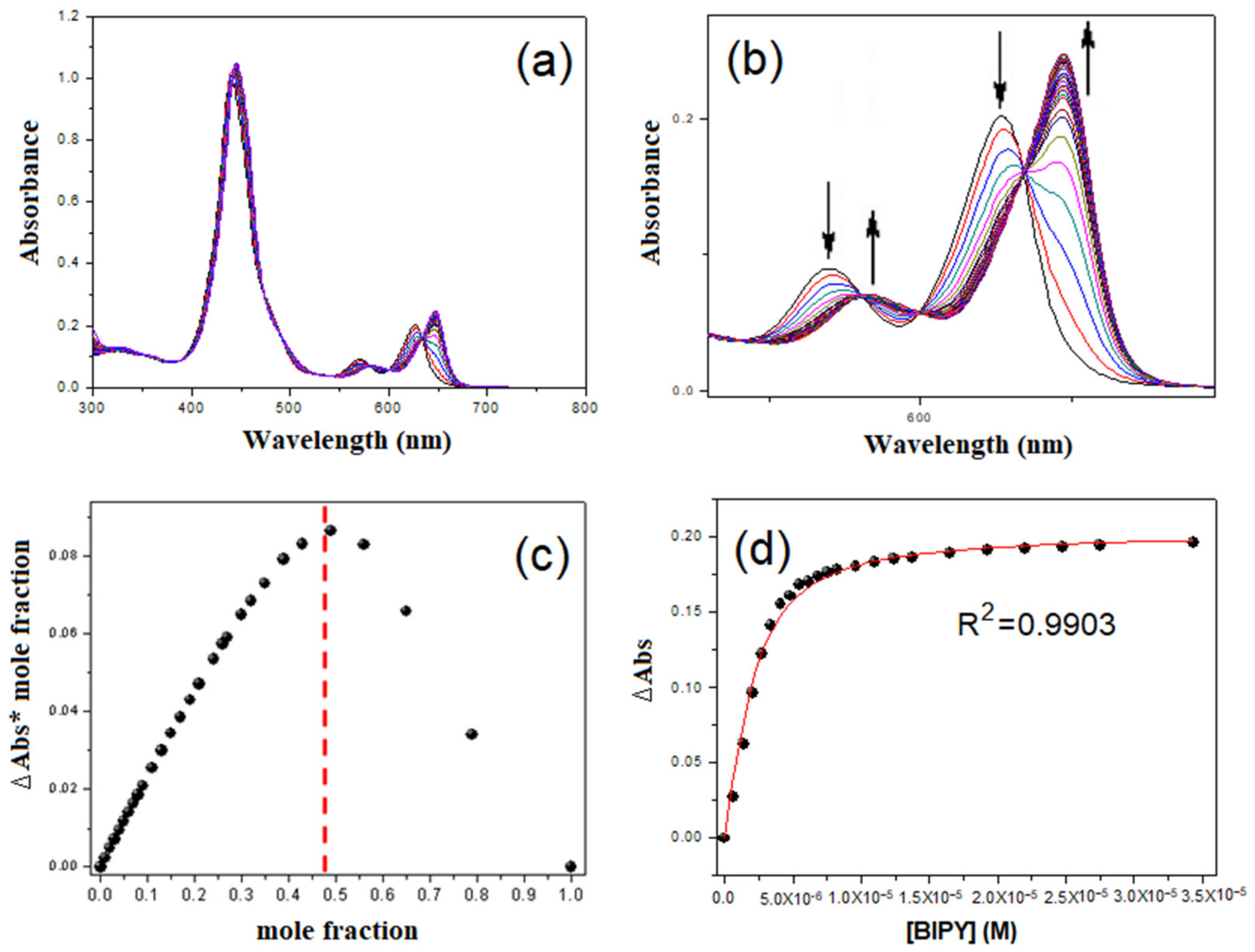
Upon the addition of 0–3 equiv. of BIPY into a toluene solution of 1, the Soret band initially shifted from 439 to 443 nm (Figure 2 and Figure S3). Such a 4 nm redshift was characteristic of the formation of a 3:2 double-decker complex, as shown in Scheme 3. With the further addition of BIPY above 3 equiv. to the solution of 1, the band at 443 nm started to decrease in intensity, and a new band appeared at 446 nm, suggesting the deconstruction of the 3:2 double-decker complex and the mono-coordination of 1 with BIPY to form a 3:1 open complex. In addition, a series of isosbestic points (441, 581, 599, and 634 nm) along with a shift in the Q-bands (570 to 583 nm and 627 to 647 nm) were also observed during the titration process. Based on the fitting of the titration data to the three species (1, 3:2 double-decker coordination complex, and 3:1 open complex) and the binding model described in Scheme 3, binding constants K32 and K31 were calculated to be (9.26 ± 0.07) × 1027 M−4 and (3.1 ± 0.13) × 1015 M−3, respectively, using global multivariate factor analysis [31,32,33,35,36,37]. On the other hand, the BIPY titration of a toluene solution of monomeric 2 resulted in a two-phase absorbance change, which appeared stable even after the addition of more than 3 equiv. of BIPY. The titration results consisted of four concomitant isosbestic points (436, 570, 592, and 611 nm) along with a shift in the Soret band (434 to 438 nm) and in the Q-bands (563–575 nm and 605–622 nm). The associated 4 nm redshift in the Soret band evidently supports forming a 1:1 2∙BIPY complex. From the titration data, it is clear that forming a 2:1 complex of Zn-monomeric 2 with BIPY was negligible under our experimental conditions. It is likely that 2:2 or 4:2 complexes were generated, which would affect the binding constant, as mentioned in the literature [32]. However, we did not consider these species owing to their negligible concentration under our experimental conditions (micromolar concentrations and room temperature). At micromolar concentrations, ladder-type 2:1 complexes are not likely to be formed during UV-visible titration [38]. The binding constant, K11 (= [BIPY∙2]/[BIPY][2]) for 2∙BIPY, was calculated to be (2.02 ± 0.03) × 104 M−1 (Figure S4). The above observation confirmed that the synergistic effect of the three conjugated porphyrin moieties in 1 was operational during the titration process [26]. This cooperative effect facilitates constructing a 3:2 double-decker coordination complex at a certain concentration of BIPY in solution.
The binding of the rigid ligand BIPY to 1 was then compared to the assembly behavior of 1 with flexible N-donor ligand, 1,3-di(4-pyridyl)propane (DPYP) in toluene. Titrimetric addition of DPYP to a toluene solution of 1 gave rise to a series of isosbestic points (439, 579, 599, and 633 nm) along with a shift in the Soret band (439 to 446 nm) and in the Q-bands (570 to 582 nm and 627 to 645 nm). The appearance of the titration profile was very similar to that of BIPY with 1 (Figure 3 and Figure 4), although the fitted curve did not perfectly match with experimental data. This deviation may be due to side reactions that form unexpected complexes.
A notable difference is that the degradation of the sandwiched 3:2 complex and forming an open 3:1 complex proceeded more easily in the case of DPYP addition to the solution of 1 compared to that of BIPY titration. Binding constants K32 and K31 were calculated to be (3.62 ± 0.16) × 1027 M−4 and (8.1 ± 0.13) × 1015 M−3, respectively. Thus, the binding constant, K32 of 1∙BIPY, was approximately 2.5 times higher than that of 1∙DPYP. This implies that rigid BIPY forms a more robust self-assembled double-decker complex than flexible DPYP. Using the above stability constants of 1∙BIPY and 1∙DPYP, and the SPECFIT program, we simulated the speciation profile considering only three species (1, 3:2 sandwiched complex, and 3:1 open structure) according to the reported methods [32,33], as shown in Figure 5. It should be mentioned that for simplicity, we neglected other species (~2:2 or 4:2 model structures mentioned in [32]), due to their negligible content at micromolar concentration. We observed some notable features from these simulated speciation profiles along with the experimental spectra in Figure 2 and Figure 3. Initially, the concentration of 1 is high (100%), as indicated by the Soret band at 439 nm. After the addition of ligands (BPY or DPYP), the Soret band is redshifted to 443 nm, and the concentration of 1 is decreased. This is because, at a low concentration of ligand, both BIPY and DPYP formed 3:2 double-decker complexes with 1. The concentration of the 3:2 double-decker complex increased upon the addition of bidentate ligands up to 3 equivalents. The 3:2 double-decker complex seems to be stable up to a certain range of ligand concentrations. After that, these double-decker complexes gradually degrade to form 3:1 open complexes as the concentration of ligands highly increases further. The Soret band was further redshifted from 443 nm to 446 nm during this process.
Several notable conclusions can be drawn from these UV-vis titration profiles. Degradation of the 3:2 double-decker 1∙BIPY complex is less favorable compared to that of 1∙DPYP. Consequently, forming the 3:1 open 1∙DPYP complex proceeded more readily than that of 1∙BIPY. It should be noted that DPYP is somewhat curved, whereas BIPY is a categorically linear ligand. Therefore, the possible geometry of the double-decker 3:2 1∙BIPY complex would be a regular sandwich (eclipsed stacked structure). On the other hand, the double-decker 3:2 1∙DPYP complex would exhibit offset sandwich geometry (displaced stacked structure).
To validate our results, we employed DABCO, a common bridging ligand, previously used to construct double-decker complexes [31,32,33,36]. Titrimetric addition of DABCO to a toluene solution of 1 afforded a series of isosbestic points (439, 578, 598, and 632 nm) along with a shift in the Soret band (439 to 446 nm), as well as in the Q-bands (570 to 582 nm and 627 to 645 nm). The appearance of the titration profile was comparable to that of BIPY titration of 1 (Figure S5). Binding constants K32 and K31 were calculated to be (1.47 ± 0.09) × 1027 M−4 and (1.7 ± 0.21) × 1015 M−3, respectively. The binding constant, K32 derived from 1∙BIPY, was higher than that from 1∙DABCO. Compared to DABCO, the longer arm of BIPY appears to avoid steric clashing with the aryl groups of 1 in forming double-decker complexes.

3. Materials and Methods

Zn-trisporphyrin conjugate 1 and monomeric Zn-porphyrin 2 were synthesized according to our previous report [34]. All other chemicals (pyridine, 4,4-bipyridine, and 1,3-di(4-pyridyl)propane) were purchased from Sigma-Aldrich and used without further purification. Steady-state UV-vis spectra were recorded on a Shimadzu UV-3600 spectrophotometer. For UV-vis titrations, we prepared the host and guest solutions in toluene at room temperature. The porphyrin (host) concentration was fixed at 1 μM, and ligand (guest) concentration was varied from 0.001 to 0.1 M. UV-vis titration experiments were performed by recording a spectrum of the host (porphyrin) in toluene at 298 K and adding the guest (ligand) solution in increments. After each addition, a new UV-vis spectrum was obtained. The data obtained from the UV-vis spectrophotometric titrations were analyzed by fitting the entire spectral series at 1 nm intervals using the SPECFIT software [39], as mentioned in earlier reports [32,33]. To determine the binding constants, the titration results were fitted using the nonlinear regression method within the Origin 9 software and generated the following equation:
y = A((1 + Kx + KP) − ((1 + Kx + KP)2 − 4K2Px)0.5/2KP
x = [guest] × (binding site)
y = ΔA = A − A0
A =ΔA at 100% complexation
K = Binding constant
P = [host]

4. Conclusions

In summary, the supramolecular assembly of conjugated Zn-trisporphyrin 1 with different types of N-donor ligands was investigated in solution at micromolar concentrations, employing UV-vis spectrophotometric titration. While pyridine, a non-bridging ligand, forms a 3:1 open complex with 1, bridging ligands, BIPY, and DPYP, form stable 3:2 double-decker complexes with 1. These double-decker complexes collapse into simple open 3:1 complexes with the addition of excess BIPY or DPYP. For DPYP (flexible ligand) complexation with 1, the 3:2 double-decker complex equilibrated with the 3:1 open complex more readily than the 1∙BIPY complex (rigid ligand). Additionally, forming a 3:2 double-decker coordination complex was similarly observed when the DABCO complexed with 1. Our results provide valuable insights for constructing a variety of metalloporphyrin-based self-assembled structures, potentially applicable in constructing novel functional materials for chemical detection, supramolecular catalysis, and photoelectronics.

Supplementary Materials

The following are available online. Figure S1: UV-vis titration spectra of Zn-trisporphyrin 1 with pyridine in toluene; Figure S2: UV-vis titration spectra of Zn-monoporphyrin 2 with pyridine in toluene; Figure S3: UV-vis titration spectra of Zn-trisporphyrin 1 with 4,4-bipyridine in toluene; Figure S4: UV-vis titration spectra of Zn-monoporphyrin 2 with 4,4-bipyridine in toluene, Figure S5: UV-vis titration spectra of Zn-trisporphyrin 1 with DABCO in toluene.

Author Contributions

Formal analysis, data curation, visualization, software, and writing, N.K.S.; Investigation, methodology, J.-W.S.; conceptualization, project administration, supervision, funding acquisition, resources, review, validation, and editing, H.-J.K. All authors have read and agreed to the published version of the manuscript.

Funding

This work was supported by the Kumoh National Institute of Technology (Grant No. 202001330001).

Data Availability Statement

Data are available in the article and Supplementary Materials.

Acknowledgments

We would like to thank S.Y. Jang for assistance with the titration experiments.

Conflicts of Interest

The authors declare no conflict of interest.

Sample Availability

Samples of the compounds are not available from the authors.

References

  1. Vantomme, G.; Meijer, E.W. The construction of supramolecular systems. Science 2019, 363, 1396–1397. [Google Scholar] [CrossRef] [PubMed]
  2. Lehn, J.-M. Perspectives in Supramolecular Chemistry—From Molecular Recognition towards Molecular Information Processing and Self-Organization. Angew. Chem. Int. Ed. Engl. 1990, 29, 1304–1319. [Google Scholar] [CrossRef]
  3. Wang, S.-P.; Lin, W.; Wang, X.; Cen, T.-Y.; Xie, H.; Huang, J.; Zhu, B.-Y.; Zhang, Z.; Song, A.; Hao, J.; et al. Controllable hierarchical self-assembly of porphyrin-derived supra-amphiphiles. Nat. Commun. 2019, 10, 1399–1411. [Google Scholar] [CrossRef] [PubMed] [Green Version]
  4. Liu, Y.; Chen, Q.; Cullen, D.A.; Xie, Z.; Lian, T. Efficient Hot Electron Transfer from Small Au Nanoparticles. Nano Lett. 2020, 20, 4322–4329. [Google Scholar] [CrossRef]
  5. Li, C.; Park, K.-M.; Kim, H.-J. Ionic assembled hybrid nanoparticle consisting of tin(IV) porphyrin cations and polyoxomolybdate anions, and photocatalytic hydrogen production by its visible light sensitization. Inorg. Chem. Commun. 2015, 60, 8–11. [Google Scholar] [CrossRef]
  6. Qi, Z.-L.; Cheng, Y.-H.; Xu, Z.; Chen, M.-L. Recent advances in porphyrin-based materials for metal ions detection. Int. J. Mol. Sci. 2020, 21, 5839. [Google Scholar] [CrossRef] [PubMed]
  7. Sun, H.; Ren, J.; Qu, X. Carbon Nanomaterials and DNA: From Molecular Recognition to Applications. Acc. Chem. Res. 2016, 49, 461–470. [Google Scholar] [CrossRef]
  8. Montaseri, H.; Kruger, C.A.; Abrahamse, H. Recent advances in porphyrin-based inorganic nanoparticles for cancer treatment. Int. J. Mol. Sci. 2020, 21, 3358. [Google Scholar] [CrossRef] [PubMed]
  9. Drain, C.M.; Varotto, A.; Radivojevic, I. Self-organized porphyrinic materials. Chem. Rev. 2009, 109, 1630–1658. [Google Scholar] [CrossRef] [Green Version]
  10. Beletskaya, I.; Tyurin, V.S.; Tsivadze, A.Y.; Guilard, R.; Stern, C. Supramolecular chemistry of metalloporphyrins. Chem. Rev. 2009, 109, 1659–1713. [Google Scholar] [CrossRef]
  11. Sun, Y.; Chen, C.; Liu, J.; Liu, L.; Tuo, W.; Zhu, H.; Lu, S.; Li, X.; Stang, P.J. Self-Assembly of Porphyrin-Based Metallacages into Octahedra. J. Am. Chem. Soc. 2020, 142, 17903–17907. [Google Scholar] [CrossRef]
  12. Kim, M.K.; Shee, N.K.; Lee, J.; Yoon, M.; Kim, H.-J. Photoinduced electron transfer upon supramolecular complexation of (porphyrinato)Sn-viologen with cucurbit[7]uril. Photochem. Photobiol. Sci. 2019, 18, 1996–2002. [Google Scholar] [CrossRef]
  13. Shee, N.K.; Kim, M.K.; Kim, H.-J. Fluorescent chemosensing for aromatic compounds by a supramolecular complex composed of tin(iv) porphyrin, viologen, and cucurbit[8]uril. Chem. Commun. 2019, 55, 10575–10578. [Google Scholar] [CrossRef]
  14. Shee, N.K.; Kim, H.-J. Self-Assembled Nanomaterials Based on Complementary Sn(IV) and Zn(II)-Porphyrins, and Their Photocatalytic Degradation for Rhodamine B Dye. Molecules 2021, 26, 3598. [Google Scholar] [CrossRef]
  15. Kim, H.-J.; Jo, H.J.; Kim, J.; Kim, S.-Y.; Kim, D.; Kim, K. Supramlecular self-assembly of tin(IV)porphyrin channels stabilizing single-file chains of water molecules. CrystEngComm 2005, 7, 417–420. [Google Scholar] [CrossRef] [Green Version]
  16. Lee, S.J.; Malliakas, C.D.; Kanatzidis, M.G.; Hupp, J.T.; Nguyen, S.T. Amphiphilic porphyrin nanocrystals: Morphology tuning and hierarchical assembly. Adv. Mater. 2008, 20, 3543–3549. [Google Scholar] [CrossRef]
  17. Liu, H.; Xu, J.; Li, Y.; Li, Y. Aggregate Nanostructures of Organic Molecular Materials. Acc. Chem. Res. 2010, 43, 1496–1508. [Google Scholar] [CrossRef]
  18. Shao, S.; Rajendiran, V.; Lovell, J.F. Metalloporphyrin nanoparticles: Coordinating diverse theranostic functions. Coord. Chem. Rev. 2019, 379, 99–120. [Google Scholar] [CrossRef] [PubMed]
  19. Magna, G.; Monti, D.; di Natale, C.; Paolesse, R.; Stefanelli, M. The assembly of porphyrin systems in well-defined nanostructures: An update. Molecules 2019, 24, 4307. [Google Scholar] [CrossRef] [PubMed] [Green Version]
  20. Wang, Z.; Medforth, C.J.; Shelnutt, J.A. Porphyrin Nanotubes by Ionic Self-Assembly. J. Am. Chem. Soc. 2004, 126, 15954–15955. [Google Scholar] [CrossRef]
  21. Lee, S.J.; Hupp, J.T.; Nguyen, S.T. Growth of narrowly dispersed porphyrin nanowires and their hierarchical assembly into macroscopic columns. J. Am. Chem. Soc. 2008, 130, 9632–9633. [Google Scholar] [CrossRef]
  22. Shee, N.K.; Kim, M.K.; Kim, H.-J. Supramolecular Porphyrin Nanostructures Based on Coordination-Driven Self-Assembly and Their Visible Light Catalytic Degradation of Methylene Blue Dye. Nanomaterials 2020, 10, 2314. [Google Scholar] [CrossRef]
  23. Oliveras-González, C.; di Meo, F.; González-Campo, A.; Beljonne, D.; Norman, P.; Simón-Sorbed, M.; Linares, M.; Amabilino, D.B. Bottom-Up Hierarchical Self-Assembly of Chiral Porphyrins through Coordination and Hydrogen Bonds. J. Am. Chem. Soc. 2015, 137, 15795–15808. [Google Scholar] [CrossRef] [PubMed]
  24. Zhang, C.; Chen, P.; Dong, H.; Zhen, Y.; Liu, M.; Hu, W. Porphyrin supramolecular 1D structures via surfactant-assisted self-assembly. Adv. Mater. 2015, 27, 5379–5387. [Google Scholar] [CrossRef]
  25. Yamamoto, K.; Imaoka, T.; Tanabe, M.; Kambe, T. New horizon of nanoparticle and cluster catalysis with dendrimers. Chem. Rev. 2019, 120, 1397–1437. [Google Scholar] [CrossRef] [PubMed]
  26. Drobizhev, M.; Stepanenko, Y.; Rebane, A.; Wilson, C.J.; Screen, T.E.O.; Anderson, H.L. Strong Cooperative Enhancement of Two-Photon Absorption in Double-Strand Conjugated Porphyrin Ladder Arrays. J. Am. Chem. Soc. 2006, 128, 12432–12433. [Google Scholar] [CrossRef] [PubMed]
  27. Smith, A.R.; Ruggles, J.L.; Yu, A.; Gentle, I.R. Multilayer Nanostructured Porphyrin Arrays Constructed by Layer-by-Layer Self-Assembly. Langmuir 2009, 25, 9873–9878. [Google Scholar] [CrossRef] [PubMed]
  28. Abd El-Mageed, A.I.A.; Ogawa, T. Supramolecular structures of terbium(iii) porphyrin double-decker complexes on a single-walled carbon nanotube surface. RSC Adv. 2019, 9, 28135–28145. [Google Scholar] [CrossRef] [Green Version]
  29. Maeda, C.; Toyama, S.; Okada, N.; Takaishi, K.; Kang, S.; Kim, D.; Ema, T. Tetrameric and Hexameric Porphyrin Nanorings: Template Synthesis and Photophysical Properties. J. Am. Chem. Soc. 2020, 142, 15661–15666. [Google Scholar] [CrossRef]
  30. Murphy, R.B.; Norman, R.E.; White, J.M.; Perkins, M.V.; Johnston, M.R. Tetra-porphyrin molecular tweezers: Two binding sites linked via a polycyclic scaffold and rotating phenyl diimide core. Org. Biomol. Chem. 2016, 14, 8707–8720. [Google Scholar] [CrossRef] [Green Version]
  31. Slagt, F.V.; van Leeuwen, P.W.N.M.; Reek, J.N.H. Multicomponent Porphyrin Assemblies as Functional Bidentate Phosphite Ligands for Regioselective Rhodium-Catalyzed Hydroformylation. Angew. Chem. Int. Ed. 2003, 42, 5619–5623. [Google Scholar] [CrossRef]
  32. Ballester, P.; Oliva, A.I.; Costa, A.; Deyà, P.M.; Frontera, A.; Gomila, R.M.; Hunter, C.A. DABCO-Induced Self-Assembly of a Trisporphyrin Double-Decker Cage: Thermodynamic Characterization and Guest Recognition. J. Am. Chem. Soc. 2006, 128, 5560–5569. [Google Scholar] [CrossRef]
  33. Huang, C.; Shen, B.; Wang, K.; Lu, J.; Sun, X. Rigid Trisporphyrin with 1,3,5-Triazine Core: Synthesis, Spectroscopy, and DABCO-Induced Self-Assembly Properties. J. Porphyrins Phthalocyanines 2020, 24, 1066–1073. [Google Scholar] [CrossRef]
  34. Seo, J.-W.; Jang, S.Y.; Kim, D.; Kim, H.-J. Octupolar trisporphyrin conjugates exhibiting strong two-photon absorption. Tetrahedron 2008, 64, 2733–2739. [Google Scholar] [CrossRef]
  35. Oliva, A.I.; Ventura, B.; Würthner, F.; Camara-Campos, A.; Hunter, C.A.; Ballester, P.; Flamigni, L. Self-Assembly of Double-Decker Cages Induced by Coordination of Perylene Bisimide with a Trimeric Zn Porphyrin: Study of the Electron Transfer Dynamics Between the Two Photoactive Components. Dalton Trans. 2009, 4023–4037. [Google Scholar] [CrossRef] [PubMed]
  36. Ballester, P.; Costa, A.; Castilla, A.M.; Deyà, P.M.; Frontera, A.; Gomila, R.M.; Hunter, C.A. DABCO-Directed Self-Assembly of Bisporphyrins (DABCO = 1,4-Diazabicyclo[2.2.2]octane). Chem. Eur. J. 2005, 11, 2196–2206. [Google Scholar] [CrossRef] [PubMed]
  37. Ercolani, G. Thermodynamics of Metal-Mediated Assemblies of Porphyrins. In Structure and Bonding; Springer: Berlin/Heidelberg, Germany, 2006; Volume 121, pp. 167–215. [Google Scholar]
  38. Taylor, P.N.; Anderson, H.L. Cooperative Self-Assembly of Double-Strand Conjugated Porphyrin Ladders. J. Am. Chem. Soc. 1999, 121, 11538–11545. [Google Scholar] [CrossRef]
  39. Binstead, R.A.; Zuberbühler, A.D.; Jung, B. SPECFIT/32, V. 3.0.23; Spectrum Software Associates: Marlborough, MA, USA, 2004. [Google Scholar]
Scheme 1. Chemical structures of Zn-porphyrins and N-donor ligands used in this study.
Molecules 26 04771 sch001
Scheme 2. Schematic representation of the species involved in the binding of Zn-trisporphyrin 1 with pyridine.
Molecules 26 04771 sch002
Figure 1. UV-vis titration spectra of Zn-trisporphyrin 1 complexation with pyridine in toluene (c = 1 μM). (a) UV-visible region, (b) Q-band zone, (c) data curve for Job’s plot at 446 nm (mole fraction of 1), (d) fitting curve for the determination of the binding constant K31 at 446 nm.
Molecules 26 04771 g001
Scheme 3. Schematic representation of the species involved in the binding of Zn-trisporphyrin 1 with BIPY or DPYP.
Molecules 26 04771 sch003
Figure 2. UV-vis titration spectra of Zn-trisporphyrin 1 with BIPY in toluene (c = 1 μM). (a) UV-vis region, (b) Q-band zone, (c) data curve for Job’s plot at 446 nm (mole fraction of 1), (d) fitting curve for the determination of the binding constant K32 at 446 nm.
Molecules 26 04771 g002
Figure 3. UV-vis titration spectra of Zn-trisporphyrin 1 with DPYP in toluene (c = 1 μM). (a) UV-visible region, (b) Soret-band zone, (c) Q-band zone, (d) fitting curve for the determination of the binding constant K32 at 446 nm.
Molecules 26 04771 g003
Figure 4. Job’s plots for the UV-vis titration of Zn-trisporphyrin 1 with DPYP in toluene (c = 1 μM). (a) mole fraction, and (b) site fraction.
Molecules 26 04771 g004
Figure 5. Simulated speciation profiles for titrations of 1 with (a) BIPY and (b) DPYP. Insets show enlargements of the lower-site-ratio region.
Molecules 26 04771 g005
Publisher’s Note: MDPI stays neutral with regard to jurisdictional claims in published maps and institutional affiliations.