Article

A Validated Ultrasound-Assisted Extraction Coupled with SPE-HPLC-DAD for the Determination of Flavonoids in By-Products of Plant Origin: An Application Study for the Valorization of the Walnut Septum Membrane

by
Natasa P. Kalogiouri
and
Victoria F. Samanidou
*
Laboratory of Analytical Chemistry, Department of Chemistry, Aristotle University of Thessaloniki, 54124 Thessaloniki, Greece
*
Author to whom correspondence should be addressed.
Molecules 2021, 26(21), 6418; https://doi.org/10.3390/molecules26216418
Submission received: 12 October 2021 / Revised: 18 October 2021 / Accepted: 22 October 2021 / Published: 24 October 2021
(This article belongs to the Special Issue Chromatography-the Ultimate Analytical Tool)

Abstract

:
Walnut byproducts have been shown to exert functional properties, but the literature on their bioactive content is still scarce. Among walnut byproducts, walnut septum is a dry ligneous diaphragm tissue that divides the two halves of the kernel, exhibiting nutritional and medicinal properties. These functional properties are owing to its flavonoid content, and in order to explore the flavonoid fraction, an ultrasound-assisted (UAE) protocol was combined with solid phase extraction (SPE) and coupled to high-performance liquid chromatography with diode array detection (HPLC-DAD) for the determination of flavonoids in Greek walnut septa membranes belonging to Chandler, Vina, and Franquette varieties. The proposed UAE-SPE-HPLC-DAD method was validated and the relative standard deviations (RSD%) of the within-day and between-day assays were lower than 6.2 and 8.5, respectively, showing good precision, and high accuracy ranging from 90.8 (apigenin) to 97.5% (catechin) for within-day assay, and from 88.5 (myricetin) to 96.2% (catechin) for between-day assay. Overall, seven flavonoids were determined (catechin, rutin, myricetin, luteolin, quercetin, apigenin, and kaempferol) suggesting that the walnut septum is a rich source of bioactive constituents. The quantification results were further processed using ANOVA analysis to examine if there are statistically significant differences between the concentration of each flavonoid and the variety of the walnut septum.

1. Introduction

The development of analytical methodologies to characterize natural ingredients and produce high added value food products is a field of research that has attracted the interest of the scientific community, industry, and consumers, as well. During the last decades, there has been an increasing demand for foods that not only have high nutritional and sensorial quality but also deliver health promoting benefits through certain ingredients, namely “bioactive” or “functional” [1]. Moreover, emphasis is given on the valorization of food byproducts that combine nutritional value, flavor, and health benefits, as well. Towards this direction and in order to protect both public health and environment agri-food byproducts, generated in large amounts worldwide, can be exploited as a promising source of valuable compounds for novel food applications.
Walnuts (Juglans regia L.) are a valuable nutritional source with pleasant taste, consumed on a global scale. Walnut has been cultivated since 1000 BC, and it has naturally diverged to several cultivars, such as Chander, Vina, Franquette, Mellanaise, Lara, Marbot, Hartley Mayette, Serr, Tulare, Sorento, etc. [2]. Apart from the nutritional benefits, walnuts exert health-promoting properties, such as antioxidant, anti-inflammatory, antimicrobial, antidiabetic, hepatoprotective, anticancer, and cardioprotective, among others [3,4]. These benefits are owing to the presence of phytonutrients, and specifically to the phenolic compounds [5,6,7]. Even though the nutritional importance of the walnut is mainly related to the kernel, walnut byproducts, such as walnut husks, leaves, etc. [8,9], have been shown to exert functional properties, as well. Among walnut byproducts, walnut septum is a dry ligneous diaphragm tissue that divides the two halves of the kernel. Traditionally, the walnut septum has been used as a nutraceutical and medicinal material, and has been documented in the Chinese Pharmacopoeia [10]. The septum contains bioactive constituents, and it has been shown to exhibit antitumor, antioxidant, and immunoenhancement effects in vitro [11,12]. Phenolic compounds, and particularly flavonoids, are an important class of bioactive constituents that might be related to the functional properties of the walnut septum. Despite the fact that flavonoids are responsible for several pharmacological activities [13], the literature on the flavonoid content of walnut septum is still scarce.
Thus, the phenolic fraction of this unexplored byproduct has to be assessed with analytical methodologies. The generic analytical procedure for the determination of flavonoids in agricultural products involves first sample preparation, and then separation, detection, and quantification. Several traditional extraction techniques have been proposed for the extraction of flavonoids from natural products, including percolation, maceration, hydro-distillation, boiling, reflux, Soxhlet [14,15]. These techniques, however, present several disadvantages such as a lot of labor and time, large amounts of organic solvents, low selectivity, low extraction yield, and high cost [16]. For this reason, advanced green microextraction techniques are continually being developed to overcome these limitations. Green extraction techniques involve ultrasound-assisted extraction (UAE), microwave assisted extraction (MAE), supercritical fluid extraction (SFE), and solid phase extraction (SPE), among others. The current state-of-the art on the green extraction of natural constituents from food products has already been critically reviewed [4,17].
The main analytical technique used for the separation and determination of phenolic compounds is traditionally liquid chromatography (LC) coupled to UV, diode array detection (DAD), or mass spectrometric detectors (MS) [18,19,20,21]. The recent trends in analytical determination also involve the development of high-resolution mass spectrometric (HRMS) techniques, which provide separation efficiency and high accuracy in identifications, as it has already been reviewed [4,15,22,23]. Among these techniques, HPLC-DAD is a rapid analytical technique that enables the separation, identification, and quantification of flavonoids, offering several advantages in terms of sensitivity, specificity, ruggedness, and low cost of analysis. The accurate determination of the flavonoids and the further quantification of such analytes that exist in trace levels in food matrices is a challenging task. The further processing of the quantification results with statistical tools enhances the conclusions derived from the experimental data, allowing the discovery of trends and behaviors among the samples.
In this work, a green UAE-SPE-HPLC-DAD methodology was developed and validated for the determination of flavonoids in walnut septum belonging to different varieties (Chandler, Vina, and Franquette) cultivated in Greece. The determined analytes were quantified, and the results were further analyzed by one-way ANOVA to examine if there are statistically significant differences between the analytes’ concentrations and the walnut variety. To the best of our knowledge, this is the first report of assessing the flavonoid profile of Greek walnut septa.

2. Results and Discussion

2.1. Method Development and Validation

An HPLC-DAD methodology was developed and validated to assess the flavonoid profile of walnut septum and all the analytical parameters, including the calibration curves, linear range, the determined coefficients (r2), accuracy and precision, limits of detection (LODs), and limits of quantification (LOQs) are presented in Table 1. The analytical curves presented an adequate fit when submitted to the lack-of-fit test (Fcalculated was less than Ftabulated in all cases), with r2 above 0.99, proving that they can be used for the quantification of the flavonoids. The LOQs were found to range between 0.30 μg/g and 0.90 μg/g, while the LODs were calculated over the range 0.10 μg/g to 0.30 μg/g. The RSD% of the within-day (n = 6) and between-day assays (n = 3 × 3) were lower than 6.2, and 8.5, respectively, showing adequate precision. The accuracy was assessed by means of relative percentage of recovery (%R) at low, medium, and maximum concentration levels of 1, 5, and 10 μg/g, and the results were acceptable, ranging from 90.8 (apigenin, at 10 μg/g concentration level) to 97.5% (catechin, at 10 μg/g concentration level) for within-day assay (n = 6) (Table 2), and from 88.5 (myricetin, at 1 μg/g concentration level) to 96.2% (catechin, at 5 μg/g concentration level) for between-day assay (n = 3 × 3) (Table 3).

2.2. Walnut Septum Analysis

Twenty-four walnut septum membranes belonging to the varieties Chandler, Vina, and Franquette were analyzed in triplicate and the flavonoids: catechin, rutin, myricetin, luteolin, quercetin, apigenin, and kaempferol were determined. The chromatographic identification results, including the retention times (Rts) of the analytes, and their respective maximum absorption wavelengths (λ, nm) are presented in Table 4. Figure 1 illustrates the chromatographic separation of the flavonoids in a walnut septum extract that was monitored at 280 nm.

2.3. Quantitative Analysis of Flavonoids

The walnut septa were analyzed in triplicate (n = 3). The identified flavonoids (catechin, rutin, myricetin, luteolin, quercetin, apigenin, and kaempferol) were quantified on the basis of their maximum absorption wavelengths. The presence of the determined flavonoids was linked to certain positive health effects and other bioactive functions that have already been reported in the literature to highlight the potential functional activity of the analyzed byproduct [4]. Table 5 presents the quantification ranges and mean values (±SD) of the determined flavonoids in the walnut septa belonging to Chandler, Vina, and Franquette variety. Box and Whisker plots were constructed to graphically illustrate the concentrations of the flavonoids and present the distributional characteristics of each variety (Chandler, Vina, Franquette). The quantification results were further analyzed with ANOVA to examine if there are statistically significant differences between the concentrations of the determined flavonoids and the varieties of the walnut septa.

2.3.1. Catechin

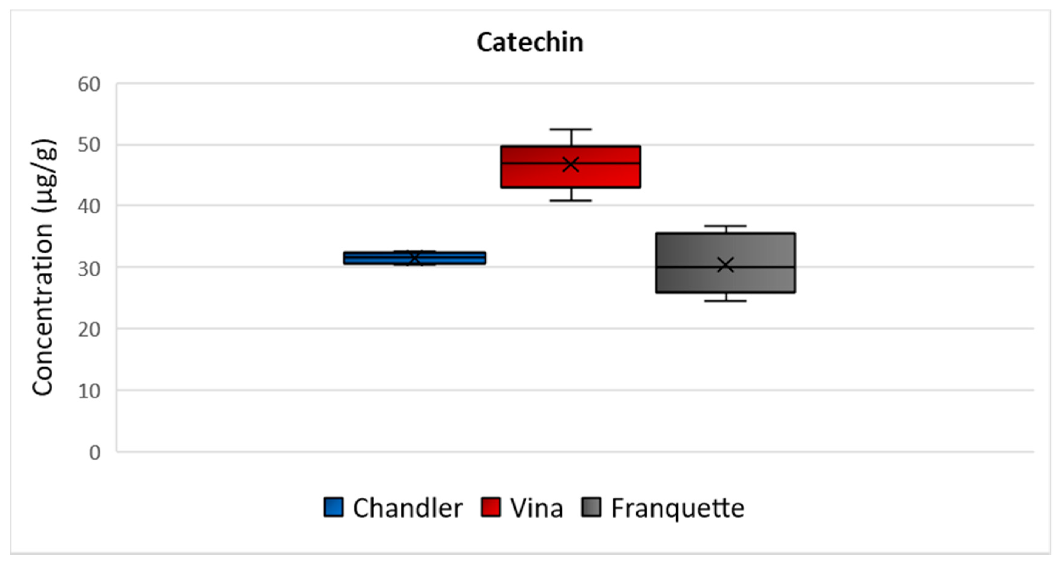
Catechin was the flavonoid detected in higher concentration in all the analyzed septa. Specifically, the highest mean concentration was determined in Vina septa (47 μg/g), and then Chandler (32 μg/g), and Franquette (31 μg/g) varieties followed (Figure 2). The ANOVA analysis exhibited a statistically significant difference in catechin concentration among all of the walnut septa of the different varieties with p = 0.04. The concentration ranges indicate that the walnut septum is rich in catechin, and it could be effectively used as a food additive to increase the antioxidant potential of food products, enrich feeds, while it could also be used as a functional ingredient of novel functional food products, including soft beverages, teas, infusions, etc. Several health properties have been associated with catechins, as they have proven to be promising protective agents against diabetes, arterial hypertension and ischemic stroke, obesity, metabolic syndrome [24]. Moreover, catechins demonstrate significant biological activities against oral cancer, breast cancer, Alzheimer’s disease, Parkinson’s disease [24]. Furthermore, catechin displays unique features responsible for several pharmacological and biological properties, as it possesses the ability to block ROS-induced chain reactions, acting as a promising antioxidant [25]. It also demonstrates significant antidiabetic function, via hepatoprotective, antineurodegenarative effects, insulin-mimetic effects, hindering amyloid formation [24].

2.3.2. Rutin

Rutin was determined over the range 1–3 μg/g in Chandler walnut septa, and over the ranges 4–6 μg/g and 3–6 μg/g in Vina and Franquette walnut septa, respectively (Figure 3). A statistically significant difference was observed for the concentration of rutin among the septa of the different varieties (p = 0.002). Rutin possesses promising antioxidant potential and exhibits significant biological properties, thus, playing an essential role in the human body’s numerous physiological functions [26]. According to the literature, rutin may provide a wide variety of therapeutic effects, such as antiallergic, antiviral, antihypertensive, vasoactive, cytoprotective, anti-inflammatory, antiprotozoal, hypolipidemic, antispasmodic, anticarcinogenic, antibacterial, and antiplatelet activities [27]. Furthermore, rutin contributes to the strengthening of the blood vessels capillaries, due to its high radical scavenging capacity, thereby preventing fragility-associated hemorrhagic disorders in humans [28].

2.3.3. Myricetin

The mean concentration values of myricetin were equal to 4 μg/g, 8 μg/g, and 7 μg/g in Chander, Vina, and Franquette walnut septa, respectively (Figure 4). Τhe ANOVA analysis showed that there is significant statistically difference among the septa of the different varieties (p = 0.002). Great scientific interest has been raised in myricetin, which has been shown to exhibit antioxidant, anti-inflammatory, antiviral, and anticarcinogen activities. It is functioning as an antineoplastic agent in human patients, since it has demonstrated strong suppressive effects on the activities of several types of cancer cells (e.g., cancer cell invasion or metastasis), thus, regulating apoptosis, and inhibitory properties on their proliferation [29,30,31,32].

2.3.4. Luteolin

The mean concentration values of luteolin were equal to 3 μg/g, 3 μg/g, and 2 μg/g in Chandler, Vina, and Franquette walnut septa, respectively (Figure 5). Statistically significant differences were observed among the luteolin concentrations determined in septa of the different varieties (p = 0.039). As for its biological action, luteolin is a naturally occurring flavonoid with a yellow crystalline appearance, and scientific research has indicated that it may display multiple cellular effects, hence, favorably affecting human health. It may exhibit antioxidant properties, protecting cells from ROS induced damage or act as an antineoplastic, anti-inflammatory, antimicrobial, or estrogenic regulatory compound and prevent liver diseases [33].

2.3.5. Quercetin

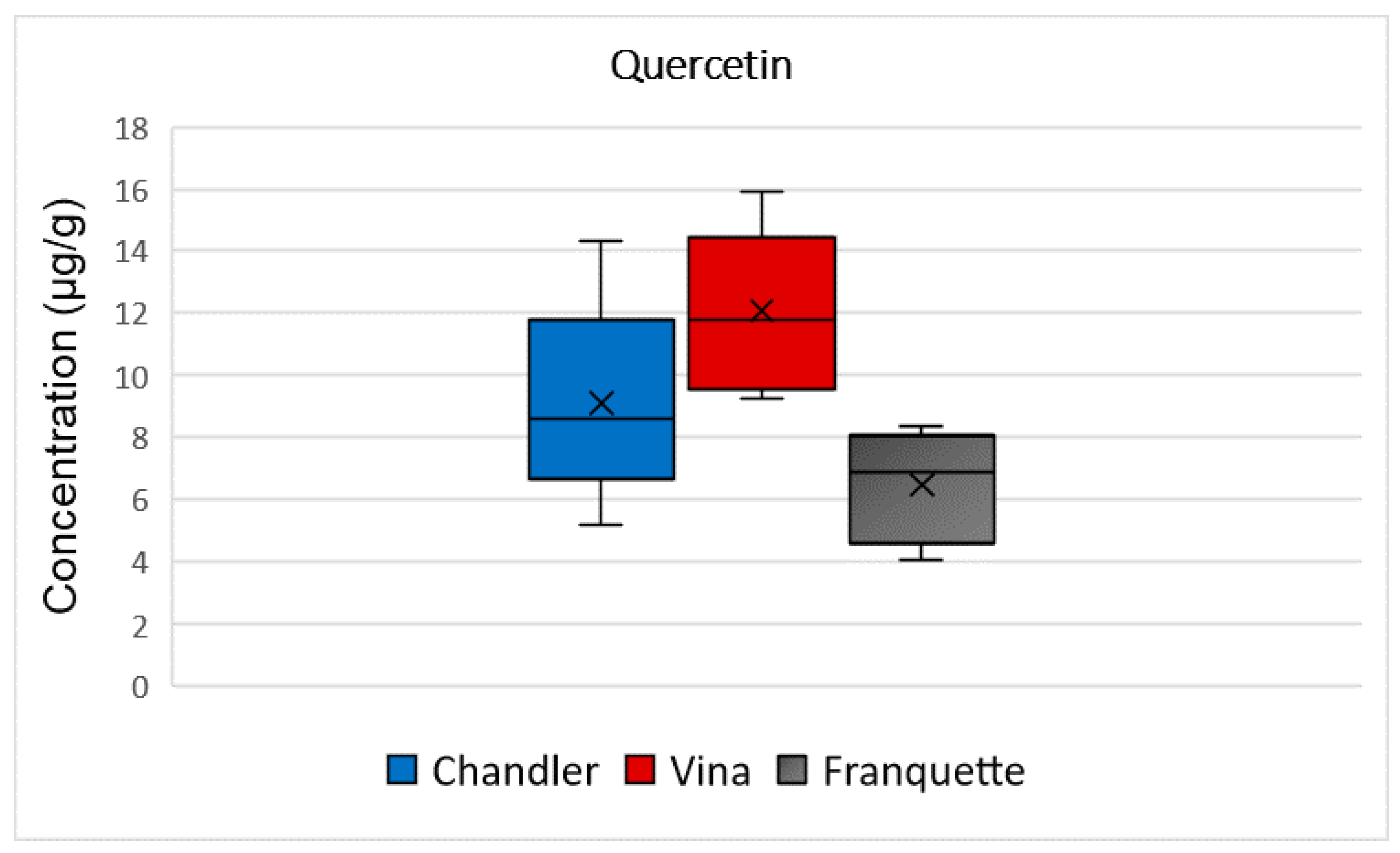
Quercetin was determined at 9 μg/g, in Chandler walnut septa. The mean concentration of quercetin was higher in walnut septa belonging to the Vina variety (12 μg/g), and lower for those belonging to the Franquette variety (6 μg/g), as it is presented in Figure 6. The ANOVA analysis showed that there is statistically significant difference among the quercetin concentration and the analyzed varieties (p = 0.0008). According to the literature, quercetin is responsible for the bitter flavor of foods, and in synergy with myricetin and other phenolic compounds has been recently used as food additives to protect meat products against bacteria’s development [34]. It demonstrates several significant health-promoting functions, including cardioprotective, anti-ulcer, antidiabetic, antioxidant properties and chemopreventive potential [35]. It may also produce anti-inflammatory and anti-allergy effects through the inhibition of the lipoxygenase and cyclooxygenase pathways, thereby reducing the production of pro-inflammatory or pro-oxidant mediators [35].

2.3.6. Apigenin

Apigenin was determined at mean concentrations of 4 μg/g, 6 μg/g, and 5 μg/g in Chandler, Vina, and Franquette walnut septa, respectively (Figure 7). The ANOVA analysis showed that there was no statistically significant difference among the apigenin concentrations in the analyzed septa of the different varieties (p = 0.147). Traditionally, extracts, oils, and teas from plants with a high content of apigenin were used for its soothing qualities as a sedative, mild analgesic and sleep medication [36], reinforcing the idea that walnut septa could be used in infusion making. Moreover, in the food industry, apigenin possesses a role as a flavoring or adjuvant agent, enhancing the human body’s response to antigens [35]. It may yield antiproliferative and antimetastatic effects, suppressing the formation and inducing malignant tumor cells [28]. Furthermore, it could prevent skin or colon cancer and acts as an anti-inflammatory, antioxidant, antiallergic, antimicrobial, antiviral, cardioprotective, and neuroprotective agent [28].

2.3.7. Kaempferol

The highest mean concentrations of kaempferol were observed in the Vina variety (5 μg/g), while Chandler variety followed with 6 μg/g, and Franquette exhibited the lowest mean concentration of 4 μg/g (Figure 8). No statistically significant difference was observed for the concentration of kaempferol among the septa of the different varieties (p = 0.06). As for its biological action, kaempferol is a natural dietary flavonoid, potentially acting as chemopreventive agent [37], protecting against oxidative stress and inflammatory chronic disorders.

2.4. Effect of the Variety on the Phenolic Content

The determination of the seven flavonoids clearly indicates that walnut septum is a byproduct that is rich in flavonoids, and, thus, could be characterized as a “nutraceutical” raw ingredient. This is also supported by the quantification ranges of each individual flavonoid identified. This fruit septum of walnuts could be utilized as a substance for medicinal purposes, while it could be also further exploited for the development of novel functional foods and beverages, and for the enrichment of feeds, as well.
Except for the functional properties that have been attributed to each individual analyte, and were discussed in detail in Section 2.3, the findings of this research also demonstrated that the flavonoid content of the walnut septum is affected by the variety. The sum of the individually quantified flavonoids showed that the walnut septum of the Vina variety exhibited the highest flavonoid content (88 ± 15 μg/g). The flavonoid contents of the septa belonging to Chandler variety, and the Franquette variety were approximately similar, with calculated concentrations of 59 ± 12 μg/g, and 60 ± 9 μg/g, respectively. The graph in Figure 9 presents the sum of the mean values of the individual concentrations of the flavonoids determined in the walnut septa belonging to Chandler, Vina, and Franquette varieties. Considering that the analyzed samples originated from the same geographical origin (Thessaly, Greece), the variation in the concentration of the flavonoids could be linked to the genetic fingerprint of each variety.
Furthermore, the ANOVA analysis showed that there is statistically significant difference between the concentrations of catechin, rutin, myricetin, luteolin, quercetin, and the walnut variety. Furthermore, PCA analysis was employed to investigate the similarities in the flavonoid content of the septa according to their variety, and the first two PCs explained the 70% of variance. The PCA score plot and loading plot are presented in the Supplementary Material in Figures S1 and S2, respectively. Even though several works have already associated the bioactive content of the walnut kernel with the geographical origin of walnuts [18,38], and the variety [39,40], these are the first reports relating the flavonoid content of the walnut septum with the variety.

3. Materials and Methods

3.1. Chemicals and Reagents

Methanol (MeOH) and acetonitrile (ACN), HPLC grade, were acquired from Merck (Darmstadt, Germany). Acetic acid 99% and trifluoroacetic acid (TFA) 99% were purchased from Sigma-Aldrich (Steinheim, Germany). The LiChrolut RP-18 (C18, 3 mL, 500 mg) SPE cartridges used were supplied by Merck (Darmstadt, Germany). Ultrapure water was provided by a Milli-Q® purification system (Millipore, Bedford, MA, USA). The flavonoids catechin 98%, rutin 98%, myricetin 98%, luteolin 98%, quercetin 98%, kaempferol 98%, and apigenin 98% were supplied by Sigma-Aldrich (Steinheim, Germany). Stock standard solutions at 1000 mg/L concentration level were prepared and stored in dark brown glass bottles at −20 °C. Working standard solutions were prepared in MeOH after appropriate dilution of the stock solutions every laboratory day, before analysis.

3.2. Instrumentation

A quaternary low-pressure gradient HPLC–DAD system by Shimadzu (Kyoto, Japan) was used for analysis. The HPLC system consisted of: (a) an FCV-10ALVP mixing system, (b) a Rheodyne 7725i injection valve (Rheodyne, Cotati, CA, USA), and a 20 µL loop for sample injection, (c) an LC- 10ADVP pump equipped with a Shimadzu SCL-10ALVP System Controller, (d) an SPD-M10AVP photodiode array detector. Real time analysis monitoring and post run processing were carried out using the software Lab Solutions-LC solutions, supplied by Shimadzu. A glass vacuum filtration apparatus, acquired by Alltech Associates (Deereld, IL, USA), and nylon 0.2 µm membrane Filters (Alltech Associates, Chicago, IL, USA) were utilized for the filtration of the mobile phase, and a DGU-10B de-gassing unit with helium was used for degassing. A vortexer purchased from FALC Instruments (Treviglio (BG), Italy) was used for sample agitation. Centrifugation was carried out using a HermLe centrifuge, model Z-230 (B. HermLe, Gosheim, Germany). An ultrasonic bath (MRC: DC-150H) by MRC (Essex, UK) was used for sample preparation. For evaporation, after SPE extraction, a ReactiVap 9-port evaporator model 18,780 by Pierce (Rockford, IL, USA) was utilized. For sample filtration, prior to the injection in the chromatographic system, Q-Max RR syringe filters (0.45 µm nylon membrane) were purchased from Frisenette ApS (Knebel, Denmark).

3.3. Chromatographic Separation and Analysis

The chromatographic separation of the flavonoids was achieved on a C18 UniverSil column (250 mm × 4.6 mm, 5 µm), supplied by Fortis Technologies Ltd. (Neston, UK). A reverse-phase HPLC assay was carried out using a gradient system with 1 mL/min flow rate, thermostated at 30 °C. The mobile phase consisted of (A) 1% acetic acid in water, and (B) ACN. The gradient elution program begun with 80:20, v/v (A: B), gradually increasing to 50:50, v/v (A: B), in the following 25 min, and then remaining constant for the next 5 min. Τhe initial conditions were restored for 10 min, prior to the next injection. The injection volume was 20 μL of solution and the total run time was less than 25 min for each injection. For peak identification, the Rts of the peaks of the real samples were compared with the Rts of the standard compounds, along with the spectral information provided by the DAD detector that operated over the range 280–400 nm. Peak monitoring and quantitation were performed at the maximum wavelength of each analyte.

3.4. Sample Collection

Twenty-four walnut septa samples were created after crushing walnuts in the laboratory using a wooden hammer, and carefully removing the walnut septa. Each walnut septum sample was a bulk sample that consisted of ten walnut septa originating from the same tree. In this way, eight bulk walnut septum samples were created in the laboratory for each variety (Chandler, Vina, Franquette). All the walnut samples were collected during the harvesting period of November 2020 from Kokkinogi, in Thessaly, Greece (40°2′2.62″ N 22°9′54.00″ E).

3.5. Sample Preparation

The samples were homogenized in a porcelain mortar and stored at −20 °C until analysis. For sample preparation, 50 mg of each homogenized bulk sample was weighted in 2-mL Eppendorf tubes, and then, 0.5 mL of 0.05% TFA in methanol: water at 60:40 ratio (v/v), was added [18]. The mixture was vortexed for 1 min and, then ultra-sound assisted extraction took place in an ultrasonic bath for 10 min at 25 °C. Each sample was centrifuged for 10 min at 10,000 rpm and, then, the supernatant was collected, according to Kalogiouri et al. [18]. The extract was further diluted with water at a final volume of 2 mL. The diluent was purified using a modified version of the SPE protocol proposed by Bajkacz et al. [41]. The LiChrolut RP-18 (C18, 3 mL, 500 mg) SPE cartridges were used for this purpose. First, the C18 column was conditioned with 2 mL MeOH, followed by 2 mL of water. Then, the diluted sample extract was passed through the sorbent at a flow rate of approximately 1 mL/min. The analytes were eluted with 3 mL MeOH and the eluates were evaporated to dryness with nitrogen. The residues were dissolved in 1 mL MeOH, and filtered through 0.22 μm nylon syringe filters. Finally, 20 μL was injected into the chromatographic system.

3.6. Method validation and Quantification

Linearity, selectivity, LODs and LOQs, within-day, and between-day accuracy and precision were evaluated. Linearity was examined by testing the lack-of-fit of the calibration calibration curves over the range 1–10 μg/g. The slopes, intercepts, and the determination coefficients of each analyte were calculated using last square linear regression analysis. LODs and LOQs were calculated by the equations:
LOD = 3.3 × Sa/b
LOQ = 10 × Sa/b
where, Sa is the standard error of the intercept α; and b is the slope of the calibration curve [42]. Accuracy was evaluated after spiking a bulk sample at 1, 5, and 10 mg/kg concentration level, and analysis was performed in triplicate. Accuracy was expressed as relative recovery, and precision was expressed as relative standard deviation (RSD%). Repeatability, expressed as within-day precision, was assessed in six replicates (n = 6), and reproducibility, expressed as between-days precision, was assessed after analyzing the spiked bulk samples within three consecutive days (n = 3 × 3). The analytes were quantified using the corresponding calibration curves of the standards. For the quantification of the analytes with high concentrations that exceeded beyond the linear range, such as catechin, the extracts were further diluted with MeOH and re-injected in the chromatographic system to ensure that their calculated concentration was within the linear range of the curves.

3.7. Chemometric Analysis

The quantification results were processed with one-way analysis of variance (ANOVA), using the data analysis tool of Microsoft Excel (Microsoft, Redmond, WA, USA). ANOVA was applied to examine potential statistically significant differences among the flavonoids’ concentrations and the different varieties of the walnut septa (Chandler, Vina, Franquette). A p-value was used for a confidence level of 95% to evaluate the results. If the p-value was calculated higher than 0.05, there was no statistically significant difference, and in the cases that the p-value was lower than 0.05, there was statistically significant difference among the samples. Principal Component Analysis (PCA) was employed to investigate the interrelationships among the determined flavonoids and the samples belonging to the different varieties (Chandler, Vina, and Franquette). PCA was created in R using the MetaboAnalystR package [43].

4. Conclusions

A UAE-SPE-HPLC-DAD analytical method was developed and validated to determine flavonoids in twenty-four walnut septa belonging to Chandler, Vina, and Franquette varieties cultivated in Greece. Overall, seven flavonoids were determined (catechin, rutin, myricetin, luteolin, quercetin, apigenin, and kaempferol), indicating that the walnut septum is rich in flavonoids, and it could be further exploited and utilized in the pharmaceutical industry, and in the food and feed industry, as well. The calculated concentrations of all the individual flavonoids were further analyzed with ANOVA, and the results indicated that there is statistically significant difference among the concentrations of catechin, rutin, myricetin, luteolin, quercetin, and the variety of the analyzed septa, demonstrating that the flavonoid content of the walnut byproduct is affected by the variety. PCA analysis was employed to investigate the similarities in the flavonoid content of the septa according to their variety, and the first two PCs explained the 70% of variance. Overall, the findings of this work suggest that the walnut septum is a promising raw ingredient with functional properties and several potential uses.

Supplementary Materials

The following are available online. Figure S1: PCA 3D score plot in color presenting pairwise correlation between PCs in the clustering between walnut septa belonging to Chandler, Vina, and Franquette variety; Figure S2: PCA loading plot showing the projection of the data set in PC1 × PC2 plane.

Author Contributions

Conceptualization, N.P.K. and V.F.S.; methodology, N.P.K.; validation, N.P.K.; formal analysis, N.P.K.; investigation, N.P.K.; resources, V.F.S.; data curation, N.P.K.; writing—original draft preparation, N.P.K.; writing—review and editing, V.F.S.; visualization, N.P.K.; supervision, V.F.S.; project administration, N.P.K. and V.F.S. All authors have read and agreed to the published version of the manuscript.

Funding

This research was co-financed by Greece and the European Union (European Social Fund, ESF) through the Operational Programme “Human Resources Development, Education and Lifelong Learning” in the context of the project “Reinforcement of Postdoctoral Researchers—2nd Cycle” (MIS5033021), implemented by the State Scholarships Foundation (IKY) with grant no. 2019-050-0503-17749.

Acknowledgments

The authors would like to thank Terra Rossa di Olimpo and Ioannis Tsirogiannis for supporting the sampling of walnuts originating from Kokkinogi in Thessaly, Greece used in this study.

Conflicts of Interest

The authors declare no conflict of interest.

Sample Availability

Samples of the compounds are available from the authors.

References

  1. Kaderides, K.; Kyriakoudi, A.; Mourtzinos, I.; Goula, A.M. Potential of pomegranate peel extract as a natural additive in foods. Trends Food Sci. Technol. 2021, 115, 380–390. [Google Scholar] [CrossRef]
  2. Martínez, M.L.; Mattea, M.A.; Maestri, D.M. Varietal and crop year effects on lipid composition of walnut (Juglans regia) genotypes. JAOCS, J. Am. Oil Chem. Soc. 2006, 83, 791–796. [Google Scholar] [CrossRef]
  3. Abdallah, I.B.; Tlili, N.; Martinez-Force, E.; Rubio, A.G.P.; Perez-Camino, M.C.; Albouchi, A.; Boukhchina, S. Content of carotenoids, tocopherols, sterols, triterpenic and aliphatic alcohols, and volatile compounds in six walnuts (Juglans regia L.) varieties. Food Chem. 2015, 173, 972–978. [Google Scholar] [CrossRef] [Green Version]
  4. Kalogiouri, N.P.; Manousi, N.; Rosenberg, E.; Zachariadis, G.A.; Samanidou, V.F. Advances in the Chromatographic Separation and Determination of Bioactive Compounds for Assessing the Nutrient Profile of Nuts. Curr. Anal. Chem. 2020, 16, 1–17. [Google Scholar]
  5. Medic, A.; Jakopic, J.; Hudina, M.; Solar, A.; Veberic, R. Identification and quantification of the major phenolic constituents in Juglans regia L. peeled kernels and pellicles, using HPLC–MS/MS. Food Chem. 2021, 352, 129404. [Google Scholar] [CrossRef] [PubMed]
  6. Slatnar, A.; Mikulic-Petkovsek, M.; Stampar, F.; Veberic, R.; Solar, A. Identification and quantification of phenolic compounds in kernels, oil and bagasse pellets of common walnut (Juglans regia L.). Food Res. Int. 2015, 67, 255–263. [Google Scholar] [CrossRef]
  7. Pereira, J.A.; Oliveira, I.; Sousa, A.; Ferreira, I.C.F.R.; Bento, A.; Estevinho, L. Bioactive properties and chemical composition of six walnut (Juglans regia L.) cultivars. Food Chem. Toxicol. 2008, 46, 2103–2111. [Google Scholar] [CrossRef] [PubMed]
  8. Medic, A.; Jakopic, J.; Solar, A.; Hudina, M.; Veberic, R. Walnut (J. regia) agro-residues as a rich source of phenolic compounds. Biology (Basel). 2021, 10, 1–24. [Google Scholar]
  9. Pereira, J.A.; Oliveira, I.; Sousa, A.; Valentão, P.; Andrade, P.B.; Ferreira, I.C.F.R.; Ferreres, F.; Bento, A.; Seabra, R.; Estevinho, L. Walnut (Juglans regia L.) leaves: Phenolic compounds, antibacterial activity and antioxidant potential of different cultivars. Food Chem. Toxicol. 2007, 45, 2287–2295. [Google Scholar] [CrossRef]
  10. Liu, P.; Li, L.; Song, L.; Sun, X.; Yan, S.; Huang, W. Characterisation of phenolics in fruit septum of Juglans regia Linn. by ultra performance liquid chromatography coupled with Orbitrap mass spectrometer. Food Chem. 2019, 286, 669–677. [Google Scholar] [CrossRef]
  11. Fizeșan, I.; Rusu, M.E.; Georgiu, C.; Pop, A.; Ștefan, M.G.; Muntean, D.M.; Mirel, S.; Vostinaru, O.; Kiss, B.; Popa, D.S. Antitussive, antioxidant, and anti-inflammatory effects of a walnut (Juglans regia L.) septum extract rich in bioactive compounds. Antioxidants 2021, 10, 119. [Google Scholar] [CrossRef] [PubMed]
  12. Meng, Q.; Wang, Y.; Chen, F.; Xiao, T.; Zhang, L. Polysaccharides from Diaphragma juglandis fructus: Extraction optimization, antitumor, and immune-enhancement effects. Int. J. Biol. Macromol. 2018, 115, 835–845. [Google Scholar] [CrossRef] [PubMed]
  13. Scarano, A.; Chieppa, M.; Santino, A. Looking at flavonoid biodiversity in horticultural crops: A colored mine with nutritional benefits. Plants 2018, 7, 98. [Google Scholar] [CrossRef] [PubMed] [Green Version]
  14. Alara, O.R.; Abdurahman, N.H.; Ukaegbu, C.I. Soxhlet extraction of phenolic compounds from Vernonia cinerea leaves and its antioxidant activity. J. Appl. Res. Med. Aromat. Plants 2018, 11, 12–17. [Google Scholar] [CrossRef]
  15. de Albuquerque Mendes, M.K.; dos Santos Oliveira, C.B.; Veras, M.D.A.; Araujo, B.Q.; Dantas, C.; Chaves, M.H.; Júnior, C.A.L.; Vieira, E.C. Application of multivariate optimization for the selective extraction of phenolic compounds in cashew nuts (Anacardium occidentale L.). Talanta 2019, 205, 120100. [Google Scholar] [CrossRef]
  16. Manousi, N.; Raber, G.; Papadoyannis, I. Recent Advances in Microextraction Techniques of Antipsychotics in Biological Fluids Prior to Liquid Chromatography Analysis. Separations 2017, 4, 18. [Google Scholar] [CrossRef] [Green Version]
  17. Bodoira, R.; Maestri, D. Phenolic Compounds from Nuts: Extraction, Chemical Profiles, and Bioactivity. J. Agric. Food Chem. 2020, 68, 927–942. [Google Scholar] [CrossRef] [PubMed]
  18. Kalogiouri, N.P.; Samanidou, V.F. HPLC Fingerprints for the Characterization of Walnuts and the Detection of Fraudulent Incidents. Foods 2021, 10, 2145. [Google Scholar] [CrossRef]
  19. Motilva, M.J.; Serra, A.; Macià, A. Analysis of food polyphenols by ultra high-performance liquid chromatography coupled to mass spectrometry: An overview. J. Chromatogr. A 2013, 1292, 66–82. [Google Scholar] [CrossRef]
  20. Delgado-Zamarreño, M.M.; Fernández-Prieto, C.; Bustamante-Rangel, M.; Pérez-Martín, L. Determination of tocopherols and sitosterols in seeds and nuts by QuEChERS-liquid chromatography. Food Chem. 2016, 192, 825–830. [Google Scholar] [CrossRef] [PubMed]
  21. Bolling, B.W.; Chen, C.Y.O.; McKay, D.L.; Blumberg, J.B. Tree nut phytochemicals: Composition, antioxidant capacity, bioactivity, impact factors. A systematic review of almonds, Brazils, cashews, hazelnuts, macadamias, pecans, pine nuts, pistachios and walnuts. Nutr. Res. Rev. 2011, 24, 244–275. [Google Scholar] [CrossRef] [PubMed] [Green Version]
  22. Pyrzynska, K.; Sentkowska, A. Recent Developments in the HPLC Separation of Phenolic Food Compounds. Crit. Rev. Anal. Chem. 2015, 45, 41–51. [Google Scholar] [CrossRef]
  23. Ghisoni, S.; Lucini, L.; Rocchetti, G.; Chiodelli, G.; Farinelli, D.; Tombesi, S.; Trevisan, M. Untargeted metabolomics with multivariate analysis to discriminate hazelnut (Corylus avellana L.) cultivars and their geographical origin. J. Sci. Food Agric. 2020, 100, 500–508. [Google Scholar] [CrossRef] [PubMed]
  24. Bahadori, M.B.; Zengin, G.; Dinparast, L.; Eskandani, M. The health benefits of three Hedgenettle herbal teas (Stachys byzantina, Stachys inflata, and Stachys lavandulifolia) - profiling phenolic and antioxidant activities. Eur. J. Integr. Med. 2020, 36, 101134. [Google Scholar] [CrossRef]
  25. Fusi, F.; Trezza, A.; Tramaglino, M.; Sgaragli, G.; Saponara, S.; Spiga, O. The beneficial health effects of flavonoids on the cardiovascular system: Focus on K+ channels. Pharmacol. Res. 2020, 152, 104625. [Google Scholar] [CrossRef]
  26. Kritikou, E.; Kalogiouri, N.P.; Kolyvira, L.; Thomaidis, N.S. Target and Suspect HRMS Metabolomics for the Determination of Functional Ingredients in 13 Varieties of Olive Leaves and Drupes from Greece. Molecules 2020, 25, 4889. [Google Scholar] [CrossRef] [PubMed]
  27. Babou, L.; Hadidi, L.; Grosso, C.; Zaidi, F.; Valentão, P.; Andrade, P.B. Study of phenolic composition and antioxidant activity of myrtle leaves and fruits as a function of maturation. Eur. Food Res. Technol. 2016, 242, 1447–1457. [Google Scholar] [CrossRef]
  28. Zheng, J.; Lu, B.; Xu, B. An update on the health benefits promoted by edible flowers and involved mechanisms. Food Chem. 2021, 340, 127940. [Google Scholar] [CrossRef]
  29. Alañón, M.E.; Pérez-Coello, M.S.; Marina, M.L. Wine science in the metabolomics era. TrAC - Trends Anal. Chem. 2015, 74, 1–20. [Google Scholar] [CrossRef]
  30. Kalogiouri, N.P.; Samanidou, V.F. Liquid chromatographic methods coupled to chemometrics: A short review to present the key workflow for the investigation of wine phenolic composition as it is affected by environmental factors. Environ. Sci. Pollut. Res. 2020. [Google Scholar] [CrossRef]
  31. Arceusz, A.; Wesolowski, M.; Ulewicz-Magulska, B. Flavonoids and Phenolic Acids in Methanolic Extracts, Infusions and Tinctures from Commercial Samples of Lemon Balm. Nat. Prod. Commun. 2015, 10, 8–12. [Google Scholar] [CrossRef] [Green Version]
  32. Martínez-Poveda, B.; Torres-Vargas, J.A.; Ocaña, M.D.C.; García-Caballero, M.; Medina, M.Á.; Quesada, A.R. The mediterranean diet, a rich source of angiopreventive compounds in cancer. Nutrients 2019, 11, 2036. [Google Scholar] [CrossRef] [PubMed] [Green Version]
  33. Jegal, K.H.; Kim, E.O.; Kim, J.K.; Park, S.M.; Jung, D.H.; Lee, G.H.; Ki, S.H.; Byun, S.H.; Ku, S.K.; Cho, I.J.; et al. Luteolin prevents liver from tunicamycin-induced endoplasmic reticulum stress via nuclear factor erythroid 2-related factor 2-dependent sestrin 2 induction. Toxicol. Appl. Pharmacol. 2020, 399, 115036. [Google Scholar] [CrossRef]
  34. Tamkutė, L.; Gil, B.M.; Carballido, J.R.; Pukalskienė, M.; Venskutonis, P.R. Effect of cranberry pomace extracts isolated by pressurized ethanol and water on the inhibition of food pathogenic/spoilage bacteria and the quality of pork products. Food Res. Int. 2019, 120, 38–51. [Google Scholar] [CrossRef]
  35. Nour, V.; Trandafir, I.; Cosmulescu, S. Bioactive compounds, antioxidant activity and nutritional quality of different culinary aromatic herbs. Not. Bot. Horti Agrobot. Cluj-Napoca 2017, 45, 179–184. [Google Scholar] [CrossRef] [Green Version]
  36. Srivastava, J.K.; Shankar, E.; Gupta, S. Chamomile: A herbal medicine of the past with a bright future (review). Mol. Med. Rep. 2010, 3, 895–901. [Google Scholar]
  37. Dormán, G.; Flachner, B.; Hajdú, I.; András, C.D. Target identification and polypharmacology of nutraceuticals. Nutraceuticals Effic. Saf. Toxic. 2016, 263–286. [Google Scholar]
  38. Esteki, M.; Farajmand, B.; Amanifar, S.; Barkhordari, R.; Ahadiyan, Z.; Dashtaki, E.; Mohammadlou, M.; Vander Heyden, Y. Classification and authentication of Iranian walnuts according to their geographical origin based on gas chromatographic fatty acid fingerprint analysis using pattern recognition methods. Chemom. Intell. Lab. Syst. 2017, 171, 251–258. [Google Scholar] [CrossRef]
  39. Vu, D.C.; Vo, P.H.; Coggeshall, M.V.; Lin, C.H. Identification and Characterization of Phenolic Compounds in Black Walnut Kernels. J. Agric. Food Chem. 2018, 66, 4503–4511. [Google Scholar] [CrossRef]
  40. Jaćimović, V.; Adakalić, M.; Ercisli, S.; Božović, D.; Bujdoso, G. Fruit quality properties of walnut (Juglans regia l.) genetic resources in Montenegro. Sustain. 2020, 12, 1–19. [Google Scholar]
  41. Bajkacz, S.; Baranowska, I.; Buszewski, B.; Kowalski, B.; Ligor, M. Determination of Flavonoids and Phenolic Acids in Plant Materials Using SLE-SPE-UHPLC-MS/MS Method. Food Anal. Methods 2018, 11, 3563–3575. [Google Scholar] [CrossRef] [Green Version]
  42. Zakeri-Milani, P.; Islambulchilar, Z.; Majidpour, F.; Jannatabadi, E.; Lotfpour, F.; Valizadeh, H. A study on enhanced intestinal permeability of clarithromycin nanoparticles. Brazilian J. Pharm. Sci. 2014, 50, 121–129. [Google Scholar] [CrossRef] [Green Version]
  43. Pang, Z.; Chong, J.; Zhou, G.; de Lima Morais, D.A.; Chang, L.; Barrette, M.; Gauthier, C.; Jacques, P.-É.; Li, S.; Xia, J. MetaboAnalyst 5.0: Narrowing the gap between raw spectra and functional insights. Nucleic Acids Res. 2021, 49, 388–396. [Google Scholar] [CrossRef]
Figure 1. Characteristic chromatogram of a walnut septum extract; monitored at 280 nm.
Molecules 26 06418 g001
Figure 2. Box and Whisker plot for the concentration of catechin among walnut septa, belonging to the Chandler, Vina, and Franquette varieties.
Molecules 26 06418 g002
Figure 3. Box and Whisker plot for the concentration of rutin among walnut septa belonging to the Chandler, Vina, and Franquette varieties.
Molecules 26 06418 g003
Figure 4. Box and Whisker plot for the concentration of myricetin among walnut septa belonging to the Chandler, Vina, and Franquette varieties.
Molecules 26 06418 g004
Figure 5. Box and Whisker plot for the concentration of luteolin among walnut septa belonging to the Chandler, Vina, and Franquette varieties.
Molecules 26 06418 g005
Figure 6. Box and Whisker plot for the concentration of quercetin among walnut septa belonging to the Chandler, Vina, and Franquette varieties.
Molecules 26 06418 g006
Figure 7. Box and Whisker plot for the concentration of apigenin among walnut septa belonging to the Chandler, Vina, and Franquette varieties.
Molecules 26 06418 g007
Figure 8. Box and Whisker plot for the concentration of kaempferol among walnut septa belonging to the Chandler, Vina, and Franquette varieties.
Molecules 26 06418 g008
Figure 9. Average flavonoid concentration of walnut septum belonging to Chandler, Vina, and Franquette varieties.
Molecules 26 06418 g009
Table 1. HPLC-DAD method analytical parameters.
CompoundCalibration Equation
y = (a ± Sa) + (b ± Sb)x
(Linear Range: 1–10 μg/g)
r2
Fcalc

Ftab
LOD
(μg/g)
LOQ
(μg/g)
catechiny = (1095 ± 1115) + (11808 ± 305)x 0.9977.9 × 10−90.23340.310.94
rutiny = (389 ± 1200) + (19857± 204)x 0.9951.9 × 10−90.23340.200.60
myricetiny = (989 ± 1450) + (20005 ± 424)x 0.9935.6 × 10−90.23340.240.72
luteoliny = (1017 ± 1608) + (17008 ± 440)x 0.9952.9 × 10−90.23340.200.60
quercetiny = (−1032 ± 1128) + (18404 ± 153)x 0.9936.5 × 10−90.23340.200.60
apigeniny = (1732 ± 152) + (1745 ± 665)x 0.9944.6 × 10−70.23340.290.87
kaempferoly = (1710 ± 54.3) + (19045 ± 685)x 0.9961.7 × 10−90.23340.290.90
Ftab: Ftabulated, Fcalc: Fcalculated, LOD: limit of detection, LOQ: limit of quantitation.
Table 2. %Recoveries (%R, n = 6) for the evaluation of repeatability.
Compound%R
Low Conc. Level
(1 μg/g)
%RSD%R
Medium Conc. Level (5 μg/g)
%RSD%R
Maximum Conc. Level (10 μg/g)
%RSD
catechin97.15.396.46.297.55.3
rutin93.55.192.54.598.42.5
myricetin91.84.394.45.291.21.7
luteolin94.23.895.64.693.63.9
quercetin93.63.498.84.294.14.2
apigenin94.45.491.76.190.85.1
kaempferol92.12.893.53.293.52.9
Conc.: Concentration.
Table 3. %Recoveries (%R, n = 3 × 3) for the evaluation of intermediate precision.
Compound%R
Low Conc. Level (1 μg/g)
%RSD%R
Medium Conc. Level (5 μg/g)
%RSD%R
Maximum Conc. Level (10 μg/g)
%RSD
catechin95.15.296.2 4.892.16.4
rutin93.57.195.75.995.25.2
myricetin88.56.293.46.594.46.1
luteolin91.85.889.27.593.58.5
quercetin94.57.890.38.192.36.9
apigenin93.76.391.17.494.25.4
kaempferol95.55.888.96.695.47.3
Conc.: Concentration.
Table 4. Retention time and maximum absorption wavelength of the determined flavonoids.
CompoundChemical StructureRtλ (nm)
catechin Molecules 26 06418 i0015.8278
rutin Molecules 26 06418 i00210.1353
myricetin Molecules 26 06418 i00316.5370
luteolin Molecules 26 06418 i00421.1356
quercetin Molecules 26 06418 i00522.7378
apigenin Molecules 26 06418 i00624.5360
kaempferol Molecules 26 06418 i00726.1360
Rt: retention time.
Table 5. Quantification results of the flavonoids determined in walnuts septa belong to Chandler, Vina, and Franquette varieties (samples analyzed in triplicate, n = 3).
VarietyChandlerVinaFranquette
CompoundConcentration Range (μg/g)Mean Value (μg/g ± SD)Concentration Range (μg/g)Mean Value
(μg/g ± SD)
Concentration Range (μg/g)Mean Value
(μg/g ± SD)
catechin30–3332 ± 541–5347 ± 325–3731 ± 1
rutin1–32.4 ± 0.23–65 ± 23–64 ± 2
myricetin1–63.8 ± 0.83–118 ± 25–97 ± 2
luteolin1–42.6 ± 0.22–53.4 ± 0.91–32.4 ± 0.3
quercetin5–149 ± 19–1612 ± 44–86 ± 2
apigenin1–64 ± 14–96 ± 22–105.5 ± 0.7
kaempferol2–95 ± 32–86 ± 12–53.7 ± 0.9
Publisher’s Note: MDPI stays neutral with regard to jurisdictional claims in published maps and institutional affiliations.