Article

Highly Sensitive, Selective, Flexible and Scalable Room-Temperature NO2 Gas Sensor Based on Hollow SnO2/ZnO Nanofibers

1
Key Laboratory of Material Physics, Ministry of Education, School of Physics and Microelectronics, Zhengzhou University, Zhengzhou 450052, China
2
State Key Laboratory of New Ceramics and Fine Processing, School of Materials Science and Engineering, Tsinghua University, Beijing 100084, China
3
Department of Basic Sciences, Air Force Engineering University, Xi’an 710051, China
4
Beijing National Research Center for Information Science and Technology (BNRist), Institute of Microelectronics, Tsinghua University, Beijing 100084, China
*
Authors to whom correspondence should be addressed.
Molecules 2021, 26(21), 6475; https://doi.org/10.3390/molecules26216475
Submission received: 6 August 2021 / Revised: 13 October 2021 / Accepted: 20 October 2021 / Published: 27 October 2021

Abstract

:
Semiconducting metal oxides can detect low concentrations of NO2 and other toxic gases, which have been widely investigated in the field of gas sensors. However, most studies on the gas sensing properties of these materials are carried out at high temperatures. In this work, Hollow SnO2 nanofibers were successfully synthesized by electrospinning and calcination, followed by surface modification using ZnO to improve the sensitivity of the SnO2 nanofibers sensor to NO2 gas. The gas sensing behavior of SnO2/ZnO sensors was then investigated at room temperature (~20 °C). The results showed that SnO2/ZnO nanocomposites exhibited high sensitivity and selectivity to 0.5 ppm of NO2 gas with a response value of 336%, which was much higher than that of pure SnO2 (13%). In addition to the increase in the specific surface area of SnO2/ZnO-3 compared with pure SnO2, it also had a positive impact on the detection sensitivity. This increase was attributed to the heterojunction effect and the selective NO2 physisorption sensing mechanism of SnO2/ZnO nanocomposites. In addition, patterned electrodes of silver paste were printed on different flexible substrates, such as paper, polyethylene terephthalate and polydimethylsiloxane using a facile screen-printing process. Silver electrodes were integrated with SnO2/ZnO into a flexible wearable sensor array, which could detect 0.1 ppm NO2 gas after 10,000 bending cycles. The findings of this study therefore open a general approach for the fabrication of flexible devices for gas detection applications.

1. Introduction

Nitrogen dioxide (NO2), one of the most hazardous gases, poses a great threat to humans, animals, and plants [1,2,3]. It may induce various illnesses in humans even at very low concentrations. In addition, the excessive emission of NO2 gas causes numerous environmental problems, such as surface water acidification and photochemical smog [4,5]. Therefore, the detection of NO2 gas is critical for human health and environmental conservation. The interest in semiconducting metal oxides has grown in recent years owing to their good performance in the optical, electronic and gas sensor fields. Semiconducting metal oxides such as SnO2, ZnO, In2O3, TiO2, and NiO have the advantages of good chemical stability, excellent sensitivity, low cost, and easy fabrication. Therefore, these semiconductors have been widely investigated and applied in NO2 gas detection [6,7,8,9,10,11]. There are various ways of improving the detection efficiency of these materials, such as the application of an electrostatic field [12], doping with other nanomaterials [13], and ultraviolet (UV) illumination [14,15]. Of these techniques, nanomaterials doping is attracting increasing attention as an effective strategy.
SnO2 and ZnO, as typical n-type semiconductors, have been widely regarded as effective and practical gas sensing materials over the past decade [16]. However, most studies on the gas sensing properties of SnO2 and ZnO have been carried out at high temperature. Accordingly, it is still challenging to fabricate SnO2- and ZnO-based NO2 gas sensors with excellent sensing performances at room temperature. Therefore, several reasonable approaches have been used with the aim of improving their sensing properties, including doping with metals [17] and carbon materials [18], and surface modification using Pt, Ag particles [19,20]. SnO2 and ZnO nanocomposites are deemed to be effective materials for improving gas sensing properties [16,21,22]. Sunghoon et al. fabricated SnO2-Core/ZnO-shell nanowires and found a response of about 239% towards 1 ppm of NO2 gas [20]. Yang et al. also fabricated ZnO-SnO2 heterojunction nanobelts that showed a faster response (1.8 s)/recovery (18 s) speed to triethylamine [22]. Although ZnO-SnO2 nanocomposites have been prepared and modified to improve sensing performance, room temperature chemical sensors still face great challenges during application. In this study, a novel SnO2/ZnO nanocomposite with excellent sensing performance at room temperature was prepared. Its sensing properties and mechanism at room temperature were then investigated. Moreover, due to the increased development of wearable electronic devices, it is logical to study flexible wearable gas sensors [23,24]. However, flexible gas sensors usually involve transfer of the prepared substrate layers or even the whole device onto special flexible substrates, as well as the attachment of target gases, limiting their practical applications. Therefore, it would be necessary to develop a facile, flexible, and scalable gas sensor preparation method.
Herein, hollow SnO2/ZnO nanofibers were synthesized by a facile electrospinning and calcination method. The gas sensing performance and mechanism of gas sensors based on pristine SnO2 and SnO2/ZnO nanocomposite were also investigated at room temperature. The results revealed that the SnO2/ZnO nanofibers coated on titanium/gold interdigital electrodes exhibited high sensitivity and selectivity to NO2 gas sensing at ppb level, and at a minimum detection limit of 0.1 ppm during testing. SnO2/ZnO sensors exhibited high sensitivity to 0.5 ppm NO2, with a response value of 336% and a fast response time of <2 min, all of which relied on both physisorption and chemisorption-based charge transfer. Furthermore, patterned silver paste electrodes were printed on different flexible substrates including paper, polyethylene terephthalate (PET) and polydimethylsiloxane (PDMS) by using screen printing. These were then integrated with SnO2/ZnO nanofibers as sensing layers into a novel flexible and wearable gas sensor. After 10,000 bending cycles, the SnO2/ZnO flexible gas sensor did not lose its high sensitivity properties for the detection of NO2 gas.

2. Experimental Section

2.1. Materials

N-dimethylformamide (DMF), stannous chloride (SnCl4·2H2O), and zinc nitrate hexahydrate (Zn(NO3)2·6H2O) were purchased from Alfa Aesar. Poly (vinylpyrrolidone) (PVP, Mw=1,300,000) was obtained from Sigma-Aldrich. Ethanol and silver paste were supplied by Sinopharm Chemical Reagents (Shanghai, China), and Beijing NANOTOP Co. Ltd. (Beijing, China), respectively.

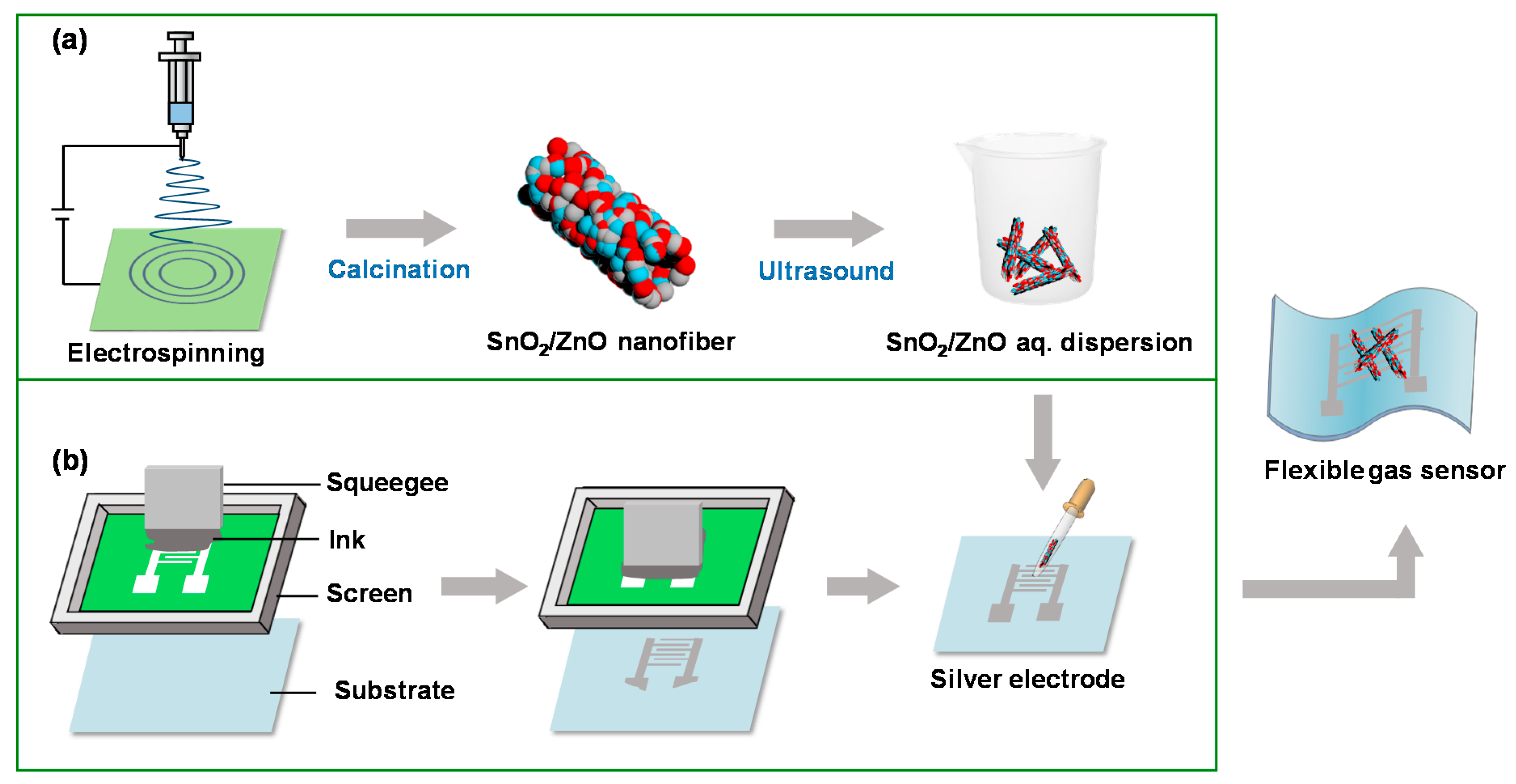
2.2. Synthesis of Hollow SnO2 and SnO2/ZnO Nanofibers

The transparent precursor solution for the synthesis of SnO2 nanofibers was prepared by addition of 0.6 g SnCl4·2H2O into 10 mL ethanol/DMF (volume ratio 1:1) solvent mixture, in which 0.8 g PVP had already been dissolved. In the preparation of SnO2/ZnO nanofibers, 0.005 g, 0.01g and 0.03 g Zn(NO3)2·6H2O were respectively added into two bottles of the precursor solution mentioned above, and the corresponding prepared samples labeled SnO2/ZnO-1, SnO2/ZnO-2, and SnO2/ZnO-3, respectively. The precursor solution was magnetically stirred at room temperature for 3 h until a clear and transparent solution was formed. A 2 mL volume of precursor solution was then drawn using a 5 mL syringe with a stainless steel needle for electrospinning. The needle was connected to the positive pole, and the negative pole was connected to an aluminum foil, which was placed 20 cm away from the collector, as shown in Figure 1a. The electrospinning parameters used were as described below: the voltage was 20 kV, the humidity range was 40–50% relative humidity (RH), and the collection time was 2 h. The electrospun fibers were then calcinated at 600 °C for 3 h at a heating rate of 10 °C/min.

2.3. Fabrication of Flexible Patterned Electrodes

A screen plate with patterned electrodes was fixed on a commercial screen-printing press. The different substrates (paper, PET, PDMS) were placed about 5 cm below the screen plate. Silver paste was then pressed along the patterns using a squeegee at a constant speed to form a clear and flat flexible electrode traces on the substrate, as shown in Figure 1b. The flexible substrates with silver electrodes were dried in the oven at 120 °C for 0.5 h after fabrication.

2.4. Preparation and Test of Gas Sensors

Titanium/gold (Ti/Au) interdigital electrodes, with gaps and finger widths of both 10μm, were fabricated on silicon/silicon dioxide (Si/SiO2) substrates by lithography. The flexible electrodes deposited on paper, PET and PDMS by screen printing had a gap and finger widths of 0.1 mm. Sensing nanofibers were then dispersed in deionized (DI) water, followed by drop-coating onto the electrodes (Figure 1b). The gas sensing properties were evaluated using a homemade system at room temperature (~20 °C). This system could monitor the changes in resistance in dynamic variation process of gas concentration controlled by mass flow controller. The resistance values were recorded using Keithley 2700, China. The response values were then calculated using Equation (1):
S (%) = (RaRg)/Ra × 100%
where Ra is the initial resistance of air and Rg is the resistance of the target gas. The response and recovery time were 90% of the time when the resistance reached its maximum in the target gas and the minimum in the air, respectively.

2.5. Characterizations

The hollow SnO2 nanofibers, SnO2/ZnO composite nanofibers were characterized by scanning electron microscopy (SEM, JSM-7401F, JEOL, Akishima, Japan), transmission electron microscopy (TEM, JEM-2010, JEOL, Tokyo, Japan), electron dispersive spectroscopy (EDS) installed in TEM, X-ray photoelectron spectroscopy (XPS, PHI-5300, Perkin-Elmer, Waltham, MA, USA) using AlKα radiation (hν = 1486.6 eV), and specific surface area analyzer (Autosorb-iQ2-MP, Quantachrome, Shanghai, China). The change in resistance of the silver electrodes during bending cycles was recorded using a stretcher (INSTRON 5943, Instron, Shanghai, China).

3. Results and Discussion

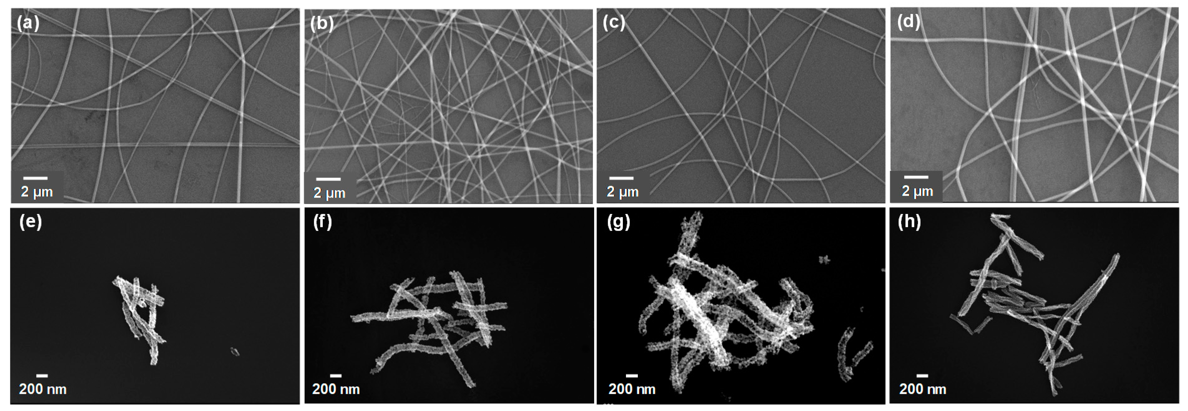
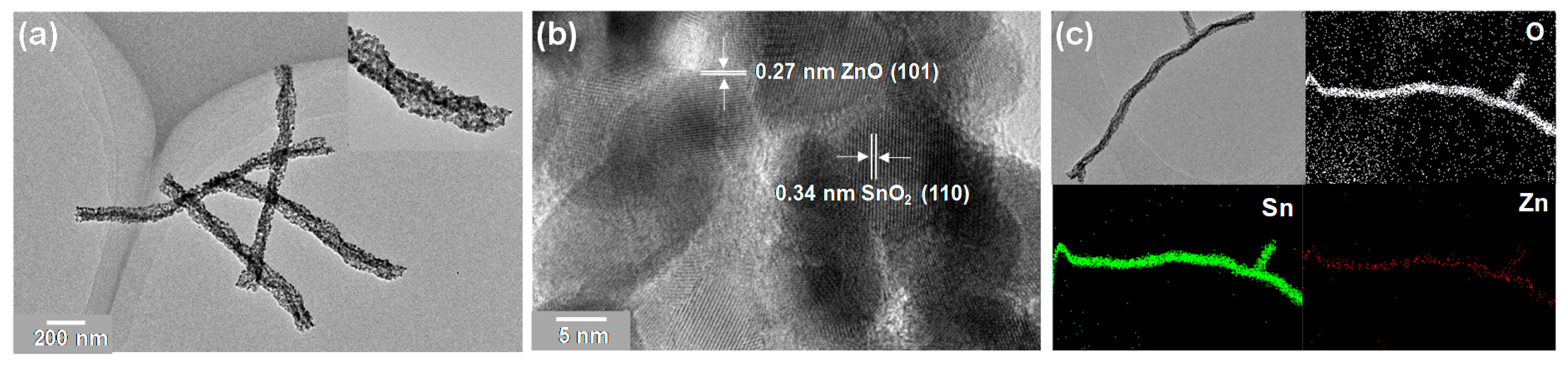
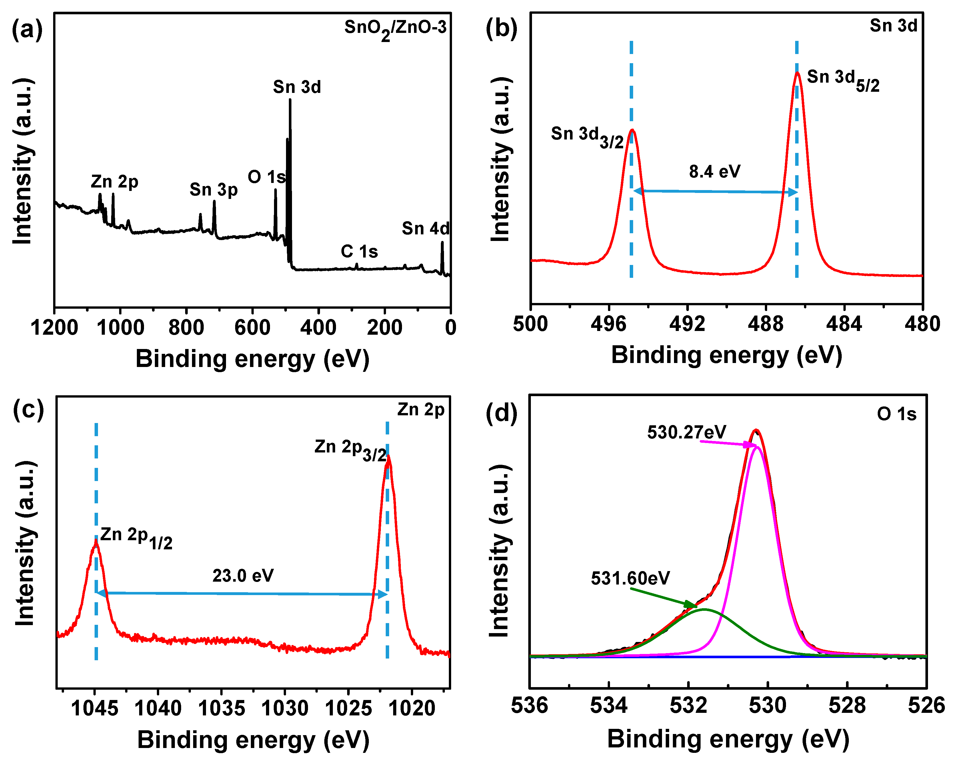
3.1. Characterizations of SnO2, SnO2/ZnO Nanofibers

The process of SnO2/ZnO composite nanofiber preparation is illustrated in Figure 1a. Humidity is a very important factor in the electrospinning process. Low humidity causes the blockage of electrospinning needle, whereas high humidity makes it difficult to collect samples at the collector. Figure 1b displays the fabrication process of flexible electrodes through screen printing using silver paste on different substrates. The fabrication process was strongly influenced by the physical properties of silver paste, especially the viscosity and the organic solvents used. The appropriate organic solvent enabled silver paste to be cured at room temperature. High viscosity was essential in preventing excessive spreading on the substrate, whereas high-viscosity silver paste was not compatible with other printing techniques, such as gravure [25] and inkjet [26].
SEM images of the electrospun SnO2/PVP, SnO2/ZnO/PVP-1, SnO2/ZnO/PVP-2, and SnO2/ZnO/PVP-3 nanofibers are shown in Figure 2a–d. The diameters of these fibers were found to be similar to each other, and their surface morphology appeared to be smooth because of the features of the polymer used. SEM images of hollow SnO2, SnO2/ZnO-1, SnO2/ZnO-2, and SnO2/ZnO-3 nanofibers after calcination were as shown in Figure 2e–h. The surface of the nanofibers was clearly concave–convex and porous after calcination treatment, and the nanofibers consisted of nanoparticles. By comparing these images, it was revealed that the morphologies of all the hollow nanofibers were porous, and their diameters exhibited almost no significant difference (d = 180 ± 20 nm). Therefore, it can be inferred that the slight difference in diameters between SnO2, SnO2/ZnO nanofibers may not have an appreciable effect on their sensing performance.
To further verify the structure of SnO2/ZnO composite nanofibers, TEM images were taken from the as-prepared SnO2/ZnO-3 sample. The TEM images in Figure 3a and its inset show porous structures with a mean diameter of 112.8 nm. As shown in Figure 3b, the interplanar spacing was 0.34 nm for the SnO2 (110) plane and 0.27 nm for the ZnO (100) plane, which were in accordance to a previous study [26]. This result illustrated that SnO2/ZnO composite nanofibers fabricated consisted of SnO2 and ZnO nanoparticles of different sizes (d = 10 ± 5 nm), which had a significant effect on the gas sensing properties of the nanofibers. The response performance decreased with the increase of grain size and exited from the optimal grain size range [27]. The grain size of SnO2 and ZnO nanoparticles prepared in this study was just in the optimal range. To investigate the chemical composition of SnO2/ZnO composite nanofibers, element mapping images were also measured. As shown in Figure 3c, the O (white), Sn (green) and Zn (red) atoms were uniformly distributed throughout the SnO2/ZnO nanofibers. Moreover, the percentage of Sn atoms was found to be more than that of O and Zn, with Zn being the least.
The surface chemical composition of SnO2/ZnO-2 composite nanofibers was further characterized using XPS. As shown in Figure 4a, the peaks of Sn, Zn, O, and C were all observed, and no other impurities were detected. The two peaks that emerged in Figure 4b, located at 486.4 and 494.8 eV with a separation of 8.4 eV, corresponded to Sn 3d5/2 and Sn 3d3/2 of Sn4+ ions, respectively. The two peaks that appeared at a binding energy of 1044.9 and 1021.9 eV in Figure 4c corresponded to Zn 2p1/2 and Zn 2p3/2 of Zn2+ ions, respectively [28,29]. From Figure 4d, it can be seen that the O 1s spectrum consisted of two different components at 530.27 eV and 531.60 eV, corresponding to the lattice oxygen in ZnO and the lattice oxygen in SnO2, respectively. These results further demonstrate that SnO2/ZnO composite nanofibers were formed from SnO2 and ZnO nanoparticles [30].
The fabrication process of hollow SnO2 nanofibers has been previously outlined [31]. During the annealing process, due to the decomposition of PVP template, Sn precursors are rapidly oxidized and redistributed through surface diffusion to form SnO2 nanoparticles, which is a component of the hollow nanofibers. Similarly, Sn and Zn precursors will be oxidized to form SnO2 and ZnO nanoparticles during the calcination process. The data shown in Table 1 point to the conclusion that moderate doping of ZnO remarkably promotes the Sn precursors oxidation, leading to the formation of more SnO2 nanoparticles. Moreover, the surface area of SnO2/ZnO composite nanofibers is larger than that of pure SnO2 nanofibers, and thus it is prone to providing more active sites for the adsorption and desorption of gas molecules [28]. Additionally, the surface area of SnO2/ZnO increases with the concentration of ZnO. Therefore, the SnO2/ZnO composite nanofibers were expected to have enhanced gas sensing properties.

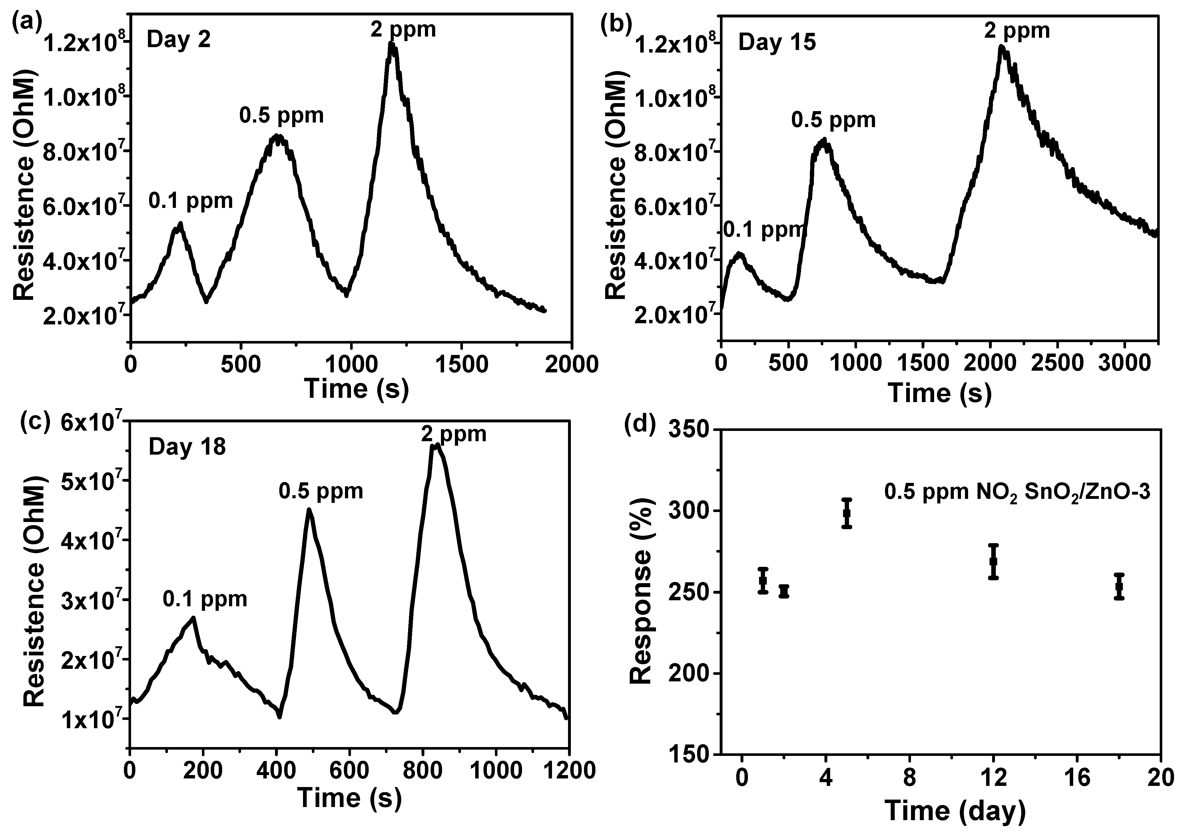
3.2. Gas Sensing Properties of SnO2, SnO2/ZnO Composite Nanofibers

The gas sensing properties of SnO2, SnO2/ZnO-1, SnO2/ZnO-2 and SnO2/ZnO-3 at different concentrations of NO2 gas at room temperature were studied. Figure 5 shows that the resistance increased upon exposure to NO2 gas and recovered completely to the initial resistance value upon the removal of NO2. This indicates that the SnO2 and SnO2/ZnO gas sensors had good and stable reversibility. The response values of pure SnO2 gas sensor exposed to 0.1, 0.5, 5, 10 and 20 ppm of NO2 gas were 8%, 13%, 24%, 34% and 45%, respectively. Meanwhile, the response values of the SnO2/ZnO-1 sensor, successively, were 13.2%, 22.9%, 49.0, 61.1%, 95.5%. For the SnO2/ZnO-2 sensor, the response values obtained were 21%, 28%, 68%, 116% and 173%, respectively, which is almost three times higher than the values obtained for the pure SnO2 gas sensor. In comparison to SnO2/ZnO-1 and SnO2/ZnO-2, the SnO2/ZnO-3 sensor possessed the best sensing performance, with a response value of 1945% to 20 ppm of NO2 at room temperature. Figure 5d shows the response values of the SnO2/ZnO-3 gas sensor to 0.1, 0.5, 5 and 10 ppm of NO2, which were 122%, 336%, 895% and 1384%, respectively.
The responses of SnO2, SnO2/ZnO-1, SnO2/ZnO-2 and SnO2/ZnO-3 gas sensors are shown in Figure 6a. The response of SnO2/ZnO was found to be better than that of pure SnO2 nanofibers, and it had the tendency of rising more rapidly when NO2 gas concentration increased. A comparison of NO2 gas sensing properties of different materials is shown in Table 2. From the table, it can be seen that SnO2/ZnO sensors exhibited high sensitivity to 0.5 ppm of NO2 with a response value of 336% and a faster response time of <2 min. Therefore, the response performance of SnO2/ZnO gas sensor was found to be remarkably higher than those of reported NO2 gas sensors. To further visually observe the NO2 sensing properties of the SnO2/ZnO-3 gas sensors at room temperature, the linear range for NO2 detection is as displayed in Figure 6b. From this figure, it can be seen that with a rise in gas concentration, there was a gradual increase in response. From Figure 6c, the response and recovery times of SnO2/ZnO-3 gas sensors to 0.5 ppm of NO2 at room temperature were 2.1 and 4.0 min, respectively. The response and recovery time of gas sensors based on as-prepared samples to 0.5 ppm of NO2 was as presented in Figure 6d. From this figure, it can be seen that the response/recovery time gradually decreased with the increase in the amount of ZnO doped. When compared with existing gas sensors, these results are ideal for the development of room temperature gas sensor [32].
To investigate the repeatability of SnO2/ZnO composites, the response performance of SnO2/ZnO-3 sensor to 0.3 ppm of NO2 was tested for four successive cycles at room temperature. As can be seen from Figure 7a, the baseline was fully capable of returning to its original position, and there was no significant difference in response values, an indication that SnO2/ZnO-3 gas sensor had excellent repeatability. Figure 7b shows the enlargement of response and recovery time of SnO2/ZnO-3 sensor to 0.3 ppm of NO2. It was found that the response value was 135.7%, and the response/recovery times were 1.6 and 4.0 min, respectively. Selectivity is another fundamental characteristic of gas sensors. Figure 7c shows the selectivity of SnO2/ZnO-3 gas sensor to 0.5 ppm of NO2 and 150 ppm other gases under the same measurement conditions, including HCHO, CH4, SO2, C8H10, NH3, and CO. The results indicate that the gas sensors based on SnO2/ZnO-3 had low sensitivity (response value < 5) to other gases except when compared to the values obtained for NO2.
When gas sensors are operated at room temperature, the effect of relative humidity on sensing properties should also be studied. To investigate the effect of humidity on both SnO2 sensors and SnO2/ZnO sensors, SnO2 and SnO2/ZnO-1 were tested in 5 ppm of NO2 gas under 25–96% RH at room temperature (shown in Figure 7d). Both SnO2 and SnO2/ZnO sensors worked well and had a relatively stable sensing ability, demonstrating the good humidity resistance of sensors semiconductors. Moreover, the sensing measurements were repeated every few days at room temperature to investigate the stability of SnO2/ZnO sensors. The results are shown in Figure 8, where the electrical signals of SnO2/ZnO-3 sensors did not change dramatically after 18 days when detecting NO2 gas. As can be seen in Figure 8d, after about ten days, the response value rose from about 250% at the beginning to about 300%, then gradually recovered and stabilized at about 250% with increasing number of days, an indication of the relatively good stability of the sensors.

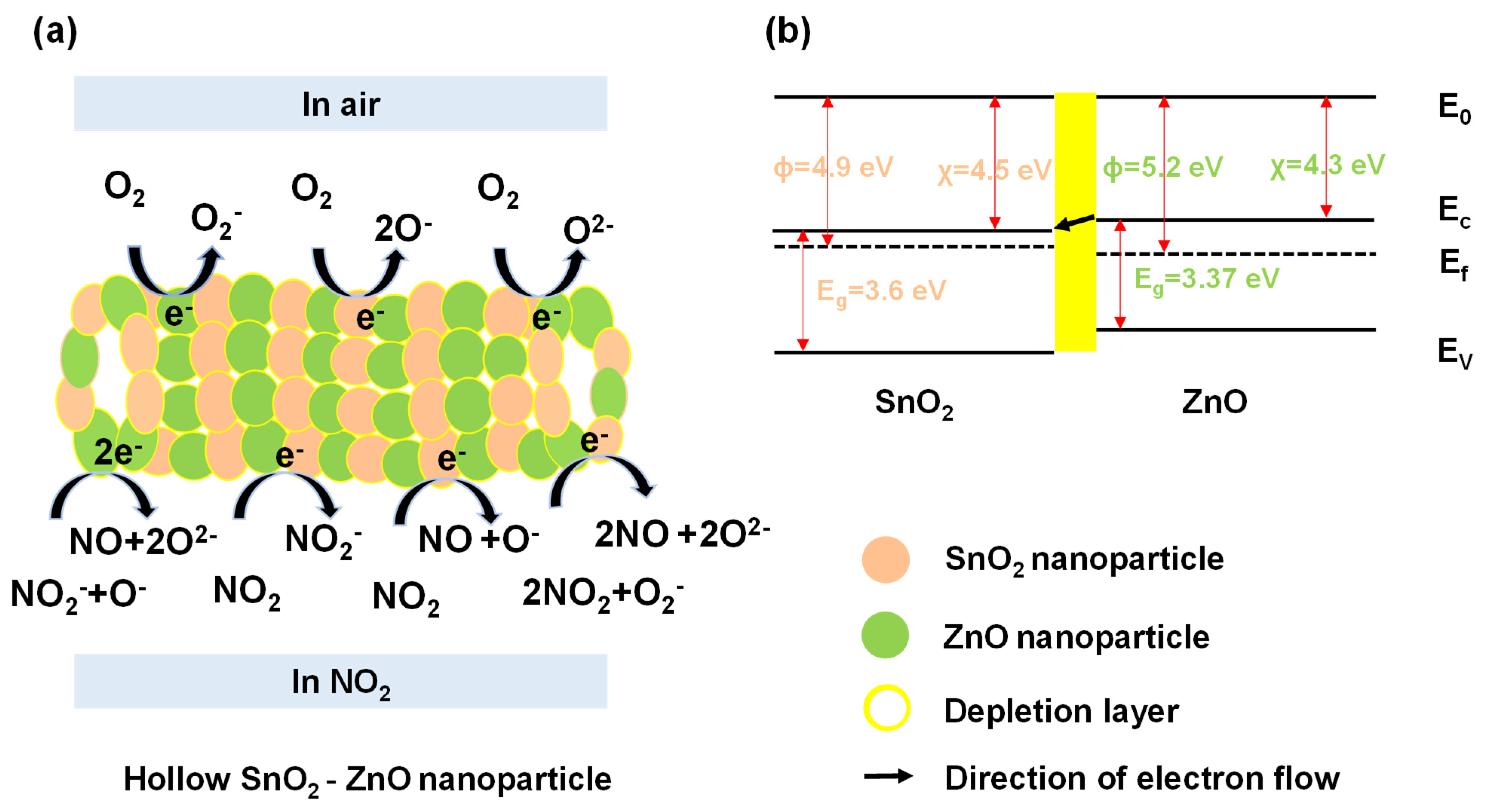
3.3. Sensing Mechanism

During the process of NO2 molecule adsorption, charge transfer can occur depending on the relative band positions of SnO2/ZnO and NO2, which can cause hybridization of NO2 gas molecules state with SnO2/ZnO nanocomposite orbitals. Such a charge transfer affects the resistance of SnO2/ZnO, which can be facilely measured using a low-cost resistive transducing device. More importantly, physisorption of NO2 molecules can occur at room temperature. When compared to pure SnO2, SnO2/ZnO has a larger electronegativity that could potentially enhance its gas adsorption sites [40,41]. On the other hand, the process of chemisorption can also modulate the resistance of sensing materials. A schematic diagram of the sensing mechanism of SnO2/ZnO composite nanofibers to NO2 gas is given in Figure 9. The significant improvement in sensing properties of SnO2/ZnO composite nanofibers can be attributed to two factors. Firstly, the surface morphology, which is a parameter in sensing. The SnO2/ZnO composite nanofibers contain more SnO2 and ZnO nanoparticles than that of pure SnO2 nanofibers. This led to larger surface area (38.7 m2/g) in SnO2/ZnO composite nanofibers, providing more adsorption sites for gas molecules. Secondly, the n-n heterojunction formed at the interface of SnO2/ZnO composite nanofibers was also a reason for the enhanced gas sensing performance [42,43,44,45]. This phenomenon is illustrated in Figure 9a,b.
As shown in Figure 9a, the SnO2/ZnO composite nanofibers were composed of SnO2 and ZnO nanoparticles with different grain sizes. There was an energy barrier at the n–n heterojunction, which modulated the transport of electrons because of electron trapping. Figure 9b illustrates the energy band structure of SnO2 and ZnO, in which Ef represents the Fermi level, Eg represents the energy band gap, and Φ, χ are working function and affinity, respectively. The resistance of SnO2/ZnO gas sensor can be described using Equation (2):
R = R 0 e x p ( Δ Φ k b T )
where R0 is a constant, kb is the Boltzmann’s constant, T is the absolute temperature, and ΔΦ is the effective potential barrier, including heterojunction and homojunction barrier [30].
In this context, oxygen molecules extracted electrons from the surface of SnO2/ZnO nanofibers and formed oxygen ions (like O, O2, O2−) in the air, thereby leading to the formation of an electron depletion layer on SnO2 and ZnO nanoparticles. The exact equations can be described using Equations (3)–(5):
O 2 + e O 2
O 2 + e 2 O
O + e O 2
When NO2 gas molecules were present, they extracted electrons from SnO2, ZnO nanoparticles and oxygen ions because of stronger affinity to SnO2/ZnO. This process widened the depletion layer and increased the resistance of the gas sensor. The surface electrochemical reaction was described using Equations (6)–(9):
NO 2 + e NO 2
NO 2 + e NO + O
NO 2 + O + 2 e NO + 2 O 2
2 NO 2 + O 2 + e 2 NO + 2 O 2
Therefore, it can be concluded that physisorption and chemisorption play an important role in controlling the gas-sensing performance of SnO2/ZnO.

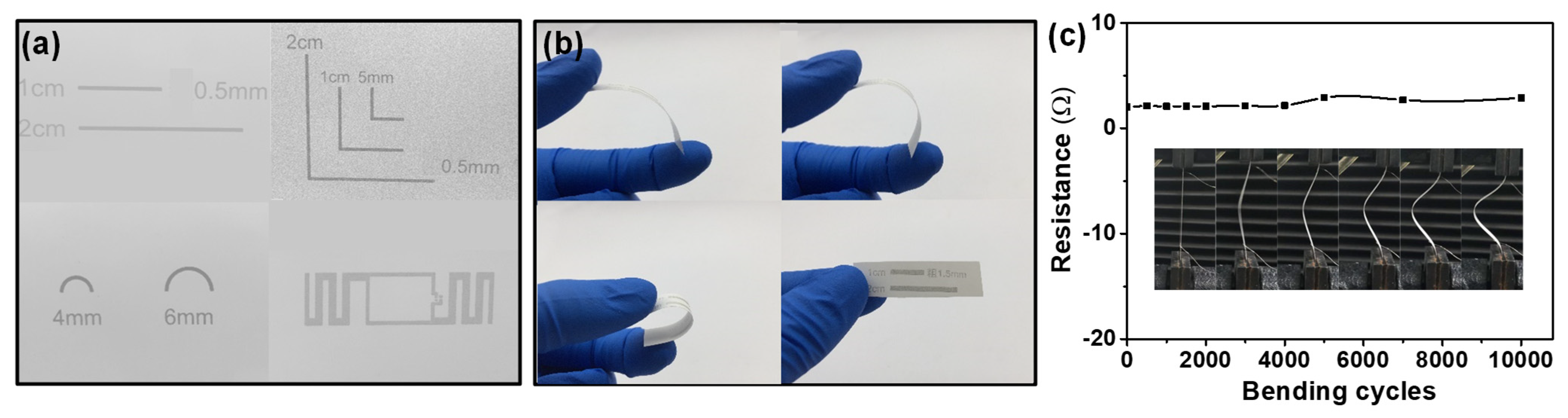
3.4. Integration and Gas Sensing Properties of Flexible Gas Sensors

The flexible electrodes were fabricated onto different flexible substrates integrated with SnO2/ZnO composite nanofibers to form flexible wearable gas sensors. Figure 10a shows the shapes of the electrodes prepared by screen printing with silver paste on paper. The linear silver electrodes were bent to angles of 45°, 90° and 180° and then recovered (Figure 10b) during testing for adhesion between the silver lines and paper. The results obtained showed that the adhesion was strong enough to sustain bending at any angle. Furthermore, as seen in Figure 10c, the resistance of linear screen-printed electrodes only changed from 2.0 to 2.8 Ω after bending 10,000 times to an angle of 45°. Figure 11a–c show the photographs of flexible silver electrodes for gas sensor prepared by screen printing on PDMS, paper, and PET. The silver electrode was found to have closely bonded to the substrate. The edge of the line was also clear, proving that this study had successfully prepared flexible sensors.
Figure 11d depicts the response and recovery curves of SnO2/ZnO-3 gas sensors with flexible silver electrodes to 0.1 ppm of NO2 before and after bending for 5000 and 10,000 cycles. The response values were 56%, 43% and 26%, respectively. Since the precision of screen printing is lower than that of the lithographic technique, the spacing of the interfingered electrodes was slightly larger, thereby increasing the difficulty of drop coating process. However, the idea of preparing flexible electrodes using screen printing could be extended to many other fields, such as EMI shielding [46], solar cells [47], and permanent memory devices [48]. The precision problems of screen printing can be improved through various methods such as filtration-assisted deposition [49].

4. Conclusions

Hollow SnO2 nanofibers and SnO2/ZnO composite nanofibers were successfully prepared through electrospinning and calcination in this work. When compared to pure SnO2, gas sensors based on SnO2/ZnO have higher sensitivity and selectivity to 0.5 ppm of NO2 at room temperature, with a response value of 336%. This can be attributed to the heterojunction effect and the selective NO2 physisorption sensing mechanism of SnO2/ZnO nanocomposites. In addition, the increase of the specific surface area of SnO2/ZnO-3 compared with pure SnO2 also had a positive impact on the detection sensitivity. The response and recovery time of the SnO2/ZnO sensors were two times shorter than those of pristine SnO2 sensors. In addition, flexible electrodes were fabricated using screen printing and integrated with SnO2/ZnO into a flexible gas sensor, then tested after 10,000 bending cycles. The flexible SnO2/ZnO gas sensor was able to detect 0.1 ppm of NO2 with a high response value of 56% at room temperature. This therefore shows that the fabrication strategy employed in this study is suitable for the development of flexible wearable sensing devices.

Author Contributions

Data curation, J.G. and Y.L.; Formal analysis, J.G. and H.H.; Methodology, J.G., W.L. and M.W.; Software, J.G. and X.Z.; Supervision, D.X., Y.Z. and H.Z.; Writing—original draft, J.G.; Writing—review & editing, J.G. All authors have read and agreed to the published version of the manuscript.

Funding

This work was financially supported by the Science and Technology Research Key Project of Henan Educational Committee. (No. 14A140023, 14A430012).

Acknowledgments

This work made use of the resources of the Beijing National Center for Electron Microscopy at Tsinghua University. We also acknowledge the Super computer Center in Zhengzhou University for its support.

Conflicts of Interest

The authors declare no conflict of interest.

Sample Availability

Samples of the compounds are not available from the authors.

References

  1. Xiao, B.; Song, S.; Wang, P.; Zhao, Q.; Chuai, M.; Zhang, M. Promoting effects of Ag on In2O3 nanospheres of sub-ppb NO2 detection. Sens. Actuators B Chem. 2017, 241, 489–497. [Google Scholar] [CrossRef]
  2. Kabcum, S.; Kotchasak, N.; Channei, D.; Tuantranont, A.; Wisitsoraat, A.; Phanichphant, S.; Liewhiran, C. Highly sensitive and selective NO2 sensor based on Au-impregnated WO3 nanorods. Sens. Actuators B Chem. 2017, 252, 523–536. [Google Scholar] [CrossRef]
  3. Zhang, J.; Zeng, D.; Zhu, Q.; Wu, J.; Huang, Q.; Zhang, W.; Xie, C. Enhanced room temperature NO2 response of NiO-SnO2 nanocomposites induced by interface bonds at the p-n heterojunction. Phys. Chem. 2016, 18, 5386–5396. [Google Scholar] [CrossRef] [PubMed]
  4. Wang, Y.; Liu, C.; Wang, Z.; Song, Z.; Zhou, X.; Han, N.; Chen, Y. Sputtered SnO2: NiO thin films on self-assembled Au nano-particle arrays for MEMS compatible NO2 gas sensors. Sens. Actuators B Chem. 2019, 278, 28–38. [Google Scholar] [CrossRef]
  5. Yuan, L.; Hu, M.; Wei, Y.; Ma, W. Enhanced NO2 sensing characteristics of Au modified porous silicon/thorn-sphere-like tung-sten oxide composites. Appl. Surf. Sci. 2016, 389, 824–834. [Google Scholar] [CrossRef]
  6. Zhang, H.; Feng, J.; Fei, T.; Liu, S.; Zhang, T. SnO2 nanoparticles-reduced graphene oxide nanocomposites for NO2 sensing at low operating temperature. Sens. Actuators B Chem. 2014, 190, 472–478. [Google Scholar] [CrossRef]
  7. Kumar, R.; Al-Dossary, O.; Kumar, G.; Umar, A. Zinc oxide nanostructures for NO2 gas–sensor applications: A review. Nano-Micro Lett. 2015, 7, 97–120. [Google Scholar] [CrossRef] [PubMed] [Green Version]
  8. Zhang, D.; Liu, Z.; Li, C.; Tang, T.; Liu, X.; Han, S.; Lei, A.B.; Zhou, C. Detection of NO2 down to ppb Levels Using Individual and Multiple In2O3 Nanowire Devices. Nano Lett. 2004, 4, 1919–1924. [Google Scholar] [CrossRef]
  9. Ruiz, A.M.; Sakai, G.; Cornet, A.; Shimanoe, K.; Morante, J.R.; Yamazoe, N. Cr-doped TiO2 gas sensor for exhaust NO2 moni-toring. Sens. Actuators B Chem. 2003, 93, 509–518. [Google Scholar] [CrossRef]
  10. Bao, M.; Chen, Y.; Li, F.; Ma, J.; Lv, T.; Tang, Y.; Chen, L.; Xu, Z.; Wang, T. Plate-like p–n heterogeneous NiO/WO3 nanocomposites for high performance room temperature NO2 sensors. Nanoscale 2014, 6, 4063–4066. [Google Scholar] [CrossRef] [PubMed]
  11. Malik, R.; Tomer, V.K.; Mishra, Y.K.; Lin, L. Functional gas sensing nanomaterials: A panoramic view. Appl. Phys. Rev. 2020, 7, 021301. [Google Scholar] [CrossRef] [Green Version]
  12. Zhang, Y.; Kolmakov, A.; Lilach, Y.; Moskovits, M. Electronic control of chemistry and catalysis at the surface of an individual tin oxide nanowire. J. Phys. Chem. B 2005, 109, 1923–1929. [Google Scholar] [CrossRef] [PubMed]
  13. Choi, K.J.; Hwang, I.-S.; Park, J.-G.; Lee, J.-H. Novel fabrication of an SnO2nanowire gas sensor with high sensitivity. Nanotechnology 2008, 19, 095508. [Google Scholar] [CrossRef]
  14. Comini, E.; Cristalli, A.; Faglia, G.; Sberveglieri, G. Light enhanced gas sensing properties of indium oxide and tin dioxide sensors. Sens. Actuators B Chem. 2000, 65, 260–263. [Google Scholar] [CrossRef]
  15. Li, W.; Guo, J.; Cai, L.; Qi, W.; Sun, Y.; Xu, J.-L.; Sun, M.; Zhu, H.; Xiang, L.; Xie, D.; et al. UV light irradiation enhanced gas sensor selectivity of NO2 and SO2 using rGO functionalized with hollow SnO2 nanofibers. Sens. Actuators B Chem. 2019, 290, 443–452. [Google Scholar] [CrossRef]
  16. Choi, K.I.; Hübner, M.; Haensch, A.; Kim, H.J.; Weimar, U.; Barsan, N.; Lee, J.H. Ambivalent effect of Ni loading on gas sensing per-formance in SnO2 based gas sensor. Sens. Actuators B Chem. 2013, 183, 401–410. [Google Scholar] [CrossRef]
  17. Wan, G.X.; Ma, S.Y.; Li, X.B.; Li, F.M.; Bian, H.Q.; Zhang, L.P.; Li, W.Q. Synthesis and acetone sensing properties of Ce-doped ZnO nano-fibers. Mater. Lett. 2014, 114, 103–106. [Google Scholar] [CrossRef]
  18. Li, W.; Teng, C.; Sun, Y.; Cai, L.; Xu, J.L.; Sun, M.; Li, X.; Yang, X.; Xiang, L.; Xie, D.; et al. Sprayed, scalable, wearable, and portable NO2 sensor array using fully flexible AgNPs-All-Carbon nanostructures. ACS Appl. Mater. Inter. 2018, 10, 34485–34493. [Google Scholar] [CrossRef]
  19. Miao, Y.-E.; He, S.; Zhong, Y.; Yang, Z.; Tjiu, W.W.; Liu, T. A novel hydrogen peroxide sensor based on Ag/SnO2 composite nanotubes by electrospinning. Electrochimica Acta 2013, 99, 117–123. [Google Scholar] [CrossRef]
  20. Wang, K.; Zhao, T.; Lian, G.; Yu, Q.; Luan, C.; Wang, Q.; Cui, D. Room temperature CO sensor fabricated from Pt-loaded SnO2 porous nanosolid. Sensors Actuators B Chem. 2013, 184, 33–39. [Google Scholar] [CrossRef]
  21. Park, S.; An, S.; Mun, Y.; Lee, C. UV-enhanced NO2 gas sensing properties of SnO2-core/ZnO-shell nanowires at room temperature. ACS Appl. Mater. Inter. 2013, 5, 4285–4292. [Google Scholar] [CrossRef] [PubMed]
  22. Yang, T.; Gu, K.; Zhu, M.; Lu, Q.; Zhai, C.; Zhao, Q.; Yang, X.; Zhang, M. ZnO-SnO2 heterojunction nanobelts: Synthesis and ultraviolet light irradiation to improve the triethylamine sensing properties. Sens. Actuators B Chem. 2019, 279, 410–417. [Google Scholar] [CrossRef]
  23. Song, J.; Son, D.; Kim, J.; Yoo, Y.J.; Lee, G.J.; Wang, L.; Choi, M.K.; Yang, J.; Lee, M.; Do, K.; et al. Wearable force touch sensor array using a flexible and transparent electrode. Adv. Funct. Mater. 2017, 27, 1–9. [Google Scholar] [CrossRef]
  24. Uddin, A.I.; Yaqoob, U.; Phan, D.-T.; Chung, G.-S. A novel flexible acetylene gas sensor based on PI/PTFE-supported Ag-loaded vertical ZnO nanorods array. Sens. Actuators B Chem. 2016, 222, 536–543. [Google Scholar] [CrossRef]
  25. Secor, E.B.; Lim, S.; Zhang, H.; Frisbie, C.D.; Francis, L.F.; Hersam, M.C. Gravure printing of graphene for large-area flexible electronics. Adv. Mater. 2014, 26, 4533–4538. [Google Scholar] [CrossRef]
  26. Hyun, W.J.; Secor, E.B.; Rojas, G.A.; Hersam, M.C.; Francis, L.F.; Frisbie, C.D. All-printed, foldable organic thin-film transistors on glassine paper. Adv. Mater. 2015, 27, 7058–7064. [Google Scholar] [CrossRef]
  27. Zhang, J.; Wang, S.; Wang, Y.; Wang, Y.; Zhu, B.; Xia, H.; Guo, X.; Zhang, S.; Huang, W.; Wu, S. NO2 sensing performance of SnO2 hol-low-sphere sensor. Sens. Actuators B Chem. 2009, 135, 610–617. [Google Scholar] [CrossRef]
  28. Ju, D.-X.; Xu, H.-Y.; Qiu, Z.-W.; Zhang, Z.-C.; Xu, Q.; Zhang, J.; Wang, J.-Q.; Cao, B.-Q. Near room temperature, fast-response, and highly sensitive triethylamine sensor assembled with au-loaded ZnO/SnO2 core–shell nanorods on flat alumina substrates. ACS Appl. Mater. Interfaces 2015, 7, 19163–19171. [Google Scholar] [CrossRef]
  29. Lu, Z.; Zhou, Q.; Wang, C.; Wei, Z.; Xu, L.; Gui, Y. Electrospun ZnO-SnO2 composite nanofibers and enhanced sensing properties to SF6 decomposition byproduct H2S. Front. Chem. 2018, 6, 540. [Google Scholar] [CrossRef] [Green Version]
  30. Zhang, Z.; Xu, M.; Liu, L.; Ruan, X.; Yan, J.; Zhao, W.; Yun, J.; Wang, Y.; Qin, S.; Zhang, T. Novel SnO2@ZnO hierarchical nanostructures for highly sensitive and selective NO2 gas sensing. Sens. Actuators B Chem. 2018, 257, 714–727. [Google Scholar] [CrossRef]
  31. Fan, H.J.; Knez, M.; Scholz, R.; Hesse, D.; Nielsch, K.; Zacharias, M.; Gösele, U. Influence of surface diffusion on the formation of hollow nanostructures induced by the kirkendall effect: The basic concept. Nano Lett. 2007, 7, 993–997. [Google Scholar] [CrossRef]
  32. Joshi, N.; Hayasaka, T.; Liu, Y.; Liu, H.; Oliveira, O.N.; Lin, L. A review on chemiresistive room temperature gas sensors based on metal oxide nanostructures, graphene and 2D transition metal dichalcogenides. Microchim. Acta 2018, 185, 213. [Google Scholar] [CrossRef]
  33. Cho, S.Y.; Lee, Y.; Koh, H.J.; Jung, H.; Kim, J.S.; Yoo, H.W.; Kim, J.; Jung, H.T. Superior chemical sensing performance of black phosphorus: Comparison with MoS2 and graphene. Adv. Mater. 2016, 28, 7020–7028. [Google Scholar] [CrossRef]
  34. Zhang, J.; Zeng, D.; Zhao, S.; Wu, J.; Xu, K.; Zhu, Q.; Zhang, G.; Xie, C. Room temperature NO2 sensing: What advantage does the rGO–NiO nanocomposite have over pristine NiO? Phys. Chem. Chem. Phys. 2015, 17, 14903–14911. [Google Scholar] [CrossRef]
  35. Long, H.; Harley-Trochimczyk, A.; Pham, T.; Tang, Z.; Shi, T.; Zettl, A.; Carraro, C.; Worsley, M.A.; Maboudian, R. High surface area MoS2/Graphene hybrid aerogel for ultrasensitive NO2Detection. Adv. Funct. Mater. 2016, 26, 5158–5165. [Google Scholar] [CrossRef] [Green Version]
  36. Xia, Y.; Wang, J.; Xu, J.-L.; Li, X.; Xie, D.; Xiang, L.; Komarneni, S. Confined formation of ultrathin ZnO nanorods/reduced graphene oxide mesoporous nanocomposites for high-performance room-temperature NO2 sensors. ACS Appl. Mater. Interfaces 2016, 8, 35454–35463. [Google Scholar] [CrossRef]
  37. Shishiyanu, S.T.; Shishiyanu, T.S.; Lupan, O. Sensing characteristics of tin-doped ZnO thin films as NO2 gas sensor. Sens. Actuators B Chem. 2005, 107, 379–386. [Google Scholar] [CrossRef]
  38. Deng, S.; Tjoa, V.; Fan, H.M.; Tan, H.R.; Sayle, D.C.; Olivo, M.; Mhaisalkar, S.; Wei, J.; Sow, C.H. Reduced graphene oxide con-jugated Cu2O nanowire mesocrystals for high-performance NO2 gas sensor. J. Am. Chem. Soc. 2012, 134, 4905–4917. [Google Scholar] [CrossRef]
  39. Chen, N.; Li, X.; Wang, X.; Yu, J.; Wang, J.; Tang, Z.; Akbar, S. Enhanced room temperature sensing of Co3O4-intercalated reduced graphene oxide based gas sensors. Sens. Actuators B Chem. 2013, 188, 902–908. [Google Scholar] [CrossRef]
  40. Ou, J.Z.; Ge, W.; Carey, B.; Daeneke, T.; Rotbart, A.; Shan, W.; Wang, Y.; Fu, Z.; Chrimes, A.F.; Wlodarski, W.; et al. Physisorption-based charge transfer in two-dimensional SnS2 for selective and reversible NO2 gas sensing. ACS Nano. 2015, 9, 10313–10323. [Google Scholar] [CrossRef]
  41. Khan, H.; Zavabeti, A.; Wang, Y.; Harrison, C.J.; Carey, B.J.; Mohiuddin, M.; Chrimes, A.F.; De Castro, I.A.; Zhang, B.Y.; Sabri, Y.M.; et al. Quasi physisorptive two dimensional tungsten oxide nanosheets with ex-traordinary sensitivity and selectivity to NO2. Nanoscale 2017, 9, 19162–19175. [Google Scholar] [CrossRef]
  42. Li, W.; Ma, S.; Li, Y.; Yang, G.; Mao, Y.; Luo, J.; Gengzang, D.; Xu, X.; Yan, S. Enhanced ethanol sensing performance of hollow ZnO-SnO2 core–shell nanofibers. Sens. Actuators B Chem. 2015, 211, 392–402. [Google Scholar] [CrossRef]
  43. Huang, H.; Gong, H.; Chow, C.L.; Guo, J.; White, T.; Tse, M.S.; Tan, O.K. Low-temperature growth of SnO2 Nanorod arrays and tunable n-p-n sensing response of a ZnO/SnO2 Heterojunction for exclusive hydrogen sensors. Adv. Funct. Mater. 2011, 21, 2680–2686. [Google Scholar] [CrossRef]
  44. Rakshit, T.; Santra, S.; Manna, I.; Ray, S.K. Enhanced sensitivity and selectivity of brush-like SnO2 nanowire/ZnO nanorod heterostructure based sensors for volatile organic compounds. RSC Adv. 2014, 4, 36749–36756. [Google Scholar] [CrossRef]
  45. Luo, Y.; Dong, Z.; Chen, Y.; Zhang, Y.; Lu, Y.; Xia, T.; Wang, L.; Li, S.; Zhang, W.; Xiang, W.; et al. Self-powered NiO@ZnO-nanowire-heterojunction ultraviolet micro-photodetectors. Opt. Mater. Express 2019, 9, 2775–2784. [Google Scholar] [CrossRef]
  46. Dijith, K.S.; Pillai, S.; Surendran, K.P. Screen printed silver patterns on La0.5Sr0.5CoO3−δ—Epoxy composite as a strategy for many-fold increase in EMI shielding. Surf. Coat. Technol. 2017, 330, 34–41. [Google Scholar] [CrossRef]
  47. Hösel, M.; Søndergaard, R.R.; Angmo, D.; Krebs, F.C. Comparison of fast roll-to-roll flexographic, inkjet, flatbed, and rotary screen printing of metal back electrodes for polymer solar cells. Adv. Eng. Mater. 2013, 15, 995–1001. [Google Scholar] [CrossRef]
  48. Park, J.-H.; Park, M.-J.; Lee, J.-S. Dry writing of highly conductive electrodes on papers by using silver nanoparticle–graphene hybrid pencils. Nanoscale 2017, 9, 555–561. [Google Scholar] [CrossRef] [Green Version]
  49. Juang, Y.-J.; Li, W.-S.; Chen, P.-S. Fabrication of microfluidic paper-based analytical devices by filtration-assisted screen printing. J. Taiwan Inst. Chem. Eng. 2017, 80, 71–75. [Google Scholar] [CrossRef]
Figure 1. (a) Schematic diagrams of the preparation process of SnO2/ZnO composite nanofibers by electrospinning. (b) Fabrication process of flexible electrodes by screen printing with silver paste on substrates.
Molecules 26 06475 g001
Figure 2. SEM images of (a) pristine SnO2/PVP, (b) SnO2/ZnO/PVP-1, (c) SnO2/ZnO/PVP-2, (d) SnO2/ZnO/PVP-3 before calcination. (eh) Corresponding SEM images of porous hollow SnO2, SnO2/ZnO-1, SnO2/ZnO-2, SnO2/ZnO-3 nanofibers after calcination, respectively.
Molecules 26 06475 g002
Figure 3. (a,b) TEM images of SnO2/ZnO-3 composite nanofibers. The inset of (a) is the enlargement. (c) Elemental mappings of SnO2/ZnO-3 composite nanofibers.
Molecules 26 06475 g003
Figure 4. (a) Survey, (b) Sn 3d, (c) Zn 2p, (d) O 1s XPS spectra of SnO2/ZnO-3 composite nanofibers.
Molecules 26 06475 g004
Figure 5. Dynamic responses of (a) pristine SnO2, (b) SnO2/ZnO-1, (c) SnO2/ZnO-2, (d) SnO2/ZnO-3 nanofibers to different concentrations of NO2 gas (0.1, 0.5, 5, 10, 20 ppm) at room temperature.
Molecules 26 06475 g005
Figure 6. (a) Dynamic response curves to 0.1–20 ppm NO2 gas at room temperature. (b) The linear range for NO2 detection of SnO2/ZnO-3 gas sensors. (c) Response and recovery time of SnO2/ZnO-3 gas sensors to 0.5 ppm NO2. (d) Response and recovery time of gas sensors based on as-prepared samples to 0.5 ppm NO2.
Molecules 26 06475 g006
Figure 7. (a) Dynamic response of SnO2/ZnO-3 gas sensors to 0.3 ppm NO2 for four successive cycles. (b) Response and recovery time of SnO2/ZnO-3 gas sensors to 0.3 ppm NO2. (c) Selectivity of as-prepared samples sensors to 0.5 ppm NO2 and 150 ppm other gases (HCHO, CH4, SO2, C8H10, NH3, CO) under the same measurement condition. (d) Response vs. relative humidity of SnO2, SnO2/ZnO-3 sensors in 0.3 ppm NO2.
Molecules 26 06475 g007
Figure 8. Resistance value of SnO2/ZnO-3 sensors to 0.1, 0.5, 2 ppm NO2 gas on the (a) second day, (b) 15th day, (c) 18th day. (d) The stability of SnO2/ZnO-3 sensors to 0.1, 0.5, 2 ppm NO2 gas within about 18 day.
Molecules 26 06475 g008
Figure 9. (a) Surface topography model of SnO2/ZnO composite nanofibers. (b) Energy band structure of SnO2/ZnO composite nanofibers.
Molecules 26 06475 g009
Figure 10. (a) Different shapes of electrodes prepared by screen printing with silver paste on paper. (b) Photographs of linear screen-printed silver electrodes bended at angles of 45°, 90°, 180° and recovered. (c) Resistance changes of linear screen-printed silver electrodes at angle of 45° bended for 10,000 times.
Molecules 26 06475 g010
Figure 11. (a) Photograph of silver electrodes on PDMS. (b) Photograph of silver electrodes on paper. (c) Photograph of silver electrodes on PET. (d) Response and recovery curves of SnO2/ZnO-3 gas sensors on flexible silver electrodes before and after 5000, 10,000 bending cycles in 0.1 ppm NO2.
Molecules 26 06475 g011
Table 1. The content of Sn, Zn, O, and C elements, and the specific surface area of SnO2, SnO2/ZnO-1, SnO2/ZnO-2, and SnO2/ZnO-3.
SamplesSn (at.%)Zn (at.%)O (at.%)C (at.%)Specific Surface Area (m2/g)
SnO218.6-51.829.628.3
SnO2/ZnO-123.91.358.416.431.2
SnO2/ZnO-223.93.559.113.535.8
SnO2/ZnO-322.98.255.613.338.7
Table 2. Comparison of the NO2 sensing properties of different materials.
MaterialsMethod°CResponse (%)Concentration (ppm)Response TimeRef.
Au-WO3modified precipitation/impregnation250836.6564.2 s[2]
Black Phosphoruschemical exfoliationRT801200 s[33]
rGO-NiOhydrothermal methodRT10015300 s[34]
MoS2/Grapheneannealing process10012.50.510 min[35]
rGO-ZnOsolution synthesisRT11912.4 min[36]
Sn-doped ZnOsuccessive ionic layer adsorption15010.51.520 min[37]
SnO2/ZnOelectrospinRT336.150.5126 sThis work
SnO2/ZnOthermal evaporationRT2391-[21]
SnO2/ZnOtwo–step hydrothermal1500.25 ppb60 s[30]
SnO2/rGOhydrothermal treatment503.315135 s[6]
rGO-Cu2Ononclassic crystallizationRT67.821000 s[38]
rGO-Co3O4hydrothermal methodRT80602 min[39]
Publisher’s Note: MDPI stays neutral with regard to jurisdictional claims in published maps and institutional affiliations.

