
Novel Thiosemicarbazone Derivatives: In Vitro and In Silico Evaluation as Potential MAO-B Inhibitors

 , , , and
Abstract
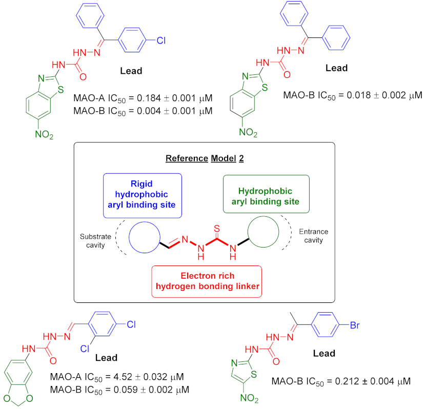
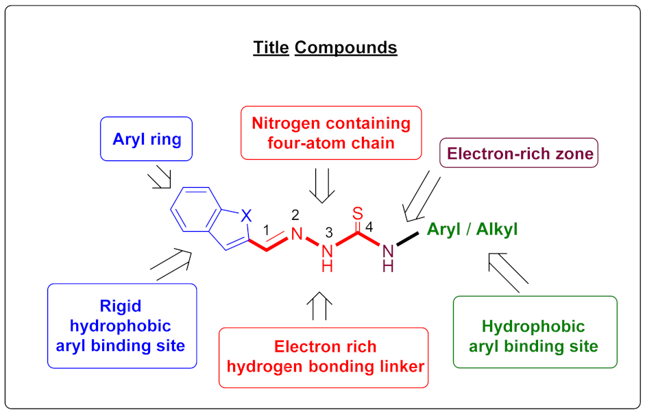
:1. Introduction
2. Results and Discussion
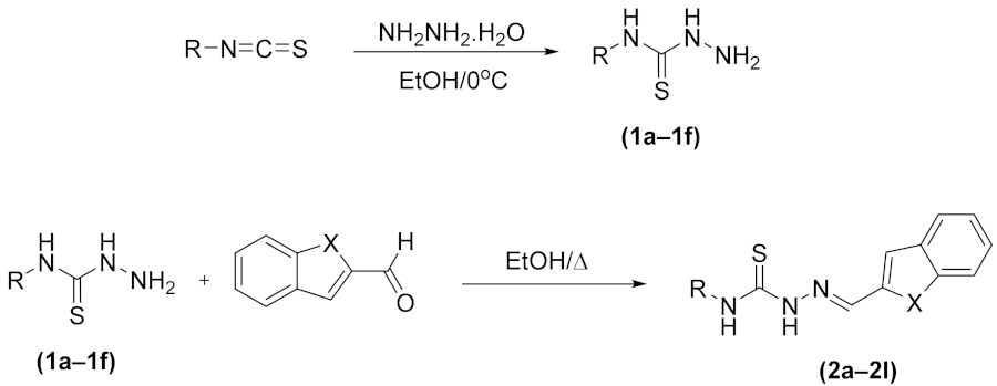
2.1. Chemistry
2.2. MAO Inhibition
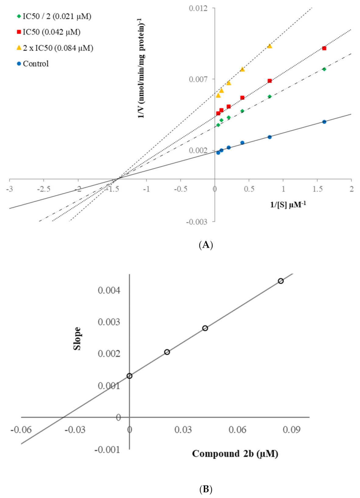
2.3. Kinetic Studies of Enzyme Inhibition
2.4. Cytotoxicity Assay
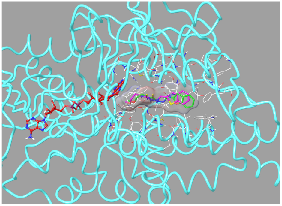
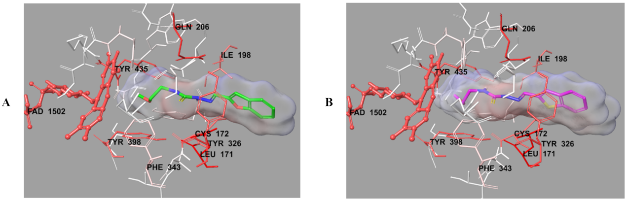
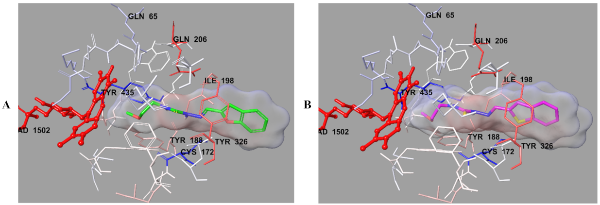
2.5. Molecular Docking
3. Materials and Methods
3.1. Chemistry
3.2. MAO-A and MAO-B Inhibition Assay
3.3. Enzyme Kinetic Studies
3.4. Cytotoxicity Assay
3.5. Molecular Docking
4. Conclusions
Supplementary Materials
Author Contributions
Funding
Institutional Review Board Statement
Informed Consent Statement
Data Availability Statement
Acknowledgments
Conflicts of Interest
Sample Availability
References
- Narayan, P.; Ehsani, S.; Lindquist, S. Correction: Corrigendum: Combating neurodegenerative disease with chemical probes and model systems. Nat. Chem. Biol. 2015, 11, 172. [Google Scholar] [CrossRef]
- Trippier, P.C.; Labby, K.J.; Hawker, D.D.; Mataka, J.J.; Silverman, R.B. Target-and mechanism-based therapeutics for neurodegenerative diseases: Strength in numbers. J. Med. Chem. 2013, 56, 3121–3147. [Google Scholar] [CrossRef] [PubMed] [Green Version]
- Hong, R.; Li, X. Discovery of monoamine oxidase inhibitors by medicinal chemistry approaches. MedChemComm 2019, 10, 10–25. [Google Scholar] [CrossRef]
- Thase, M.E. The role of monoamine oxidase inhibitors in depression treatment guidelines. J. Clin. Psychiatry 2012, 73 (Suppl. 1), 10–16. [Google Scholar] [CrossRef]
- Youdim, M.B.; Lavie, L. Selective MAO-A and B inhibitors, radical scavengers and nitric oxide synthase inhibitors in Parkinson’s desease. Life Sci. 1994, 55, 2077–2082. [Google Scholar] [CrossRef]
- Hagenow, J.; Hagenow, S.; Grau, K.; Khanfar, M.; Hefke, L.; Proschak, E.; Stark, H. Reversible small molecule inhibitors of MAO A and MAO B with anilide motifs. Drug Des. Dev. Ther. 2020, 14, 371. [Google Scholar] [CrossRef] [PubMed] [Green Version]
- Ramsay, R.R.; Albreht, A. Kinetics, mechanism, and inhibition of monoamine oxidase. J. Neural Transm. 2018, 125, 1659–1683. [Google Scholar] [CrossRef] [PubMed] [Green Version]
- Tong, J.; Meyer, J.H.; Furukawa, Y.; Boileau, I.; Chang, L.-J.; Wilson, A.A.; Houle, S. Distribution of monoamine oxidase proteins in human brain: Implications for brain imaging studies. J. Cereb. Blood Flow Metab. 2013, 33, 863–871. [Google Scholar] [CrossRef] [Green Version]
- Finberg, J.P.; Rabey, J.M. Inhibitors of MAO-A and MAO-B in psychiatry and neurology. Front. Pharmacol. 2016, 7, 340. [Google Scholar] [CrossRef] [Green Version]
- Youdim, M.B.; Edmondson, D.; Tipton, K.F. The therapeutic potential of monoamine oxidase inhibitors. Nat. Rev. Neurosci. 2006, 7, 295–309. [Google Scholar] [CrossRef] [PubMed]
- Nagatsu, T. Progress in monoamine oxidase (MAO) research in relation to genetic engineering. Neurotoxicology 2004, 25, 11–20. [Google Scholar] [CrossRef]
- Pacher, P.; Kecskemeti, V. Trends in the development of new antidepressants. Is there a light at the end of the tunnel? Curr. Med. Chem. 2004, 11, 925–943. [Google Scholar] [CrossRef] [PubMed] [Green Version]
- Hubálek, F.; Binda, C.; Khalil, A.; Li, M.; Mattevi, A.; Castagnoli, N.; Edmondson, D.E. Demonstration of isoleucine 199 as a structural determinant for the selective inhibition of human monoamine oxidase B by specific reversible inhibitors. J. Biol. Chem. 2005, 280, 15761–15766. [Google Scholar] [CrossRef] [Green Version]
- Pålhagen, S.; Heinonen, E.; Hägglund, J.; Kaugesaar, T.; Mäki-Ikola, O.; Palm, R.; the Swedish Parkinson Study Group. Selegiline slows the progression of the symptoms of Parkinson disease. Neurology 2006, 66, 1200–1206. [Google Scholar] [CrossRef]
- Tripathi, R.K.; Goshain, O.; Ayyannan, S.R. Design, synthesis, in vitro MAO-B inhibitory evaluation, and computational studies of some 6-nitrobenzothiazole-derived semicarbazones. ChemMedChem 2013, 8, 462–474. [Google Scholar] [CrossRef] [PubMed]
- Youdim, M.; Fridkin, M.; Zheng, H. Novel bifunctional drugs targeting monoamine oxidase inhibition and iron chelation as an approach to neuroprotection in Parkinson’s disease and other neurodegenerative diseases. J. Neural Transm. 2004, 111, 1455–1471. [Google Scholar] [CrossRef]
- Reis, J.; Manzella, N.; Cagide, F.; Mialet-Perez, J.; Uriarte, E.; Parini, A.; Borges, F.; Binda, C. Tight-binding inhibition of human monoamine oxidase B by chromone analogs: A kinetic, crystallographic, and biological analysis. J. Med. Chem. 2018, 61, 4203–4212. [Google Scholar] [CrossRef] [PubMed]
- Filip, V.; Kolibas, E. Selegiline in the treatment of Alzheimer’s disease: A long-term randomized placebo-controlled trial. Czech and Slovak Senile Dementia of Alzheimer Type Study Group. J. Psychiatry Neurosci. 1999, 24, 234. [Google Scholar] [PubMed]
- Sano, M.; Ernesto, C.; Thomas, R.G.; Klauber, M.R.; Schafer, K.; Grundman, M.; Woodbury, P.; Growdon, J.; Cotman, C.W.; Pfeiffer, E. A controlled trial of selegiline, alpha-tocopherol, or both as treatment for Alzheimer’s disease. N. Engl. J. Med. 1997, 336, 1216–1222. [Google Scholar] [CrossRef] [Green Version]
- Fitton, A.; Faulds, D.; Goa, K.L. Moclobemide. Drugs 1992, 43, 561–596. [Google Scholar] [CrossRef]
- Sharma, T.; Anand, R.; Hartman, R.; Rossetti, S. Overlap of cognitive deficits in Parkinson’s (PD) and Alzheimer’s (AD) diseases: Potential use of safinamide: 323. Mov. Disord. 2007, 22, 323. [Google Scholar]
- Cambria, A.; Raudino, A.; Castelli, F.; Raciti, G.; Mazzone, P.; Buemi, G.; Pignatello, R.; Mazzone, G. Structure-activity studies on monoamine oxidase inhibitors by calorimetric and quantum mechanical calculations. J. Enzym. Inhib. 1996, 10, 215–229. [Google Scholar] [CrossRef] [PubMed]
- Gritsch, S.; Guccione, S.; Hoffmann, R.; Cambria, A.; Raciti, G.; Langer, T. A 3D QSAR study of monoamino oxidase-B inhibitors using the chemical function based pharmacophore generation approach. J. Enzym. Inhib. 2001, 16, 199–215. [Google Scholar] [CrossRef] [PubMed]
- La Regina, G.; Silvestri, R.; Artico, M.; Lavecchia, A.; Novellino, E.; Befani, O.; Turini, P.; Agostinelli, E. New pyrrole inhibitors of monoamine oxidase: Synthesis, biological evaluation, and structural determinants of MAO-A and MAO-B selectivity. J. Med. Chem. 2007, 50, 922–931. [Google Scholar] [CrossRef]
- La Regina, G.; Silvestri, R.; Gatti, V.; Lavecchia, A.; Novellino, E.; Befani, O.; Turini, P.; Agostinelli, E. Synthesis, structure–activity relationships and molecular modeling studies of new indole inhibitors of monoamine oxidases A and B. Bioorganic Med. Chem. 2008, 16, 9729–9740. [Google Scholar] [CrossRef]
- Roth, M.; Mountjoy, C.; Amrein, R.; The International Collaborative Study Group. Moclobemide in elderly patients with cognitive decline and depression: An international double-blind, placebo-controlled trial. Br. J. Psychiatry 1996, 168, 149–157. [Google Scholar] [CrossRef] [Green Version]
- Silvestri, R.; La Regina, G.; De Martino, G.; Artico, M.; Befani, O.; Palumbo, M.; Agostinelli, E.; Turini, P. Simple, potent, and selective pyrrole inhibitors of monoamine oxidase types A and B. J. Med. Chem. 2003, 46, 917–920. [Google Scholar] [CrossRef]
- Tripathi, R.K.; Sasi, V.M.; Gupta, S.K.; Krishnamurthy, S.; Ayyannan, S.R. Design, synthesis, and pharmacological evaluation of 2-amino-5-nitrothiazole derived semicarbazones as dual inhibitors of monoamine oxidase and cholinesterase: Effect of the size of aryl binding site. J. Enzym. Inhib. Med. Chem. 2018, 33, 37–57. [Google Scholar] [CrossRef] [Green Version]
- Tripathi, R.K.; Rai, G.K.; Ayyannan, S.R. Exploration of a Library of 3,4-(Methylenedioxy)aniline-Derived Semicarbazones as Dual Inhibitors of Monoamine Oxidase and Acetylcholinesterase: Design, Synthesis, and Evaluation. ChemMedChem 2016, 11, 1145–1160. [Google Scholar] [CrossRef]
- Mathew, G.E.; Oh, J.M.; Mohan, K.; Kumar, K.S.; Jayanthi, S.; Kim, H.; Mathew, B. Inhibitions of monoamine oxidases and acetylcholinesterase by 1-methyl, 5-phenyl substituted thiosemicarbazones: Synthesis, biochemical, and computational investigations. Process Biochem. 2020, 99, 246–253. [Google Scholar] [CrossRef]
- Son, S.-Y.; Ma, J.; Kondou, Y.; Yoshimura, M.; Yamashita, E.; Tsukihara, T. Structure of human monoamine oxidase A at 2.2-Å resolution: The control of opening the entry for substrates/inhibitors. Proc. Natl. Acad. Sci. USA 2008, 105, 5739–5744. [Google Scholar] [CrossRef] [PubMed] [Green Version]
- De Colibus, L.; Li, M.; Binda, C.; Lustig, A.; Edmondson, D.E.; Mattevi, A. Three-dimensional structure of human monoamine oxidase A (MAO A): Relation to the structures of rat MAO A and human MAO B. Proc. Natl. Acad. Sci. USA 2005, 102, 12684–12689. [Google Scholar] [CrossRef] [PubMed] [Green Version]
- Can, N.Ö.; Osmaniye, D.; Levent, S.; Sağlık, B.N.; Inci, B.; Ilgın, S.; Özkay, Y.; Kaplancıklı, Z.A. Synthesis of new hydrazone derivatives for MAO enzymes inhibitory activity. Molecules 2017, 22, 1381. [Google Scholar] [CrossRef] [PubMed]
- Can, N.Ö.; Osmaniye, D.; Levent, S.; Sağlık, B.N.; Korkut, B.; Atlı, Ö.; Özkay, Y.; Kaplancıklı, Z.A. Design, synthesis and biological assessment of new thiazolylhydrazine derivatives as selective and reversible hMAO-A inhibitors. Eur. J. Med. Chem. 2018, 144, 68–81. [Google Scholar] [CrossRef] [PubMed]
- Can, Ö.D.; Osmaniye, D.; Özkay, Ü.D.; Sağlık, B.N.; Levent, S.; Ilgın, S.; Baysal, M.; Özkay, Y.; Kaplancıklı, Z.A. MAO enzymes inhibitory activity of new benzimidazole derivatives including hydrazone and propargyl side chains. Eur. J. Med. Chem. 2017, 131, 92–106. [Google Scholar] [CrossRef]
- Ilgın, S.; Osmaniye, D.; Levent, S.; Sağlık, B.N.; Acar Çevik, U.; Çavuşoğlu, B.K.; Özkay, Y.; Kaplancıklı, Z.A. Design and synthesis of new benzothiazole compounds as selective hMAO-B inhibitors. Molecules 2017, 22, 2187. [Google Scholar] [CrossRef] [Green Version]
- Sağlık, B.N.; Çavuşoğlu, B.K.; Osmaniye, D.; Levent, S.; Çevik, U.A.; Ilgın, S.; Özkay, Y.; Kaplancıklı, Z.A.; Öztürk, Y. In vitro and in silico evaluation of new thiazole compounds as monoamine oxidase inhibitors. Bioorganic Chem. 2019, 85, 97–108. [Google Scholar] [CrossRef] [PubMed]
- Tok, F.; Sağlık, B.N.; Özkay, Y.; Ilgın, S.; Kaplancıklı, Z.A.; Koçyiğit-Kaymakçıoğlu, B. Synthesis of new hydrazone derivatives and evaluation of their monoamine oxidase inhibitory activity. Bioorganic Chem. 2021, 114, 105038. [Google Scholar] [CrossRef] [PubMed]
- Tok, F.; Uğraş, Z.; Sağlık, B.N.; Özkay, Y.; Kaplancıklı, Z.A.; Koçyiğit-Kaymakçıoğlu, B. Novel 2, 5-disubstituted-1, 3, 4-oxadiazole derivatives as MAO-B inhibitors: Synthesis, biological evaluation and molecular modeling studies. Bioorganic Chem. 2021, 112, 104917. [Google Scholar] [CrossRef] [PubMed]
- Binda, C.; Wang, J.; Pisani, L.; Caccia, C.; Carotti, A.; Salvati, P.; Edmondson, D.E.; Mattevi, A. Structures of human monoamine oxidase B complexes with selective noncovalent inhibitors: Safinamide and coumarin analogs. J. Med. Chem. 2007, 50, 5848–5852. [Google Scholar] [CrossRef] [PubMed]
- Schrödinger, L. Glide, version 7.1; Schrödinger, LLC: New York, NY, USA, 2016. [Google Scholar]
- Sağlık, B.N.; Çavuşoğlu, B.K.; Çevik, U.A.; Osmaniye, D.; Levent, S.; Özkay, Y.; Kaplancıklı, Z.A. Novel 1,3,4-thiadiazole compounds as potential MAO-A inhibitors–design, synthesis, biological evaluation and molecular modelling. RSC Med. Chem. 2020, 11, 1063–1074. [Google Scholar] [CrossRef]
- Çevik, U.A.; Osmaniye, D.; Sağlik, B.N.; Çavuşoğlu, B.K.; Levent, S.; Karaduman, A.B.; Ilgin, S.; Karaburun, A.Ç.; Özkay, Y.; Kaplancikli, Z.A. Multifunctional quinoxaline-hydrazone derivatives with acetylcholinesterase and monoamine oxidases inhibitory activities as potential agents against Alzheimer’s disease. Med. Chem. Res. 2020, 29, 1000–1011. [Google Scholar] [CrossRef]
- Ichimori, K.; Stuehr, D.J.; Atkinson, R.N.; King, S.B. Synthesis and evaluation of new sulfur-containing L-arginine-derived inhibitors of nitric oxide synthase. J. Med. Chem. 1999, 42, 1842–1848. [Google Scholar] [CrossRef]
- Basheer, S.M.; Willis, A.C.; Pace, R.J.; Sreekanth, A. Spectroscopic and TD-DFT studies on the turn-off fluorescent chemosensor based on anthraldehyde N (4) cyclohexyl thiosemicarbazone for the selective recognition of fluoride and copper ions. Polyhedron 2016, 109, 7–18. [Google Scholar] [CrossRef]
- Huang, H.; Chen, Q.; Ku, X.; Meng, L.; Lin, L.; Wang, X.; Zhu, C.; Wang, Y.; Chen, Z.; Li, M. A series of α-heterocyclic carboxaldehyde thiosemicarbazones inhibit topoisomerase IIα catalytic activity. J. Med. Chem. 2010, 53, 3048–3064. [Google Scholar] [CrossRef] [PubMed]
- Serra, S.; Moineaux, L.; Vancraeynest, C.; Masereel, B.; Wouters, J.; Pochet, L.; Frédérick, R. Thiosemicarbazide, a fragment with promising indolamine-2,3-dioxygenase (IDO) inhibition properties. Eur. J. Med. Chem. 2014, 82, 96–105. [Google Scholar] [CrossRef]
- Misra, V.; Saxena, A. Potential Antiviral and Antituberculous Compounds. III. N-1-(4-methoxy naphthylidene/2-benzofurylidene)-N-4-(aryl) thiosemicarbazides; 5-carboxymethyl, 3-aryl, thiazolidine-2, 4-dione hydrazones; 3-aryl, thiazolidine-2, 4-dione hydrazones and N-1-(4-methoxy naphthylidene/2-benzofurylidene)-N-4-(aryl) amino guanidines. J. für Praktische Chemie 1967, 36, 260–264. [Google Scholar]
- Qiao, Y.; Che, Y.; Yu, Y.; Tang, Y.; Liu, L.; Zhao, X.; Zhao, J. Solid-state photochromic properties, mechanism and electrospun membranes of (E)-2-(benzo [b] thiophen-2-ylmethylene)-N-ethylhydrazine-1-carbothioamide. Dyes Pigment. 2018, 156, 326–331. [Google Scholar] [CrossRef]
- Sağlık, B.N.; Ilgın, S.; Özkay, Y. Synthesis of new donepezil analogues and investigation of their effects on cholinesterase enzymes. Eur. J. Med. Chem. 2016, 124, 1026–1040. [Google Scholar] [CrossRef]
- Özkay, Ü.D.; Can, Ö.D.; Sağlık, B.N.; Çevik, U.A.; Levent, S.; Özkay, Y.; Ilgın, S.; Atlı, Ö. Design, synthesis, and AChE inhibitory activity of new benzothiazole–piperazines. Bioorganic Med. Chem. Lett. 2016, 26, 5387–5394. [Google Scholar] [CrossRef]
- Patel, S.; Gheewala, N.; Suthar, A.; Shah, A. In-vitro cytotoxicity activity of Solanum nigrum extract against Hela cell line and Vero cell line. Int. J. Pharm. Pharm. Sci. 2009, 1, 38–46. [Google Scholar]

| Compounds | X | R |
|---|---|---|
| 2a | –O | –Ethyl |
| 2b | –O | –2-Methoxyethyl |
| 2c | –O | –Butyl |
| 2d | –O | –Cyclohexyl |
| 2e | –O | –Phenyl |
| 2f | –O | –2-Chlorophenyl |
| 2g | –S | –Ethyl |
| 2h | –S | –2-Methoxyethyl |
| 2i | –S | –Butyl |
| 2j | –S | –Cyclohexyl |
| 2k | –S | –Phenyl |
| 2l | –S | –2-Chlorophenyl |


| Compounds | MAO-A % Inhibition | MAO-B % Inhibition | ||
|---|---|---|---|---|
| 10−3 M | 10−4 M | 10−3 M | 10−4 M | |
| 2a | 34.128 ± 0.927 | 22.314 ± 0.824 | 74.159 ± 1.412 | 46.356 ± 0.997 |
| 2b | 32.951 ± 0.955 | 28.357 ± 0.899 | 91.625 ± 1.652 | 87.357 ± 1.388 |
| 2c | 30.357 ± 0.902 | 26.735 ± 0.820 | 76.345 ± 1.623 | 43.451 ± 0.728 |
| 2d | 37.518 ± 0.897 | 20.856 ± 0.834 | 82.624 ± 1.318 | 41.052 ± 0.956 |
| 2e | 36.735 ± 0.910 | 23.402 ± 0.832 | 85.486 ± 1.528 | 40.828 ± 0.827 |
| 2f | 31.057 ± 0.994 | 24.174 ± 0.711 | 82.366 ± 1.332 | 47.398 ± 0.930 |
| 2g | 38.935 ± 0.854 | 28.692 ± 0.803 | 78.951 ± 1.702 | 44.774 ± 0.758 |
| 2h | 36.751 ± 0.741 | 21.873 ± 0.750 | 90.412 ± 1.602 | 84.362 ± 1.325 |
| 2i | 35.654 ± 0.863 | 29.162 ± 0.816 | 84.654 ± 1.628 | 46.035 ± 0.931 |
| 2j | 33.667 ± 0.729 | 23.041 ± 0.863 | 82.923 ± 1.621 | 42.957 ± 0.965 |
| 2k | 31.065 ± 0.911 | 27.859 ± 0.815 | 80.451 ± 1.475 | 46.651 ± 0.899 |
| 2l | 36.741 ± 0.966 | 24.389 ± 0.721 | 83.642 ± 1.597 | 42.249 ± 0.763 |
| Clorgiline | 99.411 ± 1.955 | 98.257 ± 1.824 | - | - |
| Selegiline | – | – | 99.387 ± 1.385 | 95.629 ± 1.456 |
| Compounds | MAO-B % Inhibition | IC50 (µM) | Selectivity Index | ||||||
|---|---|---|---|---|---|---|---|---|---|
| 10−3 M | 10−4 M | 10−5 M | 10−6 M | 10−7 M | 10−8 M | 10−9 M | |||
| 2b | 91.625 ± 1.652 | 87.357 ± 1.388 | 81.547 ± 1.722 | 74.951 ± 1.634 | 66.379 ± 0.975 | 41.513 ± 0.856 | 25.820 ± 0.765 | 0.042 ± 0.002 | >23.812 |
| 2h | 90.412 ± 1.602 | 84.362 ± 1.325 | 80.792 ± 1.045 | 72.188 ± 1.388 | 64.970 ± 1.107 | 42.826 ± 0.948 | 30.945 ± 0.782 | 0.056 ± 0.002 | >17.857 |
| Selegiline | 99.387 ± 1.385 | 95.629 ± 1.456 | 86.205 ± 1.200 | 78.324 ± 1.108 | 66.871 ± 1.056 | 42.875 ± 0.865 | 16.748 ± 0.596 | 0.037 ± 0.001 | – |
| Compounds | IC50 (µM) NIH3T3 Cell Line | IC50 (µM) MAO-B Enzyme |
|---|---|---|
| 2b | 0.042 ± 0.002 | 129.631 ± 0.235 |
| 2h | 0.056 ± 0.002 | 81.266 ± 0.158 |
Publisher’s Note: MDPI stays neutral with regard to jurisdictional claims in published maps and institutional affiliations. |
© 2021 by the authors. Licensee MDPI, Basel, Switzerland. This article is an open access article distributed under the terms and conditions of the Creative Commons Attribution (CC BY) license (https://creativecommons.org/licenses/by/4.0/).
Share and Cite
Osmaniye, D.; Kurban, B.; Sağlık, B.N.; Levent, S.; Özkay, Y.; Kaplancıklı, Z.A. Novel Thiosemicarbazone Derivatives: In Vitro and In Silico Evaluation as Potential MAO-B Inhibitors. Molecules 2021, 26, 6640. https://doi.org/10.3390/molecules26216640
Osmaniye D, Kurban B, Sağlık BN, Levent S, Özkay Y, Kaplancıklı ZA. Novel Thiosemicarbazone Derivatives: In Vitro and In Silico Evaluation as Potential MAO-B Inhibitors. Molecules. 2021; 26(21):6640. https://doi.org/10.3390/molecules26216640
Chicago/Turabian StyleOsmaniye, Derya, Berkant Kurban, Begüm Nurpelin Sağlık, Serkan Levent, Yusuf Özkay, and Zafer Asım Kaplancıklı. 2021. "Novel Thiosemicarbazone Derivatives: In Vitro and In Silico Evaluation as Potential MAO-B Inhibitors" Molecules 26, no. 21: 6640. https://doi.org/10.3390/molecules26216640