Review

Genetic Modification Approaches for Parasporins Bacillus thuringiensis Proteins with Anticancer Activity

by
Miguel O. Suárez-Barrera
1,2,3,4,
Lydia Visser
3,
Paola Rondón-Villarreal
1,
Diego F. Herrera-Pineda
1,
Juan S. Alarcón-Aldana
1,
Anke Van den Berg
3,
Jahir Orozco
4,
Efraín H. Pinzón-Reyes
1,
Ernesto Moreno
5 and
Nohora J. Rueda-Forero
1,*
1
Facultad de Ciencias Médicas y de la Salud, Instituto de Investigación Masira, Universidad de Santander, Bucaramanga 680003, Colombia
2
Corporación de Ciencias Básicas Biomédicas, Universidad de Antioquia, Medellín 050010, Colombia
3
Department of Pathology and Medical Biology, University Medical Center Groningen, University of Groningen, 9700 RB Groningen, The Netherlands
4
MaxPlanck Tandem Group in Nanobioengieneering, Institute of Chemistry, Faculty of Natural and Exact Sciences, University of Antioquia, Medellin 050010, Colombia
5
Facultad de Ciencias Básicas, Universidad de Medellín, Medellín 050030, Colombia
*
Author to whom correspondence should be addressed.
Molecules 2021, 26(24), 7476; https://doi.org/10.3390/molecules26247476
Submission received: 6 November 2021 / Revised: 2 December 2021 / Accepted: 6 December 2021 / Published: 10 December 2021

Abstract

:
Bacillus thuringiensis (Bt) is a bacterium capable of producing Cry toxins, which are recognized for their bio-controlling actions against insects. However, a few Bt strains encode proteins lacking insecticidal activity but showing cytotoxic activity against different cancer cell lines and low or no cytotoxicity toward normal human cells. A subset of Cry anticancer proteins, termed parasporins (PSs), has recently arisen as a potential alternative for cancer treatment. However, the molecular receptors that allow the binding of PSs to cells and their cytotoxic mechanisms of action have not been well established. Nonetheless, their selective cytotoxic activity against different types of cancer cell lines places PSs as a promising alternative treatment modality. In this review, we provide an overview of the classification, structures, mechanisms of action, and insights obtained from genetic modification approaches for PS proteins.

1. Background

Bacillus thuringiensis (Bt) is an endospore-forming aerobic bacterium with a high capacity to resist elevated temperatures and desiccation conditions, characterized by producing parasporal toxins [1]. Bt was first identified in 1901 by Shigetane Ishiwata, who reported that this microorganism had an infective capacity toward Bombyx mori. This plague caused severe damage to the silk industry in Japan [2]. At that time, the author termed the bacteria Bacillus sotto. A decade later, Berliner isolated a Gram-positive bacterium in Ephesitia kuehniella larvae in the state of Thuringia (Germany). Ignoring the identification given by Ishiwata, Berliner designated the bacterium as Bacillus thuringiensis, and this nomenclature has remained ever since [3].
Bt toxins, such as the Cry and Cytolytic (Cyt) proteins, are present in crystal form, with toxic effects in several pest vectors. A subset of the Cry proteins present in the crystals are parasporins (PSs), proteins with cytotoxic activity in human cancer cell lines [4]. The wide spectrum of the potential applications of PSs in biotechnological and biological medicine research has made Bt one of the most essential microorganisms used as a bio-controller and, more recently, as a producer of non-insecticidal parasporal proteins [5].
Previous studies on proteins produced by Bt have predominantly focused on quantifying their insecticidal potential, disregarding their possible uses in other fields of biotechnology and health. However, in recent years, new properties, such as cytotoxic activity against cancer cells, antiprotozoal activity, and lectin function, have been described for crystals extracted from various Bt strains [1,6,7]. Among these functionalities, the anticancer activity is of particular relevance, being the focus of this review.

2. Overview of the Classification and Structure of Parasporins Found in Bacillus thuringiensis Strains

In the search for new therapeutic agents to treat cancer, bacterial proteins have become a focus of attention in the last two decades. In 1999, Mizuki et al. conducted a bioprospecting study using around 1700 Bt isolates by selecting 42 candidates with no hemolytic and cytotoxic activity to test their activity against MOLT-4 cells (human lymphoblastic leukemia) [6,7]. Prasad, Seki, and their teams described the existence of a 13-kDa parasporal toxin from a Bt strain with dual activity, i.e., insecticidal action against Bombyx mori and antitumor activity in colon and blood cancer cell lines [1,8]. This new promising protein, termed a parasporin, is among the Bt proteins with biological activities that potentially allow for medical applications.
The Parasporin Classification and Nomenclature Committee defined the term “parasporins” in 2006 as “parasporal proteins of Bt and related bacteria that are non-hemolytic but are preferentially able to kill cancer cells” [9]. To date, six parasporin families (PS1–PS6) including 19 PSs produced by at least 11 Bt strains have been identified mainly in four countries (Japan, Vietnam, India, and Canada) (Table 1) [10,11]. PSs are divided into those of higher molecular mass (PS1, PS3, and PS6), approximately 80 kDa, which are processed into active 60 kDa molecules, and those with lower molecular mass (PS2, PS4, and PS5), originated from precursors of 33 to 37 kDa, which are processed by proteolytic cleavage to 30 kDa molecules. When a proteolytic cleavage is made by a serine protease, such as Proteinase K, at the C- terminal and N- terminal residues of the precursor, the toxin of 60 kDa and 30 kDa is active with cytocidal activity [9,10,12]. All the PS family members are characterized by a conserved structure consisting of three domains (Figure 1A,B). A more detailed structural and sequence analysis is presented by Xu et al. [9].
There are six families of parasporins described so far, from PS1 to PS6. Table 1 summarizes the families and its representative protein. Further information can be found at http://parasporin.fitc.pref.fukuoka.jp/list.html (last accessed on 5 December 2021) [20].
PS1Aa1, also known as Cry31Aa1, contains 723 amino acid residues yielding a molecular weight of 81 kDa. The crystal structure of the activated PS1Aa1 form was determined at a 1.76-Å resolution [5], revealing the typical three-domain structure established for Cry toxins. Domains I, II, and III have chain folds that make each unique, i.e., showing a set of seven α-helices, a β prism, and a lectin-like β sandwich, respectively. Cleavage of PSAa1 in the exposed loop connecting the third and fourth α-helices of domain I results in two polypeptides of 15 kDa and 56 kDa [5]. PS1 exerts strong cytotoxic effects against cell lines such as HeLa [21], HL-60 [10], and MOLT-4 [22], and a moderate effect toward Sawano, Caco-2, and Jurkat [9] cells. The IC50 values for each of the tested cell lines are summarized in Table 1.
According to Ito et al., PS2Aa1 (Cry46Aa1) and PS2Aa2 (Cry46Aa2) are 338-amino-acid polypeptides with a deduced molecular weight of 37 kDa [21]. Unlike PS1Aa1, PS2Aa1 lacks the conserved blocks found in Cry proteins. The structure of PS2 shows homology with the aerolysin of A. hydrophila and the alpha-toxin of C. perfringens [13,23], which, similarly to the PS1 and Cry insecticides, has three domains. The processing of PS2Aa1 from a 37-kDa precursor protein results in an active 30-kDa toxin [21]. In its active form, which is formed after treatment with proteinase K [24], PS2 is toxic to HepG2, Caco-2, MOLT-4, Jurkat, and HL-60 cell lines (Table 1) [14,18] but does not present any toxicity to normal cells [25].
PS3Aa1 (Cry41Aa1) consists of 825 amino-acid residues with a deduced molecular weight of 93.68 kDa and low sequence similarity with insecticidal Cry proteins. However, it contains three domains with five conserved repetitive blocks, a typical structure for PS proteins [14]. PS3Aa1 requires proteolytic digestion at its N- and C-termini for activation, thus converting the 81-kDa precursor protein into an active 64-kDa protein, with cytotoxic effects on various cancer cell lines, such as HL-60 and HepG2 (Table 1) [14].
PS4Aa1 (Cry45Aa1) is a β-pore-forming aerolysin-type protein comprising 275 amino acids, with a predicted molecular weight of 30.07 kDa [14]. No repetitive sequence blocks, as observed in PS1Aa1 and PS3Aa1, have been reported in this protein. PS4Aa1 has three domains, whose structures do not resemble those of Cry proteins [14]. The 31-kDa protoxin is activated by cleavage in its C-terminal domain by pepsins in acidic conditions, resulting in a fully active 27-kDa toxin [26]. The active protein exhibits cytotoxic activity against human cancer cell lines, e.g., Caco-2, Sawano, MOLT-4, TCS (human cervical cancer), and HL60 cells (Table 1) [9,14,17].
PS5 and PS6 are the most scarcely studied parasporins. PS5 has been isolated from Bt strain A1100. Sequence analysis has revealed that PS5Aa1 (Cry64Aa) is an epsilon protein that acts synergistically with the drug methotrexate [18]. Its gene sequence is 918 bp in length, encoding a 305-amino-acid polypeptide with an expected molecular weight of 38 kDa. Its C-terminus is cleaved by proteinase K, producing an active 30-kDa protein [18]. PS6Aa1 (Cry63Aa) has been isolated from the Bt M109 and CP84 strains [19]. Sequence analysis suggests a protein of three domains, closely related to Cry insecticides, with homology with and similarity to Cry2 of 21.9 and 56.4, respectively [19]. As for cytotoxic activity, PS5 has been shown to be active against MOLT-4, Caco-2, HepG2, TCS, HeLa, and Sawano cells, while PS6 shows weak cytotoxicity toward HepG2 cells (Table 1).
Like Cry proteins, Parasporins share structural and functional features with pore-forming toxins (PFTs), considering that its cytotoxic activity is due to the pore formation in the cell membrane [9]. There are two larger groups of PFTs, Alpha-PFTs (α-PFTs) and Beta-PFTs (β-PFTs), based on how the secondary structure of their membrane-spanning elements are composed of α-helices and β-Barrels, respectively. Within the β-PFTs, the aerolysin family includes several parasporins [27].

β-Type-Like Pore-Forming Parasporins

The nontoxic protein of the A1470 Bt strain and the toxic PS2Aa1 have similar structures, especially in domain II. Both molecules form a sandwich that comprises a β-hairpin (S6 and S7 for the nontoxic protein; S8 and S9 for PS2Aa) and an anti-parallel five-stranded β-sheet (S3, S9, S12, S5, and S8 for the nontoxic protein; S5, S6, S7, S10, S11, and S13 in PS2Aa) (Figure 1C–E) [28,29]. In PS2 family members, the β-hairpin forms a hydrophobic core with the inner surface of the β-sheet. This surface is covered with hydrophilic residues that surround the nucleus and are stabilized by hydrogen bonds. The arrangement of hydrophobic and hydrophilic residues is a critical factor involved in folding the protein [9]. In addition, three of the five strands of the β-sheet rearrange with the S1 strand to form a four-stranded β-sheet near the border of domain I, associated with the helices of domain I through hydrophobic interactions [28]. Site-directed mutagenesis was performed in the amphipathic β-strand of an epsilon toxin [9]. These mutations altered the characteristics of the channel formed in the lipid bilayer, suggesting that domain II is involved in the insertion and formation of lytic pores [9].
Domain III is also involved in pore formation and has a loop that crosses the membrane [30]. It plays a vital role in the interaction between the individual monomers in the oligomer. In PS2, the five-stranded β-sheet is rearranged into a three-stranded β-sheet and two anti-parallel strands (S6, S11, S13/S14, S7, and S10) (Figure 1). These two β-sheets form a β-sandwich, similar to the β-sandwich in domain II [28]. The β-sheet structure is quite similar to the structure of the Bt A1470 nontoxic protein, which ends in two β-sheets: one with three strands and the other one with two (S4, S9, and S12; S5 and S8) (Figure 1) [29]. In PS2Aa1, some hydrophobic residues within the β-sandwich are exposed on both sides of the domain, forming small hydrophobic surface patches along the β-strands. A pair of anti-parallel β-strands (S4–S8 and S9–S11) (Figure 2) returns to the distal end of the domain, forming two loops [28]. The C-terminal residue is next to that pair of loops, pointing to its outer terminus. This residue is removed during proteolytic digestion, being a part of the hydrophobic nucleus inside the β-sandwich exposed to the solvent, creating a hydrophobic zone in the β-strand. These findings demonstrate the critical role of the C-terminal residue in oligomerization [9].
Sequence analysis using position-specific iterative (PSI)-BLAST indicated that the sequences of PS2, PS4, and PS5 are homologous to those of the β-PFT family [23,25,31], but they show different cytotoxic effects on normal cells [18]. Whereas PS4 did not show cytotoxic activity for any of the normal cell lines investigated [32], PS5 showed moderate cytotoxicity against the normal cell lines MRC-5 and UtSMC [18].
The high-molecular-weight PSs (Figure 1A) are more closely related to the insecticidal Cry proteins than the lower-molecular-weight PSs (Figure 1B). The bulk of the experimental data published to date related to high-molecular-weight PSs, and data from the determination of Bt have been a powerful tool for elucidating its role as a bio-controller of insect pests and disease vectors [33]. Therefore, it is imperative to consolidate the current information to characterize the great importance of the lower-molecular-weight PSs of the aerolysin type in medicine and biotechnology.

3. Effects of Parasporins on Cancer Cells

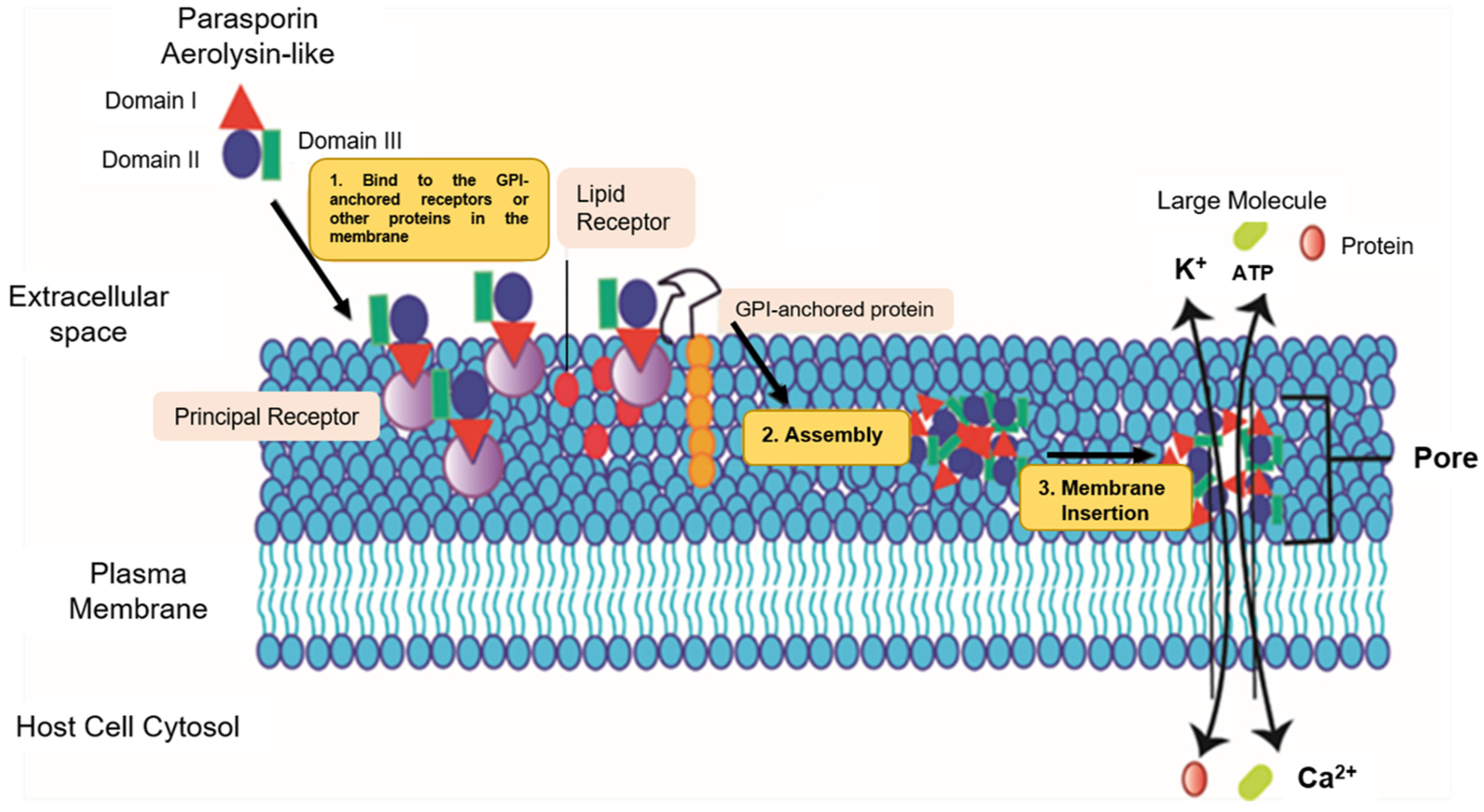
The mechanism of action of pore-forming proteins (PFPs) is dynamic, with three main steps: (1) the formation of water solubility, (2) self-assembly, and (3) insertion into the membrane, which leads to a pore suspected to be highly destructive for membrane integrity [34]. The points at which these proteins anchor to the membrane probably occur at specific receptors located in the microdomains rich in cholesterol and sphingolipids (lipid rafts), since these are requirements for GPI-anchored proteins, and the glucan region may be required for the binding and assembly in the membrane (Figure 2, part 1) [9]. Similarly, it was reported that the cell membrane receptor Beclin-1 could be important in the binding of three-domain parasporins (Parts 2 and 3, Figure 2) and that the Beclin-1 receptor is present in the mammary epithelium and epithelial carcinoma cells (Figure 2) [9,34,35].
The rearrangement of the domains typical for the classic protein model of three pore-forming domains does not occur for PS1 [36]. Therefore, its activity is not oriented to forming pores in the membrane [21,36]. PS1 was proposed to function as an activator of the apoptotic signaling pathway [14,19,37]. Selective cytotoxicity has been reported for the HeLa, Sawano, HepG2, HL-60, and MOLT-4 cell lines after PS1’s proteolytic activation by trypsin (Table 1) [6,38]. The activity of PS1 mainly involves modulating the influx of Ca2+ levels [6,21,39].
The PS2 mechanism of action likely starts with recognizing and binding to a receptor located in the cancer cells’ membranes [24], identifying lipid rafts, and anchoring the protein monomers in the periphery. The oligomers, resistant to sodium dodecyl sulfate (SDS), are embedded in the membrane, leading to its permeabilization [18,24]. Although PS2 is considered a selective pore-forming toxin, its primary receptors have not been fully elucidated [18].
Cells exposed to PS2 show morphological changes, including inflammation, blisters and lysis, microtubule disassembly, actin filament coiling, and fragmentation of the mitochondria and endoplasmic reticulum. PS2 resides in the plasma membrane and has been shown to activate apoptosis through caspases [14], triggering increased permeability [14,18,31]. These effects are induced by the accumulation of PS2 by large oligomers in the membrane’s lipid rafts [8,18,24,39]. In turn, association of PS2 with GPI is required for cytolytic action. By contrast, membrane cholesterol slightly affects the efficiency of oligomerization [1]. The activation of PS2 induced by proteinase K [25] leads to the exposure of specific regions that bind to the receptor [18,25].
PS3 acts as a pore former in cancer cells, thereby increasing cellular permeability [12,40]. Although PS3 is structurally similar to the Cry proteins, containing the five conserved blocks that characterize Cry [7,40], the PS3 and Cry proteins are fundamentally different due to a castor domain [7,40,41], which is present in many unrelated proteins and is presumed to enhance/induce carbohydrate-binding capacity [40]. Similar to the above-described PS, the mechanisms of action of PS3 remain largely unknown. Krishnan et al. suggested that PS3 is most likely pore forming [16], which leads to an imbalance in ATP, increased cell size, and membrane damage [40,41]. Its cytotoxic activity was evident in the HL-60 and HepG2 cell lines [7,40,41], but it did not affect HeLa cells [41].
Studies on PS3, PS4, PS5, and PS6 are limited compared to those on PS1 and PS2, and many action modes remain undetermined. PS4 shows homology with both Cry and pore-forming β-type aerolysin. It has been reported to be cytotoxic to the Caco-2, Sawano, and MOLT cell lines [5,41]. Its structure mainly comprises β-sheet domains, and its pore-forming activity is not dependent on cholesterol [21,41]. Cells treated with this protein show an increase in size due to an increase in the cytoplasmic compartment and shrinkage of the nucleus, leading to the rupture of the cytoplasmic membrane and cell death [42]. PS5 and PS6 are the most recently discovered PS proteins. They have three domains, similar to PS1 and PS2, and presumably have pore-forming activity. They have been reported to show cytotoxic activity in liver and cervical cancer cell lines. However, there is no further information on their mechanisms of action [14].

4. Perspectives on the Improvement of Bt Parasporins as an Innovative Strategy for Controlling Cancer Cells

By deciphering structure–function relationships, proteins with improved properties, e.g., desired thermal activity, selectivity, specificity, or folding, can be designed [43]. For example, engineered proteins with various substitutions of amino acids are used in receptor- and channel-protein-binding studies [44]. Protein engineering is called the synthesis of proteins with enhanced functionality in vitro and in vivo due to altered physical, chemical, or biological properties through genomic and post-genomic strategies. Genetic improvement is closely linked to complementary computational methods, which aim to optimize the generation of mutant libraries by simulating the experimental conditions of directed mutagenesis techniques [45,46,47,48]. In addition, other computational methods are oriented toward predicting protein structures and designing models that allow the prediction of molecular interactions and pinpoint amino-acid residues or regions at crucial positions in natural and mutant proteins [43,49]. The computational technique most widely used for studying the possible interactions of Bt Cry toxins with insect receptors is molecular docking, followed by molecular dynamics, which has proven to help predict the stability of the interactions and analyze the molecular mechanisms of action.
Florez et al. [50] obtained five Cry11 variants by DNA shuffling and showed the toxic activity against Aedes aegypti and Culex quinquefasciatus for three of them. Molecular docking simulations were performed for these three variants, and the amino acids with possible interactions were identified. BenFarhat-Touzri et al. [51] cloned and sequenced the Cry1D-133 toxin and determined its toxicity against S. littoralis larvae. Molecular docking simulations were performed to explain the enhanced toxicity of this toxin and showed that the number of toxin–receptor interactions was higher than that of the interactions exhibited by the Cry1D toxin.
The use of computational techniques based on molecular dynamics has enabled researchers to study the mechanisms of action of Cry toxins. The study of molecular dynamics has provided novel insights into the oligomerization of Cry toxins at a molecular level. Sriwimol et al. simulated the Cry4Ba structure with a three-dimensional reconstructed map for trimeric protein states. For the first time, they showed the need for membrane-induced conformational changes in Cry4Ba toxin monomers to allow the molecular assembly of a pre-pore trimer, which can be inserted into the target membranes to generate a lytic pore [52].
Other molecular dynamic studies have been applied to investigate the residue interactions relevant to the toxicity of the Bt Cry toxin family. Pacheco et al. discovered the importance of salt-bridge formation between α-helix residues from adjacent monomers for the toxicity and oligomerization of the Cry1Ab and Cry5Ba toxins by molecular dynamics’ simulations [53]. They showed a critical role for the salt bridge between the E101 and R99 residues of Cry1Ab [54]. Site-directed mutagenesis experiments confirmed decreased oligomerization and toxicity potential for Cry1Ab-E101K and Cry1Ab-R99E mutants.
Interestingly, the R99–E101 salt bridge is not fully conserved in Cry proteins, with both or one of the residues being different in Cry5Ba. However, Pacheco et al. showed that additional salt bridges with similar structural functions could also be formed in these Cry proteins. In conclusion, the computational analysis highlighted the importance of salt-bridge formation between the α-3 helices of adjacent monomers for inducing/facilitating a conformational change crucial for Cry toxicity [53].

Genetic Improvement of Cry Protein as a Model to Be Followed for Parasporins

Bt is an excellent candidate for producing both natural and genetically enhanced PS proteins [55]. Studies on Bt have been in progress for over 100 years since the discovery of Bt in Japan [56]. During this time, several studies have established associations between the crystal morphology, protein sequences, and molecular weight, and the specific effects against its insecticidal targets [57]. Bt continues to be of great scientific interest, and it is one of the most studied biotechnological alternatives of biological origin on Earth. By contrast, PS remains a scarcely explored option, as few researchers have awakened their curiosity in PS and anticancer pore-forming proteins.
Various approaches have been taken to modify Bt toxins’ binding specificity and affinity, with the ultimate goal of producing genetically modified toxins that target new pest species and counteract the resistance developed in the field. The alteration of the binding affinity and specificity of the Bt toxin can come from domain exchanges, site-directed mutagenesis, truncation, and the generation and subsequent visualization of parasporal proteins in phage libraries containing mutant toxins [55].
Although there are no reports on the genetic improvement of PS-like parasporal proteins, here we review the technology applied to other types of Bt crystal proteins, which can serve as a methodological and scientific basis for obtaining PS proteins with improved cytotoxic activity against cancer cells. Table 2 presents several examples of the directed evolution techniques that have been successful for Bt proteins and might also be used shortly for PS modification and studies.
Domain exchanges between related proteins might have an increased toxicity towards the target cells. For Cry proteins, several examples are presented in Table 2. Site-directed mutagenesis at specific Domains or Loops is an efficient technique for obtaining potent toxins with enhanced toxicity. An example is Cry19A, for which, by substitutions in Domain II loops 1 and 2, the toxicity increased up to 42,000 times, concerning the parental [64]. Other techniques, such as truncated toxins or filtering of promising candidates with increased receptor affinity through Phage-display libraries, are presented in Table 2. Genetic improvement of 3D-Cry toxins is reviewed extensively by Susana Vilchez [75].
PSs require extensive structural and functional studies, which will help to unravel their complex but elegant mechanism of action leading to the cytotoxic effects upon cancer cell lines. Studies addressing this knowledge gap will help to elucidate the toxic action of PSs and define the structure–function relationship. Acknowledging its potential as anticancer molecules, PSs’ site-directed evolution studies might focus as well on analyzing the cellular permeability and the potency of its toxicity to cells as well as the selectivity. In the long term, the generation of PS variants might allow new alternatives to address the threat of cancer to the well-being of humans and the burden on the health system. The current knowledge highlights the potential role that bacterial proteins might play in generating novel anticancer molecules [9].

5. Conclusions

The low-molecular-weight PS proteins, such as the pore-forming toxin PS-2Aa1, induce apoptosis in target cells and have cytotoxic activity against several human cancer cell lines. Remarkably, low-molecular-weight PS proteins do not show detectable cytotoxic activity against normal cells in most cases [12]. Therefore, PS proteins have emerged as a viable, efficient, and natural alternative for combating cancer, one of the types of diseases with the highest mortality rate in humans. However, limited knowledge on these toxins at the molecular level, including the mechanism of action and the receptors targeted on cancer cells, is available. PS2 is a single protein with a proposed action mode based on its structure. Therefore, there is an urgent need to establish the structural properties and mechanism of action to scale in in vivo studies and take the next step toward developing valuable products for human health [75].
In addition, there is a need to identify and evaluate new Bt native strains with improved cytotoxic activity toward cancer cells and select candidates for further genetic improvement to obtain toxins with enhanced activity. PS proteins are in the process of being consolidated as a viable alternative for cancer treatment. Compared to other current approaches, they hold the potential to produce fewer side effects, improving both the treatment outcomes and quality of life of cancer patients.

Author Contributions

Conceptualization, M.O.S.-B. and N.J.R.-F.; writing—original draft preparation, M.O.S.-B., D.F.H.-P., J.S.A.-A. and N.J.R.-F.; writing—review and editing L.V., P.R.-V., E.H.P.-R., A.V.d.B., J.O., E.M. and M.O.S.-B.; supervision M.O.S.-B. and N.J.R.-F. All authors have read and agreed to the published version of the manuscript.

Funding

The work has been funded by MINCIENCIAS, MINEDUCACIÓN, MINCIT and ICETEX, through the Program Ecosistema Científico Cod. FP44842-211-2018, Project number 58668.

Institutional Review Board Statement

Not applicable.

Informed Consent Statement

Not applicable.

Data Availability Statement

Not applicable.

Acknowledgments

All co-authors are very thankful to their respective Dept. and School at University Institution for all the moral support and encouragement.

Conflicts of Interest

The authors declare no conflict of interest. The funders had no role in the design of the study; in the collection, analyses, or interpretation of data; in the writing of the manuscript; or in the decision to publish the results.

Sample Availability

Not applicable.

References

  1. Portela-Dussán, D.D.; Chaparro-Giraldo, A.; López-Pazos, S.A. Bacillus thuringiensis biotechnology in agriculture. Nova 2013, 11, 87–96. [Google Scholar] [CrossRef]
  2. Melo, A.L.D.A.; Soccol, V.T.; Soccol, C.R. Bacillus thuringiensis: Mechanism of action, resistance, and new applications: A review. Crit. Rev. Biotechnol. 2016, 36, 317–326. [Google Scholar] [CrossRef] [PubMed]
  3. Akao, T.; Mizuki, E.; Yamashita, S.; Kim, H.S.; Lee, D.W.; Ohba, M. Specificity of lectin activity of Bacillus thuringiensis parasporal inclusion proteins. J. Basic Microbiol. 2001, 41, 3–6. [Google Scholar] [CrossRef]
  4. Akiba, T.; Okumura, S. Parasporins 1 and 2: Their structure and activity. J. Invertebr. Pathol. 2017, 142, 44–49. [Google Scholar] [CrossRef] [PubMed]
  5. Mizuki, E.; Ohba, M.; Akao, T.; Yamashita, S.; Saitoh, H.; Park, Y.S. Unique activity associated with non-insecticidal Bacillus thuringiensis parasporal inclusions: In vitro cell-killing action on human cancer cells. J. Appl. Microbiol. 1999, 86, 477–486. [Google Scholar] [CrossRef] [PubMed] [Green Version]
  6. Ammons, D.R.; Short, J.D.; Bailey, J.; Hinojosa, G.; Tavarez, L.; Salazar, M.; Rampersad, J.N. Anti-cancer Parasporin toxins are associated with different environments: Discovery of two novel Parasporin 5-like genes. Curr. Microbiol. 2016, 72, 184–189. [Google Scholar] [CrossRef]
  7. Moazamian, E.; Bahador, N.; Azarpira, N.; Rasouli, M. Anti-cancer Parasporin toxins of new Bacillus thuringiensis against human colon (HCT-116) and blood (CCRF-CEM) cancer cell lines. Curr. Microbiol. 2018, 75, 1090–1098. [Google Scholar] [CrossRef]
  8. Xu, C.; Wang, B.C.; Yu, Z.; Sun, M. Structural insights into Bacillus thuringiensis Cry, Cyt and parasporin toxins. Toxins 2014, 6, 2732–2770. [Google Scholar] [CrossRef] [Green Version]
  9. Kitada, S.; Abe, Y.; Shimada, H.; Kusaka, Y.; Matsuo, Y.; Katayama, H.; Okumura, S.; Akao, T.; Mizuki, E.; Kuge, O.; et al. Cytocidal actions of parasporin-2, an anti-tumor crystal toxin from Bacillus thuringiensis. J. Biol. Chem. 2006, 281, 26350–26360. [Google Scholar] [CrossRef] [Green Version]
  10. Gonzalez, E.; Granados, J.C.; Short, J.D.; Ammons, D.R.; Rampersad, J. Parasporins from a Caribbean Island: Evidence for a globally dispersed Bacillus thuringiensis strain. Curr. Microbiol. 2011, 62, 1643–1648. [Google Scholar] [CrossRef]
  11. Brasseur, K.; Auger, P.; Asselin, E.; Parent, S.; Cote, J.C.; Sirois, M. Parasporin-2 from a new Bacillus thuringiensis 4R2 strain induces caspases activation and apoptosis in human cancer cells. PLoS ONE 2015, 10, e0135106. [Google Scholar] [CrossRef]
  12. Mizuki, E.; Park, Y.S.; Saitoh, H.; Yamashita, S.; Akao, T.; Higuchi, K.; Ohba, M. Parasporin, a human leukemic cell-recognizing parasporal protein of Bacillus thuringiensis. Clin. Diagn. Lab. Immunol. 2000, 7, 625–634. [Google Scholar] [CrossRef] [Green Version]
  13. Okassov, A.; Nersesyan, A.; Kitada, S.; Ilin, A. Parasporins as new natural anticancer agents: A review. J. BUON 2015, 20, 5–16. [Google Scholar] [PubMed]
  14. Hayakawa, T.; Kanagawa, R.; Kotani, Y.; Kimura, M.; Yamagiwa, M.; Yamane, Y.; Takebe, S.; Sakai, H. Parasporin-2Ab, a newly isolated cytotoxic crystal protein from Bacillus thuringiensis. Curr. Microbiol. 2007, 55, 278–283. [Google Scholar] [CrossRef]
  15. Krishnan, V.; Domanska, B.; Elhigazi, A.; Afolabi, F.; West, M.J.; Crickmore, N. The human cancer cell active toxin Cry41Aa from Bacillus thuringiensis acts like its insecticidal counterparts. Biochem. J. 2017, 474, 1591–1602. [Google Scholar] [CrossRef] [PubMed]
  16. Ohba, M.; Mizuki, E.; Uemori, A. Parasporin, a new anticancer protein group from Bacillus thuringiensis. Anticancer Res. 2009, 29, 427–433. [Google Scholar] [PubMed]
  17. Ekino, K.; Okumura, S.; Ishikawa, T.; Kitada, S.; Saitoh, H.; Akao, T.; Oka, T.; Nomura, Y.; Ohba, M.; Shin, T.; et al. Cloning and characterization of a unique cytotoxic protein parasporin-5 produced by Bacillus thuringiensis A1100 strain. Toxins 2014, 6, 1882–1895. [Google Scholar] [CrossRef] [Green Version]
  18. Nagamatsu, Y.; Okamura, S.; Saitou, H.; Akao, T.; Mizuki, E. Three cry toxins in two types from bacillus thuringiensis strain M019 preferentially kill human hepatocyte cancer and uterus cervix cancer cells. Biosci. Biotechnol. Biochem. 2010, 74, 494–498. [Google Scholar] [CrossRef]
  19. Okumura, S.; Ohba, M.; Mizuki, E.; Crickmore, N.; Côté, J.-C.; Nagamatsu, Y.; Kitada, S.; Sakai, H.; Harata, K.; Shin, T. List of Parasporins. Available online: http://parasporin.fitc.pref.fukuoka.jp/list.html (accessed on 29 November 2021).
  20. Ito, A.; Sasaguri, Y.; Kitada, S.; Kusaka, Y.; Kuwano, K.; Masutomi, K.; Mizuki, E.; Akao, T.; Ohba, M. A Bacillus thuringiensis crystal protein with selective cytocidal action to human cells. J. Biol. Chem. 2004, 279, 21282–21286. [Google Scholar] [CrossRef] [Green Version]
  21. Katayama, H.; Kusaka, Y.; Yokota, H.; Akao, T.; Kojima, M.; Nakamura, O.; Mekada, E.; Mizuki, E. Parasporin-1, a novel cytotoxic protein from Bacillus thuringiensis, induces Ca2+ influx and a sustained elevation of the cytoplasmic Ca2+ concentration in toxin-sensitive cells. J. Biol. Chem. 2007, 282, 7742–7752. [Google Scholar] [CrossRef] [Green Version]
  22. Moniatte, M.; Van Der Goot, F.G.; Buckley, J.T.; Pattus, F.; Van Dorsselaer, A. Characterisation of the heptameric pore-forming complex of the Aeromonas toxin aerolysin using MALDI-TOF mass spectrometry. FEBS Lett. 1996, 384, 269–272. [Google Scholar] [CrossRef] [Green Version]
  23. Kuroda, S.; Begum, A.; Saga, M.; Hirao, A.; Mizuki, E.; Sakai, H.; Hayakawa, T. Parasporin 1Ac2, a novel cytotoxic crystal protein isolated from Bacillus thuringiensis B0462 strain. Curr. Microbiol. 2013, 66, 475–480. [Google Scholar] [CrossRef] [PubMed]
  24. Nagahama, M.; Hara, H.; Fernandez-Miyakawa, M.; Itohayashi, Y.; Sakurai, J. Oligomerization of Clostridium perfringens ε-toxin is dependent upon membrane fluidity in liposomes. Biochemistry 2006, 45, 296–302. [Google Scholar] [CrossRef]
  25. Okumura, S.; Koga, H.; Inouye, K.; Mizuki, E. Toxicity of Parasporin-4 and health effects of pro-parasporin-4 diet in mice. Toxins 2014, 6, 2115–2126. [Google Scholar] [CrossRef] [PubMed] [Green Version]
  26. Peraro, M.D.; Van Der Goot, F.G. Pore-forming toxins: Ancient, but never really out of fashion. Nat. Rev. Microbiol. 2016, 14, 77–92. [Google Scholar] [CrossRef]
  27. Akiba, T.; Abe, Y.; Kitada, S.; Kusaka, Y.; Ito, A.; Ichimatsu, T.; Katayama, H.; Akao, T.; Higuchi, K.; Mizuki, E.; et al. Crystal structure of the Parasporin-2 Bacillus thuringiensis toxin that recognizes cancer cells. J. Mol. Biol. 2009, 386, 121–133. [Google Scholar] [CrossRef]
  28. Akiba, T.; Higuchi, K.; Mizuki, E.; Ekino, K.; Shin, T.; Ohba, M.; Kanai, R.; Harata, K. Nontoxic crystal protein from Bacillus thuringiensis demonstrates a remarkable structural similarity to beta-pore-forming toxins. Proteins 2006, 63, 243–248. [Google Scholar] [CrossRef] [PubMed]
  29. Iacovache, I.; Degiacomi, M.T.; Pernot, L.; Ho, S.; Schiltz, M.; Dal Peraro, M.; van der Goot, F.G. Dual chaperone role of the C-terminal propeptide in folding and oligomerization of the pore-forming toxin aerolysin. PLoS Pathog. 2011, 7, e1002135. [Google Scholar] [CrossRef] [Green Version]
  30. Okumura, S.; Saitoh, H.; Wasano, N.; Katayama, H.; Higuchi, K.; Mizuki, E.; Inouye, K. Efficient solubilization, activation, and purification of recombinant Cry45Aa of Bacillus thuringiensis expressed as inclusion bodies in Escherichia coli. Protein Expr. Purif. 2006, 47, 144–151. [Google Scholar] [CrossRef] [PubMed]
  31. Okumura, S.; Saitoh, H.; Ishikawa, T.; Wasano, N.; Yamashita, S.; Kusumoto, K.; Akao, T.; Mizuki, E.; Ohba, M.; Inouye, K. Identification of a novel cytotoxic protein, Cry45Aa, from Bacillus thuringiensis A1470 and its selective cytotoxic activity against various mammalian cell lines. J. Agric. Food. Chem. 2005, 53, 6313–6318. [Google Scholar] [CrossRef]
  32. Pandian, G.N.; Ishikawa, T.; Togashi, M.; Shitomi, Y.; Haginoya, K.; Yamamoto, S.; Nishiumi, T.; Hori, H. Bombyx mori midgut membrane protein P252, which binds to Bacillus thuringiensis Cry1A, is a chlorophyllide-binding protein, and the resulting complex has antimicrobial activity. Appl. Environ. Microbiol. 2008, 74, 1324–1331. [Google Scholar] [CrossRef] [PubMed] [Green Version]
  33. Aldeewan, A.; Zhang, Y.; Su, L. Bacillus thuringiensis Parasporins functions on cancer cells. Int. J. Pure Appl. Biosci. 2014, 4, 67–74. [Google Scholar]
  34. Prasad, S.S.; Shethna, Y.I. Purification, crystallization and partial characterization of the antitumour and insecticidal protein subunit from the delta-endotoxin of Bacillus thuringiensis var. thuringiensis. Biochim. Biophys. Acta 1974, 362, 558–566. [Google Scholar] [CrossRef]
  35. Patyar, S.; Joshi, R.; Byrav, D.P.; Prakash, A.; Medhi, B.; Das, B. Bacteria in cancer therapy: A novel experimental strategy. J. Biomed. Sci. 2010, 17, 21. [Google Scholar] [CrossRef] [Green Version]
  36. Katayama, H.; Yokota, H.; Akao, T.; Nakamura, O.; Ohba, M.; Mekada, E.; Mizuki, E. Parasporin-1, a novel cytotoxic protein to human cells from non-insecticidal parasporal inclusions of Bacillus thuringiensis. J. Biochem. 2005, 137, 17–25. [Google Scholar] [CrossRef]
  37. Sanahuja, G.; Banakar, R.; Twyman, R.M.; Capell, T.; Christou, P. Bacillus thuringiensis: A century of research, development and commercial applications. Plant Biotechnol. J. 2011, 9, 283–300. [Google Scholar] [CrossRef] [Green Version]
  38. Okumura, S.; Saitoh, H.; Ishikawa, T.; Mizuki, E.; Inouye, K. Identification and characterization of a novel cytotoxic protein, parasporin-4, produced by Bacillus thuringiensis A1470 strain. Biotechnol. Annu. Rev. 2008, 14, 225–252. [Google Scholar] [CrossRef]
  39. Chubicka, T.; Girija, D.; Deepa, K.; Salini, S.; Meera, N.; Raghavamenon, A.C.; Divya, M.K.; Babu, T.D. A parasporin from Bacillus thuringiensis native to Peninsular India induces apoptosis in cancer cells through intrinsic pathway. J. Biosci. 2018, 43, 407–416. [Google Scholar] [CrossRef] [PubMed]
  40. Nelson, K.L.; Brodsky, R.A.; Buckley, J.T. Channels formed by subnanomolar concentrations of the toxin aerolysin trigger apoptosis of T lymphomas. Cell. Microbiol. 1999, 1, 69–74. [Google Scholar] [CrossRef] [PubMed]
  41. Maagd, R.A.; Bravo, A.; Berry, C.; Crickmore, N.; Schnepf, H.E. Structure, diversity, and evolution of protein toxins from spore-forming entomopathogenic bacteria. Annu. Rev. Genet. 2003, 37, 409–433. [Google Scholar] [CrossRef]
  42. Balabanova, L.; Golotin, V.; Podvolotskaya, A.; Rasskazov, V. Genetically modified proteins: Functional improvement and chimeragenesis. Bioengineered 2015, 6, 262–274. [Google Scholar] [CrossRef] [Green Version]
  43. Kim, S.B.; Izumi, H. Functional artificial luciferases as an optical readout for bioassays. Biochem. Biophys. Res. Commun. 2014, 448, 418–423. [Google Scholar] [CrossRef]
  44. He, L.; Friedman, A.M.; Bailey-Kellogg, C. Algorithms for optimizing cross-overs in DNA shuffling. BMC Bioinform. 2012, 13, S3. [Google Scholar] [CrossRef] [PubMed] [Green Version]
  45. Wedge, D.C.; Rowe, W.; Kell, D.B.; Knowles, J. In silico modelling of directed evolution: Implications for experimental design and stepwise evolution. J. Theor. Biol. 2009, 257, 131–141. [Google Scholar] [CrossRef] [Green Version]
  46. Pinzon, E.H.; Sierra, D.A.; Suarez, M.O.; Orduz, S.; Florez, A.M. DNA secondary structure formation by DNA shuffling of the conserved domains of the Cry protein of Bacillus thuringiensis. BMC Biophys. 2017, 10, 1–10. [Google Scholar] [CrossRef] [PubMed] [Green Version]
  47. Stimple, S.D.; Smith, M.D.; Tessier, P.M. Directed evolution methods for overcoming trade-offs between protein activity and stability. AIChE J. 2020, 66, e16814. [Google Scholar] [CrossRef] [PubMed]
  48. Basit, N.; Wechsler, H. Prediction of enzyme mutant activity using computational mutagenesis and incremental transduction. Adv. Bioinform. 2011, 2011, 1–9. [Google Scholar] [CrossRef]
  49. Florez, A.M.; Suarez-Barrera, M.O.; Morales, G.M.; Rivera, K.V.; Orduz, S.; Ochoa, R.; Guerra, D.; Muskus, C. Toxic activity, molecular modeling and docking simulations of Bacillus thuringiensis Cry11 toxin variants obtained via DNA shuffling. Front. Microbiol. 2018, 9, 2461. [Google Scholar] [CrossRef] [PubMed]
  50. BenFarhat-Touzri, D.; Driss, F.; Jemli, S.; Tounsi, S. Molecular characterization of Cry1D-133 toxin from Bacillus thuringiensis strain HD133 and its toxicity against Spodoptera littoralis. Int. J. Biol. Macromol. 2018, 112, 1–6. [Google Scholar] [CrossRef]
  51. Sriwimol, W.; Aroonkesorn, A.; Sakdee, S.; Kanchanawarin, C.; Uchihashi, T.; Ando, T.; Angsuthanasombat, C. Potential prepore trimer formation by the Bacillus thuringiensis mosquito-specific toxin: Molecular insights into a critical prerequisite of membrane-bound monomers. J. Biol. Chem. 2015, 290, 20793–20803. [Google Scholar] [CrossRef] [Green Version]
  52. Pacheco, S.; Gómez, I.; Sánchez, J.; García-Gómez, B.I.; Czajkowsky, D.M.; Zhang, J.; Soberón, M.; Bravo, A. Helix α-3 inter-molecular salt bridges and conformational changes are essential for toxicity of Bacillus thuringiensis 3D-Cry toxin family. Sci. Rep. 2018, 8, 10331. [Google Scholar] [CrossRef]
  53. Phillips, J.C.; Braun, R.; Wang, W.; Gumbart, J.; Tajkhorshid, E.; Villa, E.; Chipot, C.; Skeel, R.D.; Kalé, L.; Schulten, K. Scalable molecular dynamics with NAMD. J. Comput. Chem. 2005, 26, 1781–1802. [Google Scholar] [CrossRef] [Green Version]
  54. Deist, B.R.; Rausch, M.A.; Fernandez-Luna, M.T.; Adang, M.J.; Bonning, B.C. Bt toxin modification for enhanced efficacy. Toxins 2014, 6, 3005–3027. [Google Scholar] [CrossRef] [PubMed] [Green Version]
  55. Sansinenea, E. Discovery and Description of Bacillus thuringiensis. In Bacillus Thuringiensis Biotechnology; Springer: Dordrecht, The Netherlands, 2012; pp. 3–18. ISBN 9789400730212. [Google Scholar]
  56. López-Meza, J.E.; Ibarra, J.E. Characterization of a novel strain of Bacillus thuringiensis. Appl. Environ. Microbiol. 1996, 62, 1306–1310. [Google Scholar] [CrossRef] [Green Version]
  57. Walters, F.S.; deFontes, C.M.; Hart, H.; Warren, G.W.; Chen, J.S. Lepidopteran-active variable-region sequence imparts coleopteran activity in eCry3.1Ab, an engineered Bacillus thuringiensis hybrid insecticidal protein. Appl. Environ. Microbiol. 2010, 76, 3082–3088. [Google Scholar] [CrossRef] [Green Version]
  58. Shah, J.V.; Yadav, R.; Ingle, S.S. Engineered Cry1Ac-Cry9Aa hybrid Bacillus thuringiensis delta-endotoxin with improved insecticidal activity against Helicoverpa armigera. Arch. Microbiol. 2017, 199, 1069–1075. [Google Scholar] [CrossRef] [PubMed]
  59. de Maagd, R.A.; Weemen-Hendriks, M.; Stiekema, W.; Bosch, D. Bacillus thuringiensis delta-endotoxin Cry1C domain III can function as a specificity determinant for Spodoptera exigua in different, but not all, Cry1-Cry1C hybrids. Appl. Environ. Microbiol. 2000, 66, 1559–1563. [Google Scholar] [CrossRef] [Green Version]
  60. Karlova, R.; Weemen-Hendriks, M.; Naimov, S.; Ceron, J.; Dukiandjiev, S.; de Maagd, R.A. Bacillus thuringiensis delta-endotoxin Cry1Ac domain III enhances activity against Heliothis virescens in some, but not all Cry1-Cry1Ac hybrids. J. Invertebr. Pathol. 2005, 88, 169–172. [Google Scholar] [CrossRef] [PubMed]
  61. Naimov, S.; Weemen-Hendriks, M.; Dukiandjiev, S.; De Maagd, R.A. Bacillus thuringiemis delta-endotoxin Cry1 hybrid proteins with increased activity against the Colorado Potato Beetle. Appl. Environ. Microbiol. 2001, 67, 5328–5330. [Google Scholar] [CrossRef] [Green Version]
  62. Abdullah, M.A.F.; Alzate, O.; Mohammad, M.; McNall, R.J.; Adang, M.J.; Dean, D.H. Introduction of Culex toxicity into Bacillus thuringiensis Cry4Ba by protein engineering. Appl. Environ. Microbiol. 2003, 69, 5343–5353. [Google Scholar] [CrossRef] [Green Version]
  63. Abdullah, M.A.F.; Dean, D.H. Enhancement of Cry19Aa mosquitocidal activity against Aedes aegypti by mutations in the putative loop regions of domain II. Appl. Environ. Microbiol. 2004, 70, 3769–3771. [Google Scholar] [CrossRef] [Green Version]
  64. McNeil, B.C.; Dean, D.H. Bacillus thuringiensis Cry2Ab is active on Anopheles mosquitoes: Single D block exchanges reveal critical residues involved in activity. FEMS Microbiol. Lett. 2011, 325, 16–21. [Google Scholar] [CrossRef] [Green Version]
  65. Liu, X.S.; Dean, D.H. Redesigning Bacillus thuringiensis Cry1Aa toxin into a mosquito toxin. Protein Eng. Des. Sel. 2006, 19, 107–111. [Google Scholar] [CrossRef] [Green Version]
  66. Gómez, I.; Ocelotl, J.; Sánchez, J.; Lima, C.; Martins, E.; Rosales-Juárez, A.; Aguilar-Medel, S.; Abad, A.; Dong, H.; Monnerat, R.; et al. Enhancement of Bacillus thuringiensis Cry1Ab and Cry1Fa toxicity to Spodoptera frugiperda by domain III mutations indicates there are two limiting steps in toxicity as defined by receptor binding and protein stability. Appl. Environ. Microbiol. 2018, 84, e01393-18. [Google Scholar] [CrossRef] [PubMed] [Green Version]
  67. Craveiro, K.I.C.; Júnior, J.E.G.; Silva, M.C.M.; Macedo, L.L.P.; Lucena, W.A.; Silva, M.S.; Júnior, J.D.A.d.S.; Oliveira, G.R.; Magalhães, M.T.Q.d.; Santiago, A.D.; et al. Variant Cry1Ia toxins generated by DNA shuffling are active against sugarcane giant borer. J. Biotechnol. 2010, 145, 215–221. [Google Scholar] [CrossRef]
  68. Soberon, M.; Pardo-Lopez, L.; Lopez, I.; Gomez, I.; Tabashnik, B.E.; Bravo, A. Engineering modified Bt toxins to counter insect resistance. Science 2007, 318, 1640–1642. [Google Scholar] [CrossRef] [PubMed]
  69. Tabashnik, B.E.; Huang, F.; Ghimire, M.N.; Leonard, B.R.; Siegfried, B.D.; Rangasamy, M.; Yang, Y.; Wu, Y.; Gahan, L.J.; Heckel, D.G.; et al. Efficacy of genetically modified Bt toxins against insects with different genetic mechanisms of resistance. Nat. Biotechnol. 2011, 29, 1128–1131. [Google Scholar] [CrossRef] [PubMed]
  70. Park, H.W.; Bideshi, D.K.; Federici, B.A. Molecular genetic manipulation of truncated Cry1C protein synthesis in Bacillus thuringiensis to improve stability and yield. Appl. Environ. Microbiol. 2000, 66, 4449–4455. [Google Scholar] [CrossRef] [Green Version]
  71. Fujii, Y.; Tanaka, S.; Otsuki, M.; Hoshino, Y.; Endo, H.; Sato, R. Affinity maturation of Cry1Aa toxin to the Bombyx mori cadherin-like receptor by directed evolution. Mol. Biotechnol. 2013, 54, 888–899. [Google Scholar] [CrossRef] [PubMed]
  72. Oliveira, G.R.; Silva, M.C.; Lucena, W.A.; Nakasu, E.Y.; Firmino, A.A.; Beneventi, M.A.; Souza, D.S.; Gomes, J.E., Jr.; de Souza, J.D., Jr.; Rigden, D.J.; et al. Improving Cry8Ka toxin activity towards the cotton boll weevil (Anthonomus grandis). BMC Biotechnol. 2011, 11, 85. [Google Scholar] [CrossRef] [Green Version]
  73. Shao, E.; Lin, L.; Chen, C.; Chen, H.; Zhuang, H.; Wu, S.; Sha, L.; Guan, X.; Huang, Z. Loop replacements with gut-binding peptides in Cry1Ab domain II enhanced toxicity against the brown planthopper, Nilaparvata lugens (Stål). Sci. Rep. 2016, 6, 20106. [Google Scholar] [CrossRef] [PubMed] [Green Version]
  74. Vílchez, S. Making 3D-cry toxin mutants: Much more than a tool of understanding toxins mechanism of action. Toxins 2020, 12, 600. [Google Scholar] [CrossRef] [PubMed]
  75. Velásquez, L.-F.; Rojas, D.; Cerón, J. Proteínas de Bacillus thuringiensis con actividad citotóxica: Parasporinas. Rev. Colomb. Biotecnol. 2018, 20, 89–100. [Google Scholar] [CrossRef]
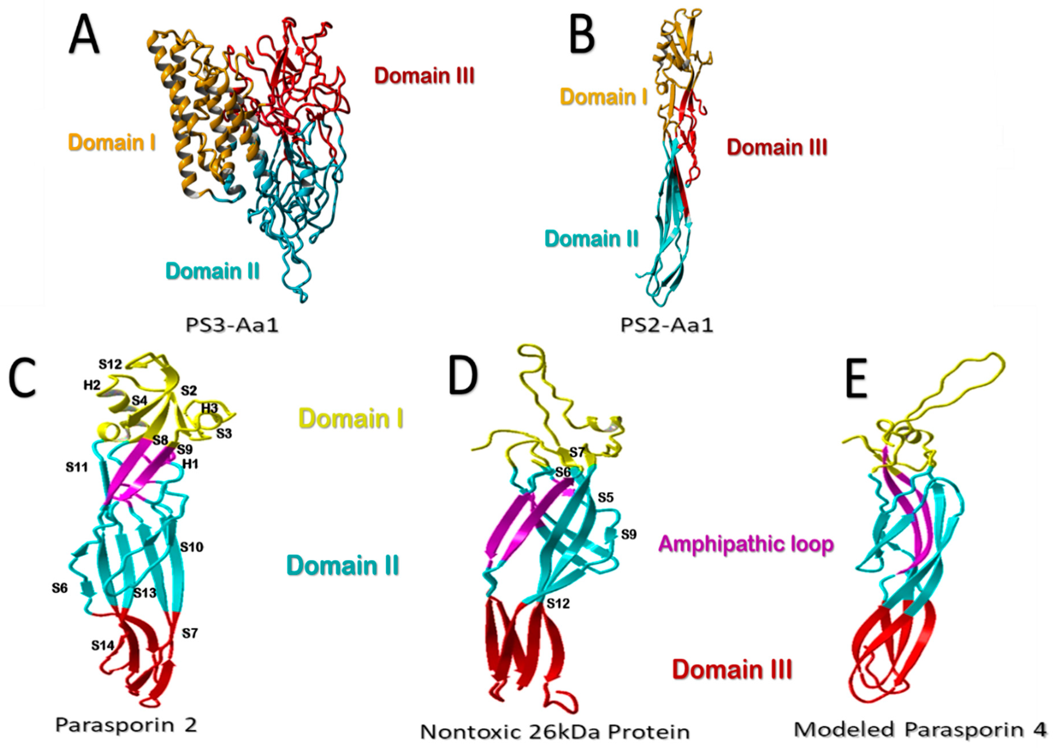
Figure 1. Structural comparison of parasporins. (A) Structural model of higher-molecular-weight PS3Aa1 with its three domains. (B) Low-molecular-weight PS2Aa1 structural model. (CE) Structural comparison between parasporin-2, the 26-kDa nontoxic protein, and aerolysin-like β-PFT. Membrane-binding-related domain I is colored yellow. The membrane-insertion and pore-formation regions are colored blue (domain II) and red (Domain III). It is suggested that the purple amphipathic β-hairpin is necessary for pore formation (CE). Parasporin 4 (PS4) was modeled using the 26-kDa nontoxic protein as an adapted template from Xu et al. [9], modified by the authors.
Molecules 26 07476 g001
Figure 2. Action mode of aerolysin-like parasporins (PS2Aa). Figure adapted from [9]. According to this model, the mechanism of action could be as follows: 1. The solubilized protein binds to the GPI-anchored receptors at the N-terminus. 2. After C-terminal proteolytic digestion, the activated protein monomers assemble (oligomerization). 3. Through reorganization, a transmembrane β-barrel is formed.
Molecules 26 07476 g002
Table 1. Parasporin families, family-containing strains, molecular weights, target cells, cytotoxic activity, and references.
ParasporinStrain (Bt)Molecular Mass
(kDa)
Target Cell LineCytotoxic Activity
IC50 (μg/mL)
Ref.
PS1Aa1A119081MOLT-4
HL-60
HepG2
HeLa
Jurkat
Sawano
Caco-2
A549
2.2
0.32
3.0
0.12
>10
>10
>10
>10
[9,13,14]
PS2Aa1A154737MOLT-4
Jurkat
HL-60
HepG2
Sawano
Caco-2
HCT116
CCRF-CEM
0.022
0.018
0.019
0.019
0.0017
0.013
10
27
[8,9,14,15]
PS3Aa1A146293MOLT-4
Jurkat
HL-60
HepG2
HeLa
Sawano
>10
>10
1.32
2.8
>10
>10
[9,14,16]
PS4Aa1A147034MOLT-4
HL-60
HepG2
Sawano
TCS
Caco-2
0.472
0.725
1.90
0.245
0.719
0.124
[9,14,17]
PS5Aa1A110031MOLT-4
Caco-2
HepG2
TCS
HeLa
Sawano.
0.075
0.30
0.049
0.046
0.08
0.065
[14,18]
PS6Aa1M109/CP8473HepG2
HeLa
Caco-2
2.3
7.2
>10
[14,19]
Table 2. Modifications made to Bt toxins to improve their efficacy.
Type of ModificationBt ToxinTarget InsectIncrease or Decrease in ToxicityReference
Domain exchanges
Domain III Exchange
For Domain III of Cry1Ab.
mCry3AaDiabrotica virgiferaThe toxicity increased ≥19%.[58]
Domain III, II, I Exchange
For Domains of Cry1Ac.
Cry9AaHelicoverpa armigeraThe toxicity increased between 4.9 and 5.1 times, concerning parentals.[59]
Domain III Exchange
For Domain III of Cry1Ca.
Cry1Ab; Cry1Ac; Cry1Ba; Cry1Ea; Cry1FaSpodoptera exiguaIncreased up to 5.5 times for Cry1Fa.[60]
Domain III Exchange
For Domain III of Cry1CAc
Cry1Ca; Cry1Fb; Cry1Ba; Cry1Da; Cry1EaHeliothis virescensThe toxicity increased 172 and 69.6 times more for Cry1Ca and Cry1Fb, respectively. [61]
Domain exchanges of Domains II and III, between Cry1Ia and Cry1Ba.Cry1Ia; Cry1BaLeptinotarsa decemlineataThe toxicity increased up to 1127 and 4.2 times, compared to Cry1Ba and Cry1Ia, correspondingly.[62]
Site-directed mutagenesis
Loops 1, 2, and 3, domain II substitution.
Cry4BaCulex pipiens; Culex quinquefasciatusThe toxicity increased up to 700 times.[63]
Loops 1 and 2 domain II substitution
Cry19AaAedes aegyptiThe toxicity increased up to 42,000 times, concerning the parental.[64]
Substitution in the domain IICry2AbAnopheles gambiaeThe toxicity increased up to 6.75 times.[65]
Loops 1 and 2 domain II substitution and deletions.
Cry1AaCulex pipiensChange in insect target.[66]
Substitution in the domain III Cry1AbSpodoptera frugiperdaThe toxicity increased up to 44 times, correspondingly to the parental.[67]
Truncated toxins
Truncation and selection of mutants, derived from a phage library Cry1IaTelchin licusThe toxicity increased, showing mortality of 50% for approach.[68]
Helix α-1 domain I truncation.Cry1APectinophora gossypiellaThe toxicity increased up to 100 and 150 times for Cry1Ab and CryAc, respectively. [69]
Helix α-1 domain I truncation.Cry1APlutella xylostella; Ostrinia nubilalisThe toxicity increased ≥350 times, against resistant insects.[70]
C-terminal truncationCry1CSpodoptera exiguaThe toxicity increased up to 4 times.[71]
Phage-display library
Selection of mutant toxins from a phage-display library based on their potential of binding.Cry1AaBombyx moriIncreased the receptor affinity potential up to 16 and 50 times more, contrasting the parentals. [72]
Selection of mutant toxins from a phage-display library based on their potential of binding.Cry8KaAnthonomus grandisIncreased the toxicity up to 3.2 times, contrasting the parental.[73]
Selection of mutant toxins from a phage-display library based on their potential of binding in the domain II.Cry1AaNilaparvata lugensThe toxicity increased between 1.4 and 8.9 times, concerning parentals.[74]
Publisher’s Note: MDPI stays neutral with regard to jurisdictional claims in published maps and institutional affiliations.