Article

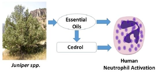
Innate Immunomodulatory Activity of Cedrol, a Component of Essential Oils Isolated from Juniperus Species

by
Gulmira Özek
1,
Igor A. Schepetkin
2,
Moldir Yermagambetova
3,4,
Temel Özek
1,5,
Liliya N. Kirpotina
2,
Shyryn S. Almerekova
3,4,
Saule I. Abugalieva
3,4,
Andrei I. Khlebnikov
6 and
Mark T. Quinn
2,*
1
Department of Pharmacognosy, Faculty of Pharmacy, Anadolu University, Eskisehir 26470, Turkey
2
Department of Microbiology and Cell Biology, Montana State University, Bozeman, MT 59717, USA
3
Institute of Plant Biology and Biotechnology, Almaty 050040, Kazakhstan
4
Faculty of Biology and Biotechnology, Al-Farabi Kazakh National University, Almaty 050040, Kazakhstan
5
Medicinal Plant, Drug and Scientific Research and Application Center (AUBIBAM), Anadolu University, Eskişehir 26470, Turkey
6
Kizhner Research Center, Tomsk Polytechnic University, 634050 Tomsk, Russia
*
Author to whom correspondence should be addressed.
Molecules 2021, 26(24), 7644; https://doi.org/10.3390/molecules26247644
Submission received: 22 November 2021 / Revised: 10 December 2021 / Accepted: 12 December 2021 / Published: 16 December 2021
(This article belongs to the Special Issue Chemistry of Natural Organic Compounds)

Abstract

:
Little is known about the immunomodulatory activity of essential oils isolated from Juniperus species. Thus, we isolated essential oils from the cones and leaves of eight juniper species found in Montana and in Kazakhstan, including J. horizontalis, J. scopolorum, J. communis, J. seravschanica, J. sabina, J. pseudosabina, J. pseudosabina subsp. turkestanica, and J. sibirica. We report here the chemical composition and innate immunomodulatory activity of these essential oils. Compositional analysis of the 16 samples of Juniper essential oils revealed similarities and differences between our analyses and those previously reported for essential oils from this species. Our studies represent the first analysis of essential oils isolated from the cones of four of these Juniper species. Several essential oil samples contained high levels of cedrol, which was fairly unique to three Juniper species from Kazakhstan. We found that these essential oils and pure (+)-cedrol induced intracellular Ca2+ mobilization in human neutrophils. Furthermore, pretreatment of human neutrophils and N-formyl peptide receptor 1 and 2 (FPR1 and FPR2) transfected HL60 cells with these essential oils or (+)-cedrol inhibited agonist-induced Ca2+ mobilization, suggesting these responses were desensitized by this pretreatment. In support of this conclusion, pretreatment with essential oils from J. seravschanica cones (containing 16.8% cedrol) or pure (+)-cedrol inhibited human neutrophil chemotaxis to N-formyl peptide. Finally, reverse pharmacophore mapping predicted several potential kinase targets for cedrol. Thus, our studies have identified cedrol as a novel neutrophil agonist that can desensitize cells to subsequent stimulation by N-formyl peptide.

Graphical Abstract

1. Introduction

The genus Juniperus L. consists of 75 species, which grow mainly in the northern hemisphere [1]. Juniperus is one of the main genera of the Cupressaceae family [2], and according to the generally accepted classification, the genus is divided into three sections distributed throughout the world [1,3,4]. These evergreen coniferous trees or shrubs thrive under a variety of environmental conditions. Some juniper species grow in the Arctic zone and the alpine belt, while others are found in subtropical and tropical zones [1,5,6,7,8].
Among the least studied Juniperus are those growing in Kazakhstan. In Kazakhstan, the local names for Juniperus species are “arsha” (in Kazakh) and “mojjevelnik” (in Russian) [9,10]. Kazakhstan’s latitude ranges from 40°7′ N to 54°9′ N, and therefore, is geographically similar to the northern states of the USA, including Montana (44°2′ N to 49°0′ N). As in Kazakhstan, a large part of Montana is occupied by steppes (approx. 60%), deserts, and semi-deserts (20%). About 10% of both Montana and Kazakhstan are covered by mountain systems where Juniperus species are an important woodland component [11,12,13]. According to the Flora of Kazakhstan [14], there are 10 Juniperus species in this country (J. communis L., J. sibirica Burgsd., J. pseudosabina Fisch. et. C. A. Mey., J. turkestanica Kom., J. intermedia Drob., J. seravschanica Kom., J. sabina L., J. semiglobosa Regel, J. talassica Lipsky and J. drobovii Sumn.). However, further studies by Abdulina [15] reduced them to seven species (J. communis, J. × media, J. pseudosabina, J. sabina, J. semiglobosa, J. sibirica and J. seravschanica), which was supported by molecular taxonomy indicating that J. turkestanica and J. pseudosabina belonged to a single species named J. pseudosabina [12]. Therefore, the former species was renamed as J. pseudosabina subsp. turkestanica. In Kazakhstan, juniper is one of the important components of forests in the high mountain ranges of Altai, Tarbagatai, Dzhungarskiy Alatau, Tien Shan, and in the boron forests of Northern Kazakhstan [16,17,18,19]. In comparison, there are three Juniperus species native to the state of Montana, USA (J. communis, J. horizontalis, J. scopulorum) [1]. Hence, only J. communis is common to both Kazakhstan and Montana. In both regions, the soil composition depends on climatic conditions and changes with variations in geography. Accordingly, the chemical composition of the vegetation also changes. Therefore, a comparative assessment of the chemical composition of juniper species growing in various eco-geographical niches of Kazakhstan and Montana is important to understanding their potential as sources of novel therapeutics, such as essential oils.
Juniperus species are a source of essential oils that have been used in the cosmetic industry. Essential oils serve as a source of aromatic substances to improve perfume properties and increase resistance to oxidation [20,21]. Juniper oil is also used in medicine as an effective antiseptic [22,23]. Besides essential oils, juniper extracts are rich in lipids, alkaloids, glucosides, polyphenols, and ascorbic acid. Juniper needles contain a significant amount of chlorophylls and carotenoids, which have antioxidant properties [24,25,26]. Representatives of the genus Juniperus have also been used in traditional medicine, gastronomy [27], pharmaceuticals [28], and in alcoholic beverages [29,30].
In traditional medicine, extracts from Juniperus species have been used for their stomachic, diuretic, antiseptic, antirheumatic, antispasmodic, astringent, carminative, and rubificant properties to treat dyspepsia, cystitis, arthritis, gout, and other inflammatory conditions [31,32,33]. One of the most important representatives of the genus is J. communis L., and the dried ripe cones of J. communis and essential oils obtained by steam distillation from the ripe, non-fermented cones of J. communis are included in the European Pharmacopoeia [34]. In addition a monograph about Juniper Tar, which is the empyreumatic volatile oil obtained from the woody portions of J. oxycedrus L., is registered in the United States Pharmacopoeia [35]. Juniper cones are the source of essential oils and the subjects of Pharmacopoeial monographs in the 5th Polish National Pharmacopoeia [36], German Pharmacopoeia (Deutches Arzneibuch) [37] and in Martindale Complete Drug Reference [38].
The essential oil composition of Juniperus species around the world have comprehensively been evaluated by Adams et al. [1,39,40,41,42,43,44,45]. However, reports on essential oils from Juniperus species growing in Kazakhstan have focused mainly on the investigation of essential oil yields and major volatile compounds isolated from their leaves [10,39,41,43,45,46,47,48], and little information has been reported regarding their biological properties [9,10,49,50].
The chemical composition of essential oils isolated from different Juniperus species is characterized by large variation, and it is difficult to make any common classification or grouping. In general, these essential oils are characterized by a high abundance of monoterpene hydrocarbons, mostly α-pinene, sabinene, myrcene, and limonene [41,43,44,45,47,51,52,53,54,55,56,57,58,59,60,61,62,63,64,65,66,67,68,69,70,71,72,73,74,75,76,77,78]. In addition, oxygenated monoterpenes, such as linalool, bornyl acetate, and sabinyl acetate, were also found in the essential oils of several Juniperus species [10,46,62,64]. Sesquiterpene constituents are rare in Juniperus oils [71], whereas diterpenes (manoyl oxide, abietatriene) were reported only in Algerian juniper essential oils [67]. Thus, it is clear that a broader evaluation of the chemical composition and biological activity of essential oils from plants found in various regions of the world is necessary to expand our understanding of the regional differences in these species.
In the present studies, we compared the chemical composition and immunomodulatory activity of essential oils obtained from the cones and leaves of seven juniper species collected in Kazakhstan and Montana, including J. horizontalis, J. scopolorum, J. communis, J. seravschanica, J. sabina, J. pseudosabina, J. pseudosabina subsp. turkestanica, and J. sibirica and analyzed their chemical composition and innate immunomodulatory activity. We show that the Juniper essential oils inhibited intracellular Ca2+ mobilization in neutrophils. Furthermore, we demonstrated that cedrol, which was present at high levels in some Juniper species from Kazakhstan, also inhibited human neutrophil functional responses and is likely one of the active components in these essential oils. Given the critical role of neutrophils in inflammation, our data support the possibility that cedrol could be effective therapeutic compound for the development anti-inflammatory agents.

2. Results and Discussion

2.1. Essential Oil Composition

Leaves and cones were collected from mature Juniperus shrubs and trees (Table 1) and extracted, as described under Materials and Methods. The extraction yields (v/w) of essential oils obtained from Juniperus spp. ranged from 0.4 to 2.0% (Table 1). The chemical composition of these essential oils was evaluated using simultaneous GC-FID and GC/MS. Major compounds (>2%) of the essential oils are shown in Table 2, and Supplementary Table S1 summarizes all identified compounds. The main class of compounds in all samples was monoterpenes, which ranged from 52.5 to 98.3% of the essential oil composition isolated from various Juniper species (Table 3). In addition, essential oils from the cones of J. sibirica (JSICon) were enriched in sesquiterpenes (46.4%).
The leaf oils of J. horizontalis from Turkey, Iran, USA, Egypt, and Canada (Saskatchewan) have been previously reported to contain linalool, limonene, sabinene, and bornyl acetate as their major volatile constituents [44,62,63,64,79]. In the present study, we found that JHLv also had a high content of sabinyl acetate (>16%), which was not found in previous reports (Table 2). The cone oil of J. horizontalis from Iran was reported to contain myrcene (22.6%), limonene (27.8%), and sabinene (38.0%) [79]. In our investigation, JHCon had predominantly sabinene (>77.0%) (Table 2), which may reflect the differences in ecology where these plants were collected.
We found that J. scopulorum essential oils JSLv and JSCon had high contents of sabinene (37.6% and 35.8%, respectively) (Table 2). These data are consistent with previous reports [80,81]. It should be noted that JSLv and JSCon essential oils also had high levels of terpinen-4-ol (10.0% and 23.0%, respectively). We also found that the eudesmane group of sesquiterpenes was enriched in the leaf oils of J. scopulorum, including elemol (14.4% and 6.0%, respectively) and 8-α-acetoxyelemol (11.0% and 1.0%, respectively) (Table 2). The co-occurrence of pregeijerene B in Juniperus oils was shown to be highly correlated with 8-α-acetoxyelemol and has systematic significance for this genus [82]. Indeed, we also detected pregeijerene B (2.2%) in the leaf oil of J. scropulorum. Similarly, JSCon also had similar amounts of 8-α-acetoxyelemol (1.0%) and pregeijerene B (0.2%). Previously, leaf oils of J. scopulorum from Colorado (USA) were reported to contain 8-α-acetoxyelemol (8.7%) and elemol (3.1%) [83], and J. scopulorum leaf oil from New Mexico was reported to contain thujopsene (57.9%), cuparene (6.1%), and cedrol (6.1%) [79]. Likewise, we found that JSLv essential oils had high levels of sesquiterpenes (Table 2 and Table 3).
J. communis is one of the most studied Juniperus species, and our data confirmed the relatively high content of α-pinene in the leaf oil of J. communis [51,52,54,55]. In addition, J. communis cone oil contained a high amount of myrcene (53.2%), which is consistent with previous reports on essential oils from J. communis collected in Poland [52] (Table 2).
The essential oils of J. seravschanica (JSELv and JSECon) were characterized by a high content of α-pinene (45.3% and 34.4%, respectively), myrcene (19.7% and 19.5%, respectively), and limonene (3.2% and 4.5%, respectively) (Table 2). The sesquiterpene alcohol cedrol (13.1% and 16.8%, respectively) was also present in significant amounts. These data are consistent with previously published information about J. seravschanica leaf oils from Kazakhstan, Pakistan, and Iran [42,45]. Similarly, we found that the cone essential oil JSECon also contained monoterpenes, α-pinene (34.4%), myrcene (19.5%), and limonene (4.5%), as well as cedrol (16.8%) (Table 2). In addition, the diterpene 8,13-abietadiene (3.9%) was also present in JSECon. Consistent with our findings, J. seravschanica cone oil from Uzbekistan was previously reported to contain α-pinene (29.0%), germacrene B (5.9%), and cedrol (3.1%) [73].
Analysis of J. sabina leaf essential oil composition showed that they were enriched in sabinyl acetate (30.3%), cedrol (12.2%), and sabinene (33.2%), which is consistent with previous reports [10,46]. Analysis of the J. sabina cone essential oils, which has not been performed previously, showed that they were dominated by sabinene (74.1%) (Table 2).
Sabinene (39.6%), α-pinene (15.2%), terpinen-4-ol (9.5%), and linalool (3.6%) were found to be the main constituents in J. pseudosabina subsp. turkestanica leaf essential oils (JTLv), which confirmed previous reports [43,47] (Table 2). J. pseudosabina subsp. turkestanica cone essential oils (JTCon), which have not been previously reported, were enriched in sabinene (54.2%), α-pinene (25.1%), and myrcene (4.8%) (Table 2).
The leaf essential oils of J. pseudosabina from Kazakhstan, China, and Mongolia have been reported to contain α-pinene, sabinene and cedrol, as well as high levels of germacrene-D-4-ol (14.3%) in plants from Mongolia [43,56]. Consistent with these reports, we found that JPSLv were enriched in monoterpenes, α-pinene (30.8%), sabinene (19.4%), and terpinen-4-ol (4.0%), as well as cedrol (7.0%) and elemol (3.9%) (Table 2). The cone essential oils of J. pseudosabina (JPSCon) have not been analyzed previously and were enriched in α-pinene (49.3%), sabinene (20.4%), myrcene (4.1%), β-pinene (3.7%), and cedrol (3.4%) (Table 2).
Leaf essential oils of J. sibirica from Italy, Kazakhstan, Serbia, and Russia have been reported to contain high levels of α-pinene (up to 80.0%) [46,74,75,76]. In addition, Sampietro et al. [48] reported that leaf essential oils leaf essential oils of J. sibirica from Kazakhstan were enriched in α-thujene-rich (46.3%). Here, we found that JSILv contained α-pinene (26.9%), sabinene (24.3%), terpinen-4-ol (9.3%), myrcene (2.6%), and α-terpinyl acetate (2.7%), whereas JSICon contained mainly high levels of α-pinene (44.9), as well as significant levels of germacrene D (16.7%) and 1,6-germacradien-5β-ol (4.2%) (Table 2). In addition, δ-cadinene (5.8%) was present. The only other report on J. sibirica cone essential oils was based on plants from Russia and indicated the presence of α-pinene (19.9%), β-myrcene (5.0%), α-terpineol (6.0%), as well as α-bisabolene (6.1%), τ-cadinol (5.6%), and α-cadinol (6.7%) [75]. Thus, there are clear similarities and differences with our analyses, which again may be related to the geographical location or time of collection of the plant tissues analyzed.

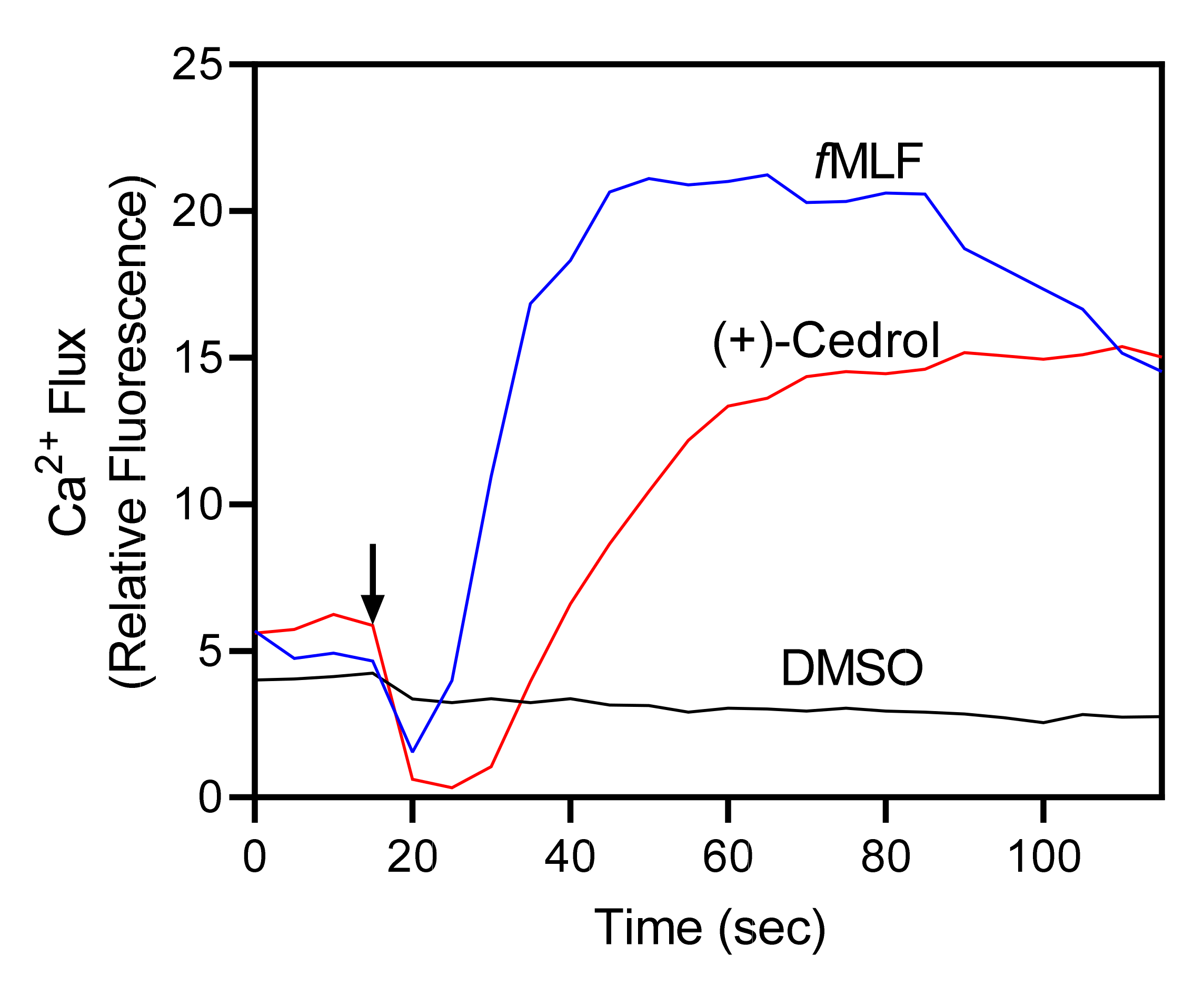
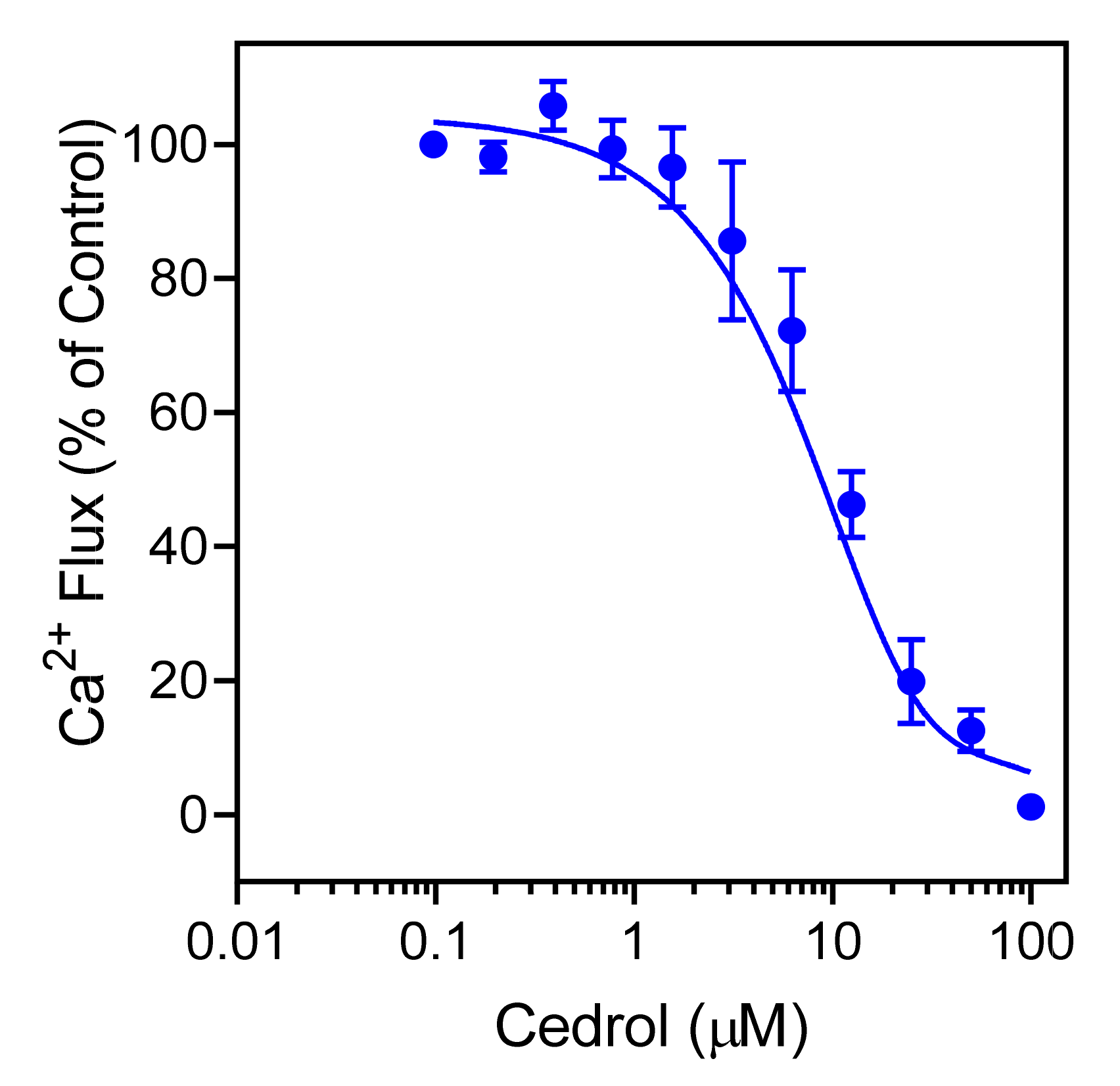
2.2. Effect of the Juniperus Essential Oils and Cedrol on Neutrophil [Ca2+]i

The innate immune system is essential for host defense and provides immediate defense against infection. Neutrophils are the most abundant leukocytes in the blood and constitute key components of the innate immunity that perform an important effector and regulatory functions in the development of human inflammatory response [84,85]. Thus, neutrophils represent an ideal pharmacological target for therapeutic development, and numerous natural products, including essential oils, have been shown to exhibit neutrophil immunomodulatory activity [86,87,88].
Juniperus essential oils were evaluated for their immunomodulatory effects on human neutrophils. Specifically, we evaluated their effects on [Ca2+]i, which is a key component of phagocyte activation [45,46]. We found that treatment of neutrophils with Juniperus essential oils increased [Ca2+]i, with EC50 values ranging from 13.9 µg/mL (JTCon) to 49.8 µg/mL (JHCon). In addition, analysis of (+) cedrol, a sesquiterpene present at levels of 13.1% (JSELv) to 16.8% (JSECon) showed that this compound also activated neutrophil [Ca2+]i (Figure 1 and Table 4). Thus, cedrol represents a novel essential oil component with innate immunomodulatory activity. Note that we evaluated (+)-cedrol (Figure 2), because this isoform is present in Juniperus essential oils [89].
Since Juniperus essential oils and cedrol stimulated human neutrophil [Ca2+]i, and it is well recognized that agonists can down regulate neutrophil responses to subsequent treatment with heterologous or homologous agonists [90], we evaluated whether Juniperus essential oils and/or cedrol could inhibit agonist-induced Ca2+ mobilization in human neutrophils and FPR-transfected HL60 cells. As shown in Table 4, Juniperus essential oils inhibited [Ca2+]i in fMLF-stimulated FPR1-HL60 cells and in WKYMVM-stimulated FPR2-HL60 cells with IC50 values in the micromolar range. Most of the essential oils also inhibited fMLF-induced Ca2+ flux in human neutrophils. However, essential oils from J. horizontalis cones (JHCon) were inactive. Note that JHCon has a minimal content of sesqueterpenes (Table 2 and Table 3), which seem to contribute to the observed biological activities of the other Juniperus essential oils. Analysis of cedrol, the unique sesquiterpene in essential oils from J. seravschanica, J. sabina, and J. pseudosabina (Table 2) showed that it also inhibited fMLF-induced neutrophil [Ca2+]i (Table 4), and this effect was dose-dependent (Figure 3), indicating that cedrol desensitized the neutrophils and FPR-transfected HL60 cells to subsequent agonist activation.
Previously, we analyzed the effects of a number of essential oil compounds on human neutrophil Ca2+ flux and found that many of the same compounds that are present in Juniperus essential oils, including α-pinene, β-pinene, sabinene, myrcene, α-terpinene, limonene, γ-terpinene, p-cymene, linalol, and terpinene-4-ol, had no effect on human neutrophils [86,87]. The only exception was germacrene D, which is present in JCCon, JCLv, and JSICon and was shown previously to active human neutrophil [Ca2+]i [91]. On the other hand, these are the first studies to evaluate the effects of cedrol on neutrophil function.

2.3. Effect of Cedrol on Neutrophil Chemotaxis

Various essential oils and their components have been reported previously to inhibit neutrophil migration [86,87,91]. We found that pretreatment with JSECon for 10 min dose-dependently inhibited fMLF-induced human neutrophil chemotaxis, with an IC50 of 3.1 ± 1.1 µg/mL (Figure 4A). Likewise, cedrol also inhibited fMLF-induced neutrophil chemotaxis, with an IC50 of 10.6 ± 3.4 µM (Figure 4B). These results are consistent with the ability of these treatments to inhibit agonist-induced [Ca2+]i in human neutrophils and again support the conclusion that cedrol is a novel innate immunomodulator.
To ensure that our analyses of neutrophil functional activity were not due to toxic effects, we evaluated cytotoxicity of the essential oils at a fairly high concentration (25 µg/mL) and cedrol (25 and 50 µM) in HL60 cells during 30 min and 90 min incubation periods. These incubation periods are comparable to the times used to measure Ca2+ mobilization (up to 30 min) and cell migration (up to 90 min). As shown in Figure 5, some of the leaf essential oils had fairly high cytotoxic effects, especially during longer incubation times (e.g., JHLv, JSLv, and JSELv). However, cedrol and JSECon, the essential oil containing the highest concentration of cedrol, had little cytotoxicity after 30 or 90 min, verifying the absence of their cytotoxicity during the Ca2+ flux and chemotaxis assays.

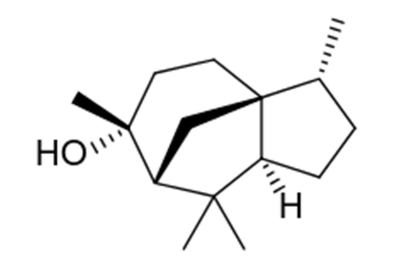
2.4. Identification of Potential Protein Targets for Cedrol

Cedrol has been reported to exhibit a number of biological activities, including anticancer [92,93,94] and anti-inflammatory. For example, cedrol was shown to have analgesic and anti-inflammatory effects in complete Freund’s adjuvant (CFA)-induced arthritis in rats [95] and in mice with collagen-induced arthritis (CIA) [96]. This sesquiterpene also attenuated neuropathic pain in chronic constriction injury of rats by inhibiting the inflammatory response [97]. Despite the various biological activities reported for cedrol, little is known about its specific cellular targets. Thus, we performed reverse-pharmacophore mapping on the molecular structures of (+)-cedrol to identify potential biological targets. PharmMapper was used to compare a large database of pharmacophore patterns with these compounds and generate target information, including normalized fitness scores and pharmacophoric characteristics. It is important to submit a compound to the PharmMapper server in the form of the proper optical isomer, as this methodology explicitly accounts for 3D structure of a molecule. Thus, we evaluated the (+)-configuration of cedrol, which is the most common enantiomer found in Juniper plants [89]. The results of PharmMapper analysis indicated that three kinases could be potential targets for cedrol: proviral integration Moloney virus kinase (PIM1), vascular endothelial growth factor receptor 2 (VEGFR2), and c-Jun N-terminal kinase 1 (JNK1) (Table 5). They are present among the 20 top-ranked targets found by PharmMapper.
We also calculated the most important physico-chemical parameters for cedrol using SwissADME [98] (Table 6 and Figure 6) and found that cedrol would be predicted to permeate the blood–brain barrier (BBB). According to the radar plot, the ADME characteristics of (+)-cedrol predict that is would exhibit high bioavailability.

3. Materials and Methods

3.1. Plant Material

J. sabina, J. pseudosabina, J. sibirica, J. pseudosabina subsp. turkestanica Kom., J. communis, and J. horizontalis are evergreen shrubs; J. seravschanica and J. scopolorum are evergreen trees. Leaves and cones were collected in 2019–2021 from mature shrubs and trees in several gorges of the Trans-Ili Alatau (Almaty region, South-Eastern Kazakhstan), Western Tien-Shan (Turkistan region, Southern Kazakhstan), Tarbagatai mountain range (Eastern Kazakhstan), and Bozeman (MT, USA) (Table 1). The collected samples were air-dried for 7–10 days at room temperature away from direct sunlight before hydrodistillation. Botanical identification of the plant material collected in Kazakhstan was performed by botanist Anna A. Ivashchenko from Ile-Alatau National Park, Almaty, Kazakhstan, who has an extensive experience in the taxonomical identification of Cupressaceae species. Botanical identification of the plant material collected in Montana was performed by botanist Robyn A. Klein from Montana State University, Bozeman, MT, USA.

3.2. Materials

Dimethyl sulfoxide (DMSO), N-formyl-Met-Leu-Phe (fMLF), Trp-Lys-Tyr-Val-Met (WKYMVM), and Histopaque 1077 were purchased from Sigma-Aldrich Chemical Co. (St. Louis, MO, USA). (+)-Cedrol was from TargetMol (Boston, MA, USA). n-Hexane was purchased from Merck (Darmstadt, Germany). Fluo-4AM was purchased from Invitrogen (Carlsbad, CA, USA). Roswell Park Memorial Institute (RPMI) 1640 medium and Dulbecco’s Modified Eagle’s Medium (DMEM):F12 medium were purchased from HyClone Laboratories (Logan, UT, USA). Fetal calf serum and fetal bovine serum were purchased from ATCC (Manassas, VA, USA). Hanks’ balanced salt solution (HBSS; 0.137 M NaCl, 5.4 mM KCl, 0.25 mM Na2HPO4, 0.44 mM KH2PO4, 4.2 mM NaHCO3, 5.56 mM glucose, and 10 mM HEPES, pH 7.4) was purchased from Life Technologies (Grand Island, NY, USA). HBSS without Ca2+ and Mg2+ is designated as HBSS; HBSS containing 1.3 mM CaCl2 and 1.0 mM MgSO4 is designated as HBSS+.

3.3. Essential Oil Extraction

Essential oils were obtained by hydrodistillation of dried plant material using a Clevenger-type apparatus, as previously described [86]. We used conditions accepted by the European Pharmacopoeia (European Directorate for the Quality of Medicines, Council of Europe, Strasbourg, France, 2014) to avoid artifacts. The essential oils collected in the graduated tube of the Clevenger-type apparatus were stored in amber vials and used for subsequent analyses. Yields were calculated based on the amount of air-dried plant material used. Stock solutions of the essential oils were prepared in DMSO (10 mg/mL) for biological evaluation and in n-hexane (10% w/v) for gas-chromatographic analysis.

3.4. Gas Chromatography-Mass Spectrometry (GC-MS) Analysis

GC-MS analysis was performed with an Agilent 5975 GC-MSD system (Agilent Technologies, Santa Clara, CA, USA), as reported previously [99]. An Agilent Innowax FSC column (60 m × 0.25 mm, 0.25 μm film thickness) was used with He as the carrier gas (0.8 mL/min). The GC oven temperature was kept at 60 °C for 10 min, increased to 220 °C at a rate of 4 °C/min, kept constant at 220 °C for 10 min, and then increased to 240 °C at a rate of 1 °C/min. The split ratio was adjusted to 40:1, and the injector temperature was 250 °C. MS spectra were monitored at 70 eV with a mass range of 35 to 450 m/z. GC analysis was carried out using an Agilent 6890N GC system. To obtain the same elution order as with GC-MS, the line was split for FID and MS detectors, and a single injection was performed using the same column and appropriate operational conditions. Flame ionization detector (FID) temperature was 300 °C. The essential oil components were identified by co-injection with standards (whenever possible), which were purchased from commercial sources or isolated from natural sources. In addition, compound identities were confirmed by comparison of their mass spectra with those in the Wiley GC/MS Library (Wiley, NY, USA), MassFinder software 4.0 (Dr. Hochmuth Scientific Consulting, Hamburg, Germany), Adams Library, and NIST Library. Confirmation was also achieved using the in-house “Başer Library of Essential Oil Constituents” database, obtained from chromatographic runs of pure compounds performed with the same equipment and conditions. A C8–C40 n-alkane standard solution (Fluka, Buchs, Switzerland) was used to spike the samples for the determination of relative retention indices (RRI). Relative percentage amounts of the separated compounds were calculated from the FID chromatograms.

3.5. Isolation of Human Neutrophils

For isolation of human neutrophils, blood was collected from healthy donors in accordance with a protocol approved by the Institutional Review Board at Montana State University (Protocol #MQ041017). Neutrophils were purified from the blood using dextran sedimentation, followed by Histopaque 1077 gradient separation and hypotonic lysis of red blood cells, as described previously [100]. Isolated neutrophils were washed twice and resuspended in HBSS. Neutrophil preparations were routinely >95% pure, as determined by light microscopy, and >98% viable, as determined by trypan blue exclusion. Neutrophils were obtained from multiple different donors (n = 8); however, the cells from different donors were never pooled during experiments.

3.6. Cell Culture

Human promyelocytic leukemia HL60 cells stably transfected with FPR1 (FPR1-HL60 cells) or FPR2 (FPR2-HL60 cells) (kind gifts from Dr. Marie-Josephe Rabiet, INSERM, Grenoble, France) were cultured in RPMI-1640 medium supplemented with 10% heat-inactivated fetal calf serum, 10 mM HEPES, 100 μg/mL streptomycin, 100 U/mL penicillin, and G418 (1 mg/mL). G418 was removed in the last round of culture before assays were performed.

3.7. Ca2+ Mobilization Assay

Changes in intracellular Ca2+ concentrations ([Ca2+]i) were measured with a FlexStation 3 scanning fluorometer (Molecular Devices, Sunnyvale, CA, USA). Briefly, human neutrophils were suspended in HBSS-, loaded with Fluo-4AM at a final concentration of 1.25 μg/mL, and incubated for 30 min in the dark at 37 °C. After dye loading, the cells were washed with HBSS-, resuspended in HBSS+, separated into aliquots, and loaded into the wells of flat-bottom, half-area well black microtiter plates (2 × 10⁵ cells/well). To assess the direct effects of test compound or pure essential oils on Ca2+ flux, the compound/oil was added to the wells (final concentration of DMSO was 1%), and changes in fluorescence were monitored (λex = 485 nm, λem = 538 nm) every 5 s for 240 s at room temperature after addition of the test compound. To evaluate inhibitory effects of the compounds on FPR1/FPR2-dependent Ca2+ flux, the compound/oil was added to the wells (final concentration of DMSO was 1%) with cells (human neutrophils or FPR1/FPR2 HL60 cells). The samples were preincubated for 10 min, followed by addition of 5 nM fMLF (for human neutrophils or FPR1-HL60 cells) or 5 nM WKYMVM (for FPR2-HL60 cells). The maximum change in fluorescence, expressed in arbitrary units over baseline, was used to determine the agonist response. Responses were normalized to the response induced by 5 nM fMLF or 5 nM WKYMVM, which were assigned as 100%. Curve fitting (at least five or six points) and calculation of median effective concentration values (EC50 or IC50) were performed by nonlinear regression analysis of the dose–response curves generated using Prism 9 (GraphPad Software, Inc., San Diego, CA, USA).

3.8. Chemotaxis Assay

Human neutrophils were resuspended in HBSS+ containing 2% (v/v) heat-inactivated fetal bovine serum (2 × 10⁶ cells/mL), and chemotaxis was analyzed in 96-well ChemoTx chemotaxis chambers (Neuroprobe, Gaithersburg, MD, USA). In brief, neutrophils were preincubated with the indicated concentrations of the test sample (essential oil or pure compound) or DMSO (1% final concentration) for 30 min at room temperature and added to the upper wells of the ChemoTx chemotaxis chambers. The lower wells were loaded with 30 µL of HBSS+ containing 2% (v/v) fetal bovine serum and the indicated concentrations of test sample, DMSO (negative control), or 1 nM fMLF as a positive control. Neutrophils were added to the upper wells and allowed to migrate through the 5.0-µm pore polycarbonate membrane filter for 60 min at 37 °C and 5% CO2. The number of migrated cells was determined by measuring ATP in lysates of transmigrated cells using a luminescence-based assay (CellTiter-Glo; Promega, Madison, WI, USA), and luminescence measurements were converted to absolute cell numbers by comparison of the values with standard curves obtained with known numbers of neutrophils. Curve fitting (at least eight to nine points) and calculation of median effective concentration values (IC50) were performed by nonlinear regression analysis of the dose-response curves generated using GraphPad Prism 9.

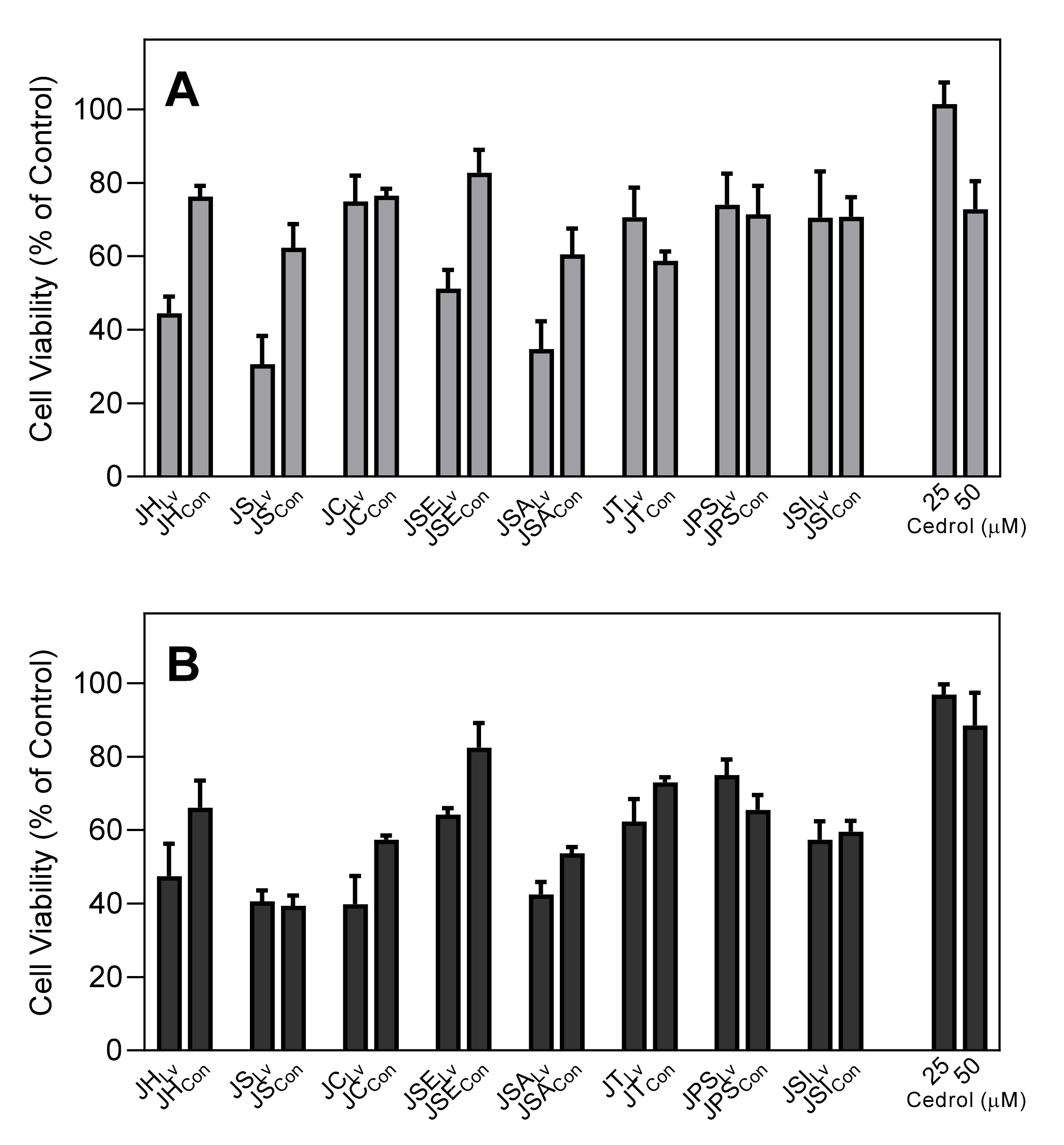
3.9. Cytotoxicity Assay

Cytotoxicity of essential oils and pure compounds in HL60 monocytic cells was analyzed with a CellTiter-Glo Luminescent Cell Viability Assay Kit (Promega), according to the manufacturer’s protocol. Briefly, HL60 cells were cultured at a density of 104 cells/well with different concentrations of essential oil or compound (final concentration of DMSO was 1%) for 30 min or 2 h at 37 °C and 5% CO2. Following treatment, substrate was added to the cells, and the samples were analyzed with a Fluoroscan Ascent FL microplate reader.

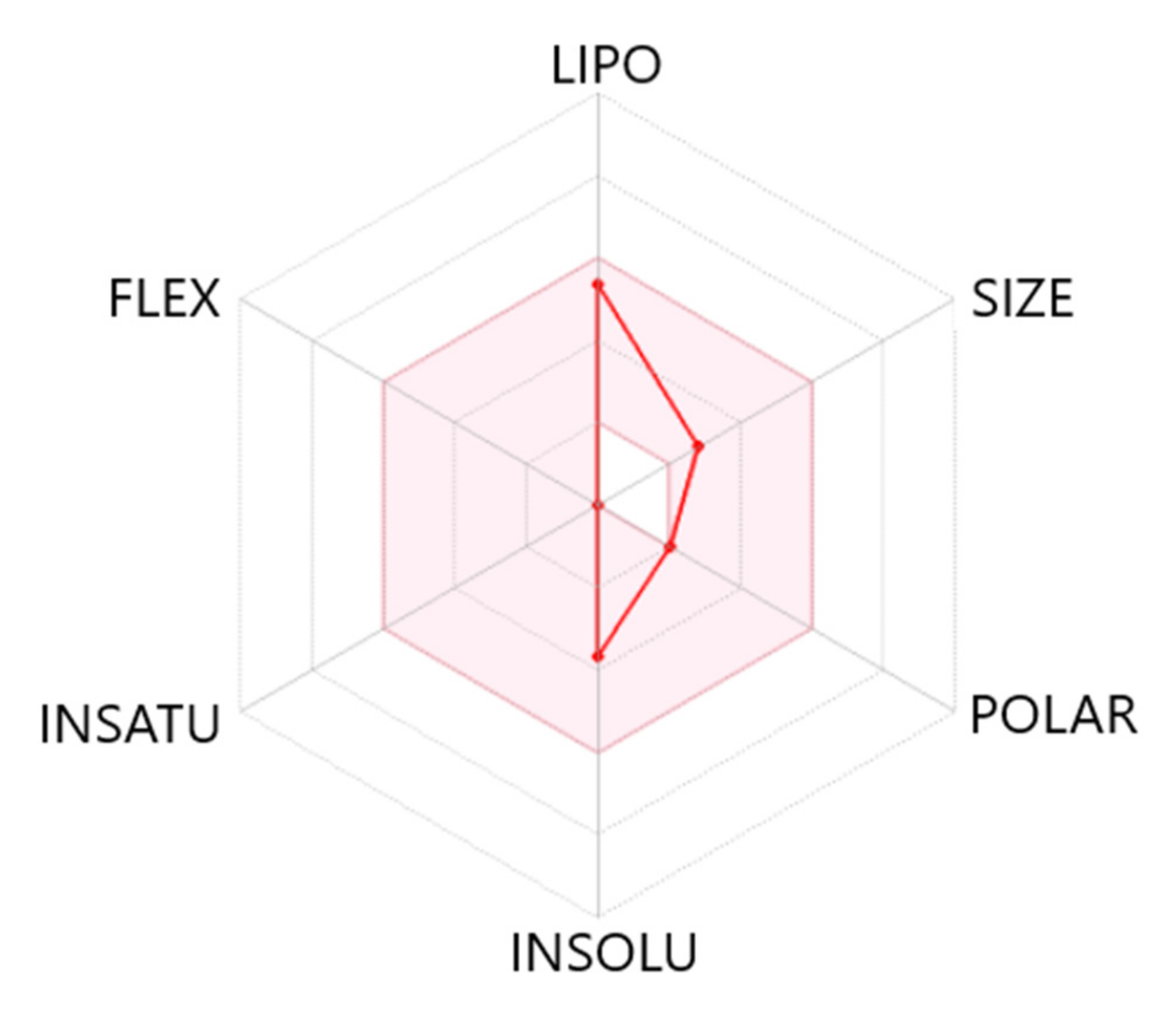
3.10. Molecular Modeling

The PharmMapper Server [101] was used for identifying the protein targets for (+)-cedrol. For a given small molecule, PharmMapper recognizes potential target possibilities using an “invert” pharmacophore mapping methodology. In several reference databases which are incorporated in the software, the protein biotargets are represented by sets of pharmacophore points that provide faster mapping. The PubChem database (https://pubchem.ncbi.nlm.nih.gov; accessed on 20 September 2021) was used as a source of initial 3D structures of the investigated compounds. The structure of (+)-cedrol (CID: 65575) was downloaded from PubChem in SDF format and further uploaded into the PharmMapper web server. Up to 300 conformers of each compound were automatically generated using a corresponding option of the software. The pharmacophore mapping was performed with the “Human Protein Targets Only” database containing 2241 targets. The top 250 potential targets per compound were retrieved and sorted by the normalized fit score. The physicochemical properties of (+)-cedrol were computed using SwissADME (http://www.swissadme.ch; accessed on 20 September 2021).

3.11. Statistical Analysis

One-way analysis of variance (ANOVA) was performed on the data sets, followed by Tukey’s pair-wise comparisons. Pair-wise comparisons with differences at p < 0.05 were considered to be statistically significant.

4. Conclusions

Compositional analysis of 16 essential oils isolated from Juniper plants revealed similarities and differences between our analyses and those previous reported for essential oils from this species. Notably, our studies represent the first time that essential oils isolated from the cones of many of these Juniper species have been analyzed, including cones from J. sabina (JSACon), J. pseudosabina (JPSCon), and J. pseudosabina subsp. turkestanica (JTCon). Interestingly, several of the essential oil samples from Juniper collected in Kazakhstan but not in Montana contained high levels of cedrol, which was fairly unique among the samples and was enriched in JSE, JSA, and JPS. Evaluation of the biological activities of the parent essential oils or pure cedrol itself showed that they induced intracellular Ca2+ mobilization in human neutrophils, which is a key component of neutrophil activation. Indeed, pretreatment of cells with these essential oils or cedrol inhibited subsequent heterologous agonist-induced Ca2+ mobilization, which is a common feature of many neutrophil agonists. Likewise, pretreatment with JSECon (16.8% cedrol content) or pure cedrol inhibited human neutrophil chemotaxis toward N-formyl peptide. Thus, these data suggest that cedrol is a neutrophil agonist that can desensitize cells to subsequent stimulation by N-formyl peptide. However, further studies are necessary to define the precise mechanisms involved in the anti-inflammatory properties of cedrol.

Supplementary Materials

The following are available online at Supplementary Table S1: Chemical composition of essential oils (%) isolated from leaves and cones of different Juniperus species.

Author Contributions

G.Ö., I.A.S. and M.T.Q. conceived and designed the project; I.A.S., L.N.K., M.Y., S.S.A. and S.I.A. collected plant material and isolated essential oils; G.Ö., I.A.S., T.Ö. and L.N.K. performed the experiments; A.I.K. conducted molecular modeling; G.Ö., I.A.S., T.Ö., L.N.K. and A.I.K. analyzed and interpreted the data; G.Ö., I.A.S., M.Y., A.I.K. and M.T.Q. drafted and revised the manuscript. All authors have read and agreed to the published version of the manuscript.

Funding

Plant material was collected with support of grants AP09259027 (2021–2023) and AP05131621 (2018–2020) from the Ministry of Education and Science of the Republic of Kazakhstan. This research was supported in part by National Institutes of Health IDeA Program Grants GM115371 and GM103474; USDA National Institute of Food and Agriculture Hatch project 1009546; the Montana State University Agricultural Experiment Station, and the Tomsk Polytechnic University Development Program.

Institutional Review Board Statement

The study was conducted according to the guidelines of the Declaration of Helsinki and approved by the Montana State University Institutional Review Board (protocol MQ041017, approved 4 January 2017).

Informed Consent Statement

Informed consent was obtained from all subjects involved in the study.

Data Availability Statement

The data that support the findings of this study are available from the authors upon reasonable request.

Conflicts of Interest

The authors declare no competing financial interest.

Sample Availability

Samples of the essential oils are available from the authors.

Abrreviations

JHLv, J. horizontalis leaves; JHCon, J. horizontalis cones; JSLv, J. scopolorum leaves; JSCon, J. scopolorum cones; JCLv, J. communis leaves; JCCon, J. communis cones; JSELv, J. seravschanica leaves; JSECon, J. seravschanica cones; JSALv, J. sabina leaves; JSACon, J. sabina cones; JTLv; J. turkestanica leaves; JTCon, J. turkestanica cones; JPSLv, J. pseudosabina leaves; JPSCon, J. pseudosabina cones; JSILv, J. sibirica leaves; JSICon, J. sibirica cones.

References

  1. Adams, R.P. Junipers of the World: The Genus Juniperus, 4th ed.; Trafford: Victoria, BC, USA, 2014; p. 422. [Google Scholar]
  2. Adams, R.P.; Schwarzbach, A.E. Taxonomy of Juniperus section Juniperus: Sequence analysis of nrDNA and five cpDNA regions. Phytologia 2012, 94, 280–297. [Google Scholar]
  3. Adams, R.P.; Demeke, T. Systematic Relationships in Juniperus Based on Random Amplified Polymorphic Dnas (Rapds). Taxon 1993, 42, 553–571. [Google Scholar] [CrossRef]
  4. Mao, K.S.; Hao, G.; Liu, J.Q.; Adams, R.P.; Milne, R.I. Diversification and biogeography of Juniperus (Cupressaceae): Variable diversification rates and multiple intercontinental dispersals. New Phytol. 2010, 188, 254–272. [Google Scholar] [CrossRef] [PubMed]
  5. Farjon, A.A. Handbook of the World’s Conifers; Koninklijke Brill NV: Leiden, The Netherlands, 2017; Volume 2. [Google Scholar]
  6. Shrestha, I.; Kunwar, R.; Hussain, W.; Jan, H.; Abbasi, A.; Bussmann, R.; Zambrana, N. Juniperus communis L. Juniperus excelsa M. Bieb. Juniperus indica Bertol. Juniperus pseudosabina var. turkestanica (Kom.) Silba Juniperus recurva Buch.-Ham. ex D. Don Juniperus sibirica Burgsd. Juniperus squamata Buch.-Ham. ex D. Don. Cupressaceae. In Ethnobotany of the Himalayas; Springer: Cham, Switzerland, 2021; pp. 1–14. [Google Scholar] [CrossRef]
  7. Thorne, R.F. Major disjunctions in the geographic ranges of seed plants. Q. Rev. Biol. 1972, 47, 365–411. [Google Scholar] [CrossRef]
  8. Farjon, A. A Monograph of Cupressaceae and Sciadopitys; Royal Botanic Gardens: Kew, UK, 2005. [Google Scholar]
  9. Demidovskaya, L.F.; Averina, V.Y.; Egeubaeva, A.L. Junipers of South Kazakhstan are perspective essential-oil plants. Proc. Inst. Bot. Acad. Sci. KazSSR. Alma-Ata 1976, 35, 49–57. [Google Scholar]
  10. Mirzagalieva, A.B.; Medeubaeva, B.Z. Chemical composition and biological activity of essential oil of Juniperus sabina from the Flora of Eastern Kazakhstan. Eurasian Sci. Coop. 2017, 9, 30–34. (In Russian) [Google Scholar]
  11. Lesica, P.; Atthowe, H.E.; Dugan, F.M. Incidence of Perenniporia fraxinophila and its effects on green ash (Fraxinus pennsylvanica) woodlands in eastern Montana, USA. For. Ecol. Manag. 2003, 182, 153–159. [Google Scholar] [CrossRef]
  12. Adams, R.P.; Turuspekov, Y. Taxonomic reassessment of some central Asian and Himalayan scale-leaved taxa of Juniperus (Cupressaceae) supported by random amplification of polymorphic DNA. Taxon 1998, 47, 75–83. [Google Scholar] [CrossRef]
  13. Ivashchenko, A.A.; Levchenko, R.A.; Zhaparova, N.K. The Red Book of Kazakhstan (Plants); ArtPrintXXI LLP: Astana, Kazakhstan, 2014. [Google Scholar]
  14. Pavlov, N.V. Flora of Kazakhstan; Nauka: Alma-Ata, USSR, 1956; pp. 71–76. [Google Scholar]
  15. Abdulina, S.A. Checklist of Vascular Plants of Kazakhstan; Academy of Sciences: Almaty, Kazakhstan, 1999. [Google Scholar]
  16. Kotukhov, Y.A.; Danilova, A.N.; Anufrieva, O.A. State of populations of Daurian juniper (Juniperus davurica Pall.) in East Kazakhstan. Bot. Res. Sib. Kazakhstan 2019, 15, 143–147. [Google Scholar]
  17. Ivashchenko, A.A.; Kotukhov, Y.A.; Utebekov, K.I. Flora of the forest belt of the Chindagatui mountain range (Flora lesnogo poyasa Chindagatuyskogo gornogo massiva). Bot. Res. Sib. Kazakhstan 2014, 20, 250–264. [Google Scholar]
  18. Ivashchenko, A.A. Materials to the flora of the Ile-Alatau National Park: Higher sporous and gymnosperms. KazNU Bull. Biol. Ser. 2015, 24, 28–35. [Google Scholar]
  19. Ivashchenko, A.A. The flora Sairam-Ugam State National Natural Park (Kazakhstan). Bot. Res. Sib. Kazakhstan 2020, 26, 52–63. [Google Scholar]
  20. Pateiro, M.; Barba, F.J.; Dominguez, R.; Sant’Ana, A.S.; Mousavi Khaneghah, A.; Gavahian, M.; Gomez, B.; Lorenzo, J.M. Essential oils as natural additives to prevent oxidation reactions in meat and meat products: A review. Food Res. Int. 2018, 113, 156–166. [Google Scholar] [CrossRef] [PubMed]
  21. Barreca, S.; La Bella, S.; Maggio, A.; Licata, M.; Buscemi, S.; Leto, C.; Pace, A.; Tuttolomondo, T. Flavouring Extra-Virgin Olive Oil with Aromatic and Medicinal Plants Essential Oils Stabilizes Oleic Acid Composition during Photo-Oxidative Stress. Agriculture 2021, 11, 266. [Google Scholar] [CrossRef]
  22. Kustova, S.D. Handbook of Essential Oils; Food Industry: Moscow, Russia, 1978; p. 175. [Google Scholar]
  23. Myrzagalieva, A.B.; Medeubaeva, B.Z. To the study of essential oil content of representatives of the family Cupressaceae Bartl. of Eastern Kazakhstan’s flora. Fundam. Res. 2014, 5, 1021–1024. [Google Scholar]
  24. Grigoryev, V.P.; Poplavskaya, L.F. Phytomass reserves of Juniperus communis L. and the content of chlorophylls, carotenoids and ascorbic acid in needles. Plant Resour. 1985, 2, 164–169. [Google Scholar]
  25. Klyshev, L.K.; Bandyukova, V.A.; Alyukina, L.S. Plant Flavonoids; Nauka: Alma-Ata, USSR, 1978. [Google Scholar]
  26. Kyosev, P.A. Complete Reference Book of Medicinal Plants; EKSMO-Press: Moscow, Russia, 2001. [Google Scholar]
  27. Motti, R. Wild plants used as herbs and spices in Italy: An ethnobotanical review. Plants 2021, 10, 563. [Google Scholar] [CrossRef]
  28. Tavares, W.R.; Seca, A.M. The current status of the pharmaceutical potential of Juniperus L. metabolites. Medicines 2018, 5, 81. [Google Scholar] [CrossRef] [PubMed] [Green Version]
  29. Falcão, S.; Bacém, I.; Igrejas, G.; Rodrigues, P.J.; Vilas-Boas, M.; Amaral, J.S. Chemical composition and antimicrobial activity of hydrodistilled oil from juniper berries. Ind. Crop. Prod. 2018, 124, 878–884. [Google Scholar] [CrossRef] [Green Version]
  30. Bussmann, R.W.; Batsatsashvili, K.; Kikvidze, Z.; Paniagua-Zambrana, N.Y.; Khutsishvili, M.; Maisaia, I.; Sikharulidze, S.; Tchelidze, D. Juniperus communis L. Juniperus depressa Raf. Juniperus hemisphaerica J. Presl & C. Presl Juniperus oblonga M. Bieb. Juniperus sabina L. Cupressaceae. In Ethnobotany of the Mountain Regions of Far Eastern Europe: Ural, Northern Caucasus, Turkey, and Iran; Springer Nature: Cham, Switzerland, 2020; pp. 523–532. [Google Scholar]
  31. Salamon, I.; Tarawneh, A. Ethnobotany of common juniper (Juniperus communis L.) in Slovakia. In Proceedings of the II International Symposium on Beverage Crops, Xi’An, China, 22–25 October 2018; Volume 1274, pp. 167–171. [Google Scholar]
  32. Dorjey, K.; Maurya, A.K. Ethnobotany of Juniperus polycarpos C. Koch (Cupressaceae) in the Himalayan cold desert of Union Territory of Ladakh, India. Indian J. Tradit. Knowl. 2021, 20, 83–90. [Google Scholar]
  33. Rashid, A.; Kuchay, R.A.H. A review on phyto-pharmacological potentials of Juniperus recurva. J. Exp. Integr. Med. 2016, 6, 93–97. [Google Scholar] [CrossRef] [Green Version]
  34. Monograph. European Pharmacopoeia. In European Directorate for the Quality of Medicine & Health Care of the Council of Europe (EDQM); Council of Europe: Strasbourg, France, 2016; Volume 1, pp. 1404–1405. [Google Scholar]
  35. USP; NF. The United States Pharmacopeia. In The United States Pharmacopeia. The National Formulary; The United States Pharmacopeial Convention; United Book Press, Inc.: Baltimore, MD, USA, 2013; Volume 3, p. 4031. [Google Scholar]
  36. Polish Pharmacopoeia Commission. Polish National Pharmacopoeia, 5th ed.; Office for Registration of Medicinal Products, Medical Devices & Biocidal Products: Warsaw, Poland, 1999.
  37. German Pharmacopoeia. Deutsches Arzneibuch; Deutscher Apotheker Verlag: Gerlingen, Germany, 1998. [Google Scholar]
  38. Sweetman, S.C. Supplementary Drugs and Other Substances. Red Cedar, 36th ed.; Pharmaceutical Press: London, UK, 2009; p. 2278. [Google Scholar]
  39. Adams, R.P.; Shatar, S.; Dembitsky, A. Comparison of the volatile leaf oils of Juniperus davurica Pall. from Mongolia, with plants cultivated in Kazakhstan, Russia and Scotland. J. Essent. Oil Res. 1994, 6, 217–221. [Google Scholar] [CrossRef]
  40. Adams, R.P.; Palma, M.M.; Moore, W.S. Volatile oils of mature and juvenile leaves of Juniperus horizontalis: Chemosystematic significance. Phytochemistry 1981, 20, 2501–2502. [Google Scholar] [CrossRef]
  41. Adams, R.P.; Nguyen, S.; Liu, J. Geographic Variation in the Leaf Essential Oils of Juniperus sabina L. and J. sabina var. arenaria (EH Wilson) Farjon. J. Essent. Oil Res. 2006, 18, 497–502. [Google Scholar] [CrossRef]
  42. Adams, R.P.; Hojjati, F. Leaf essential oils of Juniperus in central and southern Iran. Phytologia 2013, 95, 288–295. [Google Scholar]
  43. Adams, R.P.; Dembitsky, A.; Shatar, S. The leaf essential oils and taxonomy of Juniperus centrasiatica Kom., J. jarkendensis Kom., J. pseudosabina Fisch., Mey. & Ave-Lall., J. sabina L. and J. turkestanica Kom. from Central Asia. J. Essent. Oil Res. 1998, 10, 489–496. [Google Scholar]
  44. Adams, R.P. The leaf essential oil of Juniperus maritima compared with J. horizontalis, J. scopulorum and J. virginiana oils. Phytologia 2009, 91, 31–39. [Google Scholar]
  45. Adams, R.P. Systematics of multi-seeded eastern hemisphere Juniperus based on leaf essential oils and RAPD DNA fingerprinting. Biochem. Syst. Ecol. 1999, 27, 709–725. [Google Scholar] [CrossRef]
  46. Mirzagalieva, A.B.; Medeubaeva, B.Z. Investigation of essential oil bearing representatives of Cupressaceae family from the Flora of the Eastern Kazakhstan. Fundam. Res. 2014, 5, 1021–1024. [Google Scholar]
  47. Goryaev, M.; Dzhalilov, D. The essential oil of needles of Turkestan Juniper (Juniperus turkestanica). Izv. Akad. Nauk. Kaz. SSR (Ser. Him.) 1960, 2, 107–113. [Google Scholar]
  48. Sampietro, D.A.; Gomez, A.d.l.A.; Jimenez, C.M.; Lizarraga, E.F.; Ibatayev, Z.A.; Suleimen, Y.M.; Catalán, C.A. Chemical composition and antifungal activity of essential oils from medicinal plants of Kazakhstan. Nat. Prod. Res. 2017, 31, 1464–1467. [Google Scholar] [CrossRef] [PubMed]
  49. Suleimen, Y.M.; Ishmuratova, M.; Iskakova, J.B. Comprehensive phytochemical study of Juniperus sabina L. from Kazakhstan. Natural and Mathematical Sciences in the Modern World (in Russian) 2013, 12. [Google Scholar]
  50. Alimtay, G.A.; Aidarbayeva, D.K. The current state of useful plants in the Trans-Ili Alatau (Aksai and Kaskelen gorges). Ann. D’italia 2020, 6, 3–5. [Google Scholar]
  51. Ghorbanzadeh, A.; Ghasemnezhad, A.; Sarmast, M.K.; Ebrahimi, S.N. An analysis of variations in morphological characteristics, essential oil content, and genetic sequencing among and within major Iranian Juniper (Juniperus spp.) populations. Phytochemistry 2021, 186, 112737. [Google Scholar] [CrossRef] [PubMed]
  52. Ložienė, K.; Venskutonis, P.R. Juniper (Juniperus communis L.) oils. In Essential Oils in Food Preservation, Flavor and Safety; Elsevier: Amsterdam, The Netherlands, 2016; pp. 495–500. [Google Scholar]
  53. Majewska, E.; Kozłowska, M.; Kowalska, D.; Gruczynska, E. Characterization of the essential oil from cone-berries of Juniperus communis L. (Cupressaceae). Herba Pol. 2017, 63, 48–55. [Google Scholar] [CrossRef] [Green Version]
  54. Labokas, J.; Ložienė, K. Variation of essential oil yield and relative amounts of enantiomers of α-pinene in leaves and unripe cones of Juniperus communis L. growing wild in Lithuania. J. Essent. Oil Res. 2013, 25, 244–250. [Google Scholar] [CrossRef]
  55. Yaglioglu, A.S.; Eser, F.; Yaglioglu, M.S.; Demirtas, I. The antiproliferative and antioxidant activities of the essential oils of Juniperus species from Turkey. Flavour Fragr. J. 2020, 35, 511–523. [Google Scholar] [CrossRef]
  56. Adams, R.P. Systematics of the one seeded Juniperus of the eastern hemisphere based on leaf essential oils and random amplified polymorphic DNAs (RAPDs). Biochem. Syst. Ecol. 2000, 28, 529–543. [Google Scholar] [CrossRef]
  57. Shanjani, P.S.; Mirza, M. Seasonal variation of the leaf and cone oil of Juniperus excelsa MB. J. Med. Plants 2006, 5, 50–58. [Google Scholar]
  58. Semerdjieva, I.B.; Zheljazkov, V.D.; Dincheva, I.; Astatkie, T.; Kačániová, M. Chemotypes of Juniperus oxycedrus in Bulgaria and the antimicrobial activity of galbuli essential oils. Ind. Crop. Prod. 2020, 158, 113005. [Google Scholar] [CrossRef]
  59. Semerdjieva, I.B.; Shiwakoti, S.; Cantrell, C.L.; Zheljazkov, V.D.; Astatkie, T.; Schlegel, V.; Radoukova, T. Hydrodistillation extraction kinetics regression models for essential oil yield and composition in Juniperus virginiana, J. excelsa, and J. sabina. Molecules 2019, 24, 986. [Google Scholar] [CrossRef] [PubMed] [Green Version]
  60. Zheljazkov, V.D.; Cantrell, C.L.; Semerdjieva, I.; Radoukova, T.; Stoyanova, A.; Maneva, V.; Kačániová, M.; Astatkie, T.; Borisova, D.; Dincheva, I. Essential Oil Composition and Bioactivity of Two Juniper Species from Bulgaria and Slovakia. Molecules 2021, 26, 3659. [Google Scholar] [CrossRef] [PubMed]
  61. Venditti, A.; Maggi, F.; Saab, A.; Bramucci, M.; Quassinti, L.; Petrelli, D.; Vitali, L.; Lupidi, G.; El Samrani, A.; Borgatti, M. Antiproliferative, antimicrobial and antioxidant properties of Cedrus libani and Pinus pinea wood oils and Juniperus excelsa berry oil. Plant Biosyst. Int. J. Deal. All Asp. Plant Biol. 2020, 1–12. [Google Scholar] [CrossRef]
  62. Eryiğit, T.; Okut, N.; Ekici, K.; Yildirim, B. Chemical composition and antibacterial activities of Juniperus horizontalis essential oil. Can. J. Plant Sci. 2014, 94, 323–327. [Google Scholar] [CrossRef]
  63. Cantrell, C.L.; Zheljazkov, V.D.; Carvalho, C.R.; Astatkie, T.; Jeliazkova, E.A.; Rosa, L.H. Dual extraction of essential oil and podophyllotoxin from creeping juniper (Juniperus horizontalis). PLoS ONE 2014, 9, e106057. [Google Scholar]
  64. Elshafie, H.; Caputo, L.; De Martino, L.; Gruľová, D.; Zheljazkov, V.; De Feo, V.; Camele, I. Biological investigations of essential oils extracted from three Juniperus species and evaluation of their antimicrobial, antioxidant and cytotoxic activities. J. Appl. Microbiol. 2020, 129, 1261–1271. [Google Scholar] [CrossRef] [PubMed]
  65. Valentini, G.; Bellomaria, B.; Maggi, F.; Manzi, A. The leaf and female cone oils of Juniperus oxycedrus L. ssp. oxycedrus and J. oxycedrus ssp. macrocarpa (Sibth. et Sm.) Ball. from Abruzzo. J. Essent. Oil Res. 2003, 15, 418–421. [Google Scholar] [CrossRef]
  66. Hayta, S.; Bagci, E. Essential oil constituents of the leaves, bark and cones of Juniperus oxycedrus subsp. oxycedrus L. from Turkey. Acta Bot. Gall. 2014, 161, 201–207. [Google Scholar] [CrossRef]
  67. Fadel, H.; Benayache, F.; Chalchat, J.-C.; Figueredo, G.; Chalard, P.; Hazmoune, H.; Benayache, S. Essential oil constituents of Juniperus oxycedrus L. and Cupressus sempervirens L. (Cupressaceae) growing in Aures region of Algeria. Nat. Prod. Res. 2019, 35, 2616–2620. [Google Scholar] [CrossRef]
  68. Meriem, A.; Msaada, K.; Sebai, E.; Aidi Wannes, W.; Salah Abbassi, M.; Akkari, H. Antioxidant, anthelmintic and antibacterial activities of red juniper (Juniperus phoenicea L.) essential oil. J. Essent. Oil Res. 2021, 1–10. [Google Scholar] [CrossRef]
  69. Moghaddam, M.; Ghasemi Pirbalouti, A.; Farhadi, N. Seasonal variation in Juniperus polycarpos var. turcomanica essential oil from northeast of Iran. J. Essent. Oil Res. 2018, 30, 225–231. [Google Scholar] [CrossRef]
  70. Mehdizadeh, L.; Taheri, P.; Pirbalouti, A.G.; Moghaddam, M. Phytotoxicity and antifungal properties of the essential oil from the Juniperus polycarpos var. turcomanica (B. Fedsch.) RP Adams leaves. Physiol. Mol. Biol. Plants 2020, 26, 759. [Google Scholar] [CrossRef] [PubMed]
  71. Meng, X.; Li, D.; Zhou, D.; Wang, D.; Liu, Q.; Fan, S. Chemical composition, antibacterial activity and related mechanism of the essential oil from the leaves of Juniperus rigida Sieb. et Zucc against Klebsiella pneumoniae. J. Ethnopharmacol. 2016, 194, 698–705. [Google Scholar] [CrossRef]
  72. Samsonova, N.A.; Gusakova, M.A.; Bogolitsyn, K.G.; Selivanova, N.V. Chemical composition and antibacterial activity of woody greenery essential oil Juniperus communis L. from the subarctic zone of Russia. Sib. For. J. 2020, 2, 31. [Google Scholar]
  73. Basher, K.; Kurkcuoglu, M.; Demirci, B.; Gusakova, S.; Sagdullaev, S.S.; Maltzev, I.; Aripov, K.N. Essential oil and lipids from the cone berries of Juniperus seravschanica. Chem. Nat. Compd. 1999, 35, 397–400. [Google Scholar] [CrossRef]
  74. Caramiello, R.; Bocco, A.; Buffa, G.; Maffei, M. Chemotaxonomy of Juniperus communis, J. sibirica and J. intermedia. J. Essent. Oil Res. 1995, 7, 133–145. [Google Scholar] [CrossRef]
  75. Efremov, E.; Zykova, I.; Efremov, A.; Strukova, E. The composition of the essential oil from raw material and cones of Juniperus sibirica from Evenk region. Russ. J. Bioorganic Chem. 2012, 38, 790–795. [Google Scholar] [CrossRef]
  76. Nikolić, B.; Vasilijević, B.; Mitić-Ćulafić, D.; Lesjak, M.; Vuković-Gačić, B.; Dukić, N.M.; Knežević-Vukčević, J. Screening of the antibacterial effect of Juniperus sibirica and Juniperus sabina essential oils in a microtitre plate-based MIC assay. Bot. Serbica 2016, 40, 43–48. [Google Scholar]
  77. Adams, R.P.; Mumba, L.E.; James, S.A.; Pandey, R.N.; Gauquelin, T.; Badri, W. Geographic variation in the leaf oils and DNA fingerprints (RAPDs) of Juniperus thurifera L. from Morocco and Europe. J. Essent. Oil Res. 2003, 15, 148–154. [Google Scholar] [CrossRef]
  78. Kalaba, V.; Marjanović-Balaban, Ž.; Kalaba, D.; Lazić, D.; Cvjetković, V.G. Antibacterial activity of essential oil of Juniperus communis L. Qual. Life 2020, 18, 1–2. [Google Scholar] [CrossRef]
  79. Adams, R.P. Investigation of Juniperus species of the United States for new sources of cedarwood oil. Econ. Bot. 1987, 41, 48–54. [Google Scholar] [CrossRef]
  80. Zheljazkov, V.D.; Astatkie, T.; Jeliazkova, E.A.; Schlegel, V. Distillation time alters essential oil yield, composition, and antioxidant activity of male Juniperus scopulorum trees. J. Oleo Sci. 2012, 61, 537–546. [Google Scholar] [CrossRef] [PubMed]
  81. Zheljazkov, V.D.; Astatkie, T.; Jeliazkova, E.A.; Tatman, A.O.; Schlegel, V. Distillation time alters essential oil yield, composition and antioxidant activity of female Juniperus scopulorum trees. J. Essent. Oil Res. 2013, 25, 62–69. [Google Scholar] [CrossRef]
  82. Adams, R.P. The co-occurrence and systematic significance of pregeijerene B and 8-alpha-acetoxyelemol in Juniperus. Biochem. Syst. Ecol. 2004, 32, 559–563. [Google Scholar] [CrossRef]
  83. Adams, R.P. Systematics of smooth leaf margin Juniperus of the western hemisphere based on leaf essential oils and RAPD DNA fingerprinting. Biochem. Syst. Ecol. 2000, 28, 149–162. [Google Scholar] [CrossRef]
  84. Beutler, B. Innate immunity: An overview. Mol. Immunol. 2004, 40, 845–859. [Google Scholar] [CrossRef] [PubMed]
  85. Malech, H.L.; DeLeo, F.R.; Quinn, M.T. The Role of Neutrophils in the Immune System: An Overview. Methods Mol. Biol. 2020, 2087, 3–10. [Google Scholar] [CrossRef] [PubMed]
  86. Schepetkin, I.A.; Kushnarenko, S.V.; Ozek, G.; Kirpotina, L.N.; Utegenova, G.A.; Kotukhov, Y.A.; Danilova, A.N.; Ozek, T.; Baser, K.H.; Quinn, M.T. Inhibition of Human Neutrophil Responses by the Essential Oil of Artemisia kotuchovii and Its Constituents. J. Agric. Food Chem. 2015, 63, 4999–5007. [Google Scholar] [CrossRef] [PubMed] [Green Version]
  87. Schepetkin, I.A.; Kushnarenko, S.V.; Ozek, G.; Kirpotina, L.N.; Sinharoy, P.; Utegenova, G.A.; Abidkulova, K.T.; Ozek, T.; Baser, K.H.; Kovrizhina, A.R.; et al. Modulation of Human Neutrophil Responses by the Essential Oils from Ferula akitschkensis and Their Constituents. J. Agric. Food Chem. 2016, 64, 7156–7170. [Google Scholar] [CrossRef] [PubMed] [Green Version]
  88. Ozek, G.; Schepetkin, I.A.; Utegenova, G.A.; Kirpotina, L.N.; Andrei, S.R.; Ozek, T.; Baser, K.H.C.; Abidkulova, K.T.; Kushnarenko, S.V.; Khlebnikov, A.I.; et al. Chemical composition and phagocyte immunomodulatory activity of Ferula iliensis essential oils. J. Leukoc. Biol. 2017, 101, 1361–1371. [Google Scholar] [CrossRef] [Green Version]
  89. Thappa, R.K.; Aggarwal, S.G.; Kapahi, B.K.; Sarin, Y.K. Juniperus-Excelsa Leaf Oil, a New Source of Cedrol. J. Nat. Prod. 1987, 50, 323–324. [Google Scholar] [CrossRef]
  90. Ali, H.; Richardson, R.M.; Haribabu, B.; Snyderman, R. Chemoattractant receptor cross-desensitization. J. Biol. Chem. 1999, 274, 6027–6030. [Google Scholar] [CrossRef] [PubMed] [Green Version]
  91. Schepetkin, I.A.; Ozek, G.; Ozek, T.; Kirpotina, L.N.; Khlebnikov, A.I.; Quinn, M.T. Chemical Composition and Immunomodulatory Activity of Hypericum perforatum Essential Oils. Biomolecules 2020, 10, 916. [Google Scholar] [CrossRef] [PubMed]
  92. Chang, K.F.; Huang, X.F.; Chang, J.T.; Huang, Y.C.; Weng, J.C.; Tsai, N.M. Cedrol suppresses glioblastoma progression by triggering DNA damage and blocking nuclear translocation of the androgen receptor. Cancer Lett. 2020, 495, 180–190. [Google Scholar] [CrossRef] [PubMed]
  93. Zhang, S.Y.; Li, X.B.; Hou, S.G.; Sun, Y.; Shi, Y.R.; Lin, S.S. Cedrol induces autophagy and apoptotic cell death in A549 non-small cell lung carcinoma cells through the P13K/Akt signaling pathway, the loss of mitochondrial transmembrane potential and the generation of ROS. Int. J. Mol. Med. 2016, 38, 291–299. [Google Scholar] [CrossRef] [PubMed] [Green Version]
  94. Mishra, S.K.; Bae, Y.S.; Lee, Y.M.; Kim, J.S.; Oh, S.H.; Kim, H.M. Sesquiterpene Alcohol Cedrol Chemosensitizes Human Cancer Cells and Suppresses Cell Proliferation by Destabilizing Plasma Membrane Lipid Rafts. Front. Cell Dev. Biol. 2020, 8, 571676. [Google Scholar] [CrossRef] [PubMed]
  95. Zhang, Y.M.; Shen, J.; Zhao, J.M.; Guan, J.; Wei, X.R.; Miao, D.Y.; Li, W.; Xie, Y.C.; Zhao, Y.Q. Cedrol from Ginger Ameliorates Rheumatoid Arthritis via Reducing Inflammation and Selectively Inhibiting JAK3 Phosphorylation. J. Agric. Food Chem. 2021, 69, 5332–5343. [Google Scholar] [CrossRef] [PubMed]
  96. Chen, X.; Shen, J.; Zhao, J.M.; Guan, J.; Li, W.; Xie, Q.M.; Zhao, Y.Q. Cedrol attenuates collagen-induced arthritis in mice and modulates the inflammatory response in LPS-mediated fibroblast-like synoviocytes. Food Funct. 2020, 11, 4752–4764. [Google Scholar] [CrossRef] [PubMed]
  97. Sakhaee, M.H.; Sayyadi, S.A.H.; Sakhaee, N.; Sadeghnia, H.R.; Hosseinzadeh, H.; Nourbakhsh, F.; Forouzanfar, F. Cedrol protects against chronic constriction injury-induced neuropathic pain through inhibiting oxidative stress and inflammation. Metab. Brain Dis. 2020, 35, 1119–1126. [Google Scholar] [CrossRef]
  98. Daina, A.; Michielin, O.; Zoete, V. SwissADME: A free web tool to evaluate pharmacokinetics, drug-likeness and medicinal chemistry friendliness of small molecules. Sci. Rep. 2017, 7, 42717. [Google Scholar] [CrossRef] [Green Version]
  99. Ozek, G.; Ishmuratova, M.; Tabanca, N.; Radwan, M.M.; Goger, F.; Ozek, T.; Wedge, D.E.; Becnel, J.J.; Cutler, S.J.; Baser, K.H.C. One-step multiple component isolation from the oil of Crinitaria tatarica (Less.) Sojak by preparative capillary gas chromatography with characterization by spectroscopic and spectrometric techniques and evaluation of biological activity. J. Sep. Sci. 2012, 35, 650–660. [Google Scholar] [CrossRef] [PubMed]
  100. Schepetkin, I.A.; Kirpotina, L.N.; Khlebnikov, A.I.; Quinn, M.T. High-throughput screening for small-molecule activators of neutrophils: Identification of novel N-formyl peptide receptor agonists. Mol. Pharmacol. 2007, 71, 1061–1074. [Google Scholar] [CrossRef] [PubMed] [Green Version]
  101. Liu, X.; Ouyang, S.; Yu, B.; Liu, Y.; Huang, K.; Gong, J.; Zheng, S.; Li, Z.; Li, H.; Jiang, H. PharmMapper server: A web server for potential drug target identification using pharmacophore mapping approach. Nucleic Acids Res. 2010, 38, W609–W614. [Google Scholar] [CrossRef] [PubMed] [Green Version]
Figure 1. (+)-Cedrol induces neutrophil Ca2+ mobilization. Human neutrophils were treated with 25 µM (+)-cedrol, 5 nM fMLF (positive control), or 1% DMSO (negative control), and [Ca2+]i was monitored for the indicated times (arrow indicates when treatments were added). The data are from one experiment that is representative of three independent experiments.
Molecules 26 07644 g001
Figure 2. Chemical structure of (+)-cedrol.
Molecules 26 07644 g002
Figure 3. Inhibition of fMLF-induced neutrophil Ca2+ mobilization by (+)-cedrol. Human neutrophils were treated with the indicated concentrations of (+)-cedrol or 1% DMSO (negative control) for 10 min. The cells were activated by 5 nM fMLF, and [Ca2+]i was monitored, as described. The data are from one experiment that is representative of three independent experiments.
Molecules 26 07644 g003
Figure 4. Inhibition of neutrophil chemotaxis by essential oil J. seravschanica cones (JSECon) (A) and (+)-cedrol (B). Neutrophil migration toward 1 nM fMLF was measured, as described under Materials and Methods. The data are from one experiment that is representative of two independent experiments.
Molecules 26 07644 g004
Figure 5. Cytotoxicity of Juniperus essential oils and cedrol. HL60 cells were preincubated with 25 µg/mL of Juniper oil or 25 and 50 µM of pure (+)-cedrol for 30 min (A) and 90 min (B) and cell viability was analyzed, as described. Values are the mean ± SD of triplicate samples from one experiment that is representative of two independent experiments with similar results.
Molecules 26 07644 g005
Figure 6. Bioavailability radar plot of (+)-cedrol. The plot depicts the LIPO (lipophilicity), SIZE (molecular weight), POLAR (polarity), INSOLU (insolubility), INSATU (unsaturation), and FLEX (rotatable bond flexibility) parameters.
Molecules 26 07644 g006
Table 1. Location and date of collection of the plant material.
Juniperus spp.LocalityLatitude
(N)
Longitude
(E)
Altitude (m)Plant MaterialDate of CollectionYield (%) Leaves/Cones
Juniperus pseudosabina Fisch.Trans-Ili Alatau, Big Almaty gorge, Almaty region, South-Eastern Kazakhstan43.04450°76.97850°2714leavesJuly 20190.5
Juniperus pseudosabina Fisch.Trans-Ili Alatau, Kim-Asar gorge, Almaty region, South-Eastern Kazakhstan43.16250°77.09388°2264conesSeptember 20200.7
Juniperus sibirica Burgsd.Trans-Ili Alatau, Big Almaty gorge, Almaty region, South-Eastern Kazakhstan43.04450°76.97850°2714leavesSeptember 20200.7
Juniperus sibirica Burgsd.Lineisky Ridge, West Altai Nature Reserve, Eastern Kazakhstan49.2544°82.5736°1589conesAugust 20200.8
Juniperus pseudosabina subsp. turkestanica Kom.Western Tien-Shan, Aksu-Zhabagly Nature Reserve, sublatitudinal crest of the ridge in the region of the Kshi-Kaindy pass, Turkistan region, Southern Kazakhstan42.39352°70.55950°1854leaves/conesJuly 20200.4/0.6
Juniperus seravschanica Kom.Western Tien-Shan, Mashat gorge, Aksu-Zhabagly Nature Reserve, Turkistan region, Southern Kazakhstan42.41652°70.20741°1005leaves/conesAugust 20210.5/0.4
Juniperus sabina L.Trans-Ili Alatau, Kim-Asar gorge, Almaty region, South-Eastern Kazakhstan43.16250°77.09388°2264leaves/conesSeptember 20201.4/2.0
J. horizontalisBozeman, MT, USA45.66885°111.06629°1462leaves/conesAugust 20191.0/1.2
J. scopolorumSypes canyon,
Bozeman, MT, USA
45.74118°110.98698°1415leaves/conesAugust 20191.1/1.0
J. communisHyalite Canyon, Bozeman, MT, USA45.48873°111.00474°2142leaves/conesAugust 20190.6/1.0
Table 2. Main compound components of essential oils (%) isolated from leaves and cones of different Juniperus species.
NoRRICompoundJHLvJHConJSLvJSConJCLvJCConJSELvJSEConJSALvJSAConJTLvJTConJPSLvJPSConJSILvJSICon
11032α-Pinene1.23.01.34.068.722.345.334.41.24.315.225.130.849.326.944.9
21035α-Thujene0.51.81.22.3 0.71.81.11.8 1.7
31118β-Pinene0.10.20.10.23.30.60.70.60.10.21.51.62.83.71.81.1
41132Sabinene17.377.437.635.80.40.20.20.433.274.139.654.219.420.424.30.3
51159δ-3-Carene 2.2t 0.8t0.1 1.30.4
61174Myrcene9.85.30.93.53.353.219.719.52.75.53.24.82.04.12.62.8
71188α-Terpinene0.60.51.22.5t 0.10.10.30.31.50.30.70.61.3
81203Limonene4.61.5 3.24.51.21.21.01.20.81.01.8
91255γ-Terpinene1.00.92.14.8tt0.710.60.62.60.51.11.02.2t
101280p-Cymenet0.11.12.5tt0.90.20.40.10.20.60.40.21.1t
111451β-Thujone2.6t 0.1 0.9t0.1 0.1 0.4
121553Linalool0.20.60.10.10.10.61.3 1.01.83.60.12.50.31.0
131579Pregeijerene B0.1 2.20.2
141590Bornyl acetate 26.3t0.1t0.60.30.71.20.1t0.10.1t0.40.20.4
151611Terpinen-4-ol3.93.110.023.00.10.20.51.33.21.59.52.74.02.79.30.8
161658Sabinyl acetate16.8 t 30.31.0 t t
171709α-Terpinyl acetate 0.2 0.2 0.30.4 2.70.7
181726Germacrene D0.20.60.10.63.98.70.10.9 0.1 0.2 0.40.416.7
191773δ-Cadinene0.90.10.80.30.40.70.30.20.1t0.70.10.610.85.8
2020691,6-Germacradien-5β-ol 0.7 0.51.10.10.20.20.10.70.30.71.40.84.2
212096Elemol2.10.114.46.0 0.5 2.91.33.91.20.70.3
222143Cedrol 13.116.812.21.0 t7.03.4
2324798-α-Acetoxyelemol0.1 11.11.0 tt
2424928,13-Abietadiene0.10.1 0.4 0.13.90.2 0.40.10.4 t
The data are presented as relative % for each component that was identified in the essential oils. RRI, relative retention index calculated on the basis of retention of n-alkanes; %, calculated from flame ionization detector data. Trace amounts (t) were present at <0.1%. All other compounds were identified by comparison with co-injected standards. Major component compounds (>2%) are indicated in bold. Abbreviations: JHLv, J. horizontalis leaves; JHCon, J. horizontalis cones; JSLv, J. scopolorum leaves; JSCon, J. scopolorum cones; JCLv, J. communis leaves; JCCon, J. communis cones; JSELv, J. seravschanica leaves; JSECon, J. seravschanica cones; JSALv, J. sabina leaves; JSACon, J. sabina cones; JTLv; J. pseudosabina subsp. turkestanica leaves; JTCon, J. pseudosabina subsp. turkestanica cones; JPSLv, J. pseudosabina leaves; JPSCon, J. pseudosabina cones; JSILv, J. sibirica leaves; JSICon, J. sibirica cones.
Table 3. Summary of the chemical composition (%) of essential oils from Juniperus species.
CompoundsJHLvJHConJSLvJSConJCLvJCConJSELvJSEConJSALvJSAConJTLvJTConJPSLvJPSConJSILvJSICon
Monoterpene hydrocarbons37.891.985.059.684.977.972.062.241.089.46891.159.382.368.650.3
Oxygenated monoterpenes51.86.413.328.54.84.94.43.242.67.020.14.420.24.623.02.2
Sesquiterpene hydrocarbons1.50.91.41.27.314.66.97.01.50.61.20.91.93.73.233.5
Oxygenated sesquiterpenes5.10.132.69.01.71.913.718.112.51.26.42.714.08.53.912.9
All sesquiterpenes6.61.034.010.29.016.520.625.014.01.87.63.615.912.27.146.4
Diterpenes0.50.1 0.4 0.40.15.40.4 0.40.10.6 0.2
Fatty acids 0.2 0.2 0.10.10.80.10.2
Others 0.12.20.20.4 0.1 0.30.12.70.31.70.20.1
Total96.799.596.998.998.999.597.495.998.398.398.999.698.599.499.099.1
Abbreviations: JHLv, J. horizontalis leaves; JHCon, J. horizontalis cones; JSLv, J. scopolorum leaves; JSCon, J. scopolorum cones; JCLv, J. communis leaves; JCCon, J. communis cones; JSELv, J. seravschanica leaves; JSECon, J. seravschanica cones; JSALv, J. sabina leaves; JSACon, J. sabina cones; JTLv; J. pseudosabina subsp. turkestanica leaves; JTCon, J. pseudosabina subsp. turkestanica cones; JPSLv, J. pseudosabina leaves; JPSCon, J. pseudosabina cones; JSILv, J. sibirica leaves; JSICon, J. sibirica cones.
Table 4. Biological activity of essential oils isolated from leaves and cones of different Juniperus species, as well as pure cedrol.
Source of Juniperus Essential OilsEONeutrophils HL-60-FPR1HL-60-FPR2Neutrophils
EC50 (μM)IC50 (μM)
J. horizontalis leavesJHLv24.7 ± 6.211.8 ± 3.710.1 ± 2.413.8 ± 1.3
J. horizontalis conesJHCon49.8 ± 12.113.8 ± 4.418.7 ± 6.4N.A.
J. scopolorum leavesJSCLv24.8 ± 8.412.7 ± 3.612.8 ± 3.816.0 ± 2.9
J. scopolorum conesJSCon38.0 ± 9.412.1 ± 5.112.4 ± 4.527.9 ± 1.4
J. communis leavesJCLv54.0 ± 3.510.1 ± 3.411.0 ± 4.534.7 ± 6.4
J. communis conesJCCon53.6 ± 1.913.6 ± 5.2 14.8 ± 4.329.2 ± 8.0
J. seravschanica leavesJSELv43.0 ± 7.711.6 ± 4.113.4 ± 3.535.5 ± 3.8
J. seravschanica cones JSECon41.0 ± 7.116.0 ± 5.220.1 ± 6.834.2 ± 7.9
J. sabina leavesJSALv28.5 ± 9.313.6 ± 4.413.5 ± 4.223.6 ± 3.3
J. sabina conesJSACon40.7 ± 4.311.8 ± 4.314.1 ± 2.535.0 ± 7.6
J. pseudosabina subsp. turkestanica leaves JTLv43.0 ± 10.115.6 ± 5.515.4 ± 2.236.7 ± 10.8
J. pseudosabina subsp. turkestanica conesJTCon13.9 ± 4.214.1 ± 5.114.2 ± 2.829.7 ± 5.3
J. pseudosabina leavesJPSLv43.5 ± 10.611.3 ± 4.412.0 ± 4.129.4 ± 10.2
J. pseudosabina conesJPSCon45.1 ± 9.714.9 ± 5.415.1 ± 4.236.5 ± 11.1
J. sibirica leavesJSILv34.3 ± 7.37.0 ± 2.316.5 ± 6.748.7 ± 8.2
J. sibirica conesJSICon31.8 ± 3.810.2 ± 3.516.4 ± 4.920.3 ± 6.8
Cedrol15.6 ± 2.554.0 ± 3.514.3 ± 3.515.4 ± 4.3
Table 5. Potential protein targets of (+)-cedrol identified by PharmMapper.
RankPDB IDTarget NameFit ScoreRankPDB IDTarget NameFit Score
11REUBMP21112PIRAndrogen receptor0.8213
21P49Steroid sulfatase1123BL1CA20.8032
31J96AKR1C21133CJGVEGFR20.7553
41E7ESerum albumin1142OF0β-Secretase 10.75
51L6LApo A-II1151SQNProgesterone receptor0.75
61W8LPPIase A0.9633162G01JNK10.7472
72PG2KIF110.9482171ZXCADAM 170.7442
82C3IPim-10.8963181SHLCaspase-70.7399
91J78DBP0.8598191P0PCholinesterase0.7331
103EQMP450 19A10.8397201S95PPP50.7326
Abbreviations: ADAM 17, metalloprotease 17; AKR1C2, aldo-keto reductase family 1 member C2 (bile acid binding protein); Apo A-II, apolipoprotein A-II; BMP2, bone morphogenetic protein 2; CA2, carbonic anhydrase 2; DBP, vitamin D-binding protein; KIF11, kinesin-like protein; JNK1, c-Jun N-terminal kinase 1; P450 19A1, cytochrome P450 19A1; Pim-1, proto-oncogene serine/threonine-protein kinase; PPIase A, peptidyl-prolyl cis-trans isomerase A; PPP5, serine/threonine-protein phosphatase 5; VEGFR2, vascular endothelial growth factor receptor 2.
Table 6. Predicted physicochemical properties of (+)-cedrol according to SwissADME results.
Molecular DescriptorProperty
FormulaC15H26O
M.W.222.37
Heavy atoms16
Fraction Csp31.00
Rotatable bonds0
H-bond acceptors1
H-bond donors1
MR68.56
tPSA20.23
iLogP2.99
BBB permeationYes
Abbreviations: M.W., molecular weight (g/mol); MR, molar refractivity; tPSA, topological polar surface area (Å2); iLogP, lipophilicity; BBB, blood–brain barrier.
Publisher’s Note: MDPI stays neutral with regard to jurisdictional claims in published maps and institutional affiliations.

