1. Introduction
Surgical treatment of scoliosis involves spinal fusion mostly with the use of titanium-based biomaterials. Titanium is a relatively inert metal; however, a long-term exposure of implants to the biological environment leads to progressive wear and unfavourable processes, e.g., corrosion and mechanical damage caused by friction and abrasion. Consequently, titanium is gradually released into surrounding tissues and metal particles may cause local allergy and inflammation. Moreover, soluble titanium may migrate to blood and distant organs [
1]. Therefore, analysis of the titanium level in various tissues of patients with implants is a relevant research issue and has great significance from the point of view of human health [
1,
2,
3,
4].
The general requirement of modern techniques for elemental analysis, such as atomic absorption spectrophotometry and inductively coupled plasma spectrometry, is the necessity to convert a solid sample into a representative solution. This laborious and time-consuming step depends on the type of sample, the complexity of matrix, and the nature of the analyte. There are many reports on determination of titanium in patients subjected to treatment with implants; however, they are mostly focused on blood and tissues distant from the surgical place [
1]. However, paraspinal tissue adjacent to implants is specific, as it contains titanium in a soluble ionic form, as well as metallic debris. To assess both forms, the conditions of sample preparation should be carefully chosen. Firstly, the organic matrix should be fully decomposed without digestion of metallic debris to quantify the content of titanium dissolved in the tissue. Secondly, metallic solids should be dissolved to estimate the total titanium in tissue.
In literature, different strategies, including various oxidising mixtures and types of digestion, were described for preparation of tissue containing both forms of Ti for elemental analysis; however, no justification for the choice of the method was included [
3,
4,
5,
6,
7,
8,
9,
10,
11,
12]. Moreover, there are no systematic studies on the influence of the conditions used on particular Ti forms. Therefore, the aim of our work was to test experimentally the effect of different compositions of the oxidising mixture, including various concentrations of nitric acid and its mixtures with perchloric acid and hydrogen peroxide, as well as the temperature and time of the mineralization process on the effectiveness of titanium and tissue digestion. The digestion was performed in a closed mode using a microwave-assisted system and a carbon heating block.
2. Results
Before elemental analysis, the organic matrix should be decomposed by oxidation to gaseous products because non-decomposed tissue would disturb nebulisation of the solution, and the analyte would not be fully released from the tissue. Wet digestion with nitric acid and its combination with hydrogen peroxide, sulphuric acid, or perchloric acid is the most common dissolution method; however, the concentration of reagents, as well as the type and temperature of the process, are crucial factors influencing the effectiveness.
In our investigation, mineralisation in a closed system was chosen. When the process is conducted at atmospheric pressure in open systems, the boiling temperature of the mixture composed of nitric acid and water is insufficient to oxidise the organic matrix completely. The other parameters of digestion were experimentally tested. Two aspects were taken into consideration to find the proper conditions: (i) digestion of tissue without digestion of metallic titanium and (ii) digestion of metallic titanium debris. The results were expressed as a percentage of Ti found in the solution relative to Ti added to the tissue (% recovery). The minimal or maximal level of Ti recovery was regarded suitable to the aim of the study.
2.1. Digestion in the Carbon Heating Block
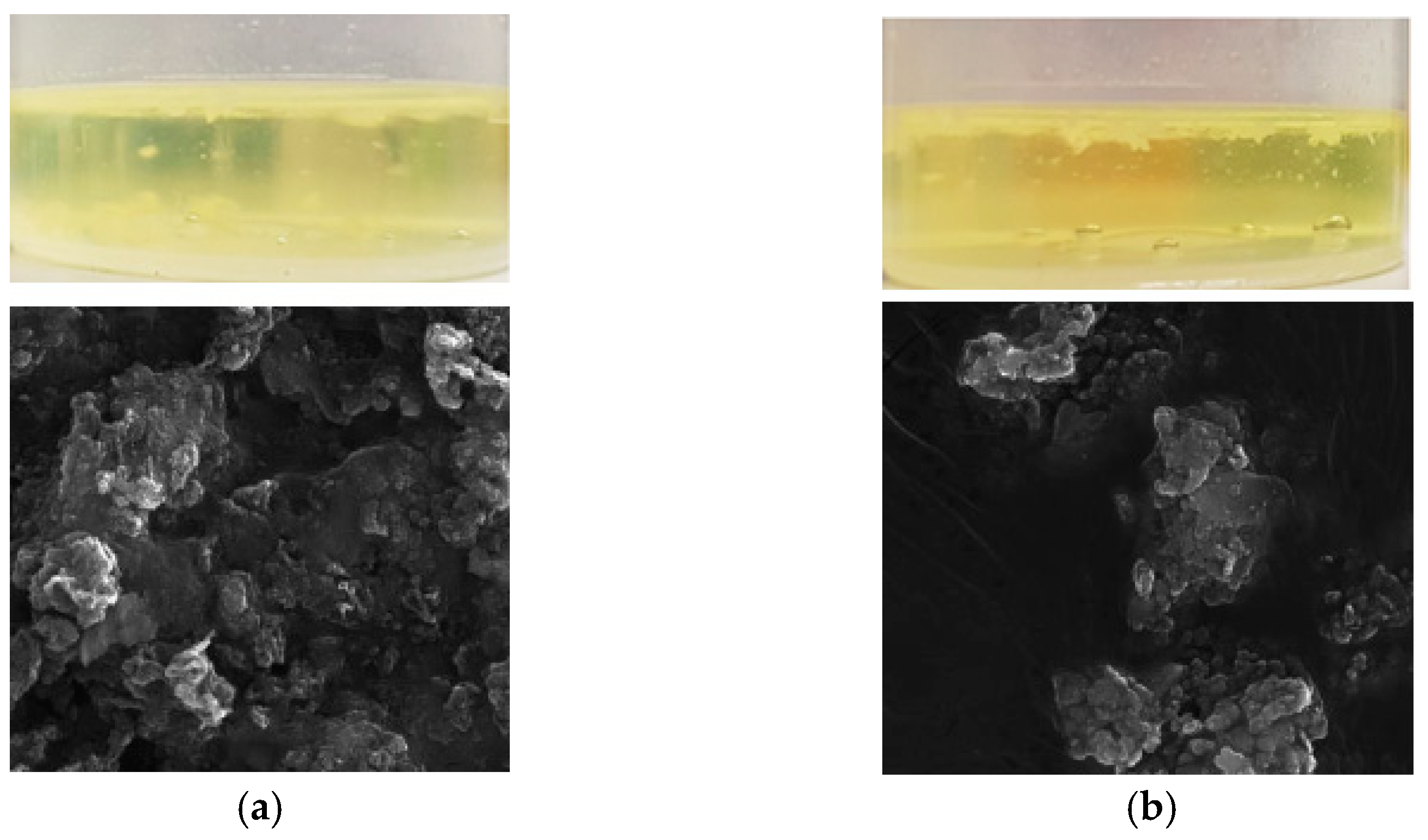
Initially, the carbon heating block was chosen for our study. It is a relatively inexpensive and easily operated system. Its great advantage is the disposable vessels with screw-on filters, which minimise the risk of contamination. The vessels were closed during mineralisation (the volume of the liquid did not exceed 5 mL to prevent an excessive increase in pressure and damage to the vessels). Different combinations of the oxidising mixture, including nitric acid and mixtures of nitric acid with hydrogen peroxide and perchloric acid in various ratios, were tested. The totality of matrix digestion was assessed visually or with scanning electron microscopy if necessary. The examples of the solutions after digestion are shown in
Figure 1.
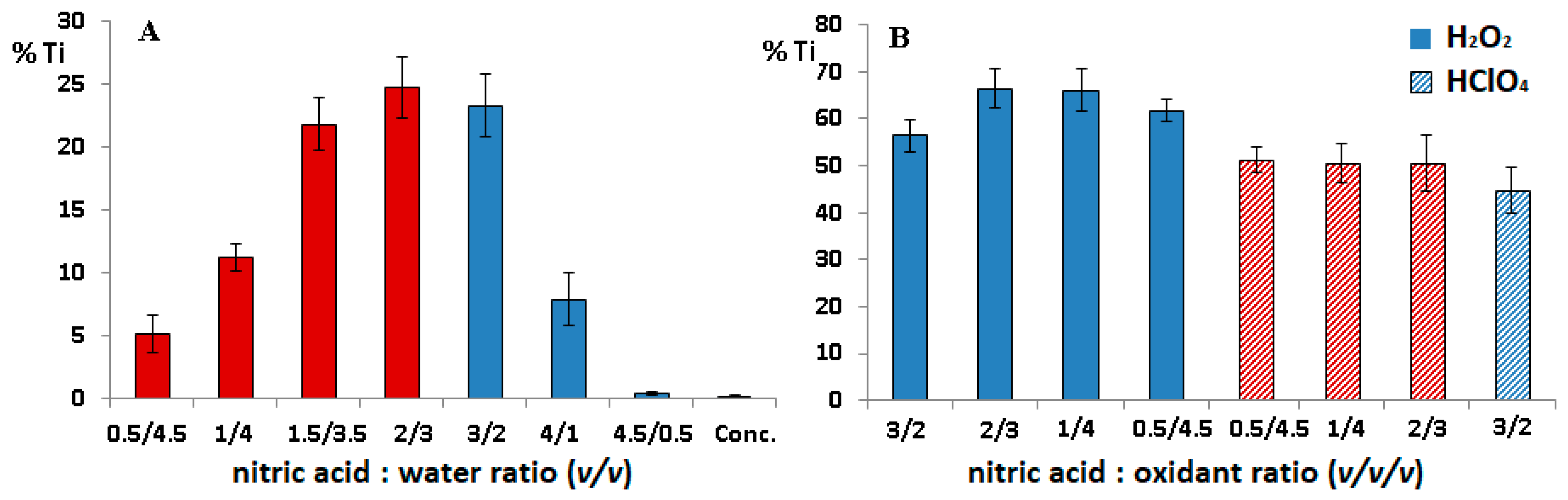
In our research, different concentrations of nitric acid were tested. Nitric acid is commonly used for digestion of various tissues; however, the concentration of the acid should be carefully chosen because nitric oxides formed during digestion are harmful for the environment, and a strongly acidic solution can damage parts of the apparatus. In turn, an insufficient concentration of acid results in incomplete decomposition of the organic matrix.
As can be seen in
Figure 2A, the recovery of titanium at the lowest concentration of nitric acid was relatively high (ca. 5%); moreover, up to 7.5 mol/L of nitric acid, the matrix was not fully decomposed (bars in
Figure 2 marked in red colour). However, the solutions obtained at the higher concentrations were transparent, and no fragments of tissue were visible under the microscope, and the recovery of titanium significantly decreased as a result of passivation of Ti particles. Thus, it can be concluded that digestion of tissue with Ti debris should be carried out with the use of concentrated nitric acid to assess the soluble titanium form.
On the other hand, the maximal level of digested titanium did not exceed 25%; thus, to increase the oxidative power of the mixture, the effect of addition of perchloric acid and hydrogen peroxide on titanium digestion was assessed. Both reagents increased the recovery of Ti significantly; however, the value did not exceed 70%, and the value was insufficient to determine total Ti content in the tissue. The examples of the results are presented in
Figure 2B. Moreover, the prolongation of the mineralisation time even up to 240 min did not enhance the digestion of titanium; therefore, the system is not suitable for preparation of tissues with metallic debris for analysis of Ti.
2.2. Microwave Assisted Mineralisation
In our further investigation, a microwave-assisted system was used to prepare titanium-containing samples for the elemental analysis. Although the cost of such equipment is relatively high, it is very effective and is recommended for hard-to-digest materials.
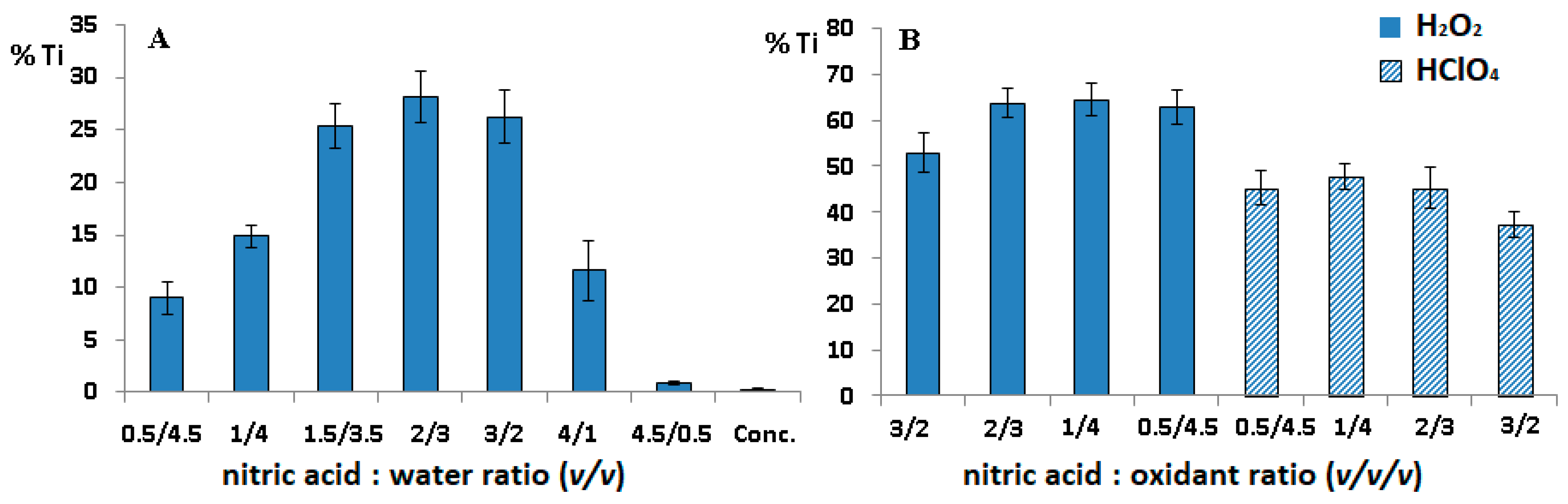
As in the procedure described in
Section 2.1, the impact of different concentrations of nitric acid and its mixtures with hydrogen peroxide and perchloric acid on the digestion of titanium debris and muscle tissue was assessed. The results are shown in
Figure 3.
As can be seen, the percentage of digested titanium was slightly higher than that for the heating block; the minimal value was ca. 0.2%, and the maximal one did not exceed 70%. This means that the soluble form of Ti can be determined when concentrated acid is used, and the conditions are not suitable for estimation of metallic titanium. However, it should be noted that the microwave-assisted process was more effective in tissue digestion even at a low acid concentration, which is beneficial from the point of view of consumption of Suprapur® reagents.
Since the oxidation potential of nitric acid strongly depends on temperature, the time-temperature working program of the apparatus was further modified. Different variants were tested, including higher/lower temperature and increased time; however, no improvement of the results were noted.
It should also be mentioned that a serious disadvantage of the microwave system is the necessity to washout and digest the Teflon vessel after each use, which prolongs the experimental procedure. Moreover, residues of the matrix or analyte sometimes penetrate into the vessel walls, resulting in memory effects, and an excessive concentration of nitric acid decreases considerably the lifetime of polymer vessels.
2.3. Digestion with the ISO Method
The samples were also digested in both systems using the ISO method dedicated to Ti. The method is based on mineralisation with sulphuric acid with addition of ammonium sulphate. However, the recovery of titanium did not exceed 60%. Moreover, the organic matrix was not fully decomposed in the heating block even if the digestion was repeated, as suggested in the procedure.
2.4. Analysis of Titanium Ions in Paraspinal Tissue of Patients with Titanium Implants
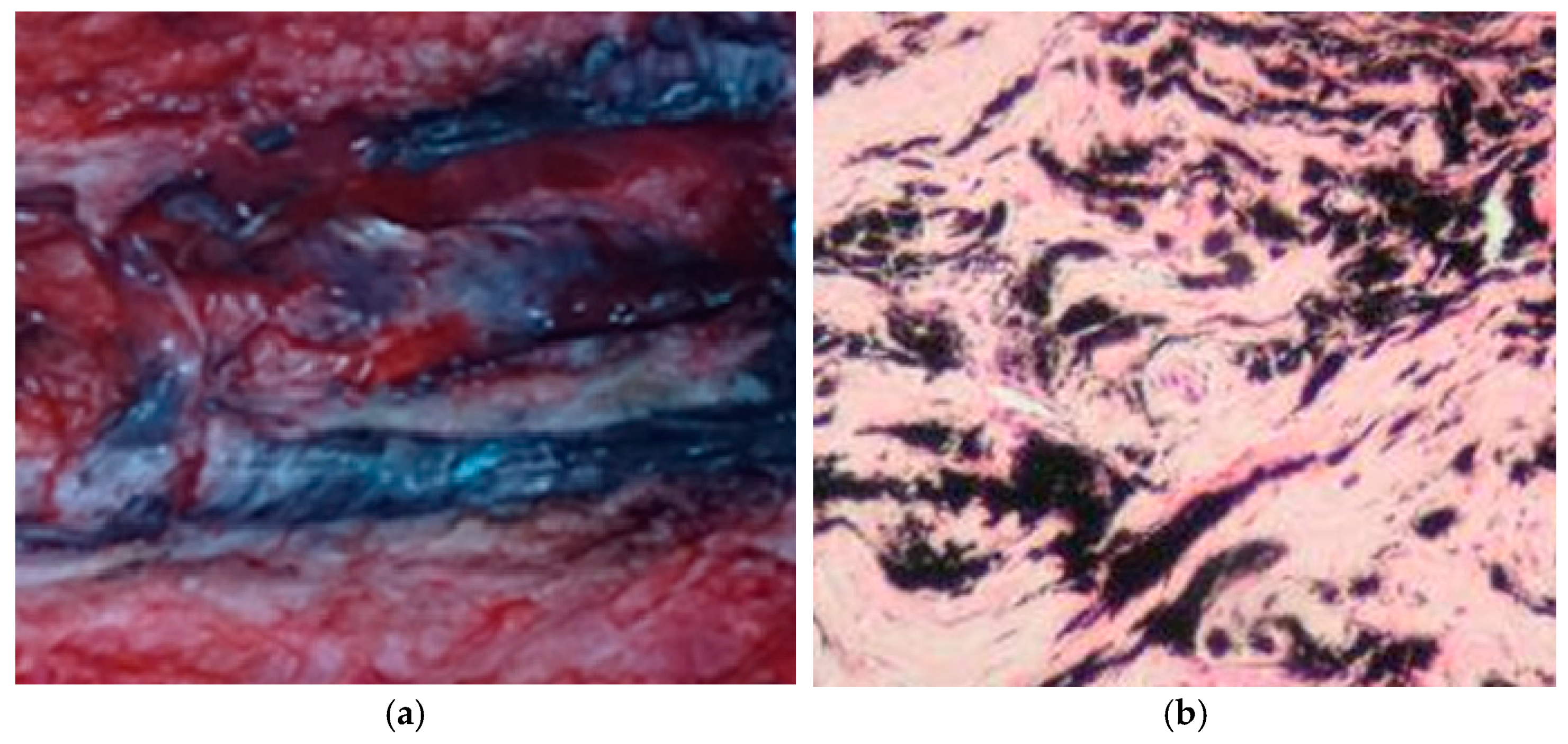
The samples of tissue of patients with different titanium-based surgical systems to correct spinal deformity (traditional growing rod (TGR), guided growth systems (GGS), and vertical expandable prosthesis titanium rib (VEPTR)) were provided by the Paediatric Orthopaedic Clinic of the Medical University of Lublin. Metallosis was clearly visible in all samples. The example is shown in
Figure 4.
Homogenised samples were digested using concentrated nitric acid in the carbon heating block and microwave systems to estimate the soluble form of titanium. All mineralisates were clear without opalescence, which indicates that the organic matrix was fully decomposed, as confirmed by the microscopic observation. The results of Ti quantification are shown in
Table 1. As can be seen, the differences between the results obtained for samples prepared using both systems were not statistically significant.
3. Discussion
There are many methods for preparation of tissues for elemental analysis, and dissolution of the tissue is usually not a big challenge. Treatment with acids or bases is the most common procedure. It is effective if appropriate concentrations of reagents and methods of digestion are chosen [
3]. However, proper preparation of tissues containing titanium as metallic particles and soluble ions is very difficult. Two approaches can be useful to resolve this problem: one of them includes full solubilisation of the sample with metallic debris, and the other one includes digestion of tissue without dissolution of Ti particles, filtration, and quantification of only the soluble form of the analyte [
3,
4,
5]. Ti is a chemically inert metal and, as shown in our study, the conversion of total Ti into the soluble ion form using standard digestion methods, such as heating and microwave mineralization, is impossible. Thus, the second resolution seems to be better. The literature review has revealed that treatment with nitric acid at different concentrations is the most common procedure for periimplant tissue; however, most reports focused on dissolving the tissue and the impact of digestion conditions on metallic Ti debris was not investigated. Our investigation proved that Ti is partially soluble, even in a mild acidic solution, when mineralisation is supported by microwaves or heating. On the other hand, heating with the use of a low concentration of acid did not decompose the tissue effectively. Concentrated nitric acid, which prevents the digestion of Ti by covering the particle surface with titanium oxide, is the best solvent for preparation of tissue to analyse only the ionic titanium form.
Treatment with strong bases is an alternative to acidic digestion; this approach was used by Kosssovsky et al. [
13] and Shanbhag et al. [
14] to decompose tissue for quantification of metallic particles; however, the high density of the solution obtained causes problems with nebulisation and reaction with quartz elements; therefore, it is not recommended for inductively coupled plasma spectrometry determination.
On the other hand, enzymatic treatment is sometimes used for digestion [
15,
16], but Campbell et al. [
17] have found that it is not fully effective to decompose the organic matrix.
Table 2 summarises the experimental conditions applied for preparation of titanium-containing tissues for elemental analysis.
4. Materials and Methods
4.1. Chemicals
Sixty-five percent nitric acid Suprapur®, 31% hydrogen peroxide Suprapur®, 60% perchloric acid EMSURE®, sulphuric acid, and ammonium sulphate were purchased from Merck (Darmstad, Germany). Deionised water (DI water) was obtained from the Direct-Q® 3 UV water purification system (Merck).
4.2. Sample Preparation
For the sample, 216 ± 12 mg of pork shoulder and 5.15 ± 0.23 mg of metallic titanium powder were weighed into digestion vessels, and 5 mL of a relevant oxidation mixture was then added. Tissue without Ti was used as a control. A microwave-assisted pressure digestion system (TOPwave, Analytik Jena, Jena, Germany) and heating block (DigiPREP, SCP Science, Baie-D’Urfé, Quebec, Canada) were used for sample digestion. Microwave digestion was carried out using CX 100 TFM-PTFE vessels and program specified in
Table 3.
Digestion in the heating block was performed for 2 h at a temperature of 120 °C using closed 50-mL DigiTUBEs (SCP Science, Baie-D’Urfé, QC, Canada). After digestion, the solution was filtered with a DigiFILTER (SCP Science) and a vacuum pump Rocker 300C into clean DigiTUBEs and filled up to 10 mL with DI water.
Final conditions used for preparation of paraspinal tissues: 5 mL of concentrated nitric acid was added to 200 mg of tissue, and two mineralisation variants were applied, including digestion in heating block (2 h at a temperature of 120 °C) and microwave digestion, according to program given in
Table 3 (in step 2, temperature was 180 °C, and time was 5 min).
4.3. Analysis of Titanium Ions
Transparent solutions were then diluted 1000 times with DI water and titanium concentrations were quantified using inductively coupled plasma optical emission spectrometry (ICP-OES) (PlasmaQuant PQ 9100 Elite, Analityk Jena, Jena, Germany). The ICP-OES working conditions are presented in
Table 4. The titanium standard solution was prepared using 1000 mg Ti, (NH
4)
2TiF
6 in water Titrisol
® (Merck).
5. Conclusions
In the present study, the conditions for preparation of paraspinal tissue from patients with titanium implants were experimentally chosen taking into account two aspects, including (i) digestion of metallic titanium present near the implants and (ii) decomposition of tissue without digestion of Ti, which allowed assessment of the total Ti and the soluble form of Ti, respectively. Our study revealed that microwave digestion was more effective in dissolution of the organic matrix at a lower concentration of nitric acid; however, microwave only slightly supported the digestion of metallic titanium. Moreover, total digestion of titanium was impossible, irrespective of the systems used and the composition of the oxidizing mixture; therefore, the quantification of metallic Ti failed. The microwave system, as well as the heating block, can be successfully used to prepare periimplant tissue for analysis of the soluble form of Ti when concentrated nitric acid is used, as it prevents the digestion of metallic debris as a result of passivation.
Author Contributions
Conceptualization, I.S., A.D. and M.W.; methodology, I.S. and J.S.; validation, K.T.-R.; formal analysis, J.S.; investigation, J.S. and A.S.-K.; writing—original draft preparation, M.W. and A.D.; writing—review and editing, I.S.; visualization, K.T.-R.; supervision, M.W. and M.L. All authors have read and agreed to the published version of the manuscript.
Funding
This research received no external funding.
Institutional Review Board Statement
The study was conducted according to the guidelines of the Declaration of Helsinki, and approved by Bioethics Committee of the Medical University of Lublin (consent no. KE-0254/105/2015).
Informed Consent Statement
Informed consent was obtained from all subjects involved in the study.
Data Availability Statement
The data presented in this study are available on request from the corresponding author.
Conflicts of Interest
The authors declare no conflict of interest.
Sample Availability
Samples are available from the authors.
References
- Ulusaloğlu, A.C.; Atici, T.; Ermutlu, C.; Akesen, S. Evaluation of titanium release from titanium alloy implants in patients with spinal instrumentation. J. Int. Med. Res. 2021, 49, 49. [Google Scholar] [CrossRef]
- Danielewicz, A.; Wójciak, M.; Sawicki, J.; Dresler, S.; Sowa, I.; Latalski, M. Comparison of Different Surgical Systems for Treatment of Early Onset Scoliosis in the Context of Release of Titanium Ions. Spine 2020. [Google Scholar] [CrossRef] [PubMed]
- Margevicius, K.J.; Bauer, T.W.; McMahon, J.T.; Brown, S.A.; Merritt, K. Isolation and characterization of debris in membranes around total joint prostheses. J. Bone Jt. Surg. Ser. A 1994, 76, 1664–1675. [Google Scholar] [CrossRef] [PubMed]
- Day, J.S.; Baxter, R.M.; Ramsey, M.L.; Morrey, B.F.; Connor, P.M.; Kurtz, S.M.; Steinbeck, M.J. Characterization of wear debris in total elbow arthroplasty. J. Shoulder Elb. Surg. 2013, 22, 924–931. [Google Scholar] [CrossRef] [PubMed]
- Hirakawa, K.; Bauer, T.W.; Stulberg, B.N.; Wilde, A.H.; Secic, M. Characterization and Comparison of Wear Debris from Failed Total Hip Implants of Different Types. J. Bone Jt. Surg. 1996, 78. [Google Scholar] [CrossRef]
- Golasik, M.; Herman, M.; Jasiewicz, B.; Tęsiorowski, M.; Piekoszewski, W. Development of a method for the determination of titanium in tissue by graphite furnace atomic absorption spectrometry for clinical analysis. J. Anal. At. Spectrom. 2014, 29, 1844–1849. [Google Scholar] [CrossRef]
- Okazaki, Y.; Gotoh, E.; Manabe, T.; Kobayashi, K. Comparison of metal concentrations in rat tibia tissues with various metallic implants. Biomaterials 2004, 25, 5913–5920. [Google Scholar] [CrossRef] [PubMed]
- Dorr, L.D.; Bloebaum, R.; Emmanual, J.; Meldrum, R. Histologic, biochemical, and ion analysis of tissue and fluids retrieved during total hip arthroplasty. Clin. Orthop. Relat. Res. 1990, 261, 82–95. [Google Scholar] [CrossRef]
- Langkamer, V.G.; Case, C.P.; Heap, P.; Taylor, A.; Collins, C.; Pearse, M.; Solomon, L. Systemic distribution of wear debris after hip replacement. A cause for concern? J. Bone Jt. Surg. Ser. B 1992, 74, 831–839. [Google Scholar] [CrossRef] [Green Version]
- Pettersson, M.; Pettersson, J.; Molin Thorén, M.; Johansson, A. Release of titanium after insertion of dental implants with different surface characteristics—An ex vivo animal study. Acta Biomater. Odontol. Scand. 2017, 3, 63–73. [Google Scholar] [CrossRef] [Green Version]
- He, X.; Reichl, F.X.; Wang, Y.; Michalke, B.; Milz, S.; Yang, Y.; Stolper, P.; Lindemaier, G.; Graw, M.; Hickel, R.; et al. Analysis of titanium and other metals in human jawbones with dental implants—A case series study. Dent. Mater. 2016, 32, 1042–1051. [Google Scholar] [CrossRef] [PubMed] [Green Version]
- Safioti, L.M.; Kotsakis, G.A.; Pozhitkov, A.E.; Chung, W.O.; Daubert, D.M. Increased Levels of Dissolved Titanium Are Associated with Peri-Implantitis—A Cross-Sectional Study. J. Periodontol. 2017, 88, 436–442. [Google Scholar] [CrossRef] [PubMed]
- Kossovsky, N.; Liao, K.; Gelman, A.; Millett, D. Size and mass quantification of sub-micron particulates in human synovial tissues recovered at revision. In Proceedings of the Transactions of the Annual Meeting of the Society for Biomaterials in Conjunction with the International Biomaterials Symposium; Society for Biomaterials: Algonquin, IL, USA, 1991; T. 14, s. 242. [Google Scholar]
- Shanbhag, A.S.; Jacobs, J.J.; Glant, T.T.; Gilbert, J.L.; Black, J.; Galante, J.O. Composition and morphology of wear debris in failed uncemented total hip replacement. J. Bone Jt. Surg. Ser. B 1994, 76, 60–67. [Google Scholar] [CrossRef]
- Maloney, W.J.; Smith, R.L.; Schmalzried, T.P.; Chiba, J.; Huene, D.; Rubash, H. Isolation and characterization of wear particles generated in patients who have had failure of a hip arthroplasty without cement. J. Bone Jt. Surg. Ser. A 1995, 77, 1301–1310. [Google Scholar] [CrossRef] [PubMed]
- Patel, J.; Lal, S.; Nuss, K.; Wilshaw, S.P.; von Rechenberg, B.; Hall, R.M.; Tipper, J.L. Recovery of low volumes of wear debris from rat stifle joint tissues using a novel particle isolation method. Acta Biomater. 2018, 71, 339–350. [Google Scholar] [CrossRef] [PubMed]
- Campbell, P.; Ma, S.; Belcher, G. A method for metal and particle isolation from periprosthetic tissues. Trans. Orthop. Reg. Soc. 1993, 18, 494. [Google Scholar]
- Musib, M.K.; Rasquinha, V.; Saha, S. Identification and characterization of polymeric and metallic wear debris from periprosthetic tissues after total hip revision surgery. J. Long. Term. Eff. Med. Implants 2011, 21, 281–290. [Google Scholar] [CrossRef]
- Agins, H.J.; Alcock, N.W.; Bansal, M.; Salvati, E.A.; Wilson, P.D.J.; Pellicci, P.M.; Bullough, P.G. Metallic Wear in Failed Titanium-Alloy Total Hip Replacements. A Histological and Quantitative Analysis. J. Bone Joint Surg. Am. 1988, 70, 347–356. [Google Scholar] [CrossRef]
- Huo, M.H.; Salvati, E.A.; Lieberman, J.R.; Betts, F.; Bansal, M. Metallic debris in femoral endosteolysis in failed cemented total hip arthroplasties. Clin. Orthop. Relat. Res. 1992, 276, 157–168. [Google Scholar] [CrossRef]
- Bianco, P.D.; Ducheyne, P.; Cuckler, J.M. Systemic titanium levels in rabbits with a titanium implant in the absence of wear. J. Mater. Sci. Mater. Med. 1997, 8, 525–529. [Google Scholar] [CrossRef]
- Lee, S.; Goh, B.T.; Lai, S.H.; Tideman, H.; Stoelinga, P.J.W.; Jansen, J.A. Peri-implant and systemic release of metallic elements following insertion of a mandibular modular endoprosthesis in Macaca fascicularis. Acta Biomater. 2009, 5, 3640–3646. [Google Scholar] [CrossRef] [PubMed]
- Rodriguez, D.; Gil, F.J.; Planell, J.A.; Jorge, E.; Alvarez, L.; García, R.; Larrea, M.; Zapata, A. Titanium levels in rats implanted with Ti6A14V treated samples in the absence of wear. J. Mater. Sci. Mater. Med. 1999, 10, 847–851. [Google Scholar] [CrossRef] [PubMed]
| Publisher’s Note: MDPI stays neutral with regard to jurisdictional claims in published maps and institutional affiliations. |
© 2021 by the authors. Licensee MDPI, Basel, Switzerland. This article is an open access article distributed under the terms and conditions of the Creative Commons Attribution (CC BY) license (https://creativecommons.org/licenses/by/4.0/).


 ,
, 


