Article

Pharmacological Potential and Chemical Composition of Crocus sativus Leaf Extracts

1
Department of Pharmaceutical Chemistry, National University of Pharmacy of Ministry of Health of Ukraine, 4-Valentinivska St., 61168 Kharkiv, Ukraine
2
Laboratory of Drug Targets Histopathology, Institute of Cardiology, Lithuanian University of Health Sciences, Sukilėlių pr. 13, LT-50162 Kaunas, Lithuania
3
Life Sciences Center, Institute of Biotechnology, Vilnius University, Saulėtekio al. 7, LT-10257 Vilnius, Lithuania
4
Graduate Institute of Natural Products, College of Pharmacy, Kaohsiung Medical University, Kaohsiung 80708, Taiwan
5
Department of Biotechnology, College of Life Science, Kaohsiung Medical University, Kaohsiung 80708, Taiwan
6
Graduate Institute of Natural Products, College of Medicine, Chang Gung University, Taoyuan 33302, Taiwan
7
Research Center for Chinese Herbal Medicine, Research Center for Food and Cosmetic Safety, Graduate Institute of Health Industry Technology, College of Human Ecology, Chang Gung University of Science and Technology, Taoyuan 33302, Taiwan
8
Department of Pharmacognosy, Faculty of Pharmacy, Ain-Shams University, Organization of African Unity Street, Abassia, Cairo 11566, Egypt
9
Department of Pharmaceutical Biology, Faculty of Pharmacy and Biotechnology, German University in Cairo, Cairo 11835, Egypt
10
The Institute of Biomedical Sciences, National Sun Yat-sen University, Kaohsiung 80424, Taiwan
11
Department of Biochemistry and Molecular Biology, College of Medicine, Chang Gung University, Taoyuan 33302, Taiwan
12
Educational and Scientific Institute of Applied Pharmacy, National University of Pharmacy of Ministry of Health of Ukraine, 12-Kylikivska St., 61000 Kharkiv, Ukraine
13
Department of Analytical and Toxicological Chemistry, Lithuanian University of Health Sciences, A. Mickevičiaus g. 9, LT-44307 Kaunas, Lithuania
14
Department of Anesthesiology, Chang Gung Memorial Hospital, Taoyuan 33305, Taiwan
15
Department of Chemical Engineering, Ming Chi University of Technology, New Taipei City 24301, Taiwan
*
Authors to whom correspondence should be addressed.
These authors contributed equally to this work.
Molecules 2022, 27(1), 10; https://doi.org/10.3390/molecules27010010
Submission received: 12 November 2021 / Revised: 6 December 2021 / Accepted: 9 December 2021 / Published: 21 December 2021

Abstract

:
Crocus sativus L. (saffron) has been traditionally used as a food coloring or flavoring agent, but recent research has shown its potent pharmacological activity to tackle several health-related conditions. Crocus sp. leaves, and petals are the by-products of saffron production and are not usually used in the medicine or food industries. The present study was designed to determine the chemical composition of the water and ethanolic extracts of C. sativus leaves and test their cytotoxic activity against melanoma (IGR39) and triple-negative breast cancer (MDA-MB-231) cell lines by MTT assay. We also determined their anti-allergic, anti-inflammatory, and anti-viral activities. HPLC fingerprint analysis showed the presence of 16 compounds, including hydroxycinnamic acids, xanthones, flavonoids, and isoflavonoids, which could contribute to the extracts’ biological activities. For the first time, compounds such as tectoridin, iristectorigenin B, nigricin, and irigenin were identified in Crocus leaf extracts. The results showed that mangiferin (up to 2 mg/g dry weight) and isoorientin (8.5 mg/g dry weight) were the major active ingredients in the leaf extracts. The ethanolic extract reduced the viability of IGR39 and MDA-MB-231 cancer cells with EC50 = 410 ± 100 and 330 ± 40 µg/mL, respectively. It was more active than the aqueous extract. Kaempferol and quercetin were identified as the most active compounds. Our results showed that Crocus leaves contain secondary metabolites with potent cytotoxic and antioxidant activities.

Graphical Abstract

1. Introduction

Cancer, one of the most devastating and complex diseases, is tackled by several techniques, including radiotherapy, immunotherapy, and chemotherapy. However, the toxicity and resistance of synthetic chemotherapeutic drugs have prompted scientists to search for natural chemotherapeutic agents obtained from plants and marine organisms. Since the 1970s, great progress has been made in the search for and development of natural anticancer drugs derived from medicinal herbs [1]. About 35,000 plant species were tested by the National Cancer Institute (NCI) for their potential anti-tumor activity, and among them, about 3000 plant species showed reproducible anti-tumor activity [2]. In addition to their potent anticancer effect, natural chemotherapeutics agents serve as drug leads for the vast majority of synthetic chemotherapeutic agents [2]. Anticancer drugs of plant origin proved to exhibit a safer safety profile in comparison with their synthetic counterparts but were less selective [3]. The interesting therapeutic properties of natural chemotherapeutic agents have encouraged scientists to search for more natural cytotoxic drugs from medicinal or edible plants. Among the edible plants that attracted attention in the last few decades is Crocus sativus (saffron).
Crocus sativus L. is a perennial underground corm that belongs to the family Iridaceae. In Ayurveda, saffron is used to treat cold, cough, asthma, arthritis, acne, and skin diseases and as an aphrodisiac and stimulant. In ancient Egypt, saffron was used to treat all types of gastrointestinal diseases. Saffron was an ingredient of laudanum or tinctura opii crocata, a “Swedish herbal mixture” for digestion [4]. Saffron is used in folk medicine to treat cancer, asthma, hysteria, convulsion, and spasms [5]. In Ukraine, saffron is used as an anticancer agent and for the treatment of eye diseases [6]. Saffron is used in oriental medicine in central and south Asia [7], and it is included in the European Pharmacopoeia as a homeopathic remedy [8].
Crocus stigmas contain many important secondary metabolites, such as crocin, crocetin, safranal, and picrocrocin, which showed potent cytotoxic and antiproliferative activity in different models and many cancer cell lines [9,10,11]. Recent scientific research indicated the potential application of Crocus stigma supplements for retinal treatment [12] and to improve metabolism in obese people [13]. Several pharmacological activities were reported for saffron and its constituents, including antioxidant, antinociceptive, anti-inflammatory, anticonvulsant, antidepressant, antitumorigenic, and immunomodulatory effects [14].
In addition to Crocus stigmas, the saffron plant has many by-products, including leaves, petals, and corms. To collect 1 kg of stigmas, about 350 kg of petals, 1500 kg of leaves, and the corms are not used and thrown away [15]. However, this biomass can be used as a potentially significant source of biologically active secondary metabolites, including flavonoids and anthocyanins. The exploitation of these by-products can significantly increase the profitability and sustainability of saffron production [16,17]. However, the data on the biological activity, especially the cytotoxic effect, of other C. sativus organs are still scarce.
In one report, it was shown that the proliferation of human adenocarcinoma cells (Caco-2) was greatly inhibited by the ethanolic extracts of C. sativus petals and leaves (EC50 = 0.42 mg/mL), while the corm extract caused some signs of toxicity and completely abolished proliferation (EC50 = 0.05 mg/mL) [15].
Mir et al. [18] reported the isolation of naturally derived crocetin β-D-glucosyl ester from the leaves of C. sativus var. cashmerianus obtained from Pampore Kashmir, India. The purified compound showed an antiproliferative effect against the human breast adenocarcinoma cell model (MCF-7), possibly by inhibiting the estrogen receptor alpha and HDAC2-mediated signaling cascade. The extracts of Crocus leaves showed antibacterial, antioxidant [19], and metal ion chelating activities [20] and mild antidepressant and anti-inflammatory efficacy [21]. The main aim of the current study was to investigate the phytochemical content of the water and ethanolic extracts from Crocus leaves grown in Ukraine and to determine their cytotoxic activity on several cancer cell lines, including melanoma (IGR39) and triple-negative breast cancer (MDA-MB-231) cells. Until now, the extracts of C. sativus leaves grown in Ukraine have not been previously studied for their cytotoxic potential.

2. Results and Discussion

Crocus waste products, such as the leaves and flowers, are gaining popularity as potential cytotoxic drugs. However, there is a lack of empirical data to support this claim. In this study, the activities of the aqueous and ethanolic extracts of Crocus leaves from Ukraine were evaluated for their cytotoxic activity and other bioactivities, including antiviral, antiallergic, and anti-inflammatory effects. The composition of the biologically active substances in the plant extracts may depend on the selected extraction method, and, most importantly, on the extractant [22], and thus we prepared the ethanolic and aqueous extracts for the selected type of raw material to be able to compare their chemical compositions. The solvent extraction method was chosen according to the most commonly used method [23]. In this study, the crude extracts of C. sativus leaves were prepared using water-ethanol (70%) and distilled water as the solvents. The effect of each extract on the viability of melanoma (IGR39) and triple-negative breast cancer (MDA-MB-231) cells was determined using the MTT assay.

2.1. Qualitative and Quantitative Analysis of the Identified Compounds

Previous phytochemical analysis of C. sativus leaves from Ukraine showed the presence of flavonoids (trans-cinnamic acid, chlorogenic acid, kaempferol, ononin, irigenin, and mangiferin), as well as amino acids and carboxylic acids in the plant raw material [24]. Therefore, further research was devoted to establishing the chemical composition of the dry C. sativus leaf extracts aiming to reveal their effect on the pharmacological activity of the extracts.
To determine the composition of the tested extracts, HPLC was used. The HPLC fingerprints of Crocus aqueous and ethanolic dry extracts are shown in Figure 1 and Table 1. All peaks in the chromatograms were identified by comparing their chromatograms with the chromatograms of the reference compounds. The compound structures were further elucidated based on their fragmentation pathways. UPLC–MS/MS analysis was applied to all the phenolic standards to identify and confirm each compound’s chemical structure. Since polyphenols contain one or more hydroxyl and/or carboxylic acid groups, MS data were acquired in the negative ionization mode. The identification of the phenolic compounds was carried out by comparing retention times (tR), calculated molecular weights, and MS/MS data of the 16 reference standards. The full spectroscopic data of the compounds are shown in Table 2. A brief description and a comparison with the data reported compounds in previous literature are provided in the Supplementary Materials.
Phenol carboxylic acid derivatives. According to the literature data, some phenol carboxylic acids (caffeic, chlorogenic, and gallic acid) were only found in the stigma aqueous extract of C. sativus [25]. Acar et al. [26] identified p-coumaric acid and rosmarinic acid in C. baytopiorum leaves. No more literature data were found regarding the study of phenol carboxylic acids in the leaves of Crocus sp. Thus, in this study, the presence of chlorogenic acid and caffeic, ferulic, and cinnamic acids in C. sativus leaf extracts was established for the first time (Table 1). The content of the chlorogenic acid in the aqueous and ethanolic extracts of Crocus leaves was the same (0.68 mg/g). Cinnamic acid, a precursor in the biosynthesis of flavonoids via the shikimate pathway, was only found in the ethanolic extract of Crocus leaves (1.42 mg/g) [27,28]. Our data indicated that Crocus leaves contained more derivatives of hydroxycinnamic acids and fewer flavonoids.
Xanthones. It is known that plants of the genus Iris (Iridaceae) accumulate various xanthones, mainly C-glycosylxanthones [29]. In the genus Crocus, mangiferin was isolated and only identified from the leaves of C. aureus and C. stellaris [30,31,32]. Therefore, mangiferin can be considered one of the markers of this family. On the chromatograms, peak C at 17.18 min was identified as mangiferin based on comparison with the UV spectrum of the reference compound. This is the first report on the identification of mangiferin from C. sativus leaf extracts. In Crocus leaves, the content of mangiferin was almost equal in both extracts (1.8–2.0 mg/g).
Flavonoids and isoflavonoids and their glycosides. Previous reports showed the presence of glycosylated derivatives of luteolin, kaempferol, quercetin, and apigenin in C. sativus leaf ethanolic extract [33]. Flavonoid glycosides are better recovered by water [34,35]. Isoorienin (luteolin-6-C-glucoside) and genistein-7-O-glucoside were identified in this experiment in the aqueous extract only of Crocus leaves. In the leaves, the content of the isoflavone glycoside tectoridin (7-glucoside tectorigenin) was higher in the ethanolic extract (0.11 mg/g).
It should be noted that the flavonoid aglycones are more hydrophobic [36] than their corresponding glycosides, and thus their solubility is low. The solubility of quercetin and kaempferol in water is very limited. Therefore, the concentration of kaempferol in the aqueous extract was low (0.033 mg/g) in comparison with the ethanolic extract that was slightly higher (0.046 mg/g).
Tectoridin and iristectorigenin B were previously only isolated or identified from the genus Iris [29,32]. This is the first report on the identification of those compounds from C. sativus leaves. Among the detected isoflavonoid anglycones, iristectorigenin B, nigricin, irigenin, and biochanin A were identified. These compounds possess from 1 to 3 hydroxy groups and 0–3 methoxy groups, which affects their solubility in the extracting solvent. The iristectorigen (3 OH groups) concentration in the aqueous extract of Crocus leaves was slightly higher (0.073 mg/g) than that in the ethanol (0.058 mg/g). Nigricin (a compound that has 1 OH-group and 2 OCH3-groups) was only found in the aqueous extract of the leaves (0.069 mg/g). Biochanin A was only identified in the aqueous extract of Crocus leaves (0.108 mg/g). Irigenin (3 OH-group, 3 OCH3 groups), like the other isoflavonoids, was found in the extracts of Crocus leaves for the first time; however, it showed the lowest concentration among all identified substances (0.028 mg/g and 0.031 mg/g for ethanolic and aqueous extracts, respectively).
It was noted that the flavonoid glycoside isoorientin showed a higher concentration (8.51 mg/g) in comparison with other components. Flavonoids are potent antiproliferative and cytotoxic agents [37]. Their composition and concentration significantly contributed to the cytotoxic activity in Crocus extracts [38].
The content of all compounds depends on climatic conditions, cultivation methods, drying, and storage [6]. Smolskaite et al. [39] analyzed 46 samples of Crocus leaves from Azerbaijan, India, New Zealand, Morocco, France, Turkey, Iran, Italy, and Spain by HPLC–UV and HPLC–MS methods. Several flavonoids were identified including kaempferol-8-C-gluco-6,3-O-diglucoside and kaempferol-8-C-gluco-6-O-glucose, which were detected for the first time. The authors noted that different samples of Crocus leaves did not differ in chemical composition but varied in the concentration of each component. The current research focused on the analysis of Crocus from Ukraine, as it is a new country in the saffron market. It should be noted that the quality of saffron grown in Ukraine is high and meets the highest ISO category [6]. This indicates an increased interest in this crop, especially since the climatic conditions of Ukraine are well suited for the cultivation of this plant. The increased demand for the cultivation of saffron results in the production of large quantities of its by-products, especially the leaves. Thus, it is important to evaluate the economic value of the leaves in terms of their biologically active secondary metabolites.

2.2. HPLC Method Validation

An HPLC method was developed for the simultaneous quantitative determination of phenolic compounds in Crocus leaf extracts. Full validation of this method was carried out. Method validation was performed to demonstrate the applicability of the developed analytical method. The following parameters were tested according to the guidelines of the International Conference on Harmonization (ICH) Q2 (R1): specificity, linearity, the limit of detection (LOD), the limit of detection (LOQ), accuracy, and precision. The results are given in Tables S1–S3 of the Supplementary Materials. The standard curves showed good linearity (R2 > 0.999) over a certain concentration range for each reference standard. The relative standard deviation (RSD) of the peak areas of the sixteen reference standards was less than 1.6%, suggesting that the instrument was in good condition. The intra- and inter-day precision and accuracy were less than 1.35% and 1.57%, respectively. The accuracy of the tested compounds was within the range of 98.08–102.88%. The LOQs and LODs were also acceptable for the simultaneous determination of all reference components, indicating that the developed HPLC method was satisfactory for the quantitative analysis of Crocus extracts.

2.3. Cytotoxic Activity of the Extracts

All tested extracts showed cytotoxic activity against the melanoma (IGR39) and triple-negative breast cancer (MDA-MB-231) cells (Figure 2). The ethanolic extract of Crocus leaves was more active than the aqueous extract for both cell lines (EC50 of the ethanolic extract against IGR39: 0.41 ± 0.10 mg/mL, against MDA-MB-231: 0.33 ± 0.04 mg/mL; EC50 of the aqueous extract against IGR39: 0.57 ± 0.11 mg/mL, against MDA-MB-231: 1.13 ± 0.09 mg/mL). It was found that the aqueous extracts significantly reduced the viability of melanoma (IGR39) cells compared with MDA-MB-231 cells.
The cytotoxic activity of the tested ethanolic extracts against breast cancer cell lines was generally higher than that of the corresponding aqueous extract (Figure 2). The potent cytotoxic activity is generally associated with the presence of various phenolic compounds.

2.4. Molecular Docking Studies

The molecular docking study aimed to analyze the biological activity of the identified compounds in Crocus extracts. In addition, it aimed to investigate the descriptors with the highest cytotoxic activity and to examine the possible mechanism of action of the selected compounds. Among the identified compounds, kaempferol, rutin, mangiferin, and quercetin were suitable for selected molecular docking experiments according to the literature data [40,41,42].
Several studies indicated that Crocus extracts exhibited cytotoxic effects because of the presence of crocin, picrocrocin, and safranal as well as phenolic compounds [15,30,31]. Kaempferol showed an antiproliferative effect on human breast carcinoma MDA-MB-453 cells [43], the mouse melanoma cell line B16F1 [44], and A375 human malignant melanoma cells [45]. To assess the most potent compounds for further studies, molecular docking experiments were performed to evaluate the affinity of individual components of the extracts in silico, as well as to investigate the activity of the most promising compounds on cancer cell lines.
For docking research on cytotoxic activity, scientists use different types of proteins depending on the nature of cancer [46]. For instance, the breast cancer proteins with BRCA1-type HRD phenotypes such as 1T15 [47], 4Y2G [48], 4RJ3 [49], estrogen receptors 2IOK [49,50,51] and 2IOG [52]; aromatase 4GL7 [53], or oncogenic kinases/phosphatases (JAK2 [54]) are the most commonly used targets for molecular docking research. Lyu et al. [55] stated the importance of human epidermal growth factor receptor HER3 in the pathogenesis of metastatic breast cancer. The enzymes chosen for docking studies were selected based on their reported binding energy for natural compounds, especially flavonoids. The interaction with c-Met kinases (PDB codes: 2RFN and 4XYF) [56] is a key mechanism of rutin’s cytotoxic activity against breast cancer. HSP90 proteins (PDB ID 4RJ3, 2IOK) were used for in silico docking analysis of curcumin and resveratrol on breast cancer proteins compared with their effect on the MCF-7 cell line [49]. Among estrogen receptors that play an important role in cancer pathogenesis, we selected the human ERα-LBD (PDB ID 3ERT) complex with 4-hydroxytamoxifen, which is an active metabolite of tamoxifen. The affinity of this enzyme to naturally occurring flavonoids such as quercetin, luteolin, myricetin, kaempferol, naringin, hesperidin, galangin, baicalein, and epicatechin was investigated as one of the factors that suppress MCF-7 cell line growth [54].
According to the docking studies, several substances identified in the active extracts showed affinity to the active sites of the selected enzymes that were similar or even better than their native ligands (Table 3).
According to the docking results, it was found that almost all the natural compounds from different chemical groups showed an affinity for the selected enzymes (expressed as a negative score function). Interestingly, some of the constituents of C. sativus extract including chlorogenic acid, isoorientin, ferulic acid, tectoridin, quercetin, cinnamic acid, genistein-7-glucoside, apigenin, kaempferol, and irigenin showed, better affinity compared with the native ligands of breast cancer proteins 4RJ3, 2IOK, and 4XYF. Moreover, the affinity of many compounds (chlorogenic acid, ferulic acid, cinnamic acid, and iristectorigenin B) to the estrogen receptor (3ERT) was comparable to hydroxytamoxifen. However, the role of each compound should be proven at least by in vitro studies. Among the constituents identified in the most active ethanolic leaf extract, we selected certain compounds for the in vitro studies, including phenolic acids—cinnamic and caffeic—as well as flavonoids—tectoridin, kaempferol, nigricin, irigenin, and mangiferin.

2.5. Cytotoxic Activity of Secondary Metabolites from C. sativus Leaf Extracts

The ethanolic extract of Crocus leaves showed higher cytotoxic activity than the aqueous extract against both cell lines. This could be due to higher levels of mangiferin (2.031 ± 0.648 mg/g), tectoridin (0.107 ± 0.045 mg/g), ferulic acid (0.251 ± 0.005 mg/g), caffeic acid (1.425 ± 0.012 mg/g), quercetin (0.255 ± 0.003 mg/g), nigricin (0.069 ± 0.003 mg/g), and kaempferol (0.046 ± 0.002 mg/g) in the leaves’ ethanolic extract (Table 1) since these compounds showed cytotoxic activity in various cell lines. Grasso R. et al. [57] showed that ferulic acid reduced the viability of U-87 cells by up to 70% in comparison with the control, and the cells treated with ferulic acid in combination with NLCs (nanostructured lipid carriers) showed a reduction in viability up to 40%. In vitro studies showed that flavonoids quercetin and kaempferol selectively suppressed the viability of brain, breast, cervix, and lung cancer cells [58,59]. Yamagata K. et al. demonstrated that chlorogenic acid significantly suppressed the viability of A549 cancer cells and regulated the expression of genes associated with apoptosis [60]. According to previous literature, caffeic acid showed a cytotoxic effect, which was attributed to its antioxidant properties by suppressing the formation of ROS and DNA damage [61]. The cytotoxic activity of the water and ethanolic extracts of Crocus leaves is affected by the concentration of the active substances. The higher levels of flavonoids, phenolic acids, and their derivatives in the ethanol extract of the leaves can be correlated with the more potent cytotoxic effect of this extract compared with the aqueous extract.
To evaluate the effect of individual compounds on the cytotoxic activity of the whole extracts, several compounds, such as quercetin, kaempferol, mangiferin, rutin, apigenin, caffeic acid, irigenin, nigericin, and tectoridin, were tested against melanoma and triple-negative breast cancer cell lines (Figure 3).
Kaempferol and quercetin were the most active compounds, but their concentrations in the extracts were not very high. Therefore, it can be assumed that the activity of extracts was due to the synergistic activity of all its cytotoxic compounds.

2.6. Antioxidant Activity

Six compounds with antioxidant properties were identified in the Crocus leaf extracts, including mangiferin, isoorientin, caffeic acid, ferulic acid, rutin, and tectoridin, by post-column HPLC accompanied with the ABTS test (Table 4). The antioxidant activity values of the leaf aqueous and ethanol extracts were 0.635 and 0.951 mmol/L, respectively, and were comparable to Trolox (0.3995 mmol/L). The leaves’ bioactivity was correlated to the presence of the following two antioxidant phenolic compounds: mangiferin and rutin. The structure of the phenolic compounds, especially the number and position of the hydroxy groups and the nature of the substitutions on the aromatic ring, significantly affects the activity of the compounds [62,63,64]. Our findings indicated that Crocus leaves, a by-product of saffron production, can serve as a valuable natural source of antioxidants.

2.7. Bioactivity Screening and Anti-Neuraminidase Activity

There is a lack of studies on the anti-neuraminidase, anti-inflammatory, anti-allergic and antiviral activities of the C. sativus leaf extracts. According to our results (Table 5), the aqueous extract at 10 µg/mL inhibited superoxide anion generation by 35.67 ± 5.62% and elastase release by 19.62 ± 2.90% in fMLF/CB-induced human neutrophils. Moreover, both the aqueous and ethanolic extracts increased the effect of NRF2 expression exclusively in normal cells (152.5% and 136.5%, respectively), while the NRF2 expression was intact in the cancer cell line at 100 µg/mL (106.2% and 104.5%, respectively). Previous studies indicated that plant extracts rich in phenolic compounds may affect the antioxidant function of cells, including NRF2 expression [65]. The C. sativus leaf aqueous and ethanolic extracts were inactive in the other bioactivity assays (Table 5), including the anti-allergic (degranulation assay, 100 µg/mL), anti-viral (influenza H1N1 and enterovirus D68, 50 µg/mL; ethanolic extract on coronavirus 229E, 10 µg/mL), and lipid droplets assay (100 µg/mL). Our results, along with the literature data, suggest that the high content of the phenolic compounds may correlate with the inhibitory effects of C. sativus leaves on superoxide anion generation in neutrophils and NRF2 activation in normal cells. A higher concentration may be needed for other bioactivity assays to show positive results.

3. Materials and Methods

3.1. Chemicals and Reagents

The acetonitrile and methanol were of HPLC grade and were purchased from Roth GmbH (Karlsruhe, Germany). The reference compounds (chlorogenic acid, caffeic acid, ferulic acid, t-cinnamic acid, mangiferin, kaempferol, tectoridin, iristectorigenin B, nigricin, irigenin, isoorientin, genistein-7-glucoside, and biochanin A) were purchased from ChromaDex (Santa Ana, CA, USA), Sigma-Aldrich (Saint Louis, MO, USA), HWI ANALYTIK GmbH, and Roth GmbH (Karlsruhe, Germany). All other chemicals were of analytical grade.

3.2. Crude Extracts Preparation

C. sativus leaves were collected from the plantation in Lyubimivka village (Ukraine) in November 2019. Botanical identification was performed by Dr. Mykhailenko, National University of Pharmacy, Ukraine. The leaves were air-dried at room temperature with suitable ventilation and were crushed to grit (1.0 mm size) for further extraction. The dried material (100 g) was extracted with boiled distilled water (1 L) or ethanol/water 70/30 (v/v) at 90 °C for 2 h three times on a horizontal water bath shaker. The extracts were filtered through Whatman no. 40 filter paper (Whatman International Ltd., Kent, UK) using a Büchner funnel. The filtered solution was evaporated to dryness under reduced pressure at 40 °C at an approximate rotation speed of 200 rpm using a rotary evaporator (Heidolph, Germany) to obtain a crude extract (18.8 g and 20.5 g for the water and ethanolic extracts, respectively). Finally, the extracts were refrigerated at 4 °C in an airtight container until further analysis. Before the bioactivity assay, the crude extracts were dissolved in DMSO (10% final concentration) and were filter-sterilized with a 0.2 µm membrane filter (Millex) to prepare a stock solution.

3.3. Sample Preparation for HPLC Analysis

A portion of the dry plant extract (0.1 g) was extracted with 10 mL methanol in an ultrasonic bath at room temperature (20 ± 2 °C) for 30 min. The solutions were filtered through a membrane filter (0.45 µm) before use. An aliquot of 10 µL was injected into the HPLC system for analysis. A standard solution of the reference compounds at a concentration of 1.0 mg/mL for each compound was prepared by dissolving it in methanol and was used for calibration. All samples were kept at 4 °C before use.

3.4. HPLC Conditions

The Shimadzu Nexera X2 LC-30AD HPLC system (Shimadzu, Kyoto, Japan) includes a quaternary pump, an online degasser, a column temperature controller, the SIL-30AC autosampler, and SPD-M20A diode array detector (DAD) (Shimadzu, Kyoto, Japan) was used for fingerprint analysis. The chromatographic separation of the phenolic compounds was carried out using an ACE C18 column (250 mm × 4.6 mm, 5.0 μm; Bellefonte, PA, USA). The mobile phase was composed of solvent A (0.1% acetic acid in water) and solvent B (acetonitrile). The elution was performed at a flow rate of 1 mL/min, and the following linear gradient program was applied: 0–8 min, 5–15% B; 8–30 min, 15–20% B; 30–48 min, 20–40% B; 48–58 min, 40–50% B; 58–65 min, 50% B; 65–66 min, 50–95% B. The column temperature was maintained at 25 °C. The injection volume of the sample solution was 10 μL. The detection wavelengths were 270 nm, 240 nm, and 310 nm. The standard solutions of the reference compounds were used for the calibration of a standard curve using an external standard method. The analyses were performed in duplicate. Chromatographic peak identification was carried out according to the analyte and reference compound retention time by comparing the UV absorption spectra of the reference compounds and analytes obtained with a diode array detector.
The validation of the HPLC method was performed according to the guideline of the International Conference on Harmonization (ICH) and included the following parameters: specificity, linearity, precision, LOD, and LOQ (Tables S1–S3 of the Supplementary Materials).

3.5. HPLC–PDA, and UPLC–MS Conditions, HPLC Post-Column Assay

HPLC–PDA and HPLC–ABTS were done using a Waters Alliance 2695 separation module system as previously described by Marksa et al. [66] with some modifications. UPLC–MS analysis was performed using the ACQUITY H-class UPLC system (Waters, Milford, MA, USA). The details are described in the Supplementary Materials section.

3.6. In Vitro Cytotoxic Activity

The human melanoma cancer cell line IGR39 and the human triple-negative breast cancer cell line MDA-MB-231 were used, and the method is described in detail in the Supplementary Materials section. These cell lines were obtained from American Type Culture Collection (ATCC, Manassas, VA, USA).

3.7. Antiallergic Activity in RBL-2H3 Cells

The methylthiazole tetrazolium (MTT) assay was used to measure the possible toxic effect of the samples on RBL-2H3 cells as previously described [67]. The C. sativus leaf aqueous and ethanolic extracts were then subjected to an anti-allergic degranulation assay based on β-hexosaminidase release in RBL-2H3 mast cells induced by calcium ionophore (A23187) or antigen (IgE plus DNP-BSA) according to the previous methodology [68,69]. The details of the assays are presented in the Supplementary Materials section.

3.8. Anti-Inflammatory Activity in Human Neutrophils

Blood was taken from healthy human donors using a protocol approved by the Chang Gung Memorial Hospital review board. Neutrophils were isolated according to the standard procedure described previously [70]. The inhibition of superoxide anion generation was measured by the reduction of ferricytochrome c, as previously described [71]. Elastase release representing degranulation from azurophilic granules was evaluated as described previously [72]. Details can be found in the Supplementary Materials section.

3.9. Lipid Droplet Assay

The lipid droplet assay was performed according to a previous method using a BSA-conjugated oleic acid system in Huh7 cells [73]. The details are described in the Supplementary Materials section.

3.10. NRF2 Activity

Nuclear transcription factor NRF2 activity was evaluated in HaCaT normal cells and Huh7 cancer cells according to previous methodology [74]. The cell line HaCaT/ARE carrying the antioxidant response element was developed using an HaCaT stable cell line carrying a fragment derived from the pGL4.37 (luc2P/ARE/Hygro) plasmid and the luciferase reporter gene luc2P. The details are described in the Supplementary Materials section.

3.11. Protective Effect of the Extracts against Influenza Virus and Enterovirus

The anti-viral assay was performed by cytopathic effects of the extracts on the cells infected by influenza H1N1 [75], and enterovirus D68 [76]. The details are described in the Supplementary Materials section.

3.12. Coronavirus 229E Assay

The protective effect of the samples against human coronavirus 229E (HCoV-229) was determined according to a previously described method [76]. The details are described in the Supplementary Materials section.

3.13. Neuraminidase Activity Assay

A baculovirus displayed neuraminidase NA9 on the surface (NA9-Bac) as a pseudotyped influenza virus was used to evaluate the neuraminidase activity. Details are described in [77] and in the Supplementary Materials section.

3.14. Molecular Docking

The docking simulations were performed with the SCIGRESS software package (Fujitsu, Fukuoka, Japan (license 742F6852C191)).

3.14.1. Ligand Preparation

All structures were designed using the ISIS DRAW 4.0 software and were saved as .mol files. The structures were imported into the SCIGRESS software and were saved in .csf format. Hydrogen atoms were added. Structure optimization was performed using the MM3 method.

3.14.2. Enzymes Preparation

The X-ray crystal structures of proteins were obtained from the Protein Data Bank (http://www.rcsb.org/pdb, accessed 15 September 2021). Hydrogen atoms were added, and structure optimization was carried out. Water and heteroatoms were removed from the structure. The native ligand was removed from the active site after its identification.
Genetic algorithm (GA) was used within automatic docking (Population size: 500; Crossover: 0.2; Elitism: 50; Max generations: 50.000; Mutation rate: 0.0; Convergence 0.1; Max iterations: 1000; and Rate: 0.01). Docking studies were carried out using fast quantum molecular docking (rigid receptor and flexible ligand). The calculated score functions of the substances investigated were compared with the ones obtained for the native ligand. Water molecules were removed, and hydrogens were added to the crystal structure of proteins before docking. Assigning charge and protonation state final refinement (energy minimization) were conducted using MM3 force field runs.

3.15. Data Analysis

The processing of HPLC data was carried out using the LabSolutions Analysis Data System (Shimadzu, Kyoto, Japan). The results were analyzed by one-way analysis of variance (ANOVA) followed by Tukey’s multiple comparison test with the software package Prism v. 5.04 (GraphPad Software Inc., La Jolla, CA, USA, chemical composition means ± SD), by Dunnet’s test (GraphPad Prism 6.0, GraphPad Software Inc., San Diego, CA, USA, anti-allergic assay; data of cytotoxicity and antioxidant assays are presented as means ± standard error (S.E.) of at least three independent experiments), or Student’s t-test (SigmaPlot, Systat Software Inc., San Jose, CA, USA, anti-inflammatory assay, means ± S.E.M.). Values with p-values below 0.05 were considered statistically significant. At least three independent measurements were performed unless otherwise specified.

4. Conclusions

The current study investigated the in vitro cytotoxic and antioxidant activity of C. sativus leaf extracts. A quality control method using a validated HPLC fingerprint analysis for C. sativus leaf extracts was developed. Further, the molecular docking studies indicated a good binding between the identified compounds and breast cancer- and melanoma-related enzymes. The cytotoxic and antioxidant activities of the major identified compounds correlated well with the bioactivities of the whole extracts. Our results indicated that the leaves of C. sativus are a rich source of biologically active compounds that can have potential therapeutic applications and should not be wasted during the saffron preparation process.

Supplementary Materials

The following are available online: Tables S1–S3. Qualitative and quantitative data of identified compounds. Figures S1 and S2. The UPLC-MS/MS-chromatograms of C. sativus leaves extracts with a short description and comparison with literature data [78,79,80,81,82,83,84,85,86,87,88]. Materials and Methods.

Author Contributions

O.M., V.P. and M.K.—conceived and designed the study; L.I., V.G. and T.-L.H.—funding acquisition; O.M., M.K., I.B., V.P., C.-H.Y., C.-F.H., D.L. and B.-H.C.—chemistry and pharmacology investigation; O.M., V.P., L.I. and M.K.—data analysis and interpretation; O.M., M.K. and M.E.-S.—writing: original draft; M.E.-S., V.G., F.-R.C. and T.-L.H.—writing: review and editing; V.G. and T.-L.H.—supervision. All authors have read and agreed to the published version of the manuscript.

Funding

This research was supported by grants from the Ministry of Science and Technology, Taiwan (MOST 108-2320-B-255-003-MY3, MOST 109-2327-B-255-001, and MOST 109-2327-B-182-002 awarded to T.-L.H.; MOST 108-2320-B-037-022-MY3 awarded to F.-R.C.; MOST 109-2320-B-037-004-MY3 awarded to B.-H.C.). This study was also supported by Chang Gung Memorial Hospital (CMRPF1G0241∼3, CMRPF1J0051∼3, and BMRP450), Chang Gung University of Science and Technology (ZRRPF3L0091), awarded to T.-L.H. In addition, this research was funded by the Drug Development and Value Creation Research Center, Kaohsiung Medical University and Department of Medical Research, Kaohsiung Medical University Hospital, Taiwan (KMU-TC108A03-11 and KMU-TC109A03-2), awarded to F.-R.C. The funders had no role in the study design, data collection, analyses, decision to publish, or preparation of the manuscript.

Institutional Review Board Statement

The study was conducted according to the guidelines of the Declaration of Helsinki, and approved by the Institutional Review Board of Chang Gung Memorial Hospital (Registration number: IRB 100 1278C).

Informed Consent Statement

Informed consent was obtained from all subjects involved in the study.

Data Availability Statement

This research contains only Supplementary Materials information.

Acknowledgments

The authors are thankful for the financial support provided by the Science Foundation of Lithuanian University of Health Sciences and Chang Gung University. The authors would like to thank the Center for Research Resources and Development, Kaohsiung Medical University, for providing instrumentation support.

Conflicts of Interest

The authors declare no conflict of interest.

References

  1. Sagbo, I.J.; Otang-Mbeng, W. Plants used for the traditional management of cancer in the eastern cape province of south africa: A review of ethnobotanical surveys, ethnopharmacological studies and active phytochemicals. Molecules 2021, 26, 4639. [Google Scholar] [CrossRef]
  2. Desai, A.G.; Qazi, G.N.; Ganju, R.K.; El-Tamer, M.; Singh, J.; Saxena, A.K.; Bedi, Y.S.; Taneja, S.C.; Bhat, H.K. Medicinal plants and cancer chemoprevention. Curr. Drug Metab. 2008, 9, 581–591. [Google Scholar] [CrossRef] [Green Version]
  3. Fridlender, M.; Kapulnik, Y.; Koltai, H. Plant derived substances with anti-cancer activity: From folklore to practice. Front. Plant Sci. 2015, 6, 799. [Google Scholar] [CrossRef]
  4. Ratsch, C.; Hofmann, A. The Encyclopedia of Psychoactive Plants: Ethnopharmacology and Its Applications; Park Street Press: Boston, MA, USA, 2005; p. 944. [Google Scholar]
  5. Basker, D.; Negbi, M. Uses of saffron. Econ. Bot. 1983, 37, 228–236. [Google Scholar] [CrossRef]
  6. Мykhailenko, O.; Desenko, V.; Ivanauskas, L.; Georgiyants, V. Standard operating procedure of Ukrainian saffron cultivation according with Good Agricultural and Collection Practices to assure quality and traceability. Ind. Crops Prod. 2020, 151, 112376–112387. [Google Scholar] [CrossRef]
  7. Ashrafi, V.; Mirshekari, B.; Dashti, S.; Khalilvand, E.; Farzaneh, S. Locating Ornamental and medicinal saffron cultivation based on Ahp analysis in Gis environment in Ardabil province. J. Ornam. Plants 2018, 8, 155–169. [Google Scholar]
  8. European Department for the Quality of Medicines. European Pharmacopoeia, 9.0 ed.; European Department for the quality of Medicines: Strasbourg, France, 2014; Volume 1, p. 1694. [Google Scholar]
  9. Gezici, S. Comparative anticancer activity analysis of saffron extracts and a principle component, crocetin for prevention and treatment of human malignancies. J. Food Sci. Technol. 2019, 56, 5435–5443. [Google Scholar] [CrossRef]
  10. Milajerdi, A.; Djafarian, K.; Hosseini, B. The toxicity of saffron (Crocus sativus L.) and its constituents against normal and cancer cells. J. Nutr. Intermed. Metab. 2016, 3, 23–32. [Google Scholar] [CrossRef] [Green Version]
  11. Mykhailenko, O.; Lesyk, R.; Finiuk, N.; Stoika, R.; Yushchenko, T.; Ocheretniuk, A.; Vaschuk, V.; Mishchenko, V.; Georgiyants, V. In vitro anticancer activity screening of Iridaceae plants. J. Appl. Pharm. Sci. 2020, 10, 59–63. [Google Scholar]
  12. Maccarone, R.; Di Marco, S.; Bisti, S. Saffron supplement maintains morphology and function after exposure to damaging light in mammalian retina. Investig. Ophthalmol. Vis. Sci. 2008, 49, 1254–1261. [Google Scholar] [CrossRef] [Green Version]
  13. Abedimanesh, N.; Bathaie, S.Z.; Abedimanesh, S.; Motlagh, B.; Separham, A.; Ostadrahimi, A. Saffron and crocin improved appetite, dietary intakes and body composition in patients with coronary artery disease. J. Cardiovasc. Thorac. Res. 2017, 9, 200–208. [Google Scholar] [CrossRef] [Green Version]
  14. Schmidt, M.; Betti, G.; Hensel, A. Saffron in phytotherapy: Pharmacology and clinical uses. Wien. Med. Wochenschr. 2007, 157, 315–319. [Google Scholar] [CrossRef] [PubMed]
  15. Sánchez-Vioque, R.; Santana-Méridas, O.; Polissiou, M.; Vioque, J.; Astraka, K.; Alaiz, M.; Herraiz-Peñalver, D.; Tarantilis, P.A.; Girón-Calle, J. Polyphenol composition and in vitro antiproliferative effect of corm, tepal and leaf from Crocus sativus L. on human colon adenocarcinoma cells (Caco-2). J. Funct. Foods 2016, 24, 18–25. [Google Scholar] [CrossRef] [Green Version]
  16. Lahmass, I.; Lamkami, T.; Delporte, C.; Sikdar, S.; Van Antwerpen, P.; Saalaoui, E.; Megalizzi, V. The waste of saffron crop, a cheap source of bioactive compounds. J. Funct. Foods 2017, 35, 341–351. [Google Scholar] [CrossRef]
  17. Santana-Méridas, O.; González-Coloma, A.; Sánchez-Vioque, R. Agricultural residues as a source of bioactive natural products. Phytochem. Rev. Proc. Phytochem. Soc. Eur. 2012, 11, 447–466. [Google Scholar] [CrossRef]
  18. Mir, M.A.; Ahmad Ganai, S.; Mansoor, S.; Jan, S.; Mani, P.; Masoodi, K.Z.; Amin, H.; Rehman, M.U.; Ahmad, P. Isolation, purification and characterization of naturally derived Crocetin beta-D-glucosyl ester from Crocus sativus L. against breast cancer and its binding chemistry with ERalpha and HDAC2. Saudi J. Biol. Sci. 2020, 27, 975–984. [Google Scholar] [CrossRef] [PubMed]
  19. Jadouali, S.; Atifi, H.; Bouzoubaa, Z.; Majourhat, K.; Gharby, S.; Achemchem, F.; Elmoslih, A.; Laknifli, A.; Mamouni, R. Chemical characterization, antioxidant and antibacterial activity of Moroccan Crocus sativus L. petals and leaves. J. Mater. Environ. Sci. 2018, 9, 113–118. [Google Scholar] [CrossRef]
  20. Sánchez-Vioque, R.; Rodríguez-Conde, M.; Reina-Urena, J.; Escolano-Tercero, M.; Herraiz-Peñalver, D.; Santana-Méridas, O. In vitro antioxidant and metal chelating properties of corm, tepal and leaf from saffron (Crocus sativus L.). Ind. Crops Prod. 2012, 39, 149–153. [Google Scholar] [CrossRef]
  21. Khan, A.; Muhamad, N.A.; Ismail, H.; Nasir, A.; Khalil, A.A.K.; Anwar, Y.; Khan, Z.; Ali, A.; Taha, R.M.; Al-Shara, B.; et al. Potential nutraceutical benefits of in vivo grown saffron (Crocus sativus L.) as analgesic, anti-inflammatory, anticoagulant, and antidepressant in mice. Plants 2020, 9, 1414. [Google Scholar] [CrossRef]
  22. Zhang, Q.-W.; Lin, L.-G.; Ye, W.-C. Techniques for extraction and isolation of natural products: A comprehensive review. Chin. Med. 2018, 13, 20. [Google Scholar] [CrossRef] [PubMed] [Green Version]
  23. Ilina, T.; Skowronska, W.; Kashpur, N.; Granica, S.; Bazylko, A.; Kovalyova, A.; Goryacha, O.; Koshovyi, O. Immunomodulatory activity and phytochemical profile of infusions from Cleavers herb. Molecules 2020, 25, 3721. [Google Scholar] [CrossRef]
  24. Mykhailenko, O.; Bezruk, I.; Ivanauskas, L.; Lesyk, R.; Georgiyants, V. Characterization of phytochemical components of Crocus sativus leaves using HPLC-MS/MS and GC-MS: A new potential by-product. Sci. Pharm. 2021, 89, 28. [Google Scholar] [CrossRef]
  25. Gismondi, A.; Serio, M.; Canuti, L.; Canini, A. Biochemical, antioxidant and antineoplastic properties of Italian saffron (Crocus sativus L.). Am. J. Plant. Sci. 2012, 3, 1573–1580. [Google Scholar] [CrossRef] [Green Version]
  26. Acar, G.; Dogan, N.M.; Duru, M.E.; Kivrak, I. Phenolic profiles, antimicrobial and antioxidant activity of the various extracts of Crocus species in Anatolia. Afr. J. Microbiol. Res. 2010, 4, 1154–1161. [Google Scholar]
  27. Kumar, N.; Goel, N. Phenolic acids: Natural versatile molecules with promising therapeutic applications. Biotechnol. Repor. 2019, 24, e00370. [Google Scholar] [CrossRef] [PubMed]
  28. Kolodziejczyk-Czepas, J.; Kozachok, S.; Pecio, Ł.; Marchyshyn, S.; Oleszek, W. Determination of phenolic profiles of Heraria polygama and Herniaria incana fractions and their in vitro antioxidant and anti-inflammatory effects. Phytochemistry 2021, 190, 112861. [Google Scholar] [CrossRef]
  29. Singab, A.N.B.; Ayoub, I.M.; El-Shazly, M.; Korinek, M.; Wu, T.Y.; Cheng, Y.-B.; Chang, F.-R.; Wubde, Y.-C. Shedding the light on Iridaceae: Ethnobotany, phytochemistry and biological activity. Ind. Crops Prod. 2016, 92, 308–335. [Google Scholar] [CrossRef]
  30. Bate-Smith, E.C. The phenolic constituents of plants and their taxonomic significance. J. Linn. Soc. 1968, 60, 325–356. [Google Scholar] [CrossRef]
  31. Harborne, J.B.; Williams, C.A. 6-Hydroxyflavones and other flavonoids of Crocus. Z. Naturforsch. 1984, 39c, 18–23. [Google Scholar] [CrossRef]
  32. Mykhailenko, O.; Kovalyov, V.; Goryacha, O.; Ivanauskas, L.; Georgiyants, V. Biologically active compounds and pharmacological activities of species of the genus Crocus: A review. Phytochemistry 2019, 162, 56–89. [Google Scholar] [CrossRef] [PubMed]
  33. Baba, S.A.; Malik, A.H.; Wani, Z.A.; Mohiuddin, T.; Shah, Z.; Abbas, N.; Ashraf, N. Phytochemical analysis and antioxidant activity of different tissue types of Crocus sativus and oxidative stress alleviating potential of saffron extract in plants, bacteria, and yeast. S. Afr. J. Bot. 2015, 99, 80–87. [Google Scholar] [CrossRef]
  34. Zhao, J.; Yang, J.; Xie, Y. Improvement strategies for the oral bioavailability of poorly water-soluble flavonoids: An overview. Int. J. Pharm. 2019, 570, 118642. [Google Scholar] [CrossRef] [PubMed]
  35. Kyslychenko, V.; Karpiuk, U.; Diakonova, I.; Abu-Darwish, M.S. Phenolic compounds and terpenes in the green parts of Glycine hispida. Adv. Environ. Biol. 2010, 4, 490–494. [Google Scholar]
  36. Zhang, H.; Wang, M.; Chen, L.; Liu, Y.; Liu, H.; Huo, H.; Sun, L.; Ren, X.; Deng, Y.; Qi, A. Structure-solubility relationships and thermodynamic aspects of solubility of some flavonoids in the solvents modeling biological media. J. Mol. Liq. 2017, 225, 439–445. [Google Scholar] [CrossRef]
  37. Manthey, J.A.; Guthrie, N. Antiproliferative activities of citrus flavonoids against six human cancer cell lines. J. Agric. Food Chem. 2002, 50, 5837–5843. [Google Scholar] [CrossRef] [PubMed]
  38. Mykhailenko, O.; Petrikaitė, V.; Korinek, M.; El-Shazly, M.; Chen, B.-H.; Yen, C.-H.; Hsieh, C.-F.; Bezruk, I.; Dabrišiūtė, A.; Ivanauskas, L.; et al. Bio-guided bioactive profiling and HPLC-DAD fingerprinting of Ukrainian saffron (Crocus sativus stigma): Moving from Correlation toward Causation. BMC Complement. Med. Ther. 2021, 21, 203. [Google Scholar] [CrossRef] [PubMed]
  39. Smolskaite, L.; Talou, T.; Fabre, N.; Venskutonis, P.R. Valorization of saffron industry by-products: Bioactive compounds from leaves. In Innovations for Food Science and Production, Proceedings of the 6th Baltic Conference on Food Science and Technology FOODBALT-2011, Jelgava, Latvia, 5–6 May 2011; Straumite, E., Ed.; Faculty of Food Technology: Riga, Latvia, 2011; pp. 67–72. [Google Scholar]
  40. Samarghandian, S.; Borji, A. Anticarcinogenic effect of saffron (Crocus sativus L.) and its ingredients. Pharm. Res. 2014, 6, 99–107. [Google Scholar] [CrossRef] [Green Version]
  41. Abd Razak, S.; Hamzah, M.S.A.; Yee, F.C.; Abdul Kadir, M.R.; Mat Nayan, N.H. A review on medicinal properties of saffron toward major diseases. J. Herbs Spices Med. Plants 2017, 23, 98–116. [Google Scholar] [CrossRef]
  42. Sudhakaran, M.; Sardesai, S.; Dosef, A.I. Flavonoids: New frontier for immuno-regulation and breast cancer control. Antioxidants 2019, 8, 103. [Google Scholar] [CrossRef] [Green Version]
  43. Choi, E.J.; Ahn, W.S. Kaempferol induced the apoptosis via cell cycle arrest in human breast cancer MDA-MB-453 cells. Nutr. Res. Pract. 2008, 2, 322–325. [Google Scholar] [CrossRef] [Green Version]
  44. Wang, J.; Fang, X.; Ge, L.; Cao, F.; Zhao, L.; Wang, Z.; Xiao, W. Antitumor, antioxidant and anti-inflammatory activities of kaempferol and its corresponding glycosides and the enzymatic preparation of kaempferol. PLoS ONE 2018, 13, e0197563. [Google Scholar] [CrossRef]
  45. Yang, J.; Xiao, P.; Sun, J.; Guo, L. Anticancer effects of kaempferol in A375 human malignant melanoma cells are mediated via induction of apoptosis, cell cycle arrest, inhibition of cell migration and downregulation of m-TOR/PI3K/AKT pathway. J. BUON Off. J. Balk. Union Oncol. 2018, 23, 218–223. [Google Scholar]
  46. Gurung, A.B.; Ali, M.A.; Lee, J.; Farah, M.A.; Al-Anazi, K.M. Molecular docking and dynamics simulation study of bioactive compounds from Ficus carica L. with important anticancer drug targets. PLoS ONE 2021, 16, e0254035. [Google Scholar] [CrossRef]
  47. Muthusamy, K.; Kirubakaran, P.; Singh, K.D.; Nagamani, S.; Sindhu, S. Molecular docking studies of Bitter melon compounds against BRCA1 protein. J. Pharm. Res. 2011, 4, 388–390. [Google Scholar]
  48. Ravichandran, R.; Sundararajan, R. In silico-based virtual drug screening and molecular docking analysis of phytochemical-derived compounds and FDA approved drugs against BRCA1 receptor. J. Cancer Prev. Curr. Res. 2017, 8, 00268–00275. [Google Scholar] [CrossRef] [Green Version]
  49. Pushpalatha, R.; Selvamuthukumar, S.; Kilimozhi, D. Comparative in silico docking analysis of curcumin and resveratrol on breast cancer proteins and their synergistic effect on MCF-7 cell line. J. Young Pharm. 2017, 9, 480–485. [Google Scholar] [CrossRef] [Green Version]
  50. Senthilraja, P.; Senthil Vinoth, K.; Sindhuraj, M.; Prakash, M. potential of marine derived compounds against breast cancer (BRCA1): And in siloco docking study. Int. J. Ayurveda Res. 2012, 3, 570–572. [Google Scholar]
  51. Afrin, S.; Uddin, N.; Mehjabin, K.Z.; Niketa, T.K.; Nesa, F.; Akther, S.; Akther, S.; Chakraborty, S.; Chowdhury, D.; Akther, N. In silico molecular docking approch of some selected isolated phytochemicals from Phyllanthus emblic against breast cancer. Biomed. J. Sci. Technol. Res. 2018, 10, 7661–7664. [Google Scholar]
  52. Roy, R.; Chun, J.; Powell, S.N. BRCA1 and BRCA2: Different roles in a common pathway of genome protection. Nat. Rev. Cancer 2011, 12, 68–78. [Google Scholar] [CrossRef] [PubMed] [Green Version]
  53. Elekofehinti, O.O. Molecular docking studies on borapetol with target aromatase related to breast cancer. Int. J. Pharma Chem. 2015, 1, 149–155. [Google Scholar]
  54. Puranik, N.V.; Srivastava, P.; Bhatt, G.; Mary, D.J.S.J.; Limaye, A.M.; Sivaraman, J. Determination and analysis of agonist and antagonist potential of naturally occurring flavonoids for estrogen receptor (eRα) by various parameters and molecular modelling approach. Sci. Rep. 2019, 9, 7450. [Google Scholar] [CrossRef] [PubMed]
  55. Lyu, H.; Han, A.; Polsdofer, E.; Liu, S.; Liu, B. Understanding the biology of HER3 receptor as a therapeutic target in human cancer. Acta Pharm. Sin. B 2018, 8, 503–510. [Google Scholar] [CrossRef] [PubMed]
  56. Elsayed, H.E.; Ebrahim, H.Y.; Mohyeldin, M.M.; Siddique, A.B.; Kamal, A.M.; Haggag, E.G.; Sayed, K.A.E. Rutin as a novel c-met inhibitory lead for the control of triple negative breast malignancies. Nutr. Cancer 2017, 69, 1256–1271. [Google Scholar] [CrossRef] [PubMed]
  57. Grasso, R.; Dell’Albani, P.; Carbone, C.M.; Spatuzza, R.; Bonfanti, G.; Sposito, G.; Puglisi, F.; Musumeci, A.; Scordino, A.; Campisi, A. Synergic pro-apoptotic effects of ferulic acid and nanostructured lipid carrier in glioblastoma cells assessed through molecular and delayed luminescence studies. Sci. Rep. 2020, 10, 4680–4693. [Google Scholar] [PubMed]
  58. Imran, M.; Salehi, B.; Sharifi-Rad, J.; Aslam Gondal, T.; Saeed, F.; Imran, A.; Shahbaz, M.; Fokou, P.V.T.; Arshad, M.U.; Khanir, H.; et al. Kaempferol: A key emphasis to its anticancer potential. Molecules 2019, 24, 2277. [Google Scholar] [CrossRef] [PubMed] [Green Version]
  59. Anand David, A.V.; Arulmoli, R.; Parasuraman, S. Overviews of biological importance of quercetin: A bioactive flavonoid. Pharm. Rev. 2016, 10, 84–89. [Google Scholar]
  60. Yamagata, K.; Izawa, Y.; Onoder, D.; Tagami, M. Chlorogenic acid regulates apoptosis and stem cell marker-related gene expression in A549 human lung cancer cells. Mol. Cell Biochem. 2018, 441, 9–19. [Google Scholar] [CrossRef] [PubMed]
  61. Espíndola, K.M.M.; Ferreira, R.G.; Narvaez, L.E.M.; Silva Rosario, A.C.R.; da Silva, A.H.M.; Silva, A.G.B.; Vieira, A.P.O.; Monteiro, M.C. Chemical and pharmacological aspects of caffeic acid and its activity in hepatocarcinoma. Front. Oncol. 2019, 9, 541. [Google Scholar] [CrossRef] [Green Version]
  62. Shanaida, M.; Hudz, N.; Korzeniowska, K.; Wieczorek, P.P. Antioxidant activity of essential oils obtained from aerial part of some Lamiaceae species. Int. J. Green Pharm. 2018, 12, 200–204. [Google Scholar]
  63. Shanaida, M.; Hudz, N.; Jasicka-Misiak, I.; Wieczorek, P.P. Polyphenols and pharmacological screening of a Monarda fistulosa L. dry extract based on a hydrodistilled residue by-product. Front. Pharmacol. 2021, 12, 563436. [Google Scholar] [CrossRef] [PubMed]
  64. Starchenko, G.; Hrytsyk, A.; Raal, A.; Koshovyi, O. Phytochemical profile and pharmacological activities of water and hydroethanolic dry extracts of Calluna vulgaris (L.) Hull. herb. Plants 2020, 9, 751. [Google Scholar] [CrossRef]
  65. Tavakkoli, A.; Iranshahi, M.; Hasheminezhad, S.H.; Hayes, A.W.; Karimi, G. The neuroprotective activities of natural products through the Nrf2 upregulation. Phytother. Res. 2019, 33, 2256–2273. [Google Scholar] [CrossRef] [PubMed]
  66. Marksa, M.; Radusiene, J.; Jakstas, V.; Ivanauskas, L.; Marksiene, R. Development of an HPLC post-column antioxidant assay for Solidago canadensis radical scavengers. Nat. Prod. Res. 2016, 30, 536–543. [Google Scholar] [CrossRef] [PubMed]
  67. Korinek, M.; Chen, K.M.; Jiang, Y.H.; El-Shazly, M.; Stocker, J.; Chou, C.K.; Hwang, T.L.; Wu, Y.C.; Chen, B.H.; Chang, F.R. Anti-allergic potential of Typhonium blumei: Inhibition of degranulation via suppression of PI3K/PLCγ2 phosphorylation and calcium influx. Phytomedicine 2016, 23, 1706–1715. [Google Scholar] [CrossRef] [PubMed]
  68. Chen, B.H.; Wu, P.Y.; Chen, K.M.; Fu, T.F.; Wang, H.M.; Chen, C.Y.J. Antiallergic potential on RBL-2H3 cells of some phenolic constituents of Zingiber officinale (ginger). Nat. Prod. 2009, 72, 950–953. [Google Scholar] [CrossRef] [PubMed]
  69. Korinek, M.; Tsai, Y.H.; El-Shazly, M.; Lai, K.H.; Backlund, A.; Wu, S.F.; Lai, W.C.; Wu, T.Y.; Chen, S.L.; Wu, Y.C.; et al. Anti-allergic hydroxy fatty acids from Typhonium blumei explored through ChemGPS-NP. Front. Pharmacol. 2017, 8, 356. [Google Scholar] [CrossRef] [Green Version]
  70. Boyum, A. Isolation of mononuclear cells and granulocytes from human blood. Isolation of monuclear cells by one centrifugation, and of granulocytes by combining centrifugation and sedimentation at 1 g. Scand. J. Clin. Lab. Investig. Suppl. 1968, 97, 77–89. [Google Scholar]
  71. Yang, S.C.; Chung, P.J.; Ho, C.M.; Kuo, C.Y.; Hung, M.F.; Huang, Y.T.; Chang, W.Y.; Chang, Y.W.; Chan, K.H.; Hwang, T.L. Propofol inhibits superoxide production, elastase release, and chemotaxis in formyl peptide-activated human neutrophils by blocking formyl peptide receptor 1. J. Immunol. 2013, 190, 6511–6519. [Google Scholar] [CrossRef] [PubMed] [Green Version]
  72. Hwang, T.L.; Su, Y.C.; Chang, H.L.; Leu, Y.L.; Chung, P.J.; Kuo, L.M.; Chang, Y.J. Suppression of superoxide anion and elastase release by C18 unsaturated fatty acids in human neutrophils. J. Lipid Res. 2009, 50, 1395–1408. [Google Scholar] [CrossRef] [PubMed] [Green Version]
  73. Yen, C.H.; Chang, H.S.; Yang, T.H.; Wang, S.F.; Wu, H.C.; Chen, Y.C.; Lin, K.J.; Wang, S. High-content screening of a Taiwanese indigenous plant extract library identifies Syzygium simile leaf extract as an inhibitor of fatty acid uptake. Int. J. Mol. Sci. 2018, 19, 2130. [Google Scholar] [CrossRef] [PubMed] [Green Version]
  74. Chen, Y.-S.; Chang, H.-S.; Hsiao, H.-H.; Chen, Y.-F.; Kuo, Y.-P.; Yen, F.-L.; Yen, C.-H. Identification of Beilschmiedia tsangii root extract as a liver cancer cell–normal keratinocyte dual-selective NRF2 regulator. Antioxidants 2021, 10, 544. [Google Scholar] [CrossRef] [PubMed]
  75. Sethy, B.; Hsieh, C.F.; Lin, T.J.; Hu, P.Y.; Chen, Y.L.; Lin, C.Y.; Tseng, S.N.; Horng, J.T.; Hsieh, P.W. Design, synthesis, and biological evaluation of itaconic acid derivatives as potential anti-influenza agents. J. Med. Chem. 2019, 62, 2390–2403. [Google Scholar] [CrossRef] [PubMed]
  76. Hsieh, C.F.; Jheng, J.R.; Lin, G.H.; Chen, Y.L.; Ho, J.Y.; Liu, C.J.; Hsu, K.Y.; Chen, Y.S.; Chan, Y.F.; Yu, H.M.; et al. Rosmarinic acid exhibits broad anti-enterovirus A71 activity by inhibiting the interaction between the five-fold axis of capsid VP1 and cognate sulfated receptors. Emerg. Microbes Infect. 2020, 9, 1194–1205. [Google Scholar] [CrossRef] [PubMed]
  77. Mykhailenko, O.; Korinek, M.; Ivanauskas, L.; Bezruk, I.; Myhal, A.; Petrikaitė, V.; El-Shazly, M.; Yen, C.H.; Chen, B.H.; Georgiyants, V.; et al. Qualitative and quantitative analysis of Ukrainian Iris species: A fresh look on their content and biological activities. Molecules 2020, 25, 4588. [Google Scholar] [CrossRef]
  78. Jeong, W.T.; Bang, J.-H.; Han, S.; Hyun, T.K.; Cho, H.; Lim, H.B.; Chung, J.-W. Establishment of a UPLC-PDA/ESI-Q-TOF/MS-based approach for the simultaneous analysis of multiple phenolic compounds in Amaranth (A. cruentus and A. tricolor). Molecules 2020, 25, 5674. [Google Scholar] [CrossRef]
  79. Chen, G.; Li, X.; Saleri, F.; Guo, M. Analysis of flavonoids in Rhamnus davurica and its antiproliferative activities. Molecules 2016, 21, 1275. [Google Scholar] [CrossRef]
  80. Schaldach, B.; Griitzmacher, H.-F. The fragmentations of substituted cinnamic acids after electron impact. Org. J. Mass Spectrom. 1980, 15, 175–181. [Google Scholar]
  81. Spínola, V.; Llorent-Martínez, E.J.; Castilho, P.C. Antioxidant polyphenols of Madeira sorrel (Rumex maderensis): How do they survive to in vitro simulated gastrointestinal digestion? Food Chem. 2018, 259, 105–112. [Google Scholar]
  82. Hossain, M.B.; Rai, D.K.; Brunton, N.P.; Martin-Diana, A.B.; Barry-Ryan, C. Characterization of phenolic composition in Lamiaceae spices by LC-ESI-MS/MS. J. Agric. Food Chem. 2010, 58, 10576–10581. [Google Scholar]
  83. Sun, J.; Liang, F.; Bin, Y.; Li, P.; Duan, C. Screening non-colored phenolics in red wines using Liquid Chromatography/Ultraviolet and Mass Spectrometry/Mass Spectrometry libraries. Molecules 2007, 12, 679–693. [Google Scholar]
  84. Yu, Q.; Qi, J.; Yu, H.-X.; Chen, L.-L.; Kou, J.-P.; Liu, S.-J.; Yu, B.-Y. Qualitative and quantitative analysis of phenolic compounds in the leaves of Aquilaria sinensis using Liquid Chromatography–Mass Spectrometry. Phytochem. Anal. 2013, 24, 349–356. [Google Scholar]
  85. Luo, L.; Gao, W.; Zhang, Y.; Liu, C.; Wang, G.; Wu, H.; Gao, W. Integrated phytochemical analysis based on uplc-ms and network pharmacology approaches to explore the quality control markers for the quality assessment of Trifolium pratense L. Molecules 2020, 25, 3787. [Google Scholar] [CrossRef]
  86. Zhao, X.; Zhang, S.; Liu, D.; Yang, M.; Wei, J. Analysis of flavonoids in Dalbergia odorifera by Ultra-Performance Liquid Chromatography with Tandem Mass Spectrometry. Molecules 2020, 25, 389. [Google Scholar] [CrossRef] [Green Version]
  87. March, R.E.; Miao, X.-S.; Metcalfe, C.D.; Stobiecki, M.; Marczak, L. A fragmentation study of an isoflavone glycoside, genistein-7-O-glucoside, using electrospray quadrupole time-of-flight mass spectrometry at high mass resolution. Int. J. Mass Spectrom. 2004, 232, 171–183. [Google Scholar]
  88. Shu, P.; Hong, J.-L.; Wu, G.; Yu, B.-Y.; Qin, M.-J. Analysis of flavonoids and phenolic acids in Iris tectorum by HPLC-DAD-ESI-MSn. Chin. J. Nat. Med. 2010, 8, 202–207. [Google Scholar]
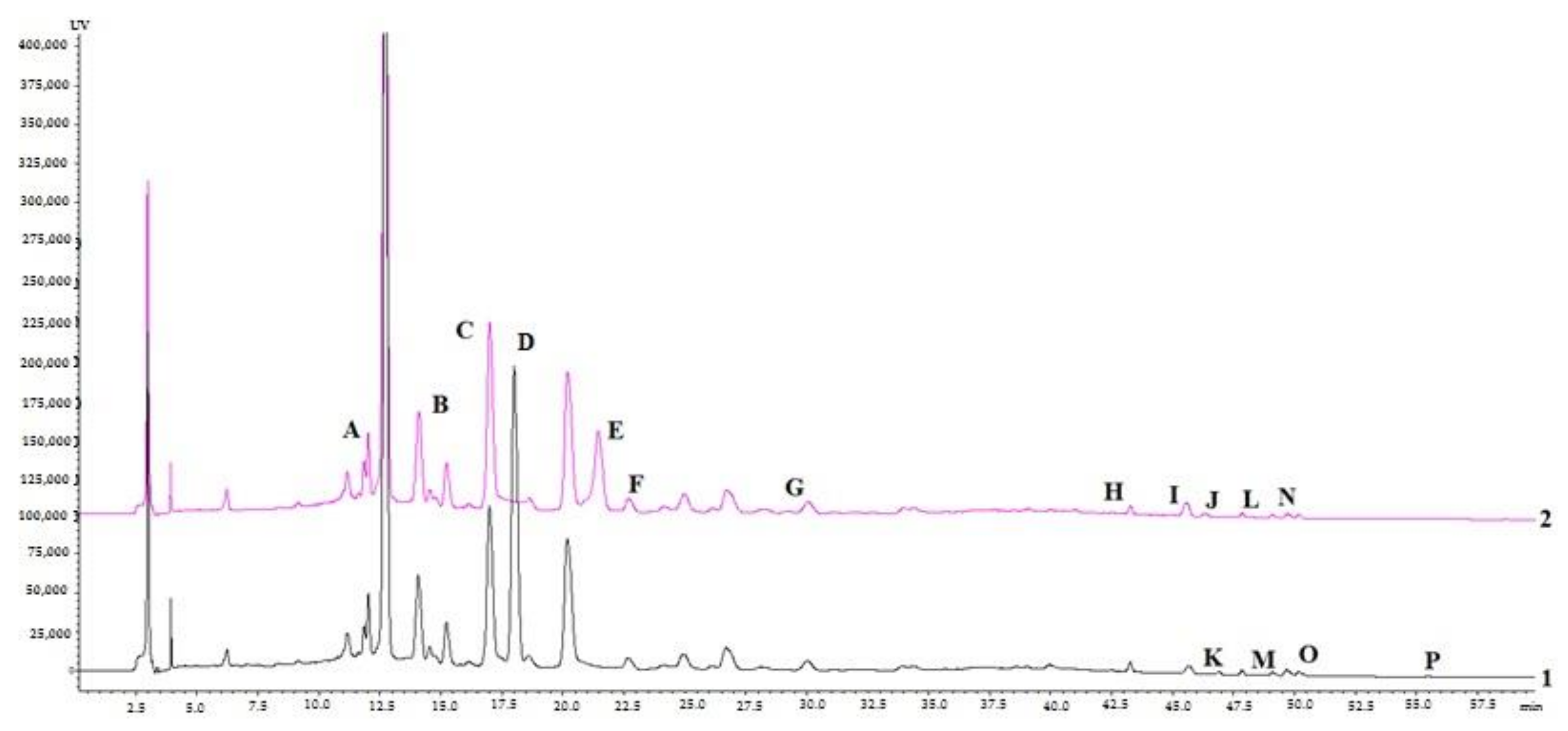
Figure 1. HPLC–DAD chromatograms of C. sativus leaves aqueous (black line 1) and ethanolic (70%, v/v) (pink line 2) dry extracts: Chlorogenic acid (A); Caffeic acid (B); Mangiferin (C); Isoorientin (D); Ferulic acid (E); Rutin (F), Tectoridin (G); Quercetin (H); Cinnamic acid (I); Nigricin (J); Genistein-7-glucoside (K); Apigenin (L); Kaempferol (M); Iristectorigenin B (N); Irigenin (O); Biochanin A (P).
Molecules 27 00010 g001
Figure 2. EC50 values of C. sativus leaf extracts against MDA-MB-231 and IGR39 cell lines after 72 h. * p < 0.05, n = 3.
Molecules 27 00010 g002
Figure 3. Cytotoxic activity of the tested individual compounds against IGR39 and MDA-MB-231 cell lines, * p < 0.05, n = 3.
Molecules 27 00010 g003
Table 1. Secondary metabolite contents (mg/g dry weight) of the aqueous and ethanolic dry extracts of C. sativus leaves.
CompoundsRt, min/λ, nmContent, mg/g
Aqueous ExtractEthanolic Extract
AChlorogenic acid11.66/3100.677 ± 0.0370.678 ± 0.004
BCaffeic acid14.18/310-1.425 ± 0.012
CMangiferin17.18/2701.823 ± 0.1242.031 ± 0.648
DIsoorientin17.66/3108.508 ± 0.001-
EFerulic acid21.64/310-0.251 ± 0.005
FRutin22.48/3100.085 ± 0.0270.100 ± 0.005
GTectoridin29.69/2700.044 ± 0.0320.107 ± 0.045
HQuercetin43.71/310-0.255 ± 0.003
It-Cinnamic acid45.22/2700.106 ± 0.0010.333 ± 0.006
JNigricin45.50/270-0.069 ± 0.003
KGenistein-7-glucoside46.07/2700.407 ± 0.015-
LApigenin47.90/340-0.088 ± 0.005
MKaempferol48.96/3100.033 ± 0.0030.046 ± 0.002
NIristectorigenin B49.15/2700.073 ± 0.0250.058 ± 0.003
OIrigenin50.03/2700.031 ± 0.0160.028 ± 0.001
PBiochanin A55.85/2700.108 ± 0.325-
Table 2. Chromatographic, UV, and mass spectroscopic data of the reference compounds.
Retention Time, min (UPLC–MS)CompoundUV λmax (nm)Mol. FormulaMol. Weight, g/mol[M−H]−(m/z)Fragment Ions (−)
3.69Chlorogenic acid218, 241, 327C16H8O9354.31353191, 179, 135
3.92Caffeic acid217, 236, 324C9H8O4180.16179161, 135
4.21Mangiferin240, 318, 257, 365C19H18O11422.30421403, 331, 301, 259, 271
4.51Isoorientin269, 349C21H20O11448.38447429, 411, 327, 297, 285
4.64Genistein-7-glucoside259, 332C21H20O10432.37431239, 268, 269, 311, 431
4.82Rutin255, 352C27H30O16610.52609301
5.09Ferulic acid218, 235, 323C10H10O4194.18193178, 149,134
6.22Apigenin237, 267, 337C15H10O5270.24269158
6.54Tectoridin263, 328C22H22O11462.41461446, 411, 341, 298
6.74Quercetin254, 369C15H10O7302.24301273, 227, 179, 151, 93
6.80trans-Cinnamic acid322, 276C9H8O2148.16147119, 103
7.41Kaempferol265, 365C15H10O6286.24285239, 187
7.46Iristectorigenin B218, 265C17H14O7330.29329314, 311, 299, 271, 255
7.58Irigenin264, 218C18H16O8360.31359344, 329, 314, 286, 258
8.20Biochanin A262, 345C16H12O5284.26283268, 267, 239, 211, 132
ND *Nigricin262, 322C17H12O6312.28NDND
* ND: compound was not detected in negative ion mode.
Table 3. Binding energies of the selected compounds identified from C. sativus leaf extracts docked against the target protein receptors.
Binding Energies in the Active Site, kcal/mol
CompoundsBreast Cancer ProteinsMelanoma
4RJ32IOK4XYF3ERT
1Chlorogenic acid−70.448−70.910−95.773−84.727
2Caffeic acid−103.721−73.071−73.432−79.224
3Mangiferin−36.321−74.476−93.985−72.074
4Isoorientin−72.180−50.336−103.029−68.522
5Ferulic acid−74.705−90.033−75.126−90.066
6Rutin−59.391−62.756−53.408−75.257
7Tectoridin−88.706−78.872−96.284−70.155
8Quercetin−77.893−56.916−94.328−66.201
9t-Cinnamic acid−81.085−72.741−60.426−84.200
10Genistein-7-Glu−76.241−67.142−96.593−72.092
11Apigenin−87.532−73.515−82.298−68.500
12Kaempferol−90.462−71.603−86.092−79.633
13Iristectorigenin B−79.516−75.179−55.603−86.510
14Nigricin−83.299−84.250−74.943−81.789
15Irigenin−86.146−80.691−80.050−56.689
Native ligands
Ligand 4RJ3−86.564
Ligand 2IOK −69.486
Hydroxytamoxifen −83.083
Ligand 4XYF −75.090
Table 4. The radical scavenging activity of individual compounds from C. sativus leaf extracts expressed as TEAC (mmol/L) using the ABTS post-column assay.
ComponentRetention TimeAqueous ExtractEthanolic Extract
Caffeic acid14.2830.172 ± 0.008
Mangiferin15.3130.405 ± 0.0170.384 ± 0.017
Isoorientin18.6930.003 ± 0.001
Ferulic acid21.8700.162 ± 0.007
Rutin22.3980.224 ± 0.0100.209 ± 0.009
Tectoridin29.7590.003 ± 0.00010.024 ± 0.001
Total 0.635 ± 0.0070.951 ± 0.010
Table 5. Neuraminidase (NA) and lipid droplets activity assays.
SampleRelative NRF2 Activity a in HacaT Cells b(%, mean ± SD)Relative NRF2 Activity a in Huh7 Cells b(%, mean ± SD)NA9 Inhibition Activity c(%, mean ± SD)Lipid Droplet Inhibition Activity d(%, mean ± SD)Superoxide Anion Generation, Human Neutrophils e (%, mean ± SEM)Elastase Release, Human Neutrophils e (%, mean ± SEM)A23187-Induced Degranulation Assay, RBL-2H3 Cells f (%, mean ± SD)Antigen-Induced Degranulation Assay, RBL-2H3 Cells f (%, mean ± SD)Protective Activity against Influenza H1N1, MDCK Cells gProtective Activity against Enterovirus 68, RD Cells gProtective Activity against Coronavirus 229E, Huh7 Cells g
C. sativus leaf aqueous extract152.5106.27.4 ± 2.986.3 ± 17.635.67 ± 5.62 **19.62 ± 2.90 **8.3 ± 7.42.0 ± 3.5inactiveinactive
C. sativus leaf ethanolic extract136.5104.56.8 ± 1.9101.3 ± 17.3ll6.3 ± 5.113.7 ± 1.5inactiveinactiveinactive
TBHQ h684.3 ± 53.3
Luteolin i23.8 ± 0.3
Zanamivir j97.4 ± 0.0
TC k16.3 ± 0.2
a Relative luciferase activity (NRF2) was calculated by normalizing luciferase activity to cell viability and presented as the fold to solvent control (n = 1). C. sativus leaves 100 µg/mL. b HacaT, a normal skin cell line. Huh7, a liver cancer cell line. c Neuraminidase inhibition assay (n = 1). C. sativus leaves 100 µg/mL. d Lipid droplet count, the average LD counts/cell of OA were used as a standard for 100% of fatty loading in the Huh7 cell line (n = 1). Saffron leaves 100 µg/mL. e Results are presented as means ± SEM (n = 3) compared with the control (fMLF/CB), ** p < 0.01. Genistein served as the positive control and inhibited 99.7% of superoxide anion generation at 10 μg/mL and 101.2% of elastase release at 30 μg/mL [30]. C. sativus leaves 10 µg/mL. f The cytotoxicity of the sample was evaluated by MTT assay (95.0 ± 8.7%). Inhibition of β-hexosaminidase release was evaluated, and the results are presented as means ± SD (n = 3) compared to the untreated control (DMSO). Dexamethasone (10 nM) was used as a positive control and inhibited 65.7 and 66.3% of A23187- and antigen-induced β-hexosaminidase release, respectively [30]. C. sativus leaves 100 µg/mL. g The protective effects were evaluated based on the viability of cells infected by the virus. Saffron stigmas 50 µg/mL for influenza and enterovirus (n = 2), 10 µg/mL for coronavirus 229E assay (n = 1). Inactive, no significant inhibition. h TBHQ, 2-(1,1-dimethylethyl)-1,4-benzenediol was used as positive control for Nrf2 activation. The drug concentration was 10 µM. i Luteolin was used as a negative control for Nrf2 activation. The drug concentration was 50 µM. j Zanamivir was used as a positive control for NA inhibition. The drug concentration was 1 µM. k TC, Triacsin C, is an inhibitor of long fatty acyl CoA synthetase and was used as a positive control for lipid droplet inhibition. The drug concentration was 1 µM. l The sample was not soluble. –, not tested.
Publisher’s Note: MDPI stays neutral with regard to jurisdictional claims in published maps and institutional affiliations.

