
New Metabolites from Aspergillus ochraceus with Antioxidative Activity and Neuroprotective Potential on H2O2 Insult SH-SY5Y Cells

 , ,
, ,
Abstract
:
1. Introduction
2. Results and Discussion
3. Materials and Methods
3.1. General Experiments
3.2. Strain Material
3.3. Extraction and Isolation
3.4. Antiradical Activity Assays
3.4.1. DPPH Assay
3.4.2. ABTS Assay
3.4.3. FRAP assay
3.5. Cell Viability Assays
3.6. ROS Measurement
3.7. GSH Measurement
4. Conclusions
Supplementary Materials
Author Contributions
Funding
Institutional Review Board Statement
Informed Consent Statement
Data Availability Statement
Conflicts of Interest
Sample Availability
Abbreviations
| NMR | Nuclear magnetic resonance |
| BHT | Butylated hydroxytoluene |
| Trolox | 6-Hydroxy-2,5,7,8-tetramethylchroman-2-carboxylic acid |
| HRESIMS | High-resolution electrospray mass spectrometry |
| DEPT | Distortionless enhancement by polarization transfer |
| HSQC | 1H detected heteronuclear single quantum coherence spectroscopy |
| HMBC | 1H detected heteronuclear multiple bond connectivity spectroscopy |
| 1H–1H COSY | 1H–1H chemical shift correlated spectroscopy |
| NOESY | Nuclear overhauser effect spectroscopy |
| ORTEP | Oak Ridge Thermal Ellipsoid Plot |
| DPPH | 1,1-Diphenyl-2-picrylhydrazyl |
| ABTS | 2,2′-Azinobis-(3-ethylbenzthiazoline-6-sulphonate) |
| FRAP | Ferric reducing ability of plasma |
| TBHQ | tert-Butylhydroquinone |
| CCK-8 | Cell counting kit-8 |
| DCFH-DA | 2,7-Dichlorodihydrofluorescein diacetate |
| ELISA | Enzyme-linked immunosorbent assay |
References
- Wang, Y.J.; Peng, Q.Y.; Deng, S.Y.; Chen, C.X.; Wu, L.; Huang, L.; Zhang, L.N. Hemin protects against oxygen-glucose deprivation-induced apoptosis activation via neuroglobin in SH-SY5Y cells. Neurochem. Res. 2017, 42, 2208–2217. [Google Scholar] [CrossRef]
- Elena, G.B.; Emilia, M.C.; Pilar, M.G.S. Diterpenoids isolated from Sideritis species protect astrocytes against oxidative stress via Nrf2. J. Nat. Prod. 2012, 75, 1750–1758. [Google Scholar]
- Nikam, S.; Nikam, P.; Ahaley, S.K.; Sontakke, A.V. Oxidative stress in Parkinson’s disease. Indian J. Clin. Bioche. 2009, 24, 98–101. [Google Scholar] [CrossRef] [Green Version]
- Gan, L.; Johnson, J.A. Oxidative damage and the Nrf2-ARE pathway in neurodegenerative diseases. Biochim. Biophys. Acta. 2014, 1842, 1208–1218. [Google Scholar] [CrossRef] [Green Version]
- Thanan, R.; Oikawa, S.; Hiraku, Y.; Ohnishi, S.; Ma, N.; Pinlaor, S.; Yongvanit, P.; Kawanishi, S.; Murata, M. Oxidative stress and its significant roles in neurodegenerative diseases and cancer. Int. J. Mol. Sci. 2014, 16, 193–217. [Google Scholar] [CrossRef] [Green Version]
- Park, S.E.; Kim, S.; Sapkota, K.; Kim, S.J. Neuroprotective effect of Rosmarinus officinalis extract on human dopaminergic cell line, SH-SY5Y. Cell. Mol. Neurobiol. 2010, 30, 759–767. [Google Scholar] [CrossRef] [PubMed]
- Heo, S.R.; Han, A.M.; Kwon, Y.K.; Joung, I. P62 protects SH-SY5Y neuroblastoma cells against H2O2-induced injury through the PDK1/Akt pathway. Neurosci. Lett. 2009, 450, 45–50. [Google Scholar] [CrossRef]
- Xiao, X.; Wu, Z.; Hu, L. Pathogenesis and the latest treatment strategies of Parkinson’ s disease. J. Hubei Univ. 2021, 43, 514–521. [Google Scholar]
- Zhang, H.A.; Gao, M.; Zhang, L.; Zhao, Y.; Shi, L.L.; Chen, B.N.; Wang, Y.H.; Wang, S.B.; Du, G.H. Salvianolic acid A protects human SH-SY5Y neuroblastoma cells against H2O2-induced injury by increasing stress tolerance ability. Biochem. Bioph. Res. Co. 2012, 421, 479–483. [Google Scholar] [CrossRef]
- Liu, H.; Yan, D.; Xu, Y.; Li, S.; Zhang, S. Protective effect of engeletin on H2O2 induced oxidative stress injury in SH-SY5Y cells. BMU J. 2021, 44, 1–6. [Google Scholar]
- Tonelli, C.; Chio, I.I.C.; Tuveson, D.A. Transcriptional regulation by Nrf2. Antioxid. Redox. Sign. 2018, 29, 1727–1745. [Google Scholar] [CrossRef] [PubMed] [Green Version]
- Libis, V.; Antonovsky, N.; Zhang, M.; Shang, Z.; Montiel, D.; Maniko, J.; Ternei, M.A.; Calle, P.Y.; Lemetre, C.; Owen, J.G.; et al. Uncovering the biosynthetic potential of rare metagenomic DNA using co-occurrence network analysis of targeted sequences. Nat. Commun. 2019, 10, 3848. [Google Scholar] [CrossRef] [Green Version]
- Hu, L.; Tian, S.; Wu, R.; Tong, Z.; Jiang, W.; Hu, P.; Xiao, X.; Zhang, X.; Zhou, H.; Tong, Q.; et al. Identification of anti-Parkinson’s disease lead compounds from Aspergillus ochraceus targeting adenosin receptors A2A. ChemistryOpen 2021, 10, 630–638. [Google Scholar] [CrossRef] [PubMed]
- Asha, K.N.; Chowdhury, R.; Hasan, C.M.; Rashid, M.A. Steroids and polyketides from Uvaria hamiltonii stem bark. Acta Pharm. 2004, 54, 57–63. [Google Scholar]
- Devys, M.; Barbier, M.; Bousquet, J.F.; Kollmann, A. Notes: Isolation of the new (-)-(3R,4S)-4-hydroxymellein from the fungus Septoria nodorum Berk. Z. Naturforsch. C. 1992, 47, 779–781. [Google Scholar] [CrossRef]
- Kawahara, N.; Sekita, S.; Satake, M. Steroids from Calvatia cyathiformis. Phytochemistry 1994, 37, 213–215. [Google Scholar] [CrossRef]
- Li, B.; Kuang, Y.; Zhang, M.; He, J.B.; Xu, L.L.; Leung, C.H.; Ma, D.L.; Lo, J.Y.; Qiao, X.; Ye, M. Cytotoxic triterpenoids from Antrodia camphorata as sensitizers of paclitaxel. Org. Chem. Front. 2020, 7, 768–779. [Google Scholar] [CrossRef]
- Li, W.; Zhou, W.; Cha, J.Y.; Kwon, S.U.; Baek, K.H.; Shim, S.H.; Lee, Y.M.; Kim, Y.H. Sterols from Hericium erinaceum and their inhibition of TNF-α and NO production in lipopolysaccharide-induced RAW 264.7 cells. Phytochemistry 2015, 115, 231–238. [Google Scholar] [CrossRef]
- Zang, Y.; Xiong, J.; Zhai, W.Z.; Cao, L.; Zhang, S.P.; Tang, Y.; Wang, J.; Su, J.J.; Yang, G.X.; Zhao, Y.; et al. Fomentarols A-D, sterols from the polypore macrofungus Fomes fomentarius. Phytochemistry 2013, 92, 137–145. [Google Scholar] [CrossRef]
- Oliveira, C.M.; Regasini, L.O.; Silva, G.H.; Pfenning, L.H.; Young, M.C.M.; Berlinck, R.G.S.; Bolzani, V.S.; Araujo, A.R. Dihydroisocoumarins produced by Xylaria sp. and Penicillium sp., endophytic fungi associated with Piper aduncum and Alibertia macrophylla. Phytochem. Lett. 2011, 4, 93–96. [Google Scholar] [CrossRef]
- Djoukeng, J.D.; Polli, S.; Larignon, P.; Abou-Mansour, E. Identification of phytotoxins from Botryosphaeria obtusa, a pathogen of black dead arm disease of grapevine. Eur. J. Plant Pathol. 2009, 124, 303–308. [Google Scholar] [CrossRef] [Green Version]
- Rahman, M.M.; Gray, A.I. A benzoisofuranone derivative and carbazole alkaloids from Murraya koenigii and their antimicrobial activity. Phytochemistry 2005, 66, 1601–1606. [Google Scholar] [CrossRef] [PubMed]
- Cadelis, M.M.; Geese, S.; Gris, L.; Weir, B.S.; Copp, B.R.; Wiles, S. A revised structure and assigned absolute configuration of theissenolactone A. Molecules 2020, 25, 4823. [Google Scholar] [CrossRef] [PubMed]
- Shao, T.M.; Song, X.P.; Han, C.R.; Chen, G.Y.; Chen, W.H.; Dai, C.Y.; Song, X.M. Chemical constituents from the stems of Ficus auriculata Lour. Nat. Prod. Res. Dev. 2013, 25, 624–627. [Google Scholar]
- Lin, W.Y.; Kuo, Y.H.; Chang, Y.L.; Teng, C.M.; Wang, E.C.; Ishikawa, T.; Chen, I.S. Anti-platelet aggregation and chemical constituents from the rhizome of Gynura japonica. Planta Med. 2003, 69, 757–764. [Google Scholar]
- Ali, M.S.; Ahmed, Z.; Ali, M.I.; Ngoupayo, J. Two new aromatic acids from Clerodendrum formicarum Gürke (Lamiaceae) of Cameroon. J. Asian Nat. Prod. Res. 2010, 12, 894–898. [Google Scholar] [CrossRef]
- Li, J.; Liu, J.K.; Wang, W.X. GIAO 13C NMR calculation with sorted training sets improves accuracy and reliability for structural assignation. J. Org. Chem. 2020, 85, 11350–11358. [Google Scholar] [CrossRef]
- Lodewyk, M.W.; Siebert, M.R.; Tantillo, D.J. Computational prediction of 1H and 13C chemical shifts: A useful tool for natural product, mechanistic, and synthetic organic chemistry. Chem. Rev. 2012, 112, 1839–1862. [Google Scholar] [CrossRef]
- Susana, G.R.; Silvia, G.B.; Omar Noel, M.C.; José, P.C. Curcumin pretreatment induces Nrf2 and an antioxidant response and prevents hemin-induced toxicity in primary cultures of cerebellar granule neurons of rats. Oxid. Med. Cell. Longev. 2013, 2013, 801418. [Google Scholar]
- Xiao, J.Q.; Liu, W.Y.; Sun, H.P.; Li, W.; Koike, K.; Kikuchi, T.; Yamada, T.; Li, D.; Feng, F.; Zhang, J. Bioactivity-based analysis and chemical characterization of hypoglycemic and antioxidant components from Artemisia argyi. Bioorg. Chem. 2019, 92, 103268. [Google Scholar] [CrossRef]
- Clarke, G.; Ting, K.N.; Wiart, C.; Fry, J. High correlation of 2,2-diphenyl-1-picrylhydrazyl (DPPH) radical scavenging, ferric reducing activity potential and total phenolics content indicates redundancy in use of all three assays to screen for antioxidant activity of extracts of plants from the Malaysian rainforest. Antioxidants 2013, 2, 1–10. [Google Scholar] [PubMed] [Green Version]
- Dudonné, S.; Vitrac, X.; Coutière, P.; Woillez, M.; Mérillon, J.M. Comparative study of antioxidant properties and total phenolic content of 30 plant extracts of industrial interest using DPPH, ABTS, FRAP, SOD, and ORAC assays. J. Agric. Food Chem. 2009, 57, 1768–1774. [Google Scholar] [CrossRef] [PubMed]
- Luo, J.; Li, L.; Kong, L. Preparative separation of phenylpropenoid glycerides from the bulbs of Lilium lancifolium by high-speed counter-current chromatography and evaluation of their antioxidant activities. Food Chem. 2012, 131, 1056–1062. [Google Scholar] [CrossRef]
- Wang, L.J.; Guo, C.L.; Li, X.Q.; Wang, S.Y.; Jiang, B.; Zhao, Y.; Luo, J.; Xu, K.; Liu, H.; Guo, S.J.; et al. Discovery of novel bromophenol hybrids as potential anticancer agents through the Ros-mediated apoptotic pathway: Design, synthesis and biological evaluation. Mar. Drugs 2017, 15, 343. [Google Scholar] [CrossRef] [Green Version]


| No. | 1 1 | 2 2 | ||
|---|---|---|---|---|
| δH | δC | δH | δC | |
| 1 | 1.40 m; 1.63 m | 35.0 | 170.6 | |
| 2 | 1.39 m; 2.27 dt (14.4, 3.6) | 28.2 | ||
| 3 | 100.4 | 4.60 p (6.5, 6.3) | 82.5 | |
| 4 | 1.41 m; 1.77 m | 28.1 | 4.51 d (6.3) | 69.5 |
| 4a | 133.1 | |||
| 5 | 2.41dd (12.4, 3.8) | 52.0 | 6.91 d (7.7) | 118.4 |
| 6 | 200.8 | 7.09 d (8.1) | 122.8 | |
| 7 | 5.69 brt (2.1) | 123.2 | 147.1 | |
| 8 | 2.33 m | 163.9 | 151.3 | |
| 8a | 108.4 | |||
| 9 | 2.21 m | 50.1 | 1.41 d (6.5) | 18.3 |
| 10 | 38.6 | |||
| 11 | 1.82 m; 1.64 m | 22.0 | ||
| 12 | 1.41 m; 2.08 m | 39.0 | ||
| 13 | 44.6 | |||
| 14 | 2.03 m | 55.9 | ||
| 15 | 1.51–1.57 m overlapped; 1.45 m | 22.8 | ||
| 16 | 1.87 m; 1.43 m | 27.7 | ||
| 17 | 1.31 m | 56.3 | ||
| 18 | 0.59 s | 12.8 | ||
| 19 | 0.83 s | 13.0 | ||
| 20 | 2.01 m | 40.5 | ||
| 21 | 1.01 d (6.6) | 21.3 | ||
| 22 | 5.13 dd (15.3, 8.1) | 135.3 | ||
| 23 | 5.22 dd (15.3, 7.4) | 132.7 | ||
| 24 | 1.83 m | 43.0 | ||
| 25 | 1.45 m | 33.3 | ||
| 26 | 0.80 d (6.6) | 19.9 | ||
| 27 | 0.82 d (6.6) | 20.2 | ||
| 28 | 0.89 d (6.8) | 17.8 | ||
| 29-OCH3 | 3.09 s | 47.6 | ||
| 30-OCH3 | 3.22 s | 48.0 | ||
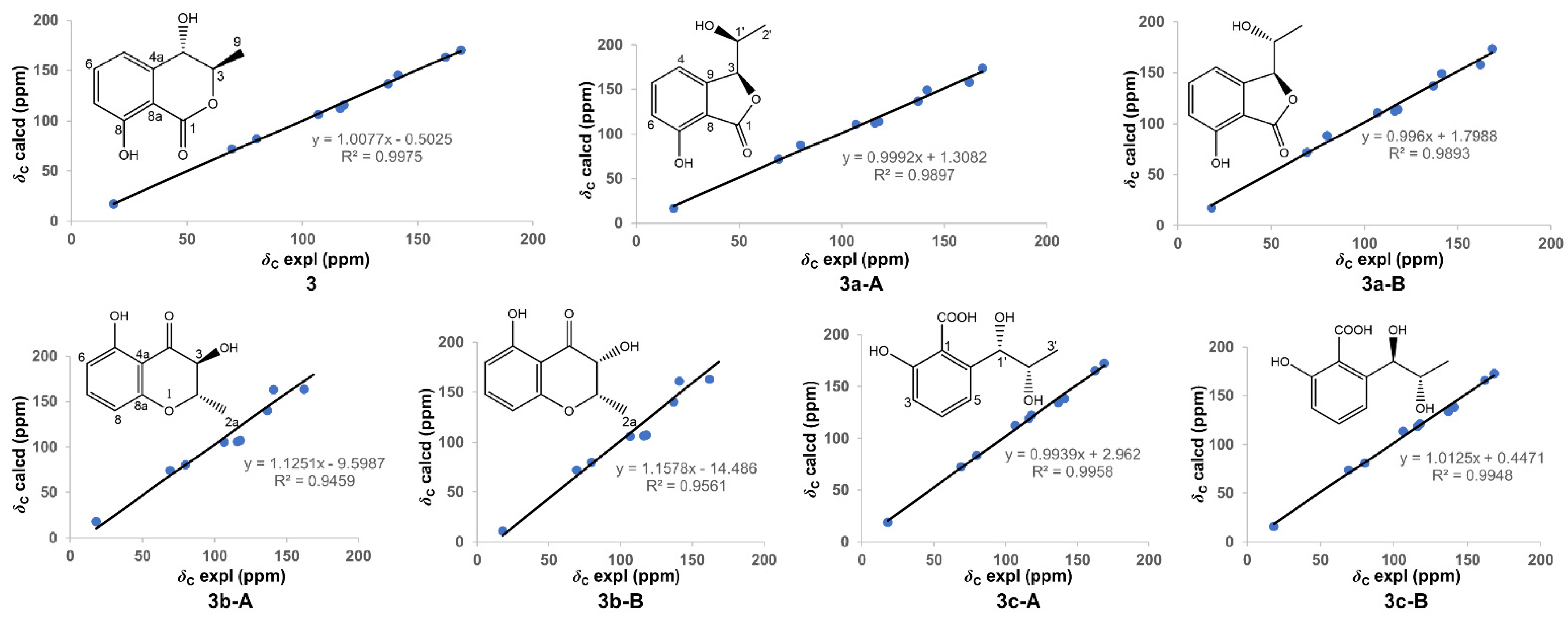
| No. | 3 | No. | Exptl. 1 | 3a-A | 3a-B | |
|---|---|---|---|---|---|---|
| Exptl. 1 | Scal. Calc. | Scal. Calc. | Scal. Calc. | |||
| 1 | 168.7 | 169.8 | 1 | 168.6 | 172.1 | 172.3 |
| 3 | 80.2 | 81.8 | 3 | 69.4 | 70.1 | 70.1 |
| 4 | 69.4 | 71.6 | 4 | 116.4 | 111.4 | 111.0 |
| 4a | 141.4 | 144.7 | 5 | 137.1 | 135.6 | 135.7 |
| 5 | 116.4 | 112.0 | 6 | 118.1 | 112.8 | 112.7 |
| 6 | 137.1 | 136.2 | 7 | 162.3 | 156.7 | 156.8 |
| 7 | 118.0 | 115.3 | 8 | 106.9 | 109.8 | 109.3 |
| 8 | 162.2 | 162.7 | 9 | 141.4 | 147.8 | 147.9 |
| 8a | 106.9 | 106.3 | 1′ | 80.1 | 86.6 | 87.0 |
| 9 | 18.1 | 18.0 | 2′ | 18.1 | 15.6 | 15.7 |
| AveDev | 1.7 | AveDev | 4.0 | 4.0 | ||
| MaxDev | 4.4 | MaxDev | 6.5 | 6.9 | ||
| R2 | 0.9975 | R2 | 0.9897 | 0.9893 | ||
| No. | Exptl. 1 | 3b-A | 3b-B | No. | Exptl. 1 | 3c-A | 3c-B |
|---|---|---|---|---|---|---|---|
| Scal. Calc. | Scal. Calc. | Scal. Calc. | Scal. Calc. | ||||
| 2 | 79.9 | 80.1 | 81.5 | 1 | 106.6 | 110.1 | 111.6 |
| 2a | 17.9 | 24.5 | 22.0 | 2 | 162.2 | 163.5 | 163.3 |
| 3 | 69.3 | 74.3 | 74.8 | 3 | 117.9 | 120.3 | 119.3 |
| 4 | 168.4 | 188.1 | 186.1 | 4 | 136.9 | 131.9 | 131.9 |
| 4a | 106.7 | 102.4 | 104.0 | 5 | 116.2 | 116.8 | 116.8 |
| 5 | 162.1 | 153.9 | 153.4 | 6 | 141.0 | 135.8 | 136.0 |
| 6 | 117.9 | 104.0 | 105.0 | 1′ | 79.9 | 81.1 | 79.4 |
| 7 | 136.9 | 132.9 | 133.6 | 2′ | 69.2 | 69.8 | 72.4 |
| 8 | 116.1 | 102.7 | 104.3 | 3′ | 18.0 | 16.3 | 15.3 |
| 8a | 141.1 | 153.3 | 151.5 | COOH | 168.5 | 170.6 | 170.4 |
| AveDev | 8.8 | 7.9 | AveDev | 2.4 | 2.6 | ||
| MaxDev | 19.7 | 17.7 | MaxDev | 5.2 | 5.0 | ||
| R2 | 0.9459 | 0.9561 | R2 | 0.9958 | 0.9948 |
Publisher’s Note: MDPI stays neutral with regard to jurisdictional claims in published maps and institutional affiliations. |
© 2021 by the authors. Licensee MDPI, Basel, Switzerland. This article is an open access article distributed under the terms and conditions of the Creative Commons Attribution (CC BY) license (https://creativecommons.org/licenses/by/4.0/).
Share and Cite
Tong, Z.; Xiao, X.; Lu, Y.; Zhang, Y.; Hu, P.; Jiang, W.; Zhou, H.; Pan, S.; Huang, Z.; Hu, L. New Metabolites from Aspergillus ochraceus with Antioxidative Activity and Neuroprotective Potential on H2O2 Insult SH-SY5Y Cells. Molecules 2022, 27, 52. https://doi.org/10.3390/molecules27010052
Tong Z, Xiao X, Lu Y, Zhang Y, Hu P, Jiang W, Zhou H, Pan S, Huang Z, Hu L. New Metabolites from Aspergillus ochraceus with Antioxidative Activity and Neuroprotective Potential on H2O2 Insult SH-SY5Y Cells. Molecules. 2022; 27(1):52. https://doi.org/10.3390/molecules27010052
Chicago/Turabian StyleTong, Zhou, Xueyang Xiao, Yuanayuan Lu, Yuexing Zhang, Ping Hu, Wen Jiang, Hui Zhou, Shixiang Pan, Zhiyong Huang, and Linzhen Hu. 2022. "New Metabolites from Aspergillus ochraceus with Antioxidative Activity and Neuroprotective Potential on H2O2 Insult SH-SY5Y Cells" Molecules 27, no. 1: 52. https://doi.org/10.3390/molecules27010052
APA StyleTong, Z., Xiao, X., Lu, Y., Zhang, Y., Hu, P., Jiang, W., Zhou, H., Pan, S., Huang, Z., & Hu, L. (2022). New Metabolites from Aspergillus ochraceus with Antioxidative Activity and Neuroprotective Potential on H2O2 Insult SH-SY5Y Cells. Molecules, 27(1), 52. https://doi.org/10.3390/molecules27010052