
The GABAergic System and Endocannabinoids in Epilepsy and Seizures: What Can We Expect from Plant Oils?

 ,
,  ,
,  , , and
Abstract
:
1. Epilepsy and Seizures
2. Role of GABA in the Pathophysiology of Epilepsy and Seizures
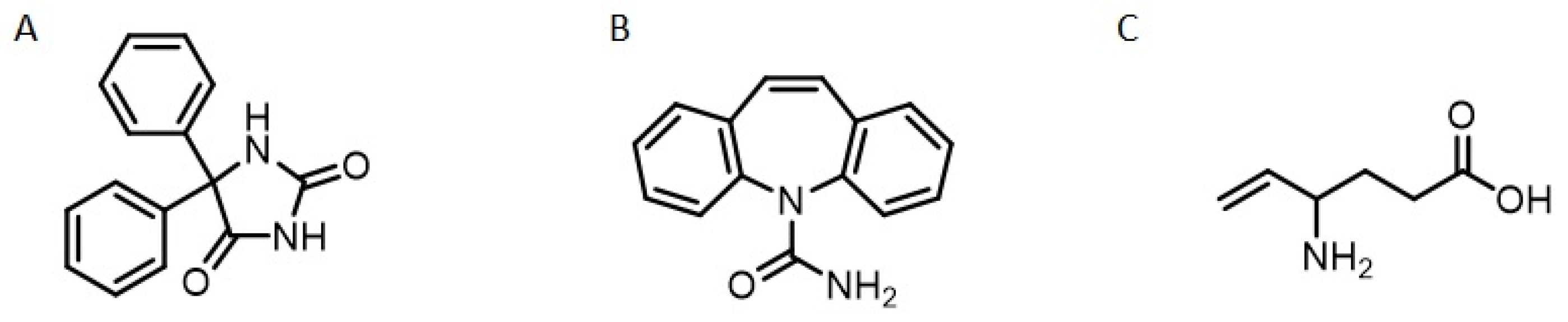
3. Treatment of Seizures and Screening of New Drugs
4. Endocannabinoids, Plant Oils, and Seizure Control
5. Endocannabinoids from Plant Oils
6. Endocannabinoids and Seizure Control
7. Conclusions
Author Contributions
Funding
Institutional Review Board Statement
Informed Consent Statement
Data Availability Statement
Conflicts of Interest
References
- Singh, G.; Sander, J.W. The global burden of epilepsy report: Implications for low- and middle income countries. Epilepsy Behav. 2020, 105, 106949–1066952. [Google Scholar] [CrossRef]
- Ravizza, T.; Boer, K.; Redeker, S.; Spliet, W.G.M.; Van Rijen, P.C.; Troost, D.; Vezzani, A.; Aronica, E. The IL-1 system in epilepsy-associated malformations of cortical development. Neurobiol. Dis. 2006, 24, 128–143. [Google Scholar] [CrossRef]
- Berg, A.T.; Berkovic, S.F.; Brodie, M.J.; Buchhalter, J.; Cross, J.H.; Van Emde BOAS, W.; Engel, J.; French, J.; Glauser, T.A.; Mathern, G.W.; et al. Revised terminology and concepts for organization of seizures and epilepsies: Report of the ILAE Commission on Classification and Terminology, 2005–2009. Epilepsia 2010, 51, 676–685. [Google Scholar] [CrossRef]
- Vezzani, A.; French, J.; Bartfai, T.; Baram, T.Z. The role of inflammation in epilepsy. Nat. Rev. Neurol. 2011, 7, 31–40. [Google Scholar] [CrossRef] [Green Version]
- Thijs, R.D.; Surges, R.; O’Brien, T.J.; Sander, J.W. Epilepsy in adults. Lancet 2019, 393, 689–701. [Google Scholar] [CrossRef]
- Xue-Ping, W.; Hai-Jiao, W.; Li-Na, Z.; Ling, L. Risk factors for drug-resistant epilepsy: A systematic review and meta-analysis. Medicine 2019, 98, e16402–e16413. [Google Scholar] [CrossRef]
- Chaudhary, U.J.; Duncan, J.S.; Lemieux, L. A dialogue with historical concepts of epilepsy from the Babylonians to Hughlings Jackson: Persistent beliefs. Epilepsy Behav. 2011, 21, 109–114. [Google Scholar] [CrossRef]
- Fisher, R.S.; Acevedo, C.; Arzimanoglou, A.; Bogacz, A.; Cross, J.H.; Elger, C.E.; Engel, J., Jr.; Forsgren, L.; French, J.A.; Glynn, M.; et al. ILAE official report: A practical clinical definition of epilepsy. Epilepsia 2014, 55, 475–482. [Google Scholar] [CrossRef] [Green Version]
- Oliveira, F.R.; Rodrigues, K.E.; Hamoy, M.; Sarquis, I.R.; Hamoy, A.O.; Lopez, M.E.C.; Ferreira, I.M.; Macchi, B.M.; do Nascimento, J.L.M. Fatty Acid Amides Synthesized from Andiroba Oil (Carapa guianensis Aublet.) exhibit Anticonvulsant Action with Modulation on GABA-A Receptor in Mice: A Putative Therapeutic Option. Pharmaceuticals 2020, 13, 43. [Google Scholar] [CrossRef] [Green Version]
- Shneker, B.F.; Fountain, N.B. Assessment of acute morbidity and mortality in nonconvulsive status epilepticus. Neurology 2003, 61, 1066–1073. [Google Scholar] [CrossRef]
- Duncan, J.S.; Sander, J.W.; Sisodiya, S.M.; Walker, M.C. Adult epilepsy. Lancet 2006, 367, 1087–1100. [Google Scholar] [CrossRef]
- Fisher, R.S.; Cross, J.H.; French, J.A.; Higurashi, N.; Hirsch, E.; Jansen, F.E.; Lagae, L.; Moshé, S.; Perez, E.R.; Scheffer, I.E.; et al. Operational classification of seizure types by the International League Against Epilepsy: Position Paper of the ILAE Commission for Classification and Terminology. Epilepsy 2017, 58, 522–530. [Google Scholar] [CrossRef] [Green Version]
- Scheffer, I.E.; Berkovic, S.; Capovilla, G. ILAE classification of the epilepsies: Position paper of the ILAE Commission for Classification and Terminology. Epilepsia 2017, 58, 512–521. [Google Scholar] [CrossRef] [Green Version]
- Terlau, J.; Yang, J.W.; Khastkhodaei, Z.; Seidenbeche, T.; Luhmann, H.J.; Pape, H.C.; Luttjohann, A. Spike-wave discharges in absence epilepsy: Segregation of electrographic components reveals distinct pathways of seizure activity. J. Physiol. 2020, 598, 2397–2414. [Google Scholar] [CrossRef]
- Armijo, J.A.; Shushtarian, M.; Valdizan, E.M.; Cuadrado, A.; de las Cuevas, I.; Adín, J. Ion Channels and Epilepsy. Curr. Pharm. Des. 2005, 11, 1975–2003. [Google Scholar] [CrossRef]
- Stafstrom, C.E. and Carmant, L. Seizures and Epilepsy: An Overview for Neuroscientists. Cold Spring Harb. Perspect. Med. 2015, 5, a022426. [Google Scholar] [CrossRef]
- Nickels, K.; Kossoff, E.H.; Eschbach, K.; Joshi, C. Epilepsy with myoclonic-atonic seizures (Doose syndrome): Clarification of diagnosis and treatment options through a large retrospective multicenter cohort. Epilepsia 2021, 62, 120–127. [Google Scholar] [CrossRef]
- Bauer, J.; Bös, M.; Reuber, M. Treatment strategies for focal epilepsy. Expert Opin. Pharm. 2009, 10, 743–753. [Google Scholar] [CrossRef]
- Pal, D.K.; Ferrie, C.; Addis, L.; Akiyama, T.; Capovilla, G.; Caraballo, R.; Saint-Martin, A.; Fejerman, N.; Guerrini, R.; Hamandi, K.; et al. Idiopathic focal epilepsies: The “lost tribe”. Epileptic Disord. 2016, 18, 252–288. [Google Scholar] [CrossRef] [Green Version]
- Garcia-Fernandez, M. Epileptic spasms in infants. Beyond hypsarrhythmia. Rev. Neurol. 2017, 17, 555–559. [Google Scholar]
- Pavone, P.; Striano, P.; Falsaperla, R.; Pavone, L.; Ruggieri, M. Infantile spasms syndrome, West syndrome and related phenotypes: What we know in 2013. Brain Dev. 2014, 36, 739–751. [Google Scholar] [CrossRef]
- Holmes, G.L. Role of Glutamate and GABA in the pathophysiology of Epilepsy mental retardation and developmental disabilities. Res. Rev. 1995, 1, 208–219. [Google Scholar]
- Cherubini, E.; Conti, F. Generating diversity at GABAergic synapses. Trends Neurosci. 2001, 24, 155–162. [Google Scholar] [CrossRef]
- Roberts, E. Disinhibition as an Organizing Principle in the Nervous System-The Role of the GABA System. Application to Neurologic and Psychiatric Disorders. In GABA in the Nerious System Funcfion; Roberts, E., Chase, T.N., Tower, D.B., Eds.; Raven Press: New York, NY, USA, 1976; pp. 515–539. [Google Scholar]
- Wang, J.; Ueda, N. Role of the endocannabinoid system in metabolic control. Curr. Opin. Nephrol. Hypertens 2008, 17, 1–10. [Google Scholar] [CrossRef]
- Ye, H.; Kaszuba, S. Inhibitory or excitatory? Optogenetic interrogation of the functional roles of GABAergic interneurons in epileptogenesis. J. Biomed. Sci. 2017, 24, 93–102. [Google Scholar] [CrossRef] [Green Version]
- Devinsky, O.; Vezzani, A.; O’Brien, T.J.; Jette, N.; Scheffer, I.E.; de Curtis, M.; Perucca, P. Epilepsy. Nat. Rev. Dis. Primers 2018, 4, 18024. [Google Scholar] [CrossRef]
- Ghit, A.; Assal, D.; Al-Shami, A.S.; Hussein, D.E.E. GABAA receptors: Structure, function, pharmacology, and related disorders. J. Genet. Eng. Biotechnol. 2021, 19, 123–138. [Google Scholar] [CrossRef]
- Tizazu, E.; Ellis, C.A.; Reichert, J.; Lancaster, E. Low rate of glutamic acid decarboxylase 65 (GAD-65) antibodies in chronic epilepsy. Seizure 2020, 80, 38–41. [Google Scholar] [CrossRef]
- Treiman, D.M. GABAergic mechanisms in epilepsy. Epilepsia 2001, 42, 8–12. [Google Scholar] [CrossRef]
- Amoras, C.G.; Aguiar, S.J.; Maués, L.A.L.; Picanço-Diniz, D.L.W.; Herculano, A.M.; do Nascimento, J.L.M. A simple and rapid method to measure γ-aminobutyric acid-transaminase (GABA-T) in the central nervous system. Neurociências 2009, 5, 135–140. [Google Scholar]
- Jembrej, M.J.; Vlainic, J. GABA Receptors: Pharmacological Potential and Pitfalls. Curr. Pharm. Des. 2015, 21, 4943–4959. [Google Scholar] [CrossRef] [Green Version]
- Terunuma, M. Diversity of structure and function of GABAB receptors: A complexity of GABAB-mediated signaling. Proc. Jpn. Acad. Ser. B Phys. Biol. Sci. 2018, 94, 390–411. [Google Scholar] [CrossRef] [Green Version]
- do Nascimento, J.L.M.; de Mello, F.G. Induced Release of y-Aminobutyric Acid by a Carrier-Mediated, High-Affinity Uptake of L-Glutamate in Cultured Chick Retina Cells. J. Neurochem. 1985, 45, 1820–1827. [Google Scholar]
- Soudijn, W.; van Wijngaarden, I. The GABA transporter and its inhibitors. Curr. Med. Chem. 2000, 7, 1063–1079. [Google Scholar] [CrossRef]
- Ethiraj, J.; Palpagama, T.H.; Turner, C.; der Werf, B.V.; Waldvogel, H.J.; Faull, R.L.M.; Kwakowsky, A. The effect of age and sex on the expression of GABA signaling components in the human hippocampus and entorhinal córtex. Sci. Rep. 2021, 11, 21470–21484. [Google Scholar] [CrossRef]
- Graus, F.; Saiz, A.; Dalmau, J. GAD antibodies in neurological disorders—Insights and challenges. Nat. Rev. Neurol. 2020, 16, 353–365. [Google Scholar] [CrossRef]
- Cossart, R.; Bernard, C.; Bem-Ari, Y. Multiple facets of GABAergic neurons and synapses: Multiple fates of GABA signalling in epilepsies. Trends Neurosci. 2005, 28, 108–115. [Google Scholar] [CrossRef]
- Margeta-Mitrovic, M.; Jan, Y.N.; Jan, L.Y. Function of GB1 and GB2 subunits in G protein coupling of GABAB receptors. Proc. Natl. Acad. Sci. USA 2001, 98, 14649–14654. [Google Scholar] [CrossRef] [Green Version]
- Perucca, P.; Scheffer, I.E.; Kiley, M. The management of epilepsy in children and adults. Med. J. Aust. 2018, 5, 226–233. [Google Scholar] [CrossRef]
- Shorvon, S.D. Drug treatment of epilepsy in the century of the ILAE: The second 50 years, 1959–2009. Epilepsia 2009, 50, 93–130. [Google Scholar] [CrossRef] [Green Version]
- Löscher, W. Animal Models of Seizures and Epilepsy: Past, Present, and Future Role for the Discovery of Antiseizure Drugs. Neurochem. Res. 2017, 42, 1873–1888. [Google Scholar] [CrossRef]
- Cook, A.M.; Bensalem-Owen, M.K. Mechanisms of action of antiepileptic drugs. Therapy 2011, 8, 307–313. [Google Scholar] [CrossRef]
- Löscher, W. Strategies for antiepileptogenesis: Antiepileptic drugs versus novel approaches evaluated in post-status epilepticus models of temporal lobe epilepsy. In Jasper’s Basic Mechanisms of the Epilepsies; Oxford University Press: Oxford, UK, 2012; pp. 1–14. [Google Scholar]
- Carpay, J.A.; Aldenkamp, A.P.; Van Donselaar, C.A. Complaints associated with the use of antiepileptic drugs: Results from a community-based study. Seizure 2005, 14, 198–206. [Google Scholar] [CrossRef] [Green Version]
- Babu, C.S.; Satishchandra, P.; Sinha, S.; Subbakrishna, D.K. Co-morbidities in people living with epilepsy: Hospital based case-control study from a resource-poor setting. Epilepsy Res. 2009, 86, 146–152. [Google Scholar] [CrossRef]
- Scott, A.J.; Sharpe, L.; Hunt, C.; Gandy, M. Anxiety and depressive disorders in people with epilepsy: A meta-analysis. Epilepsia 2017, 58, 973–982. [Google Scholar] [CrossRef]
- Lader, M. Benzodiazepine harm: How can it be reduced? Br. J. Clin. Pharmacol. 2014, 77, 295–301. [Google Scholar] [CrossRef] [Green Version]
- White, H.S. Clinical significance of animal seizure models and mechanism of action studies of potential antiepileptic drugs. Epilepsia 1997, 38, 9–17. [Google Scholar] [CrossRef]
- Kandratavicius, L.; Balista, P.A.; Lopes-Aguiar, C.; Ruggiero, R.N.; Umeoka, E.H.; Garcia-Cairasco, N.; Bueno-Junior, L.S.; Leite, J.P. Animal models of epilepsy: Use and limitations. Neuropsychiatr. Dis. Treat. 2014, 10, 1693–1705. [Google Scholar] [CrossRef] [Green Version]
- Löscher, W. Critical review of current animal models of seizures and epilepsy used in the discovery and development of new antiepileptic drugs. Seizure 2011, 20, 359–368. [Google Scholar] [CrossRef] [Green Version]
- Taiwe, G.S.; Tchoya, T.B.; Menanga, J.R.; Dabole, B.; De Waard, M. Anticonvulsant activity of an active fraction extracted from Crinum jagus L. (Amaryllidaceae), and its possible effects on fully kindled seizures, depression-like behaviour and oxidative stress in experimental rodent models. J. Ethnopharmacol. 2016, 194, 421–433. [Google Scholar] [CrossRef]
- Stumpp, L.; Smets, H.; Vespa, S.; Cury, J.; Dogue, P.; Delbeke, J.; Nonclercq, A.; Tahr, R.E. Vagus Nerve Electroneurogram-Based Detection of Acute Pentylenetetrazol. Int. J. Neural. Syst. 2021, 31, 2150024. [Google Scholar] [CrossRef]
- Ramanjaneyulu, R.; Ticku, M.K. Interactions of pentamethylenetetrazole and tetrazole analogues with the picrotoxinin site of the benzodiazepine-GABA receptor-ionophore complex. Eur. J. Pharm. 1984, 98, 337–345. [Google Scholar] [CrossRef]
- Crestani, F.; Assandr, R.; Tauber, M.; Martin, J.R.; Rudolph, U. Contribution of the alpha 1-GABAA receptor subtype to the pharmacological actions of benzodiazepine site inverse agonists. Neuropharmacology 2002, 43, 679–684. [Google Scholar] [CrossRef]
- Battista, N.; Tommaso, M.; Bari, M.; Maccarrone, M. The endocannabinoid system: An overview. Front. Behav. Neurosci. 2012, 6, 9. [Google Scholar] [CrossRef] [Green Version]
- Ezzili, C.; Otrubova, K.; Boger, D.L. Fatty acid amide signaling molecules. Bioorganic Med. Chem. Lett. 2010, 20, 5959–5968. [Google Scholar] [CrossRef] [Green Version]
- Baggelaar, M.P.; Maccarrone, M.; van der Stelt, M. 2-Arachidonoylglycerol: A signaling lipid with manifold actions in the brain. Prog. Lipid Res. 2018, 71, 1–17. [Google Scholar] [CrossRef]
- Biringer, R.G. The rise and fall of anandamide: Processes that control synthesis, degradation, and storage. Mol. Cell. Biochem. 2021, 476, 2753–2775. [Google Scholar] [CrossRef]
- Balezina, O.P.; Tarasova, E.O.; Gaydukov, A.E. Noncanonical activity of endocannabinoids and their receptors in central and peripheral synapses. Biochemistry 2021, 86, 818–832. [Google Scholar] [CrossRef]
- Cravatt, B.F.; Giang, D.K.; Mayfield, S.P.; Boger, D.L.; Lerner, R.A.; Gilula, N.B. Molecular characterization of an enzyme that degrades neuromodulatory fatty-acid amides. Nature 1996, 384, 83–87. [Google Scholar] [CrossRef]
- Cravatt, B.F.; Demarest, K.; Patricelli, M.P.; Bracey, M.H.; Giang, D.K.; Martin, B.R.; Lichtman, A.H. Supersensitivity to anandamide and enhanced endogenous cannabinoid signaling in mice lacking fatty acid amide hydrolase. Proc. Natl. Acad. Sci. USA 2001, 98, 9371–9376. [Google Scholar] [CrossRef] [Green Version]
- Egertová, M.; Cravatt, B.F.; Elphick, M.R. Comparative analysis of fatty acid amide hydrolase and CB(1) cannabinoid receptor expression in the mouse brain: Evidence of a widespread role for fatty acid amide hydrolase in regulation of endocannabinoid signaling. Neuroscience 2003, 119, 481–496. [Google Scholar] [CrossRef] [Green Version]
- Savinainen, J.R.; Saario, S.M.; Laitinen, J.T. The serine hydrolases MAGL, ABHD6 and ABHD12 as guardians of 2-arachidonoylglycerol signalling through cannabinoid receptors. Acta Physiol. 2012, 204, 267–276. [Google Scholar] [CrossRef] [PubMed]
- Ahn, K.; McKinney, M.K.; Cravatt, B.F. Enzymatic Pathways That Regulate Endocannabinoid Signaling in the Nervous System. Chem. Rev. 2008, 108, 1687–1707. [Google Scholar] [CrossRef] [PubMed] [Green Version]
- Cadas, H.; Di Tomaso, E.; Piomelli, D. Occurrence and Biosynthesis of Endogenous Cannabinoid Precursor, N-Arachidonoyl Phosphatidylethanolamine, in Rat Brain. J. Neurosci. 1997, 17, 1226–1242. [Google Scholar] [CrossRef] [PubMed] [Green Version]
- Howlett, A.C. Cannabinoid receptor signaling. Handb. Exp. Pharm. 2005, 168, 53–79. [Google Scholar]
- Katona, I.; Freund, T.F. Multiples Functions of endocannabinoid signaling in the brain. Annu. Rev. Neurosci. 2012, 35, 529–558. [Google Scholar] [CrossRef] [Green Version]
- Kubrusly, R.C.C.; Günter, A.; Sampaio, L.; Martins, R.S.; Schitine, C.S.; Trindade, P.; Fernandes, A.; Borelli-Torres, R.; Miya-Coreixas, V.S.; Rego Costa, A.C.; et al. Neuro-glial cannabinoid receptors modulate signaling in the embryonic avian retina. Neurochem. Int. 2018, 112, 27–37. [Google Scholar] [CrossRef]
- Chevaleyre, V.; Takahashi, K.A.; Castillo, P.E. Endocannabinoid-mediated synaptic plasticity in the CNS. Annu. Rev. Neurosci. 2006, 29, 37–76. [Google Scholar] [CrossRef]
- Svíženská, I.; Dubovy, P.; Sulcova, A. Cannabinoid receptors 1 and 2 (CB1 and CB2), their distribution, ligands and functional involvement in nervous system structures—A short review. Pharm. Biochem. Behav. 2008, 90, 501–511. [Google Scholar] [CrossRef]
- Pacher, P.; Kogan, N.M.; Mechoulam, R. Beyond THC and Endocannabinoids. Annu. Rev. Pharm. Toxicol. 2020, 60, 637–659. [Google Scholar] [CrossRef] [Green Version]
- Matsuda, L.A.; Lolait, S.J.; Brownstein, M.J.; Young, A.C.; Bonner, T.I. Structure of a cannabinoid receptor and functional expression of the cloned cDNA. Nature 1990, 346, 561–564. [Google Scholar] [CrossRef] [PubMed]
- Howlett, A.C. Pharmacology of cannabinoid receptors. Annu. Rev. Pharmacol. Toxicol. 1995, 35, 607–634. [Google Scholar] [CrossRef]
- Katona, I.; Freund, T.F. Endocannabinoid signaling as a synaptic circuit breaker in neurological disease. Nat. Med. 2008, 14, 923–930. [Google Scholar] [CrossRef] [PubMed]
- Gerdeman, G.; Lovinger, D.M. Emerging roles for endocannabinoids in long-term synaptic plasticity. Br. J. Pharmacol. 2003, 140, 781–789. [Google Scholar] [CrossRef] [PubMed]
- Ligresti, A.; Cascio, M.G.; Marzo, V.D. Endocannabinoid Metabolic Pathways and Enzymes. Curr. Drug Targets CNS Neurol. Disord. 2005, 4, 615–623. [Google Scholar] [CrossRef] [PubMed]
- Marsicano, G.; Lutz, B. Neuromodulatory functions of the endocannabinoid system. J. Endocrinol. Invest. 2006, 29, 27–46. [Google Scholar]
- Basu, S.; Dittel, B.N. Unraveling the complexities of cannabinoid receptor 2 (CB2) immune regulation in health and disease. Immunol. Res 2011, 51, 26–38. [Google Scholar] [CrossRef]
- Atwood, B.K.; Mackie, K. ; Mackie, K. CB2: A cannabinoid receptor with an identity crisis. Br. J. Pharmacol. 2010, 160, 467–479. [Google Scholar] [CrossRef] [Green Version]
- Ji, X.; Zeng, Y.; Wu, J. The CB2 Receptor as a Novel Therapeutic Target for Epilepsy Treatment. Int. J. Mol. Sci. 2021, 22, 8961. [Google Scholar] [CrossRef]
- Bataglion, G.A.; Silva, F.M.A.; Santos, J.M.; Santos, F.M.; Barcia, M.T.; Lourenço, C.C.; Salvador, M.J.; Godoy, H.T.; Eberlin, M.N.; Koolen, H.H.F. Comprehensive characterization of lipids from Amazonian vegetable oils by mass spectrometry techniques. Food Res. Int. 2014, 64, 472–481. [Google Scholar] [CrossRef]
- Milhomem-Paixão, S.S.R.; Fascineli, M.L.; Roll, M.M.; Longo, J.P.F.; Azevedo, R.B.; Pieczarka, J.C.; Salgado, H.L.C.; Santos, A.S.; Grisolia, C.K. The lipidome, genotoxicity, hematotoxicity and antioxidant properties of andiroba oil from the Brazilian Amazon. Genet. Mol. Biol. 2016, 39, 248–256. [Google Scholar] [CrossRef] [PubMed] [Green Version]
- Pantoja, S.S.; Conceição, L.R.V.; Costa, C.E.F.; Zamian, J.R.; Rocha Filho, G.N. Oxidative stability of biodiesels produced from vegetable oils having different degrees of unsaturation. Energy Convers. Manag. 2013, 74, 293–298. [Google Scholar] [CrossRef]
- Araujo-Lima, C.F.; Fernandes, A.S.; Gomes, E.M.; Oliveira, L.L.; Macedo, A.F.; Antoniassi, R.; Wilhelm, A.E.; Aiub, C.A.F.; Felzenszwalb, I. Antioxidant Activity and Genotoxic Assessment of Crabwood (Andiroba, Carapa guianensis Aublet) Seed Oils. Oxid Med. Cell Longev. 2018, 2, 3246719. [Google Scholar] [CrossRef] [PubMed] [Green Version]
- Silva, L.R. Propriedades físico-químicas e perfil dos ácidos graxos do óleo da andiroba. Nativa 2018, 6, 147–152. [Google Scholar] [CrossRef]
- Sousa, R.L.; Silva, S.G.; Costa, J.M.; Costa, W.A.; Maia, A.A.B.; Oliveira, M.S.; Andrade, E.H.A. Chemical profile of manually extracted andiroba oil (Carapa guianensis Aubl., Meliaceae) from Mamangal community, located in Igarapé-Miri, Pará, Brazil. Sci. Plena 2021, 17, 127201. [Google Scholar] [CrossRef]
- Deutsch, D.G. A Personal Retrospective: Elevating Anandamide (AEA) by Targeting Fatty Acid Amide Hydrolase (FAAH) and the Fatty Acid Binding Proteins (FABPs). Front. Pharmacol. 2016, 7, 370. [Google Scholar] [CrossRef]
- Tsuboi, K.; Uyama, T.; Okamoto, Y.; Ueda, N. Endocannabinoids and related N-acylethanolamines: Biological activities and metabolism. Inflamm. Regen 2018, 38, 28. [Google Scholar] [CrossRef]
- Lundberg, H.; Tinnis, F.; Selander, N.; Adolfsson, H. Catalytic amide formation from non-activated carboxylic acids and amines. Chem. Soc. Rev. 2014, 43, 2714–2742. [Google Scholar] [CrossRef] [Green Version]
- Kumar, D.; Abida, K.; Ali, A. Aminolysis of triglycerides using nanocrystalline nickel doped CaO as an efficient solid catalyst. RSC Adv. 2016, 6, 66822–66832. [Google Scholar] [CrossRef]
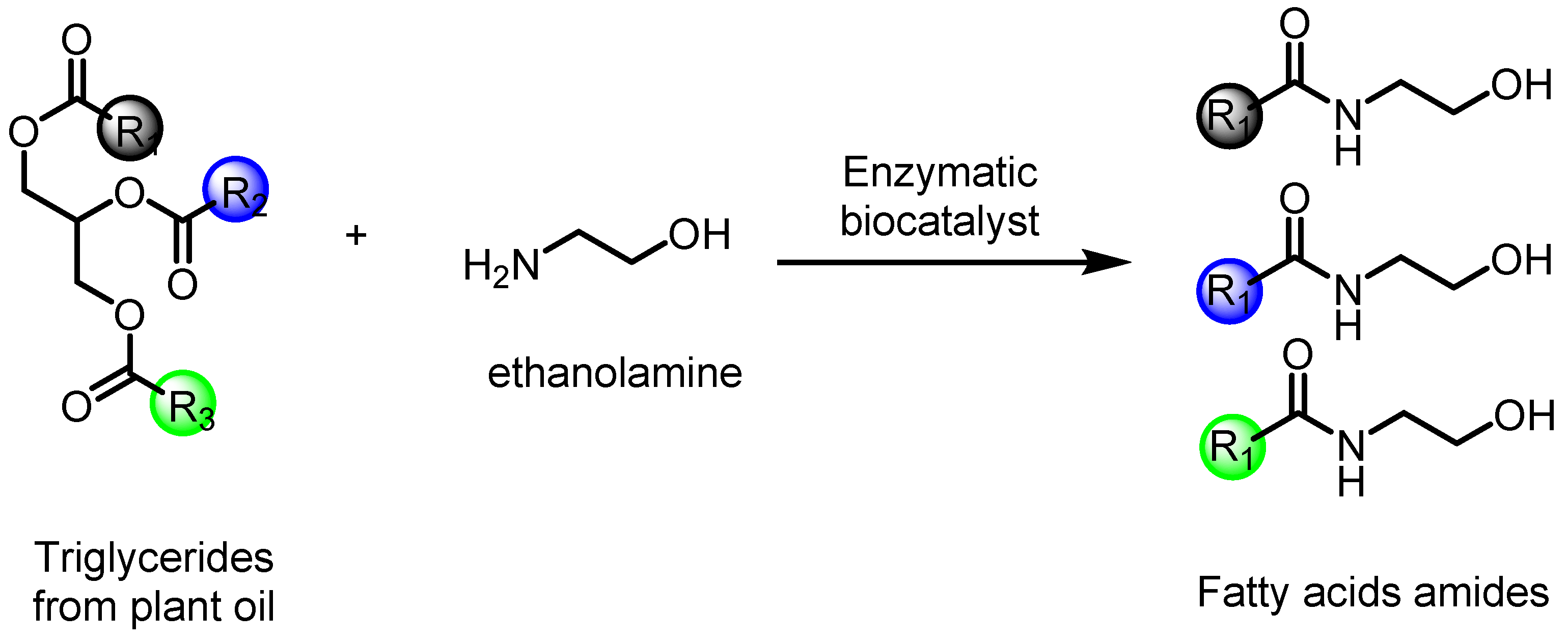
- Fernández-Pérez, M.; Otero, C. Enzymatic synthesis of amide surfactants from ethanolamine. Enzym. Microb. Technol. 2001, 28, 527–536. [Google Scholar] [CrossRef]
- Mouad, A.M.; Taupin, D.; Lehr, L.; Yvergnaux, F.; Porto, A.L.M. Aminolysis of linoleic and salicylic acid derivatives with Candida antarctica lipase B: A solvent-free process to obtain amphiphilic amides for cosmetic application. J. Mol. Catal. B Enzym. 2016, 126, 64–68. [Google Scholar] [CrossRef]
- Whitten, K.M.; Makriyannis, A.; Vadivel, S.K. Enzymatic synthesis of N-acylethanolamines: Direct method for the aminolysis of esters. Tetrahedron Lett. 2012, 53, 5753–5755. [Google Scholar] [CrossRef] [PubMed] [Green Version]
- Araújo, P.H.F.; Barata, P.H.S.; Araújo, I.F.; Curti, J.M.; Amaral, R.R.; Bereau, D.; Carvalho, J.C.T.; Ferreira, I.M. Direct and Solvent-Free Aminolysis of Triglyceride from Oenocarpus bataua (Patawa) Oil Catalyzed by Al2O3. Catal. Lett. 2018, 148, 843–851. [Google Scholar] [CrossRef]
- Jamil, M.A.R.; Siddiki, S.M.A.H.; Touchy, A.S.; Rashed, M.N.; Poly, S.S.; Jing, Y.; Ting, K.W.; Toyao, T.; Maeno, Z.; Shimizu, K.I. Selective Transformations of Triglycerides into Fatty Amines, Amides, and Nitriles by using Heterogeneous Catalysis. Chem. Sus. Chem. 2019, 12, 3115–3125. [Google Scholar] [CrossRef]
- Sun, M.; Nie, K.; Wang, F.; Deng, L. Optimization of the lipase-catalyzed selective amidation of phenylglycinol. Front. Bioeng. Biotechnol. 2020, 7, 486. [Google Scholar] [CrossRef]
- Barata, P.H.S.; Sarquis, I.R.; Carvalho, H.O.; Barros, A.S.; Rodrigues, A.B.; Galue-Parra, A.J.; Silva, E.O.; Carvalho, J.C.T.; Ferreira, I.M. Chemoenzymatic Synthesis and Anti-Inflammatory Activity of Fatty Acid Amides Prepared from Bertholletia excelsa (Brazil Nut) Triglycerides. J. Braz. Chem. Soc. 2020, 31, 1557–1565. [Google Scholar] [CrossRef]
- Wallace, M.J.; Blair, R.E.; Falenski, K.W.; Martin, B.R.; Delorenzo, R.J. The endogenous cannabinoid system regulates seizure frequency and duration in a model of temporal lobe epilepsy. J. Pharm. Exp. 2003, 307, 129–137. [Google Scholar] [CrossRef] [Green Version]
- Fezza, F.; Marrone, M.C.; Avvisati, R.; Di Tommaso, M.; Lanuti, M.; Rapino, C.; Mercuri, N.B.; Maccarrone, M.; Marinelli, S. Distinct modulation of the endocannabinoid system upon kainic acid-induced in vivo seizures and in vitro epileptiform bursting. Mol. Cell Neurosci. 2014, 62, 1–9. [Google Scholar] [CrossRef]
- Sugaya, Y.; Yamazaki, M.; Uchigashima, M.; Kobayashi, K.; Watanabe, M.; Sakimura, K.; Kano, M. Crucial roles of the endocannabinoid 2-arachidonoylglycerol in the suppression of epileptic seizures. Cell Rep. 2016, 16, 1405–1415. [Google Scholar] [CrossRef] [Green Version]
- Wallace, M.J.; Martin, B.R.; DeLorenzo, R.J. Evidence for a physiological role of endocannabinoids in the modulation of seizure threshold and severity. Eur. J. Pharm. 2002, 452, 295–301. [Google Scholar] [CrossRef]
- Fowler, C.J. Oleamide: A member of the endocannabinoid family? Br. J. Pharmacol. 2004, 141, 195–196. [Google Scholar] [CrossRef] [PubMed] [Green Version]
- Naidu, P.S.; Kinsey, S.G.; Guo, T.L.; Cravatt, B.F.; Lichtman, A.H. Regulation of Inflammatory Pain by Inhibition of Fatty Acid Amide Hydrolase. J. Pharmacol. Exp. Ther. 2010, 334, 182–190. [Google Scholar] [CrossRef] [PubMed] [Green Version]
- Boonen, J.; Bronselaer, A.; Nielandt, J.; Veryser, L.; De Tré, G.; De Spiegeleer, B. Alkamid database: Chemistry, occurrence and functionality of plant N-alkylamides. J. Ethnopharmacol. 2012, 142, 563–590. [Google Scholar] [CrossRef] [PubMed] [Green Version]
- Scuderi, C.; Stecca, C.; Esposito, G.; Steardo, L.; Valenza, M.; Carratù, M.R. Palmitoylethanolamide exerts neuroprotective effects in mixed neuroglial cultures and organotypic hippocampal slices via peroxisome proliferator-activated receptor-α. J. Neuroinflamm. 2012, 9, 49. [Google Scholar] [CrossRef] [PubMed] [Green Version]
- Hermes, D.J.; Xu, C.; Poklis, J.L.; Niphakis, M.J.; Cravatt, B.F.; Mackie, K.; Lichtman, A.H.; Ignatowska-Jankowska, B.M.; Fitting, S. Neuroprotective effects of fatty acid amide hydrolase catabolic enzyme inhibition in a HIV-1 Tat model of neuroAIDS. Neuropharmacology 2018, 141, 55–65. [Google Scholar] [CrossRef] [PubMed]
- Gomes Júnior, A.L.; Tchekalarova, J.D.; Atanasova, M.; Da Conceição Machado, K.; De Sousa Rios, M.A.; Paz, M.F.C.J.; Găman, M.A.; Găman, A.M.; Yele, S.; Shill, M.C.; et al. Anticonvulsant effect of anacardic acid in murine models: Putative role of GABAergic and antioxidant mechanisms. Biomed. Pharmacother. 2018, 106, 1686–1695. [Google Scholar] [CrossRef]
- Cione, E.; Plastina, P.; Pingitore, A.; Perri, M.; Caroleo, M.C.; Fazio, A.; Witkamp, R.; Meijerink, J. Capsaicin analogues derived from n-3 polyunsaturated fatty acids (PUFAs) reduce inflammatory activity of macrophages and stimulate insulin secretion by β-cells in vitro. Nutrients 2019, 11, 915. [Google Scholar] [CrossRef] [Green Version]
- Mazzocchi, A.; De Cosmi, V.; Risé, P.; Milani, P.; Turolo, S.; Syrén, M.L.; Sala, A.; Agostini, C. Bioactive Compounds in Edible Oils and Their Role in Oxidative Stress and Inflammation. Front. Physiol. 2021, 12, 659551. [Google Scholar] [CrossRef]
- Solomonia, R.; Nozadze, M.; Mikautadze, E.; Kuchiashvili, N.; Kiguradze, T.; Abkhazava, D.; Pkhakadze, V.; Mamulaishvili, I.; Mikeladze, E.; Avaliani, N. Effect of oleamide on pentylenetetrazole-induced seizures in rats. Bull. Exp. Biol. Med. 2008, 145, 225–227. [Google Scholar] [CrossRef]
- Guan, L.P.; Zhao, D.H.; Xiu, J.H.; Sui, X.; Piao, H.R.; Quan, Z.S. Synthesis and anticonvulsant activity of N-(2-hydroxy-ethyl)amide derivatives. Arch. Pharm. 2009, 342, 34–40. [Google Scholar] [CrossRef]
- Naderi, N.; Ahmad-Molaei, L.; Ahari, F.A.; Motamedi, F. Modulation of Anticonvulsant Effects of Cannabinoid Compounds by GABA-A Receptor Agonist in Acute Pentylenetetrazole Model of Seizure in Rat. Neurochem. Res. 2011, 36, 1520–1525. [Google Scholar] [CrossRef] [PubMed]
- Nam, H.Y.; Na, E.J.; Lee, E.; Kwon, Y.; Kim, H.J. Antiepileptic and Neuroprotective Effects of Oleamide in Rat Striatum on Kainate-Induced Behavioral Seizure and Excitotoxic Damage via Calpain Inhibition. Front. Pharmacol. 2017, 8, 817–828. [Google Scholar] [CrossRef] [PubMed] [Green Version]
- Wu, C.F.; Li, C.L.; Song, H.R.; Zhang, H.F.; Yang, J.Y.; Wang, Y.L. Selective effect of oleamide, an endogenous sleep-inducing lipid amide, on pentylenetetrazole-induced seizures in mice. J. Pharm. Pharm. 2003, 55, 1159–1162. [Google Scholar] [CrossRef]
- Yost, C.S.; Hampson, A.J.; Leonoudakis, D.; Koblin, D.D.; Bornheim, L.M.; Gray, A.T. Oleamide potentiates benzodiazepine-sensitive gamma-aminobutyric acid receptor activity but does not alter minimum alveolar anesthetic concentration. Anesth. Analg. 1998, 86, 1294–1300. [Google Scholar] [CrossRef]
- Ludányi, A.; Erőss, L.; Czirják, S.; Vajda, J.; Halász, P.; Watanabe, M.; Palkovits, M.; Maglóczky, Z.; Freund, T.F.; Katona, I. Downregulation of the CB1 cannabinoid receptor and related molecular elements of the endocannabinoid system in epileptic human hippocampus. J. Neurosci. 2008, 28, 2976–2990. [Google Scholar] [CrossRef] [PubMed] [Green Version]
- Zareie, P.; Sadegh, M.; Palizvan, M.R.; Moradi-Chameh, H. Anticonvulsive effects of endocannabinoids; an investigation to determine the role of regulatory components of endocannabinoid metabolism in the Pentylenetetrazol induced tonic-clonic seizures. Metab. Brain Dis. 2018, 33, 939–948. [Google Scholar] [CrossRef] [PubMed]
- Karanian, D.A.; Karim, S.L.; Wood, J.T.; Williams, J.S.; Lin, S.; Makriyannis, A.; Bahr, B.A. Endocannabinoid enhancement protects against kainic acidinduced seizures and associated brain damage. J. Pharmacol. Exp. Ther. 2007, 322, 1059–1066. [Google Scholar] [CrossRef]


| Fatty Acid and Structure | ||||||
|---|---|---|---|---|---|---|
| Myristic C14:0 | Palmitic C16:0 | Stearic C18:0 | Oleic C18:1 | Linoleic C18:2 | Linolenic C18:3 | References |
| 0.36 | 24.72 | 9.57 | 50.12 | 10.93 | 1.05 | Pantoja et al., 2013 [84] |
| 0.05 | 27.71 | 9.34 | 49.90 | 9.58 | 1.43 | Araujo-Lima et al., 2018 [85] |
| - | 31.02 | 10.53 | 42.71 | 12.93 | tr. | Silva, 2018 [86] |
| - | 27.30 | 12.52 | 47.19 | 9.29 | - | Sousa et al., 2021 [87] |
Publisher’s Note: MDPI stays neutral with regard to jurisdictional claims in published maps and institutional affiliations. |
© 2022 by the authors. Licensee MDPI, Basel, Switzerland. This article is an open access article distributed under the terms and conditions of the Creative Commons Attribution (CC BY) license (https://creativecommons.org/licenses/by/4.0/).
Share and Cite
de Oliveira, F.R.; da Silva, N.M.; Hamoy, M.; Crespo-López, M.E.; Ferreira, I.M.; da Silva, E.O.; de Matos Macchi, B.; do Nascimento, J.L.M. The GABAergic System and Endocannabinoids in Epilepsy and Seizures: What Can We Expect from Plant Oils? Molecules 2022, 27, 3595. https://doi.org/10.3390/molecules27113595
de Oliveira FR, da Silva NM, Hamoy M, Crespo-López ME, Ferreira IM, da Silva EO, de Matos Macchi B, do Nascimento JLM. The GABAergic System and Endocannabinoids in Epilepsy and Seizures: What Can We Expect from Plant Oils? Molecules. 2022; 27(11):3595. https://doi.org/10.3390/molecules27113595
Chicago/Turabian Stylede Oliveira, Fábio Rodrigues, Nágila Monteiro da Silva, Moisés Hamoy, Maria Elena Crespo-López, Irlon Maciel Ferreira, Edilene Oliveira da Silva, Barbarella de Matos Macchi, and José Luiz Martins do Nascimento. 2022. "The GABAergic System and Endocannabinoids in Epilepsy and Seizures: What Can We Expect from Plant Oils?" Molecules 27, no. 11: 3595. https://doi.org/10.3390/molecules27113595
APA Stylede Oliveira, F. R., da Silva, N. M., Hamoy, M., Crespo-López, M. E., Ferreira, I. M., da Silva, E. O., de Matos Macchi, B., & do Nascimento, J. L. M. (2022). The GABAergic System and Endocannabinoids in Epilepsy and Seizures: What Can We Expect from Plant Oils? Molecules, 27(11), 3595. https://doi.org/10.3390/molecules27113595