Article

Pennelliiside D, a New Acyl Glucose from Solanum pennellii and Chemical Synthesis of Pennelliisides

Research Faculty of Agriculture, Hokkaido University, Kita 9 Nishi 9, Kita-ku, Sapporo 060-8589, Japan
*
Author to whom correspondence should be addressed.
Molecules 2022, 27(12), 3728; https://doi.org/10.3390/molecules27123728
Submission received: 17 May 2022 / Revised: 3 June 2022 / Accepted: 6 June 2022 / Published: 9 June 2022

Abstract

:
Acyl glucoses are a group of specialized metabolites produced by Solanaceae. Solanum pennellii, a wild-type tomato plant, produces acyl glucoses in its hair-like epidermal structures known as trichomes. These compounds have been found to be herbicides, microbial growth inhibitors, or allelopathic compounds. However, there are a few reports regarding isolation and investigation of biological activities of acyl glucoses in its pure form due to the difficulty of isolation. Here, we report a new acyl glucose, pennelliiside D, isolated and identified from S. pennellii. Its structure was determined by 1D NMR and 2D NMR, together with FD-MS analysis. To clarify the absolute configuration of the acyl moiety of 2-methylbutyryl in the natural compound, two possible isomers were synthesized starting from β-D-glucose pentaacetate. By comparing the spectroscopic data of natural and synthesized compounds of isomers, the structure of pennelliiside D was confirmed to be 3,4-O-diisobutyryl-2-O-((S)-2-methylbutyryl)-D-glucose. Pennelliiside D and its constituent fatty acid moiety, (S)-2-methylbutanoic acid, did not show root growth-inhibitory activity. Additionally, in this study, chemical synthesis pathways toward pennelliisides A and B were adapted to give 1,6-O-dibenzylpennelliisides A and B.

Graphical Abstract

1. Introduction

Plants are considered a rich source of natural products that possess diverse structures and corresponding biological activities, such as antiherbivory, antimicrobial, and antioxidant activities [1]. Acyl sugars (sugar esters), nonvolatile secondary metabolites, are specialized natural products produced in the hair-like epidermal structures, known as trichomes, of many Solanaceae families, such as Solanum [1], Nicotina [2], Datura [3], and Petunia [4]. The backbone of acyl sugars basically consists of either a glucose or sucrose moiety attached to one or more straight or branched-chain fatty acids via O-acylation [1].
Solanum pennellii, a wild tomato species, is endemic to South America [5], and its genomic sequences [6] and introgression lines have been fully characterized [7,8]. It has been reported that S. pennellii accumulates various types of secondary metabolites in trichomes, such as terpenoids, phenylpropanoids, and acyl sugars [4,9]. Among them, acyl sugars account for 20% of the total dry weight of leaves [4,10]. Acyl sugars show insecticidal effects against aphids [11], pest repellents [12], and weed growth inhibitory activities [13] as well as allelopathic properties [14]. Most acyl glucoses produced by S. pennellii contain mono-, di-, or trifatty acid moieties ranging in carbon number from two to twelve [15,16].
Although some studies have been conducted on the biosynthesis of acyl sugars [17], their full discovery remains unclear because of the availability of vastly diverse acyl sugars [15]. This implies the potential to present considerably diverse acyl glucoses in S. pennellii as well. However, there are few reports regarding isolation and investigation of their biological activities in their pure form due to the difficulty of isolation [13,17]. That is because of α and β anomerization at the C-1 position of the glucose moiety. We recently found that α and β anomerization can be successfully control by benzylation of hydroxyl groups present at the glucose moiety. Using this strategy, three compounds, 2,3,4-O-triisobutyryl-D-glucose, 3-O-(8-methylnonanoyl)-2,4-O-diisobutyryl-D-glucose, and 3-O-decanoyl-2,4-O-diisobutyryl-D-glucose, namely, pennelliisides A-C, were reported [14].
As a part of our ongoing research, another new analogue of acyl glucose was identified from S. pennellii. To determine the absolute configuration of its fatty acid moiety, total synthesis was carried out. Additionally, in this report, chemical synthesis of previously reported 1,6-O-dibenzyl penneliisides A and B are presented. Root-growth inhibitory activity of the newly identified compound and its synthesized compound was also investigated.

2. Results and Discussion

2.1. Isolation and Identification of Pennelliiside D (1)

The aerial parts of 80-day-old S. pennellii (1.7 kg) were dipped in EtOH for 30 s, and an extract of epicuticular lipophilic wax was obtained by evaporating the organic solvent under reduced pressure. The extract was partitioned between EtOAc and sat. NaHCO3. The extract obtained from the EtOAc layer was roughly purified using silica gel column chromatography to give acyl glucoses, followed by benzylation with 2,4,6-tris(benzyloxy)-1,3,5-triazine (TriBOT) to hinder α and β anomerization as previously reported [14,18,19]. The obtained benzylated derivatives of acyl glucoses were purified using silica gel column chromatography and HPLC to give dibenzyl pennelliiside D (2, 19 mg, Figure 1B).
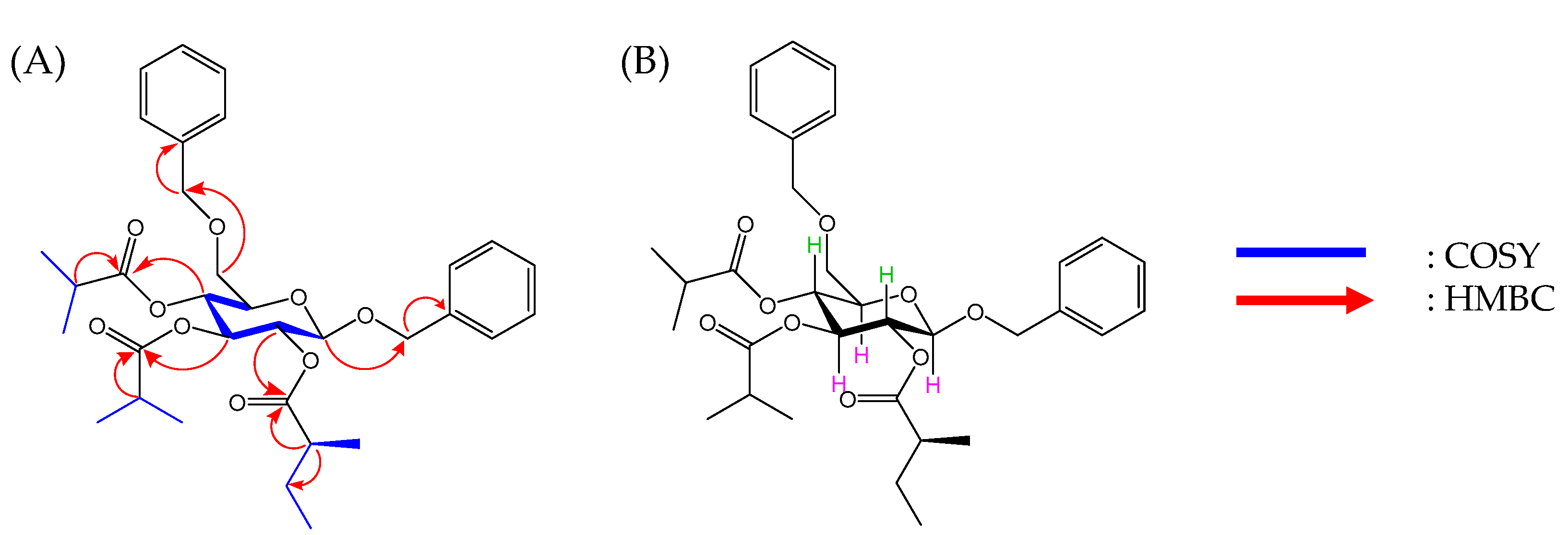
Compound 2 was obtained as a colorless oil. The molecular formula and molecular weight were found to be C33H44O9 and m/z 584.2992 [M]+ (cal. m/z 584.2985 [M]+), respectively, using HRFD-MS data (Figure S1, Supplementary Materials), indicating that 2 has 12 degrees of unsaturation. Based on 1H NMR data, signals at δH 4.38 (d, J = 7.6 Hz, 1H, H-1), 5.46 (m, 1H, H-2), 5.48 (m, 1H, H-3), 5.30 (dd, J = 10.7, 9.5 Hz, 1H, H-4), 3.42 (m, 1H, H-5), and 3.47 (m, 2H, H-6) were identified as protons related to glucopyranose (Table 1, and Figure S2, Supplementary Materials). The presence of glucopyranose was further confirmed by comparing COSY correlations between the signals at H-1/H-2, H-2/H-3, H-3/H-4, and H-4/H-5 (Figure 2A and Figure S4, Supplementary Materials) together with their corresponding coupling constants (Table 1). Meanwhile, NOESY interactions observed due to the cross-peaks of H-2/H-4 and H-1/H-3/H-5 (Figure 2B and Figure S7, Supplementary Materials), and the signal observed at δC 100.3 in 13C NMR (Table 1 and Figure S3, Supplementary Materials) also indicated that the glucose moiety exhibited a β anomeric structure.
Next, the resonances in relatively lower field at δH 7.26 (t, J = 7.4 Hz, 4H, H-3′, H-7′, H-3″, H-7″), 7.12–7.19 (m, 4H, H-4′, H-6′, H-4″, H-6″), and 7.08 (t, J = 7.3 Hz, 2H, H-5′, H-5″) (Table 1 and Figure S2, Supplementary Materials), were identified as resonances corresponding to two benzene rings. Based on the HMBC correlations (Figure 2A), signals appearing at δH 4.75 (d, J = 12.2 Hz, 1H, H-1′), 4.45 (d, J = 12.2 Hz, 1H, H-1′) and 4.33 (d, J = 5.5 Hz, 2H, H-1″) (Figure S6, Supplementary Materials) were identified as methylene protons corresponding to benzylidene attached to the C-1 and C-6 positions. Moreover, the presence of two isobutyryl ester moieties was determined according to the 1H NMR and 13C NMR spectra and COSY and HMBC correlations (Table 1 and Figures S2–S6, Supplementary Materials), and these moieties were attached to C-3 and C-4 positions in the glucose moiety (Figure 2). Similarly, the 2-methylbutyryl fatty acid moiety attached to C-2 was revealed based on COSY and HMBC correlations, as shown in Figure 2. Therefore, the detailed analysis of 2D NMR data clarified the structure of 2 to be 1,6-O-dibenzyl-3,4-O-diisobutyryl-2-O-(2-methylbutyryl)-β-D-glucose (Figure 1).
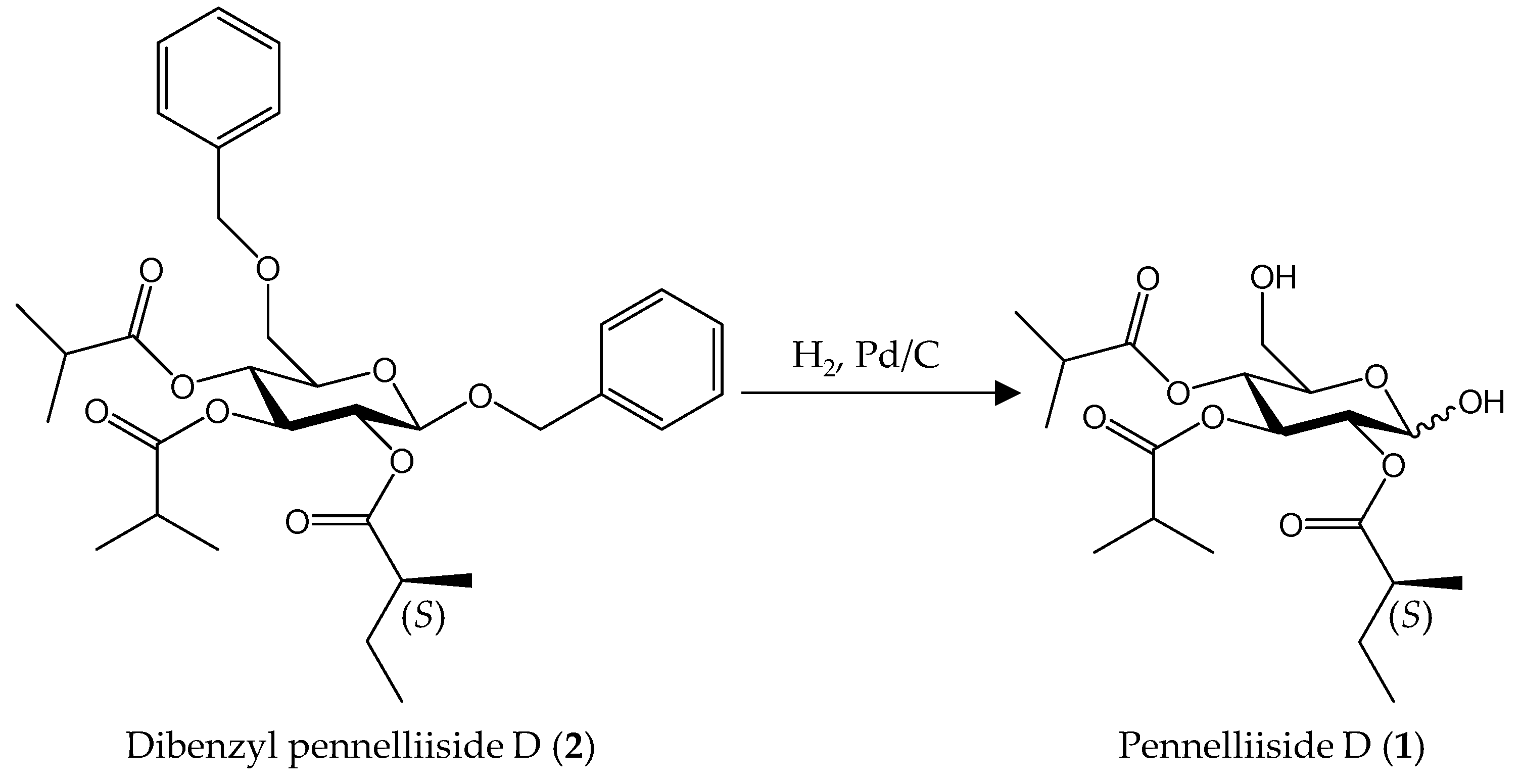
To afford 1, compound 2 was subjected to debenzylation with palladium black under a hydrogen gas atmosphere (Scheme 1). Compound 1 was obtained as a colorless oil, and the molecular formula and molecular weight were found to be C19H32O9 and m/z 405.2133 [M + H]+ (cal. m/z 405.2125 [M + H]+), respectively, using HRFD-MS data (Figure S8, Supplementary Materials), indicating that 1 has 4 degrees of unsaturation. Summarized 1H NMR and 13C NMR data of 1 are shown in Table 2. Although 1H NMR, 13C NMR, COSY, HSQC, and HMBC (Figures S9–S14, Supplementary Materials) data were complex due to the interference of α and β anomers, assignment of H and C corresponding to the α and β anomers of 1 were done partially. Assignments of α and β anomers are shown in Figure S12 in the Supplementary Materials. Based on the NMR data, the chemical structure of 1 was determined to be 3,4-O-diisobutyryl-2-O-(2-methylbutyryl)-D-glucose (Figure 1A), although the absolute configuration of the 2-methylbutyryl fatty acid moiety was still unclear [1,13,17].

2.2. Synthesis of Pennelliiside D (1)

Two possible isomers of dibenzyl pennelliiside D, 1,6-O-dibenzyl-3,4-O-diisobutyryl-2-O-((S)-2-methylbutyryl)-β-D-glucose (2) and 1,6-O-dibenzyl-3,4-O-diisobutyryl-2-O-((R)-2-methylbutyryl)-β-D-glucose (12), were synthesized to determine the absolute configuration of the fatty acid moiety, 2-methylbutyryl, attached to C-2, although the naturally available ester of 2-methylbutyryl in other natural sources is mostly in the (S) configuration [20,21].
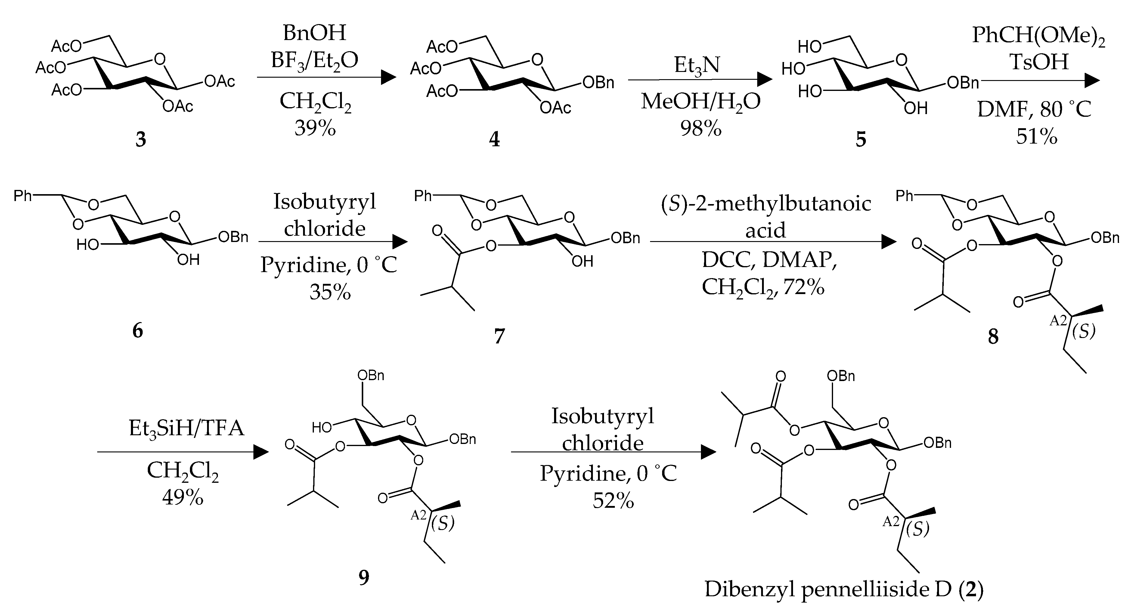
Synthesis of 2 was commenced with an available compound, β-D-glucose pentaacetate (3), by benzylation at C-1 with benzyl alcohol (Scheme 2). Removal of acetate groups followed by protection of C-4 and C-6 with benzaldehyde dimethyl acetal and p-toluenesulfonic acid resulted in 1-O-benzyl-4,6-O-benzylidine-β-D-glucose (6), as reported by Degenstein et al., 2015 [22]. Selective esterification at the C-3 position was achieved by reacting 6 with isobutyryl chloride followed by condensation with (S)-2-methylbutanoic acid under a nitrogen gas atmosphere, which offered the desired compound 8. Cleavage of 4,6-O-benzylidine moiety of 8 using triethylsilane and trifluoroacetic acid was done, which was followed by an esterification with isobutyryl chloride to give preferred dibenzyl pennelliiside D (2) having (S)-configured at the A2 position (Figures S15–S50, Supplementary Materials). Similarly, the synthesis of 12 was achieved starting with 7, which was conjugated with (R)-2-methylbutanoic acid (Scheme S1, Table S1 and Figures S60–S75, Supplementary Materials).
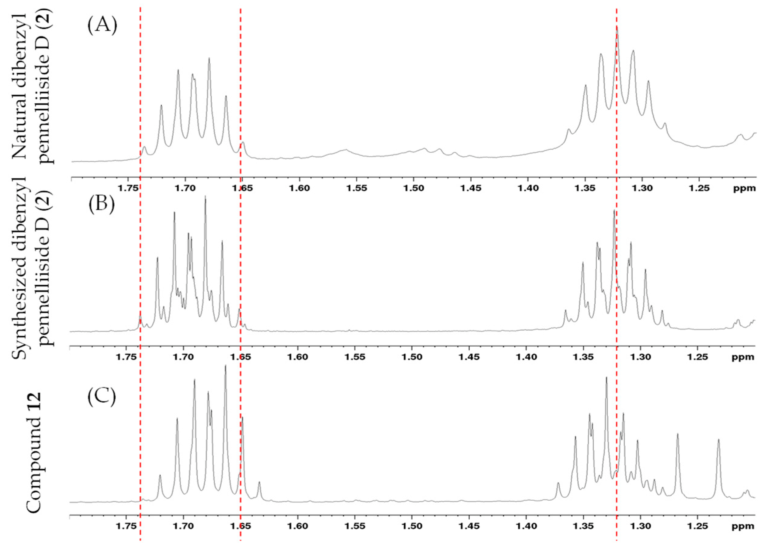
Then, we compared the 1H NMR and 13C NMR data of natural and synthesized compounds (S/R) (Table 1 and Table S1, Supplementary Materials). Synthesized (S) isomer of dibenzyl pennelliiside D (2) had good accordance with natural dibenzyl pennelliiside D (2). In the 1H NMR, the differences between synthesized (S/R) with the natural compound were found in the resonances around δ 1.65 and δ 1.32 as shown in Figure 3. Furthermore, a significant difference was shown when comparing specific rotation values with 12, while natural 2 and synthesized 2 showed almost the same value. The specific rotation values measured for natural and synthesized 2, and 12 were [α]25D = −10.5, −10.7, and −21.3 (c 0.6, MeOH), respectively. Based on the above observations, we concluded that the absolute configuration of the 2-methylbutyryl fatty acid moiety in natural 2 was (S) and confirmed its structure, as shown in Figure 1B.
By debenzylation of synthesized 2 with palladium black under a hydrogen gas atmosphere (Scheme 1), synthesized 1 (5.9 mg) was obtained as a colorless oil. The molecular formula and molecular weight were similar to those of the natural 1, which were C19H32O9 and m/z 405.2133 [M + H]+ (cal. m/z 405.2125 [M + H]+), respectively, using HRFD-MS data (Figure S51, Supplementary Materials). Summarized 1H NMR and 13C NMR data of synthesized 1 are shown in Table 3. 1H NMR, 13C NMR, COSY, HSQC, and HMBC data are shown in Figures S52–S59 in the Supplementary Materials. Similar to natural 1, partial assignment of H and C corresponding to the α and β anomers of D-glucose for synthesized 1 is shown in Figure S55 in the Supplementary Materials. Comparison of 1H NMR and 13C NMR spectra of natural and synthesized 1 also showed similar data (Table 2 and Table 3, and Figures S58 and S59, Supplementary Materials) and revealed the chemical structure of 1 to be 3,4-O-diisobutyryl-2-O-((S)-2-methylbutyryl)-D-glucose (Figure 1A).

2.3. Root Growth-Inhibitory Activity of Pennelliiside D (1)

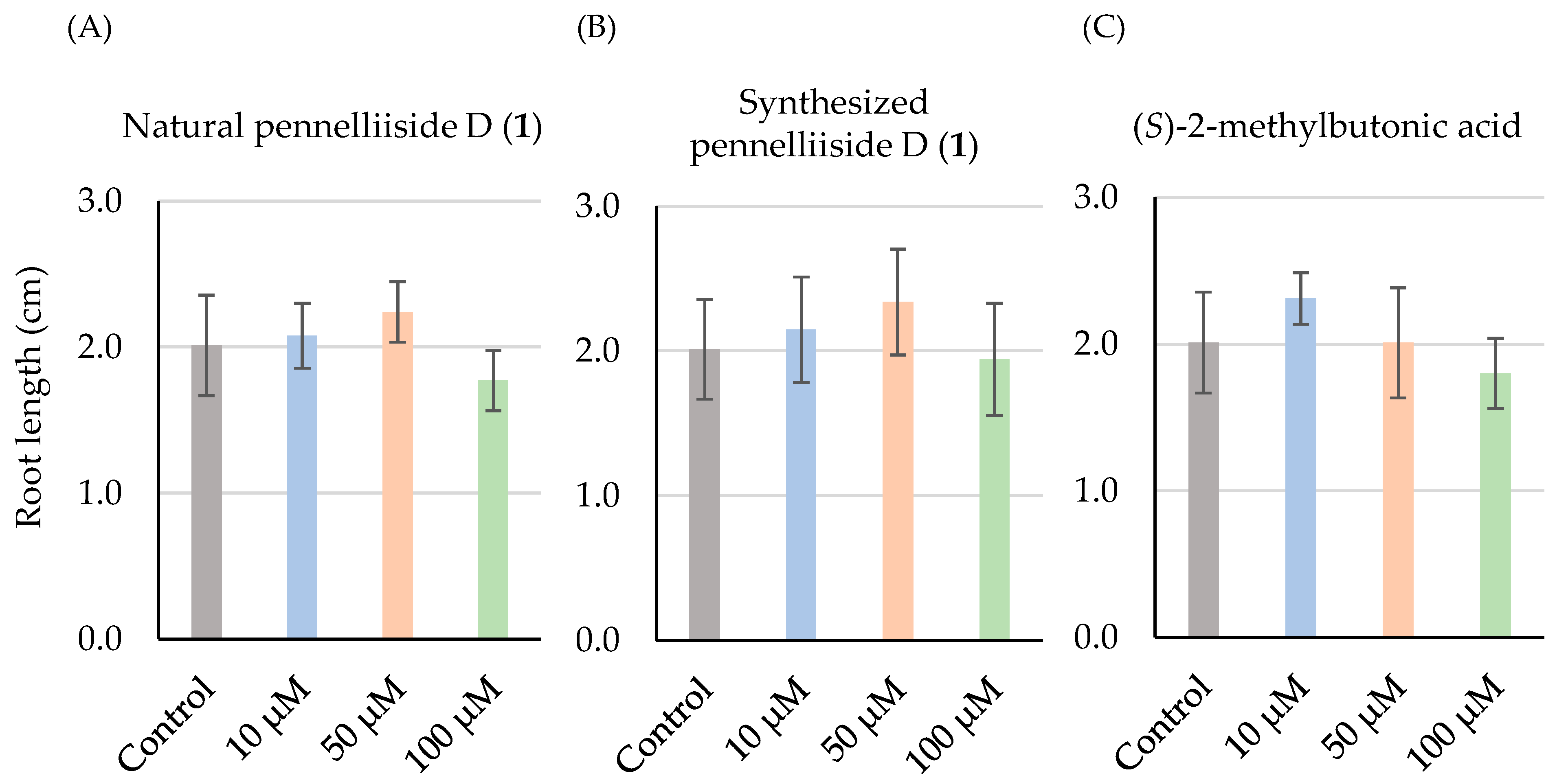
Previously, it has been reported that the acyl sucrose showed root growth-inhibitory effect on velvetleaf [23]. Therefore, root growth-inhibitory activity against natural and synthesized 1 and its constituent fatty acid, (S)-2-methylbutanoic acid, was assessed. Arabidopsis thaliana seeds and 10 µM, 50 µM, and 100 µM concentrations of compounds were used in this experiment. As the control, A. thaliana seeds were germinated in the MS medium without adding any compound. The data revealed that neither compound showed root growth-inhibitory activity at any tested concentration (Figure 4), which might support that acyl glucose contains longer chain carbon fatty acids shows root growth-inhibitory effect.

2.4. Synthesis of Dibenzyl Pennelliisides A and B

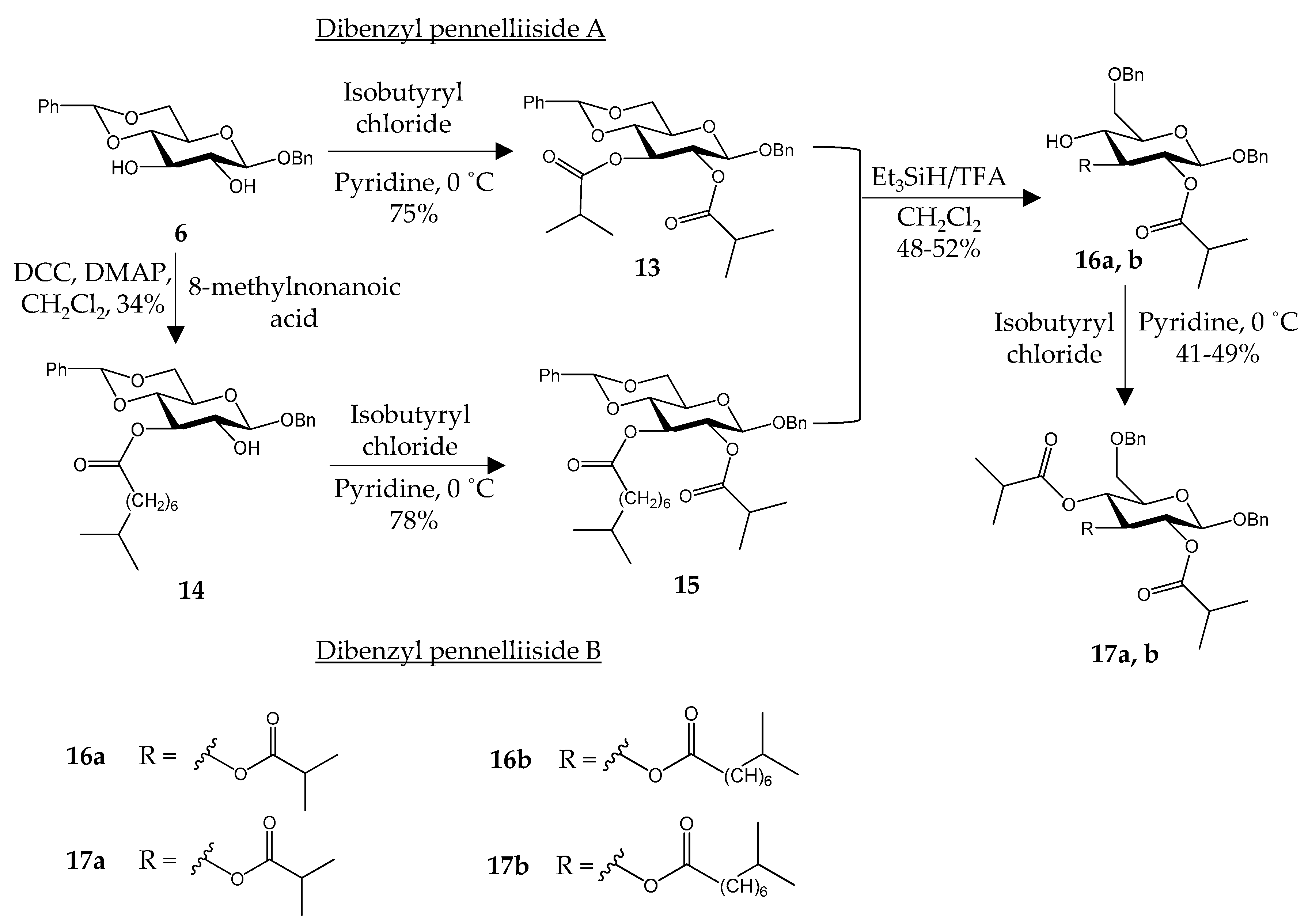
Using the same strategy of synthesis of 2, synthesis of dibenzyl pennelliisides A and B (17a, b) were conducted using 6 as the starting material (Scheme 3). Isobutyryl chloride and 8-methylnonanoic acid were used to obtain dibenzyl pennelliisides A and B (17a, b). In order to synthesize dibenzyl pennelliiside A, 6 was reacted with isobutyryl chloride to yield 13 that has two isobutyryl fatty acid moieties. Next, deprotection was carried out followed by another reaction with isobutyryl chloride to give the desired compound, dibenzyl pennelliiside A (17a). Using the same starting compound, the synthesis of dibenzyl pennelliiside B was commenced with a condensation reaction with 8-methylnonanoic acid to esterify the fatty acid moiety selectively to C-3. Then, 14 was reacted with isobutyryl chloride followed by deprotection and another esterification with isobutyryl chloride to yield dibenzyl pennelliiside B (17b). The chemical structures of all compounds were characterized using 1H NMR, 13C NMR, 2D NMR, and FD-MS (Figures S76–S102, Supplementary Materials). It has been already proven that the removal of benzyl groups can be accomplished as Scheme 1 to obtain pennelliisides A and B. Using the same synthesis pathway, it is possible to synthesize other acyl glucoses.

3. Materials and Methods

3.1. General Experimental Procedures

Optical rotations were obtained with a JASCO P-2200 polarimeter. NMR spectra were recorded in C6D6, CD3OD and CDCl3 using a JNM-EX 270 FT-NMR spectrometer (JEOL, 1H NMR: 270 MHz) and AMX 500 Bruker system (1H NMR: 500 MHz, 13C NMR: 126 MHz). Assignment of H and C was performed by obtaining 1H NMR, 13C NMR (referenced for C6D6, CD3OD and CDCl3 at δH 7.16, 3.31 and 7.24, and δC 128.4, 49.2 and 77.2, respectively), COSY, HSQC, HMBC, and NOESY spectra. FD-MS analysis was performed on a JMS-T100GCV (JEOL) instrument. Chromatographic analysis was performed using an HPLC system (InertSustain, A210max nm) equipped with a Shisheido Capcell park C18 column (4.6 × 250 nm, 5 µm, 2 mL/min, MeOH-H2O, 80:20) and a Cadenza CK-C18 column (6 × 250 nm, 3 µm, 2 mL/min, MeOH-H2O, 80:20). All moisture-sensitive reactions were performed under a nitrogen gas atmosphere. All chemicals used in the study were of analytical grade and purchased from Sigma–Aldrich, Tokyo, Japan, Kanto Chemical Co., Inc, Tokyo, Japan, and Cayman Chemical, Ann Arbor, MI, USA.

3.2. Plant Material

Seeds of S. pennellii were obtained from the National Bioresource Project (NBRP, Tsukuba). The plants were grown under 16 h of light and 8 h of dark for 80 days at 25 °C in an artificial weather room at the Faculty of Agriculture, Hokkaido University, Hokkaido, Japan.

3.3. Extraction and Isolation

To extract acyl sugars from S. pennellii, 1.7 kg of aerial parts of plants were used. Pieces of plants were roughly divided into five groups. Each group of plant material was dipped in EtOH (1 L) and shaken for 30 s separately. Then, all the solvent fractions (collectively 5 L) were combined, filtered, and concentrated using a rotary evaporator. The obtained crude material was then extracted into EtOAc (500 mL) with sat. NaHCO3 (500 mL) by liquid-liquid extraction. After drying the organic layer with MgSO4 and evaporating, the obtained crude material was separated using silica gel column chromatography (MeOH-CHCl3-CH3COOH, 5:95:0.1). Then, acyl glucoses obtained from separation were subjected to benzylation using TriBOT as mentioned previously [14,18,19]. Briefly, 300 mg of TriBOT and 35 µL of trifluoromethanesulfonic acid (TfOH) were added to a mixture of 4.5 g of obtained crude material in 100 mL of 1,4-dioxane under anhydrous conditions. The reaction mixture was stirred for 16 h at room temperature (RT). After evaporation of the organic solvent, the obtained oil compounds were subjected to silica gel column chromatography (EtOAc-n-hexane-CH3COOH, 20:80:0.1) to give subfractions. The fraction named Fr2-1 (67.7 mg) was further separated using two consecutive HPLC separations (Shisheido Capcell park C18, 4.6 × 250 nm, 5 µm, 2 mL/min, CH3CN-H2O, 80:20; Cadenza CK-C18, 6 × 250 nm, 3 µm, 2 mL/min, MeOH-H2O, 80:20) to yield colorless oil, 2 (19 mg).

3.4. Synthesis of Pennelliiside D (1)

Compounds 4, 5, 6, and 9 were synthesized according to a reported method [22].

3.4.1. Synthesis of 1-O-Benzyl-2,3,4,6-O-tetraacetyl-β-D-glucose (4)

To a mixture of β-D-glucose pentaacetate (3, 2.00 g, 5.12 mmol) in 20 mL of anhydrous CH2Cl2, benzyl alcohol (1.12 mL, 10.25 mmol) and BF3·Et2O (0.82 mL, 6.66 mmol) were added. The reaction mixture was stirred for 24 h at RT. Then, 10 mL of CH2Cl2 was added to dilute the reaction mixture. The resulting solution was partitioned between sat. NaHCO3 (30 mL × 3) and CH2Cl2. The organic layer was washed with water (30 mL × 3) and dried over Na2SO4 followed by evaporation of the organic solvent to result in a crude product. The crude material was purified using silica gel column chromatography (EtOAc-n-hexane, 30:70) to yield a colorless oil, 4 (875.5 mg, 2.00 mmol, 39%). 1H NMR (270 MHz, CDCl3, Figure S16, Supplementary Materials): δH 7.15–7.32 (m, 5H, Ar-H), 4.92–5.12 (m, 3H, H-2, H-3, H-4), 4.54 (d, J = 12.3 Hz, 1H, H-7), 4.46 (d, J = 7.6 Hz, 1H, H-7), 4.46 (d, J = 7.6 Hz, 1H, H-1), 4.19 (dd, J = 11.9, 4.67 Hz, 1H, H-6), 4.08 (dd, J = 12.7, 2.51 Hz, 1H, H-6), 3.59 (m, 1H, H-5), 2.02 (s, 3H, CH3), 1.93 (s, 3H, CH3), 1.92 (s, 3H, CH3), 1.91 (s, 3H, CH3); 13C NMR (126 MHz, CDCl3, Figure S17, Supplementary Materials): δC 170.7, 170.3, 169.4, 169.3, 136.6, 128.7–128.5, 99.3, 72.9, 71.9, 71.3, 70.8, 68.4, 62.0, 20.7, 20.6 (3C); HRFD-MS m/z 438.1516 [M]+ (calcd for C21H26O10 m/z 438.1526 [M]+) (Figure S15, Supplementary Materials).

3.4.2. Synthesis of 1-O-Benzyl-β-D-glucose (5)

Triethylamine (1.6 mL) and H2O (1.6 mL) were added to the reaction mixture containing 4 (875.5 mg, 2.00 mmol) dissolved in MeOH (13 mL). The reaction mixture was stirred at RT for 3 h and concentrated using a rotary evaporator. The resulting residue was purified using silica gel column chromatography (MeOH-CH2Cl2, 20:80) to give white powder, 5 (529.0 mg, 1.96 mmol, 98%). 1H NMR (270 MHz, CD3OD, Figure S19, Supplementary Materials): δH 7.15–7.43 (m, 5H, Ar-H), 4.89 (d, J = 11.8 Hz, 1H, H-7), 4.62 (d, J = 11.8 Hz, 1H, H-7), 4.3 (d, J = 7.8 Hz, 1H, H-1), 3.85 (dd, J = 11.8, 2.0 Hz, 1H, H-6), 3.64 (dd, J = 12.1, 5.4 Hz, 1H, H-6), 3.14–3.33 (m, 4H, H-2, H-3, H-4, H-5); 13C NMR (126 MHz, CD3OD, Figure S20, Supplementary Materials): δC 137.7, 127.9 (2C), 127.8 (2C), 127.3, 101.9, 76.7, 76.6, 73.8, 70.4, 70.3, 61.4; HRFD-MS m/z 271.1177 [M + H]+ (calcd for C13H18O6 m/z 271.1182 [M + H]+) (Figure S18, Supplementary Materials).

3.4.3. Synthesis of 1-O-Benzyl-4,6-O-benzylidine-β-D-glucose (6)

To a mixture of 5 (529.0 mg, 1.96 mmol) and benzaldehyde dimethyl acetal (PhCH(OMe)2) (0.35 mL, 2.35 mmol), p-toluenesulfonic acid (TsOH·H2O) (92.9 mg, 0.49 mmol) dissolved in dimethylformamide (DMF) (5 mL) was added. The reaction mixture was stirred for 5 min at RT, heated to 80 °C, and stirred for 4 h. Then, it was allowed to cool to RT and evaporated using a rotary evaporator. The obtained residue was subjected to liquid-liquid extraction with CH2Cl2 (20 mL) and sat. NaHCO3 (20 mL × 3). The organic layer was collected, dried over Na2SO4, and evaporated. The obtained crude material was purified with silica gel column chromatography (EtOAc-n-hexane, 50:50) to give an oil, 6 (357.7 mg, 1.00 mmol, 51%). 1H NMR (500 MHz, CDCl3, Figure S22, Supplementary Materials): δH 7.47–7.54 (m, 2H, Ar-H), 7.27–7.40 (m, 8H, Ar-H), 5.51 (s, 1H, H-7), 4.91 (d, J = 11.6 Hz, 1H, H-1′), 4.61 (d, J = 11.6 Hz, 1H, H-1′), 4.47 (d, J = 7.8 Hz, 1H, H-1), 4.34 (dd, J = 10.9, 5.0 Hz, 1H, H-6), 3.73–3.82 (m, 2H, H-3, H-6), 3.50–3.57 (m, 2H, H-2, H-4), 3.39–3.46 (m, 1H, H-5); 13C NMR (126 MHz, CDCl3, Figure S23, Supplementary Materials): δC 136.9, 136.7, 125.9–129.6, 102.1, 101.9, 80.5, 74.5, 73.1, 71.5, 68.6, 66.4; COSY, HSQC, and HMBC data are shown in Figures S24–S26, Supplementary Materials; HRFD-MS m/z 358.1408 [M]+ (calcd for C20H22O6 m/z 358.1416 [M]+) (Figure S21, Supplementary Materials).

3.4.4. Synthesis of 1-O-Benzyl-4,6-O-benzylidine-3-O-isobutyryl-β-D-glucose (7)

To 6 (357.7 mg, 1.00 mmol) dissolved in anhydrous pyridine (40 mL) at 0 °C, isobutyryl chloride (0.11 mL, 1.00 mmol) was added. The reaction mixture was stirred for 24 h, neutralized with 1 M HCl and evaporated using a rotary evaporator. The obtained crude material was partitioned between EtOAc (50 mL) and 1 M HCl (50 mL × 2) and between EtOAc (50 mL) and sat. NaHCO3 (50 mL × 2). The organic layer was washed with H2O (50 mL), dried over Na2SO4, and evaporated to give an oil, which was subjected to silica gel column chromatography (EtOAc-n-hexane, 25:75) to yield 7 (149.7 mg, 0.35 mmol, 35%). 1H NMR (270 MHz, C6D6, Figure S28, Supplementary Materials): δH 7.41–7.53 (m, 2H, Ar-H), 6.90–7.21 (m, 8H, Ar-H), 5.34 (dd, J = 10.5, 9.48 Hz, 1H, H-3), 5.07 (s, 1H, H-7), 4.64 (d, J = 11.8 Hz, 1H, H-1′), 4.25 (d, J = 11.8 Hz, 1H, H-1′), 4.12 (d, J = 7.6 Hz, 1H, H-1), 4.01 (dd, J = 10.3, 4.8 Hz, 1H, H-6), 3.55 (m, 1H, H-6), 3.25–3.42 (m, 2H, H-2, H-4), 2.88–3.20 (m, H, H-5), 2.51 (m, H, H-B2), 0.98 (d, J = 4.8 Hz, 3H, CH3), 0.95 (d, J = 4.5 Hz, 3H, CH3); 13C NMR (126 MHz, C6D6, Figure S29, Supplementary Materials): δC 176.5, 137.7, 137.5, 126.1–128.5 (10C), 102.9, 101.1, 78.8, 73.5 (2C), 71.1, 68.5, 66.1, 34.0, 18.9, 18.7; HRFD-MS m/z 429.1904 [M + H]+ (calcd for C24H28O7 m/z 429.1913 [M + H]+) (Figure S27, Supplementary Materials).

3.4.5. Synthesis of 1-O-Benzyl-4,6-O-benzylidine-3-O-isobutyryl-2-O-((S)-2-methylbutyryl)-β-D-glucose (8)

Dicyclohexylcarbodiimide (DCC) (294.3 mg, 1.40 mmol) and 4-dimethylaminopyridine (DMAP) (64.7 mg, 0.52 mmol) were added to 7 (149.7 mg, 0.35 mmol). A mixture of (S)-2-methylbutanoic acid (0.17 mL, 1.40 mmol) in anhydrous CH2Cl2 (35 mL) was added to the above mixture, and it was stirred for 24 h at RT. After evaporating volatile components in the reaction mixture, the obtained crude material was subjected to liquid-liquid extraction with CH2Cl2 (50 mL) and sat. NaHCO3 (50 mL × 2), followed by washing the organic layer with 1 M HCl (50 mL × 2) and H2O (50 mL × 2). After drying over Na2SO4 and evaporating, purification was performed using silica gel column chromatography (EtOAc-n-hexane, 30:70) to yield a pale green oil, 8 (128.9 mg, 0.25 mmol, 72%). 1H NMR (500 MHz, C6D6, Figure S31, Supplementary Materials): δH 7.56–7.61 (m, 2H, Ar-H), 7.21–7.27 (m, 2H, Ar-H), 7.12–7.18 (m, 4H, Ar-H), 7.06–7.12 (m, 2H, Ar-H), 5.57 (dd, J = 10.4, 9.6 Hz, 1H, H-3), 5.47 (dd, J = 8.5, 8.0 Hz, 1H, H-2), 5.17 (s, 1H, H-7), 4.74 (d, J = 12.1 Hz, 1H, H-1′), 4.37 (d, J = 6.6 Hz, 1H, H-1′), 4.35 (d, J = 6.9 Hz, 1H, H-1), 4.12 (dd, J = 10.0, 5.0 Hz, 1H, H-6), 3.44 (dd, J = 12.8, 10.2 Hz, 1H, H-6), 3.38 (dd, J = 10.2, 9.4 Hz, 1H, H-4), 3.10 (m, 1H, H-5), 2.47 (m, 1H, H-B2), 2.34 (m, 1H, H-A2), 1.73 (m, 1H, H-A4), 1.34 (m, 1H, H-A4), 1.07–1.15 (m, 9H, H-A3, H-B3, H-B4), 0.84 (t, J = 7.4 Hz, 3H, A5); 13C NMR (126 MHz, C6D6, Figure S32, Supplementary Materials): δC 176.1, 175.0, 139.0, 138.3, 126.5–129.8 (10C), 101.8, 101.3, 79.3, 72.4, 72.1, 71.3, 69.0, 66.8, 41.8, 34.6, 27.3, 16.9–19.7 (3C), 12.15; COSY, HSQC, HMBC, and NOESY data are shown in Figures S33–S36, Supplementary Materials; HRFD-MS m/z 511.2338 [M-H]+ (calcd for C29H36O8 m/z 511.2332 [M-H]+) (Figure S30, Supplementary Materials).

3.4.6. Synthesis of 1,6-O-Dibenzyl-3-O-isobutyryl-2-O-((S)-2-methylbutyryl)-β-D-glucose (9)

To 8 (129.0 mg, 0.25 mmol) in anhydrous CH2Cl2 (10 mL) at 0 °C, trifluoroacetic acid (25. 3 µL, 0.76 mmol) and triethylsilane (Et3SiH) (121.7 µL, 0.76 mmol) were added. The reaction was carried out at RT overnight. The reaction mixture was diluted by adding EtOAc (20 mL) and subjected to liquid-liquid extraction with EtOAC (20 mL) and sat. NaHCO3 (30 mL × 2), followed by washing the organic layer with 1 M HCl (30 mL) and H2O (30 mL). The obtained organic layer was dried over Na2SO4 and evaporated under reduced pressure. Next, purification was performed by silica gel column chromatography (EtOAc-n-hexane, 30:70) to yield an oil, 9 (63.4 mg, 0.12 mmol, 49%). 1H NMR (500 MHz, C6D6, Figure S38, Supplementary Materials): δH 7.04–7.28 (m, 10H, Ar-H), 5.43 (dd, J = 10.3, 9.4 Hz, 1H, H-2), 5.23 (dd, J = 10.5, 9.2 Hz, 1H, H-3), 4.78 (d, J = 12.5 Hz, 1H, H-1′), 4.45 (d, J = 12.2 Hz, 1H, H-1′), 4.37 (d, J = 8.0 Hz, 1H, H-1), 4.33 (d, J = 5.1 Hz, 2H, H-1″), 3.61 (m, 3H, H-4, 2H-6), 3.23 (m, 1H, H-5), 2.44 (m, 1H, H-B2), 2.31 (m, 1H, H-A2), 1.70 (m, 1H, H-A4), 1.34 (m, 1H, H-A4), 1.06–1.12 (m, 9H, H-A3, H-B3, H-B4), 0.83 (t, J = 7.4 Hz, 3H, A5); 13C NMR (126 MHz, C6D6, Figure S39, Supplementary Materials): δC 177.5, 174.9, 138.9, 138.1, 128.1–129.1 (10C), 100.5, 76.4, 75.4, 74.0, 71.7, 71.6, 70.8, 70.7, 41.9, 34.6, 27.2, 19.4 (2C), 17.2, 12.2; COSY, HSQC, HMBC, and NOESY data are shown in Figures S40–S43, Supplementary Materials; HRFD-MS m/z 513.2483 [M-H]+ (calcd for C29H38O8 m/z 513.2488 [M-H]+) (Figure S37, Supplementary Materials).

3.4.7. Synthesis of 1,6-O-Dibenzyl-3,4-O-diisobutyryl-2-O-((S)-2-methylbutyryl)-β-D-glucose (2)

To 9 (63.4 mg, 0.12 mmol) dissolved in anhydrous pyridine (10 mL) at 0 °C, isobutyryl chloride (64.7 µL, 0.62 mmol) was added. The reaction mixture was stirred for 24 h, neutralized with 1 M HCl and evaporated using a rotary evaporator. The obtained crude material was partitioned between EtOAc (30 mL) and 1 M HCl (30 mL × 2) and between EtOAc (30 mL) and sat. NaHCO3 (30 mL × 2). The organic layer was washed with H2O (30 mL), dried over Na2SO4, and evaporated to give an oil, which was subjected to silica gel column chromatography (EtOAc-n-hexane, 20:80) to yield 2 (37.5 mg, 0.06 mmol, 52%). For 1H NMR and 13C NMR, see Table S1, Figures S45–S46, and for COSY, HSQC, HMBC, and NOESY data, see Figures S47–S50, Supplementary Materials); HRFD-MS m/z 584.2995 [M]+ (calcd for C33H44O9 m/z 584.2985 [M]+) (Figure S44, Supplementary Materials).

3.4.8. Removal of Benzyl Ether

To a solution of natural 2 (13.3 mg, 0.02 mmol) in 2 mL of EtOAc, 4 mg of palladium black was added. The reaction mixture was stirred for 5 h at RT under a H2 gas atmosphere. Then, it was filtered using celite, and volatile components were evaporated under reduced pressure. The obtained crude material was purified using silica gel column chromatography (EtOAc-n-hexane, 60:40) to yield an oil, natural 1 (6 mg, 0.01 mmol, 65%). Similarly, synthesized 1 was obtained as a colorless oil (13 mg, 0.03 mmol, 54%) from synthesized 2 (35 mg, 0.06 mmol).

3.5. Synthesis of Pennelliisides A and B

3.5.1. Synthesis of 1-O-Benzyl-4,6-O-benzylidine-2,3-O-diisobutyryl-β-D-glucose (13)

To 6 (123.8 mg, 0.35 mmol) dissolved in anhydrous pyridine (5 mL) at 0 °C, isobutyryl chloride (368 µL, 3.50 mmol) was added. The reaction condition and purification method were similar to the synthesis of 7. Silica gel column chromatography (EtOAc-n-hexane, 15:85) was used to separate 13 (129.0 mg, 0.26 mmol, 75%). 1H NMR (500 MHz, C6D6, Figure S77, Supplementary Materials): δH 7.57 (d, J = 7.2 Hz, 2H, Ar-H), 7.23 (d, J = 7.5 Hz, 2H, Ar-H), 7.15 (m, 4H, Ar-H), 7.10 (m, 2H, Ar-H), 5.55 (dd, J = 9.6, 9.6 Hz, 1H, H-3), 5.45 (dd, J = 7.9, 7.9 Hz, 1H, H-2), 5.17 (s, 1H, H-7), 4.73 (d, J = 12.3 Hz, 1H, H-1′), 4.36 (d, J = 12.5 Hz, 1H, H-1′), 4.43 (d, J = 7.9 Hz, 1H, H-1), 4.10 (dd, J = 5.0, 5.0 Hz, 1H, H-6), 3.43 (dd, J = 10.2 Hz, 1H, H-6), 3.37 (dd, J = 9.6, 9.6 Hz, 1H, H-4), 3.07 (m, 1H, H-5), 2.46 (m, 2H, H-A2, B2), 1.04–1.14 (m, 12H, H-A3, A4, B3, B4); 13C NMR (126 MHz, C6D6, Figure S78, Supplementary Materials): δC 175.5, 174.8, 137.6, 137.3, 126.1–128.3 (10C), 101.1, 100.5, 78.5, 72.0, 71.5, 70.6, 68.3, 66.1, 33.9 (2C), 18.8, 18.7 (3C); HRFD-MS m/z 499.2317 [M + H]+ (calcd for C24H28O7 m/z 499.2332 [M + H]+) (Figure S76, Supplementary Materials).

3.5.2. Synthesis of 1-O-Benzyl-4,6-O-benzylidine-3-O-(8-methylnonanoyl)-β-D-glucose (14)

To a mixture of DCC (58.8 mg, 0.28 mmol) and DMAP (8.7 mg, 0.07 mmol) and 6 (100.0 mg, 0.28 mmol), 8-methylnonanoic acid (17.5 µL, 0.14 mmol) in anhydrous CH2Cl2 (10 mL) was added. The mixture was stirred for 24 h at RT. After evaporating volatile components in the reaction mixture, the obtained crude material was subjected to liquid-liquid extraction with CH2Cl2 (50 mL) and sat. NaHCO3 (50 mL × 2), followed by washing the organic layer with 1 M HCl (50 mL × 2) and H2O (50 mL × 2). After drying over Na2SO4 and evaporating, purification was performed using silica gel column chromatography (EtOAc-n-hexane, 30:70) followed by a preparative TLC (CHCl3, 100%) to yield a colorless oil, 14 (48.6 mg, 0.09 mmol, 34%). 1H NMR (500 MHz, CDCl3, Figure S89, Supplementary Materials): δH 7.46 (m, 2H, Ar-H), 7.27–7.41 (m, 8H, Ar-H), 5.53 (s, 1H, H-7), 5.23 (dd, J = 9.4, 9.4 Hz, 1H, H-3), 4.97 (d, J = 11.6 Hz, 1H, H-1′), 4.69 (d, J = 11.6 Hz, 1H, H-1′), 4.60 (d, J = 7.6 Hz, 1H, H-1), 4.41 (dd, J = 5.0, 5.0 Hz, 1H, H-6), 3.84 (dd, J = 10.2, 10.2 Hz, 1H, H-6), 3.70 (d, J = 9.5 Hz, 1H, H-4), 3.66 (dd, J = 8.3, 3.9 Hz, 1H, H-2), 3.55 (m, 1H, H-5), 2.53 (s, 1H, H-2OH), 2.40 (t, J = 7.4 Hz, 2H, H-B2), 1.65 (m, 2H, H-B3), 1.50 (m, 1H, H-B8), 1.32 (m, 2H, H-B4), 1.29 (m, 2H, H-B5), 1.23 (m, 2H, B6), 1.13 (m, 2H, H-B7), 0.86 (d, J = 6.6 Hz, 6H, H-B9, B10); 13C NMR (126 MHz, C6D6, Figure S90, Supplementary Materials): δC 173.9, 136.9, 136.6, 128.0–129.16 (10C), 102.5, 101.5, 78.5, 73.5, 73.3, 71.7, 68.7, 66.6, 38.9, 34.4, 29.5, 29.0, 27.9, 27.2, 25.1, 22.6 (2C); HRFD-MS m/z 513.2862 [M-H]+ (calcd for C30H40O7 m/z 513.2852 [M-H]+) (Figure S88, Supplementary Materials).

3.5.3. Synthesis of 1-O-Benzyl-4,6-O-benzylidine-2-O-isobutyryl-3-O-(8-methylnonanoyl)-β-D-glucose (15)

To 14 (48.6 mg, 0.09 mmol) dissolved in anhydrous pyridine (5 mL) at 0 °C, isobutyryl chloride (50.00 µL, 0.47 mmol) was added, and the reaction mixture was stirred for 24 h. Separation method was same as synthesis of 7. Compound 15 was obtained as a colorless oil (43.1 mg, 0.07 mmol, 78%). 1H NMR (500 MHz, C6D6, Figure S92, Supplementary Materials): δH 7.58 (m, 2H, Ar-H), 7.05–7.25 (m, 8H, Ar-H), 5.60 (dd, J = 9.6, 9.6 Hz, 1H, H-3), 5.46 (dd, J = 7.9, 7.9 Hz, 1H, H-2), 5.20 (s, 1H, H-7), 4.74 (d, J = 12.3 Hz, 1H, H-1′), 4.38 (d, J = 12.3 Hz, 1H, H-1′), 4.37 (d, J = 7.9 Hz, 1H, H-1), 4.10 (dd, J = 4.9, 5.0 Hz, 1H, H-6), 3.45 (dd, J = 4.1, 4.6 Hz, 1H, H-6), 3.42 (d, J = 6.1 Hz, 1H-4), 3.12 (m, 1H, H-5), 2.50 (m, 1H, H-A2), 2.23 (m, 2H, H-B2) 1.58 (m, 2H, H-B3) 1.44 (m, 1H, H-B8) 1.10–1.16 (m, 12H, H-B4, B5, B6, A3, A4) 1.07 (m, 2H, H-B7) 0.86 (d, J = 6.6, 6H, H-B9, B10); 13C NMR (126 MHz, C6D6, Figure S93, Supplementary Materials): δC 174.9, 172.2, 137.5, 137.3, 126.2–128.8 (10C), 101.3, 100.5, 78.5, 72.1, 71.6, 70.6, 68.3, 66.2, 38.9, 34.0 (2C), 29.5, 29.0, 27.9, 27.2, 24.9, 22.5 (2C), 18.8, 18.7; HRFD-MS m/z 583.3283 [M + H]+ (calcd for C34H46O8 m/z 583.3271 [M + H]+) (Figure S91, Supplementary Materials).

3.5.4. Synthesis of 16a, b and 17a, b

The same synthesis strategies as described above for 9 and 2 were employed for the synthesis of 16a, b and 17a, b respectively.
16a; 1,6-O-dibenzyl-2,3-O-diisobutyryl-β-D-glucose, (61.8 mg, 0.12 mmol, 48%), colorless oil, 1H NMR (500 MHz, C6D6 Figure S80, Supplementary Materials): δH 7.04–7.29 (m, 10H, Ar-H), 5.41 (dd, J = 8.1, 8.0 Hz, 1H, H-2), 5.22 (dd, J = 9.6, 9.6 Hz, 1H, H-3), 5.53 (s, 1H, H-7), 4.77 (d, J = 12.4 Hz, 1H, H-1′), 4.46 (d, J = 12.4 Hz, 1H, H-1′), 4.37 (d, J = 7.8 Hz, 1H, H-1), 4.33 (d, J = 4.8 Hz, 1H, H-7), 3.63 (m, 1H, H-4), 3.61 (m, 2H, H-6), 3.22 (m, 1H, H-5), 2.38–2.50 (m, 2H, H-A2, B2), 1.06–1.12 (m, 12H, H-A3, A4, B3, B4); 13C NMR (126 MHz, C6D6, Figure S81, Supplementary Materials): δC 177.1, 175.1, 138.5, 137.8, 127.7–128.7 (10C), 100.0, 76.0, 75.0, 73.6, 71.5, 70.5, 70.3, 34.3 (2C), 19.1 (4C); HRFD-MS m/z 500.2400 [M]+ (calcd for C28H36O8 m/z 500.2410 [M]+) (Figure S79, Supplementary Materials).
16b; 1,6-O-dibenzyl-2-O-isobutyryl-3-O-(8-methylnonanoyl)-β-D-glucose, (22.5 mg, 0.04 mmol, 52%), colorless oil, 1H NMR (500 MHz, C6D6, Figure S95, Supplementary Materials) δH 7.25 (m, 4H, Ar-H), 7.05–7.18 (m, 6H, Ar-H), 5.42 (dd, J = 7.9, 8.0 Hz, 1H, H-2), 5.25 (dd, J = 9.5, 9.3 Hz, 1H, H-3), 4.77 (d, J = 12.2 Hz, 1H, H-8), 4.46 (d, J = 12.3 Hz, 1H, H-8), 4.38 (d, J = 8.0 Hz, 1H, H-1), 4.31 (d, J = 5.2 Hz, 2H, H-1″), 3.64 (dd, J = 9.5, 9.5 Hz, 1H, H-4), 3.59 (dd, J = 4.7, 1.6 Hz, 2H, H-6), 3.21 (m, 1H, H-5), 2.57 (s, 1H, H-4OH) 2.49 (m, 1H, H-A2), 2.23 (m, 2H, H-B2), 1.60 (m, 2H, H-B3), 1.46 (m, 1H, H-B8), 1.18 (m, 2H, H-B4), 1.10–1.14 (m, 12H, H-B5, B6, B7, A3, A4), 0.87 (d, J = 6.6 Hz, 6H, H-B9, B10); 13C NMR (126 MHz, C6D6, Figure S96, Supplementary Materials): δC 174.8, 173.5, 138.1, 137.5, 127.2–128.4 (10C), 99.7, 75.8, 74.6, 73.3, 71.3, 70.8, 70.1, 70.0, 38.9, 34.1 (2C), 29.5, 29.1, 27.9, 27.2, 24.9, 22.5 (2C), 18.8, 18.7; HRFD-MS m/z 585.6830 [M + H]+ (calcd for C34H48O8 m/z 584.3349 [M + H]+) (Figure S94, Supplementary Materials).
17a; dibenzyl pennelliiside A, 1,6-O-dibenzyl-2,3,4-O-triisobutyryl-β-D-glucose, (28.7 mg, 0.05 mmol, 41%), yellow oil, [α]25D = +29.1 (c 0.5, CHCl3), 1H NMR (500 MHz, C6D6, Figure S83, Supplementary Materials) δH 7.21–7.28 (m, 4H, Ar-H), 7.11–7.19 (m, 4H, Ar-H), 7.07 (t, J = 7.2 Hz, 2H, Ar-H), 5.44 (dd, J = 8.5, 6.2 Hz, 1H, H-2), 5.43 (dd, J = 9.0, 8.4 Hz, 1H, H-3), 5.29 (dd, J = 10.0, 9.8 Hz, 1H, H-4), 4.74 (d, J = 12.7 Hz, 1H, H-8), 4.44 (d, J = 12.4 Hz, 1H, H-8), 4.36 (d, J = 7.1 Hz, 1H, H-1), 4.32 (d, J = 12.2 Hz, 2H, H-1″), 3.47 (m, 2H, H-6), 3.39 (m, 1H, H-5), 2.41 (m, 1H, H-A2), 2.39 (m, 2H, H-B2, C2), 1.07 (d, J = 6.8 Hz, 6H, H-A3, A4), 1.06 (d, J = 7.1 Hz, 6H, H-B3, B4), 1.01 (d, J = 7.0 Hz, 3H, H-C3), 0.97 (d, J = 7.0 Hz, 3H, H-C4); 13C NMR (126 MHz, C6D6, Figure S84, Supplementary Materials): δC 175.5, 174.6, 174.5, 138.3, 137.4, 127.2–128.4 (10C), 99.5, 73.5, 73.2, 72.9, 71.3, 70.0, 69.2 (2C), 33.9 (3C), 18.4–18.9 (6C); COSY, HSQC, and HMBC data are shown in Figures S85–S87, Supplementary Materials; HRFD-MS m/z 570.2818 [M]+) (calcd for C32H42O9 m/z 570.2829 [M]+ (Figure S82, Supplementary Materials).
17b; dibenzyl pennelliiside B, 1,6-O-dibenzyl-2,4-O-diisobutyryl-3-O-(8-methylnonanoyl)-β-D-glucose, (12.3 mg, 0.02 mmol, 49%), colorless oil, [α]25D = −13.3 (c 0.6, CHCl3), 1H NMR (500 MHz, C6D6, Figure S98, Supplementary Materials) δH 7.23–7.28 (m, 4H, Ar-H), 7.12–7.18 (m, 4H, Ar-H), 7.07 (t, J = 7.6 Hz, 2H, Ar-H), 5.49 (dd, J = 9.6, 8.2 Hz, 1H, H-3), 5.46 (dd, J = 8.8, 8.5 Hz, 1H, H-2), 5.31 (dd, J = 9.5, 8.8 Hz, 1H, H-4), 4.75 (d, J = 12.2 Hz, 1H, H-8), 4.45 (d, J = 12.3 Hz, 1H, H-8), 4.39 (d, J = 7.7 Hz, 1H, H-1), 4.33 (d, J = 12.3 Hz, 2H, H-1″), 3.48 (m, 2H, H-6), 3.42 (m, 1H, H-5), 2.46 (m, 1H, H-A2), 2.34 (m, 1H, H-C2), 2.22 (t, J = 7.5 Hz, 2H, H-B2), 1.59 (m, 2H, H-B3), 1.47 (m, 1H, H-B8), 1.13–1.23 (m, 6H, 1H-B4, B5, B6), 1.07–1.13 (m, 6H, H-A3, A4), 1.05 (d, J = 7.0 Hz, 3H, H-C3), 1.00 (d, J = 7.0 Hz, 3H, H-C4), 0.88 (d, J = 6.6 Hz, 6H, H-B9, B10); 13C NMR (126 MHz, C6D6, Figure S99, Supplementary Materials): δC 175.0, 174.9, 172.7, 138.6, 137.7, 127.7–128.6 (10C), 99.8, 73.9, 73.6, 73.3, 71.7, 70.3, 69.7, 69.6, 39.3, 34.3, 34,3, 34.2, 29.9, 29.4, 28.3, 27.5, 25.2, 22.8 (2C), 19.0, 18.8; COSY, HSQC, and HMBC data are shown in Figures S100–S102, Supplementary Materials; HRFD-MS m/z 654.3774 [M]+) (calcd for C38H54O9 m/z 654.3769 [M]+ (Figure S97, Supplementary Materials).

3.6. Root Growth-Inhibitory Activity

A. thaliana seeds were washed to remove damaged and decolorized seeds. They were planted on 1/20 MS medium separately supplemented with natural and synthesized 1, and (S)-2-methylbutanoic acid to reach final concentrations of 10 µM, 50 µM, and 100 µM of each compound. As the control, A, thaliana seeds were germinated in the MS medium without adding any compound. Compounds were added to the MS medium after autoclaving. Plates were placed vertically and grown in a 16 h light/8 h dark photoperiod. The root length of each seed was measured after 13 days.

4. Conclusions

In conclusion, new acyl glucose, pennelliiside D (1), was isolated from S. pennellii. Its structure was determined by 1D and 2D NMR, and the absolute configuration of the fatty acid moiety of 2-methylbutyryl was identified as (S) by comparing NMR data and specific rotation values between natural and synthesized compounds. The chemical structure of pennelliiside D (1) was defined as 3,4-O-diisobutyryl-2-O-((S)-2-methylbutyryl)-D-glucose. Moreover, our data showed that pennelliiside D (1) and its constituent fatty acid, (S)-2-methylbutanoic acid, did not show root growth-inhibitory activity. Additionally, chemical synthesis pathways for making 1,6-O-dibenzyl pennelliisides D were applied to give 1,6-O-dibenzyl pennelliisides A and B.

Supplementary Materials

The following Supplementary Materials can be downloaded at: https://www.mdpi.com/article/10.3390/molecules27123728/s1, 1D and 2D NMR spectra of natural and all synthesized compounds, HRFD-MS spectra, and reaction steps to synthesize 12.

Author Contributions

Conceptualization, H.M.; methodology, R.M., H.K., T.N. and Y.N.; formal analysis, R.M.; investigation, R.M. and H.K.; writing—original draft preparation, R.M.; writing—review and editing, H.M. and N.K.; visualization, R.M.; supervision, H.M. All authors have read and agreed to the published version of the manuscript.

Funding

This research was funded by JST SPRING, grant number JPMJSP2119 to R.M.

Institutional Review Board Statement

Not applicable.

Informed Consent Statement

Not applicable.

Data Availability Statement

Data are contained within the article and Supplementary Materials.

Acknowledgments

The authors would like to thank E. Fukushi and Y. Takata (Faculty of Agriculture, Hokkaido University) for assistance in obtaining the spectroscopic data. R.M. would like to thank the Ajinomoto Scholarship Foundation for their financial support.

Conflicts of Interest

The authors declare no conflict of interest.

Sample Availability

Samples of the compounds 117 are available from the authors.

References

  1. Leong, B.J.; Lybrand, D.B.; Lou, Y.R.; Fan, P.; Schilmiller, A.L.; Last, R.L. Evolution of Metabolic Novelty: A Trichome-Expressed Invertase Creates Specialized Metabolic Diversity in Wild Tomato. Sci. Adv. 2019, 5, eaaw3754. [Google Scholar] [CrossRef] [PubMed] [Green Version]
  2. Slocombe, S.P.; Schauvinhold, I.; McQuinn, R.P.; Besser, K.; Welsby, N.A.; Harper, A.; Aziz, N.; Li, Y.; Larson, T.R.; Giovannoni, J.; et al. Transcriptomic and Reverse Genetic Analyses of Branched-Chain Fatty Acid and Acyl Sugar Production in Solanum pennellii and Nicotiana benthamiana. Plant Physiol. 2008, 148, 1830–1846. [Google Scholar] [CrossRef] [PubMed] [Green Version]
  3. Kroumova, A.B.; Wagner, G.J. Different Elongation Pathways in the Biosynthesis of Acyl Groups of Trichome Exudate Sugar Esters from Various Solanaceous Plants. Planta 2003, 216, 1013–1021. [Google Scholar] [CrossRef] [PubMed]
  4. Glas, J.J.; Schimmel, B.C.J.; Alba, J.M.; Escobar-Bravo, R.; Schuurink, R.C.; Kant, M.R. Plant Glandular Trichomes as Targets for Breeding or Engineering of Resistance to Herbivores. Int. J. Mol. Sci. 2012, 13, 17077–17103. [Google Scholar] [CrossRef] [PubMed] [Green Version]
  5. Bolger, A.; Scossa, F.; Bolger, M.E.; Lanz, C.; Maumus, F.; Tohge, T.; Quesneville, H.; Alseekh, S.; Sørensen, I.; Lichtenstein, G.; et al. The Genome of the Stress-Tolerant Wild Tomato Species Solanum pennellii. Nat. Genet. 2014, 46, 1034–1038. [Google Scholar] [CrossRef]
  6. Aflitos, S.; Schijlen, E.; De Jong, H.; De Ridder, D.; Smit, S.; Finkers, R.; Wang, J.; Zhang, G.; Li, N.; Mao, L.; et al. Exploring Genetic Variation in the Tomato (Solanum Section Lycopersicon) Clade by Whole-Genome Sequencing; The 100 Tomato Genome Sequencing Consortium. Plant J. 2014, 80, 136–148. [Google Scholar] [CrossRef] [Green Version]
  7. Chitwood, D.H.; Kumar, R.; Headland, L.R.; Ranjan, A.; Covington, M.F.; Ichihashi, Y.; Fulop, D.; Jiménez-Gómez, J.M.; Peng, J.; Maloof, J.N.; et al. A Quantitative Genetic Basis for Leaf Morphology in a Set of Precisely Defined Tomato Introgression Lines. Plant Cell 2013, 25, 2465–2481. [Google Scholar] [CrossRef]
  8. Liu, Z.; Alseekh, S.; Brotman, Y.; Zheng, Y.; Fei, Z.; Tieman, D.M.; Giovannoni, J.J.; Fernie, A.R.; Klee, H.J. Identification of a Solanum pennellii Chromosome 4 Fruit Flavor and Nutritional Quality-Associated Metabolite QTL. Front. Plant Sci. 2016, 7, 1671. [Google Scholar] [CrossRef] [Green Version]
  9. Schilmiller, A.; Shi, F.; Kim, J.; Charbonneau, A.L.; Holmes, D.; Daniel Jones, A.; Last, R.L. Mass Spectrometry Screening Reveals Widespread Diversity in Trichome Specialized Metabolites of Tomato Chromosomal Substitution Lines. Plant J. 2010, 62, 391–403. [Google Scholar] [CrossRef] [Green Version]
  10. Schilmiller, A.L.; Charbonneau, A.L.; Last, R.L. Identification of a BAHD Acetyltransferase That Produces Protective Acyl Sugars in Tomato Trichomes. Proc. Natl. Acad. Sci. USA 2012, 109, 16377–16382. [Google Scholar] [CrossRef] [Green Version]
  11. Goffreda, J.C.; Mutschler, M.A.; Avé, D.A.; Tingey, W.M.; Steffens, J.C. Aphid Deterrence by Glucose Esters in Glandular Trichome Exudate of the Wild Tomato, Lycopersicon pennellii. J. Chem. Ecol. 1989, 15, 2135–2147. [Google Scholar] [CrossRef]
  12. Luu, V.T.; Weinhold, A.; Ullah, C.; Dressel, S.; Schoettner, M.; Gase, K.; Gaquerel, E.; Xu, S.; Baldwin, I.T. O-Acyl Sugars Protect a Wild Tobacco from Both Native Fungal Pathogens and a Specialist Herbivore. Plant Physiol. 2017, 174, 370–386. [Google Scholar] [CrossRef] [PubMed] [Green Version]
  13. Kroumova, A.B.M.; Zaitlin, D.; Wagner, G.J. Natural Variability in Acyl Moieties of Sugar Esters Produced by Certain Tobacco and Other Solanaceae Species. Phytochemistry 2016, 130, 218–227. [Google Scholar] [CrossRef] [Green Version]
  14. Nakashima, T.; Nambu, Y.; Inoue, Y.; Masimbula, R.; Matsuura, H. Pennelliisides A-C, 2,3,4-Trisubstituted Acyl Glucoses Isolated from Solanum pennellii. J. Nat. Prod. 2020, 83, 2337–2346. [Google Scholar] [CrossRef] [PubMed]
  15. Fan, P.; Leong, B.J.; Last, R.L. Tip of the Trichome: Evolution of Acylsugar Metabolic Diversity in Solanaceae. Curr. Opin. Plant Biol. 2019, 49, 8–16. [Google Scholar] [CrossRef]
  16. Schilmiller, A.L.; Last, R.L.; Pichersky, E. Harnessing Plant Trichome Biochemistry for the Production of Useful Compounds. Plant J. 2008, 54, 702–711. [Google Scholar] [CrossRef] [PubMed] [Green Version]
  17. Lybrand, D.B.; Anthony, T.M.; Jones, A.D.; Last, R.L. An Integrated Analytical Approach Reveals Trichome Acylsugar Metabolite Diversity in the Wild Tomato Solanum pennellii. Metabolites 2020, 10, 401. [Google Scholar] [CrossRef]
  18. Yamada, K.; Fujita, H.; Kunishima, M. A Novel Acid-Catalyzed O-Benzylating Reagent with the Smallest Unit of Imidate Structure. Org. Lett. 2012, 14, 5026–5029. [Google Scholar] [CrossRef]
  19. Yamada, K.; Yoshida, S.; Fujita, H.; Kitamura, M.; Kunishima, M. O-Benzylation of Carboxylic Acids Using 2,4,6-Tris(benzyloxy)-1,3,5-triazine (TriBOT) under Acidic or Thermal Conditions. Eur. J. Org. Chem. 2015, 2015, 7997–8002. [Google Scholar] [CrossRef]
  20. Corona-Castañeda, B.; Chérigo, L.; Fragoso-Serrano, M.; Gibbons, S.; Pereda-Miranda, R. Modulators of Antibiotic Activity from Ipomoea murucoides. Phytochemistry 2013, 95, 277–283. [Google Scholar] [CrossRef]
  21. Rosas-Ramírez, D.; Escalante-Sánchez, E.; Pereda-Miranda, R. Batatins III-VI, Glycolipid Ester-Type Dimers from Ipomoea batatas. Phytochemistry 2011, 72, 773–780. [Google Scholar] [CrossRef] [PubMed]
  22. Degenstein, J.C.; Murria, P.; Easton, M.; Sheng, H.; Hurt, M.; Dow, A.R.; Gao, J.; Nash, J.J.; Agrawal, R.; Delgass, W.N.; et al. Fast Pyrolysis of 13c-Labeled Cellobioses: Gaining Insights into the Mechanisms of Fast Pyrolysis of Carbohydrates. J. Org. Chem. 2015, 80, 1909–1914. [Google Scholar] [CrossRef] [PubMed]
  23. Peterson, J.K.; Harrison, H.F.; Chortyk, O.T. Effects of Various Synthetic Sucrose Esters on Weed Seed Germination and Crop Growth: Structure-Activity and Dose-Response Relationships. J. Agric. Food Chem. 1997, 45, 4833–4837. [Google Scholar] [CrossRef]
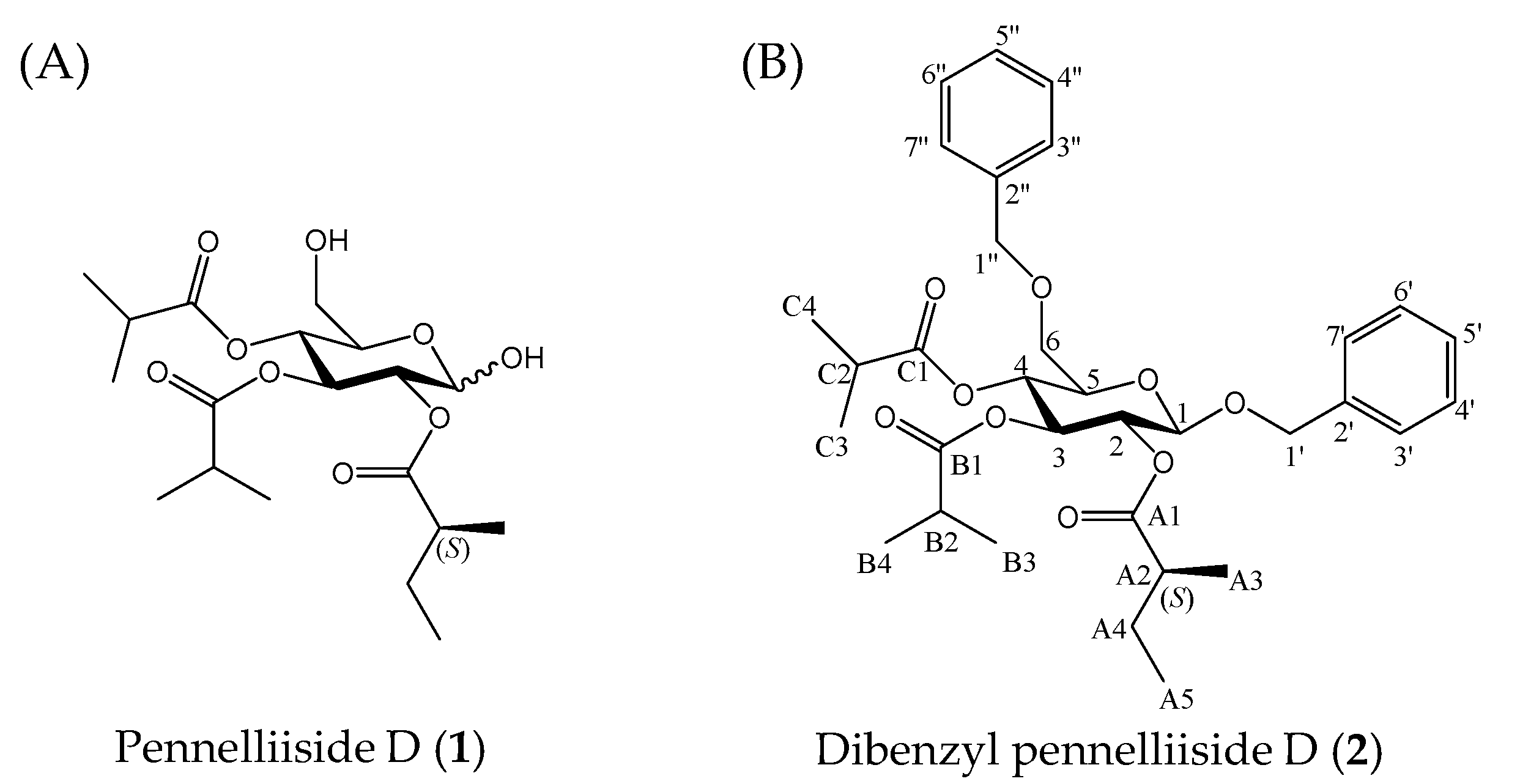
Figure 1. Structure of pennelliisides D and its benzylated derivative.
Molecules 27 03728 g001
Figure 2. Important resonances of compound 2; (A) key 1H-1H COSY and HMBC correlations; (B) key NOESY interactions observed between protons shown in the same color (pink and green).
Molecules 27 03728 g002
Scheme 1. Removal of benzyl ether.
Molecules 27 03728 sch001
Scheme 2. Total synthesis of dibenzyl pennelliiside D (2).
Molecules 27 03728 sch002
Figure 3. Expanded key 1H-NMR spectrum for the H-A4 resonances of natural dibenzyl pennelliiside D (2) and its synthesized isomers (S/R). (A) 1H-NMR spectrum of natural dibenzyl pennelliiside D (500 MHz, C6D6); (B) 1H-NMR spectrum of the synthesized isomer (S) (500 MHz, C6D6); (C) 1H-NMR spectrum of the synthesized isomer (R) (500 MHz, C6D6).
Molecules 27 03728 g003
Figure 4. Root growth inhibitory effect of pennelliiside D (1) and its constituent fatty acids. (A) and (B) root length of natural and synthesized pennelliiside D (1) and (C) root length of (S)-2-methylbutonic acid. Error bars indicate ± SE (n = 12; results are from one representative experiment); *: p < 0.05 is significantly different according to Welch’s t-test.
Molecules 27 03728 g004
Scheme 3. Synthesis of dibenzyl pennelliisides A and B.
Molecules 27 03728 sch003
Table 1. 1H NMR (500 MHz) and 13C NMR (126 MHz) spectroscopic data of natural dibenzyl pennelliiside D (2) in C6D6 (δ in ppm, J in Hz).
PositionTypeNatural Dibenzyl Pennelliiside D (2)
δCδH (J in Hz)
1CH100.34.38, d (7.6)
2CH71.95.46, m
3CH73.55.48, m
4CH70.15.30, dd (10.7, 9.5)
5CH74.33.42, m
6CH269.93.47, m
1a′CH270.74.75, d (12.2)
1b′4.45, d (12.2)
2′C138.0
3′CH128.1–128.67.26, t (7.4)
4′CH128.1–128.67.12–7.19, m
5′CH128.1–128.67.08, t (7.3)
6′CH128.1–128.67.12–7.19, m
7′CH128.1–128.67.26, t (7.4)
1C174.7
A2CH41.862.28, m
A3CH317.21.08, d (7.0)
A4CH227.21.32, 1.69, m, m
A5CH312.20.81, t (7.4)
B1C176.2
B2CH34.62.41, m
B3CH319.2–19.51.08, d (7.0)
B4CH319.2–19.51.08, d (7.0)
C1C175.3
C2CH34.52.31, m
C3CH319.2–19.51.02, d (7.0)
C4CH319.2–19.50.98, d (7.0)
1a″CH273.94.33, d (5.5)
1b″4.33, d (5.5)
2″C139.0
3″CH128.1–128.77.26, t (7.4)
4″CH128.1–128.77.12–7.19, m
5″CH128.1–128.77.08, t (7.3)
6″CH128.1–128.77.12–7.19, m
7″CH128.1–128.77.26, t (7.4)
Table 2. 1H NMR (500 MHz) and 13C NMR (126 MHz) spectroscopic data of natural pennelliiside D (1) in CDCl3 (δ in ppm, J in Hz).
Position α Anomerβ Anomer
TypeδCδH (J in Hz)δCδH (J in Hz)
1CH90.45.48, d (3.6)96.14.76, d (6.9)
2CH71.44.85, dd (6.8, 3.6)73.54.91, dd (7.7, 6.89)
3CH69.05.65, dd (10.9, 9.9)71.45.41, dd (10.4, 9.6)
4CH68.85.01, dd (10.9, 9.7)68.85.09, dd (10.4, 8.1)
5CH69.74.06, m74.73.59, m
6CH261.33.71, 3.55, m61.33.75, 3.59, m
A1C176.6 176.6
A2CH41.12.38, m41.12.42, m
A3CH316.4–19.41.03–1.17, m16.4–19.41.07–1.21, m
A4CH226.71.41, 1.62, m26.71.45, 1.66, m
A5CH311.70.85, m11.70.90, m
B1C176.0 176.0
B2CH34.22.50, m34.22.50, m
B3CH316.4–19.41.03–1.17, m16.4–19.41.07–1.21, m
B4CH316.4–19.41.03–1.17, m16.4–19.41.07–1.21, m
C1C176.9 176.9
C2CH34.22.56, m34.22.56, m
C3CH316.4–19.41.03–1.17, m16.4–19.41.07–1.21, m
C4CH316.4–19.41.03–1.17, m16.4–19.41.07–1.21, m
Table 3. 1H NMR (500 MHz) and 13C NMR (126 MHz) spectroscopic data of synthesized (S) isomer of pennelliiside D (1) in CDCl3 (δ in ppm, J in Hz).
Position.Typeα Anomerβ Anomer
δCδH (J in Hz)δCδH (J in Hz)
1CH90.45.48, d (3.6)96.14.72, d (6.9)
2CH71.44.85, dd (6.8, 3.6)73.54.87, dd (7.7, 6.89)
3CH69.05.65, dd (10.9, 9.9)71.45.37, dd (10.4, 9.6)
4CH68.85.01, dd (10.9, 9.7)68.85.05, dd (10.4, 8.1)
5CH69.74.06, m74.73.55, m
6CH261.33.71, 3.55, m61.33.71, 3.55, m
A1C176.6 176.6
A2CH41.12.24–2.42, m41.12.24–2.42, m
A3CH316.4–19.41.03-1.17, m16.4–19.41.03-1.17, m
A4CH226.71.41, 1.62, m26.71.41, 1.62, m
A5CH311.70.85, m11.70.85, m
B1C176.0 176.0
B2CH34.22.50, m34.22.50, m
B3CH316.4–19.41.03–1.17, m16.4–19.41.03–1.17, m
B4CH316.4–19.41.03–1.17, m16.4–19.41.03–1.17, m
C1C176.9 176.9
C2CH34.22.56, m34.22.52, m
C3CH316.4–19.41.03–1.17, m16.4–19.41.03–1.17, m
C4CH316.4–19.41.03–1.17, m16.4–19.41.03–1.17, m
Publisher’s Note: MDPI stays neutral with regard to jurisdictional claims in published maps and institutional affiliations.