Abstract
Prostaglandin (PG) A2, a cyclopentenone PG, induced apoptosis in both HCT116 and HCT116 p53 −/− cells. Although PGA2-induced apoptosis in HCT116 cells was dependent on the p53-DR5 pathway, the mechanism underlying PGA2-induced apoptosis in HCT116 p53 −/− cells remains unknown. In this study, we observed that PGA2 caused an increase of mRNA expression of DR5 and protein expression even in HCT116 p53 −/− cells, accompanied by caspase-dependent apoptosis. Knockdown of DR5 expression by RNA interference inhibited PGA2-induced apoptosis in HCT116 p53 −/− cells. Parallel to the induction of apoptosis, PGA2 treatment upregulated expression of genes upstream of DR5 such as ATF4 and CHOP. Knockdown of CHOP prevented DR5-dependent cell death as well as the expression of DR5 protein. Furthermore, knockdown of ATF4 by RNA interference decreased both mRNA and protein levels of CHOP and DR5, thereby suppressing PGA2-induced cell death. Consistently, the DR5 promoter activity increased by PGA2 was not stimulated when the CHOP binding site in the DR5 promoter was mutated. These results collectively suggest that PGA2 may induce DR5-dependent apoptosis via the ATF4-CHOP pathway in HCT116 p53 null cells.
1. Introduction
Prostaglandin A2 (PGA2), one of cyclopentenone PGs (cycPGs), has been reported to induce apoptosis via multiple pathways depending on cell types [1]. Although it has been reported that PGA2-induced apoptosis is dependent on de novo protein synthesis and is associated with the expression of BAX, SOX4, and c-Myc in cervical, breast and liver cancer cell lines [2,3,4], the molecular mechanisms in detail involved in PGA2-induced apoptosis have not yet been completely resolved.
DR5 is a member of the tumor necrosis factor receptor (TNFR) superfamily that possesses a cytoplasmic death domain (DD) [5]. Binding of tumor necrosis factor (TNF)-related apoptosis-inducing ligand (TRAIL) to death receptors, DR4 and DR5, promotes recruitment of Fas-associated death domain (FADD) via their cytoplasmic DDs and caspase-8 to form the death-inducing signaling complex (DISC), thereby inducing extrinsic apoptosis. Expression of DR5 can be modulated by specific transcription factors such as p53, specificity protein 1 (Sp1), nuclear factor κ-light-chain-enhancer of activated B cells (NF-κB), and C/EBP homologous protein (CHOP), whose transcriptional activities are increased by diverse triggers such as DNA damage, TNF-α, reactive oxygen species (ROS), and endoplasmic reticulum (ER) stress [6,7,8,9,10,11]. DR5 expression can also be augmented at the level of epigenetics. Interestingly, high expression of DR5 induced by anti-cancer therapeutics through diverse mechanisms leads to activation of extrinsic apoptosis via its oligomerization, even in the absence of TRAIL [7,12,13]. After it was known that the death receptor apoptotic pathway induced by TRAIL is highly selective for tumor cells over normal cells, DR5 has been intensively pursued as a target of anti-cancer therapy development [14,15].
In a previous report, PGA2 was shown to induce apoptosis dependent on DR5, whose expression was stimulated by DNA-dependent protein kinase (DNA-PK)-p53 pathway in HCT116 cells [16]. However, we also observed that PGA2 induced apoptosis in HCT116 p53 null cells (HCT116 p53 −/−). Although the potency of PGA2-induced apoptosis in HCT116 p53 −/− cells was less than that in parental HCT116 cells, it implied that PGA2 could also induce p53-independent apoptosis. Accordingly, this study aimed to investigate the mechanism underlying p53-independent apoptosis by PGA2 using HCT116 p53 −/− cells which is isogenic with HCT116 cells.
2. Results
2.1. Induction of Caspase-Dependent Apoptosis in HCT116 p53 −/− Cells by PGA2
To determine whether PGA2 can induce p53-independent apoptosis, HCT116 p53 −/− cells were treated with PGA2 and then subjected to annexin V (AV) and propidium iodide (PI) assay. As shown in Figure 1A, AV-positive HCT116 p53 −/− cells increased significantly after PGA2 treatment, accompanied by activation of caspase-8, -3, and -9, and PARP1 cleavage (Figure 1B). In addition, when HCT116 p53 −/− cells were pretreated by z-VAD-Fmk, a pan-caspase inhibitor, the increase of AV-positive cells was almost completely prevented (Figure 1C). Thus, these findings indicate that PGA2 induces caspase-dependent and p53-independent apoptosis in HCT116 p53 −/− cells.
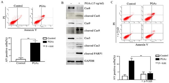
Figure 1.
Induction of caspase-dependent apoptosis in HCT116 p53 −/− cells by PGA2. (A) HCT116 p53 −/− cells were treated by PGA2 (15 μg/mL) for 30 h. Cells were then stained with annexin V and propidium iodide, which were subjected to flow cytometric analysis. A representative image of three independent annexin V assays was shown (upper panel), and the quantitative result was presented as mean ± SEM (lower panel). (B) Whole cell lysates (WCL) of HCT116 p53 −/− cells treated the same as described in (A) were subjected to immunoblot analysis against procaspase-8 (Cas8), cleaved caspase-8 (cleaved Cas8), procaspase-9 (Cas9), cleaved caspase-9 (cleaved Cas9), procaspase-3 (Cas3), cleaved caspase-3 (cleaved Cas3), and cleaved PARP1 (cleaved PARP1). GADPH was used as an internal reference protein for normalization. Densitometric measurement of three independent immunoblot analyses was represented as mean ± SEM in supplementary files. (C) HCT116 p53 −/− cells incubated in the presence of vehicle or z-VAD-Fmk for 1 h were treated with vehicle or 15 μg/mL of PGA2 for another 30 h. Cells were then analyzed by annexin V assay. The picture is representative of three independent experiments (upper panel), and their quantitative results were presented as mean ± SEM (lower panel).
2.2. Induction of DR5-Dependent Apoptosis in HCT116 p53 −/− Cells by PGA2
Since PGA2-induced apoptosis is usually dependent on de novo protein synthesis and is reported to be dependent on the expression of DR5 gene in HCT116 cells [16], we first analyzed the expression change of DR5. As shown in Figure 2A, the mRNA and protein levels of DR5 significantly all increased, implying the causative effect of DR5 in this apoptosis. Consistently with this implication, knockdown of DR5 using small interfering RNA (siRNA) suppressed PGA2-induced increase of AV-positive cells and the cleavage of PARP-1 as well (Figure 2B,C). These results collectively suggest that PGA2-induced apoptosis in HCT116 p53 −/− cells is dependent on DR5 expression.
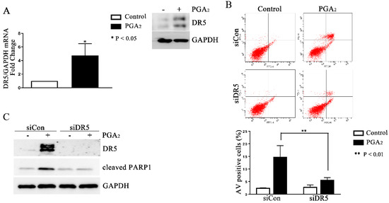
Figure 2.
Induction of DR5-dependent apoptosis in HCT116 p53 −/− cells by PGA2. (A) Total cellular RNA extracted from HCT116 p53 −/− cells treated with PGA2 (15 μg/mL) for 30 h were subjected to qPCR against indicated genes using GADPH as an internal reference gene for normalization. (B) HCT116 p53 −/− cells were transfected with scrambled RNA (siCon) or siRNA targeting DR5 (siDR5) for 24 h and incubated in the presence of vehicle or PGA2 (15 μg/mL) for an additional 30 h. Cells were then subjected to annexin V assay. The result is representative of three independent experiments (upper panel), and their quantitative result was presented as mean ± SEM (lower panel). (C) Cells treated the same as described in (B) were subjected to immunoblot analysis against DR5 and cleaved PARP-1 using GAPDH as an internal reference protein. Densitometric measurement of three independent immunoblot analyses was represented as mean ± SEM in supplementary files.
2.3. Involvement of CHOP in PGA2-Induced Apoptosis and Expression of DR5
It is generally known that the most important transcription factors that enhance the transcription of DR5 are Sp1, p53, CHOP, NF-κB, and yin-yang 1 (YY1) [6]. Since the cell line used in this study is p53 deleted, and it was reported that cycPGs accumulate in the endoplasmic reticulum (ER), thereby inducing the expression of ER stress-related genes [17,18], we expected that CHOP might be the key transcription factor to stimulate DR5 expression in PGA2-treated HCT116 p53 −/− cells. And as expected, CHOP expression was up-regulated by PGA2 at the level of both mRNA and protein along with an increase of DR5 protein (Figure 3A), and knockdown of CHOP prevented the increase of DR5 mRNA and protein expression (Figure 3B). Besides, whereas PGA2 stimulated the DR5 promoter activity, DR5 promoter with the CHOP binding site mutated was not responded to by PGA2, indicating that PGA2-induced DR5 expression is mediated by CHOP at the level of transcription (Figure 3C). Consistently with the expression change, PGA2-induced apoptosis was suppressed by knockdown of CHOP as shown in AV/PI and immunoblot analyses (Figure 3D,E). Taken together, these data suggest that PGA2 induces expression of CHOP, which in turn stimulates the transcription of DR5, resulting in apoptosis in HCT116 p53 −/− cells.
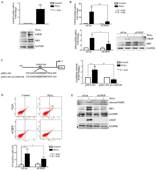
Figure 3.
Involvement of CHOP in PGA2-induced apoptosis and expression of DR5. (A) HCT116 p53 −/− cells were treated with vehicle or PGA2 (15 μg/mL) for 30 h. Cells were then subjected to qPCR against CHOP gene using GADPH as an internal reference gene for normalization (upper) or immunoblot analysis against indicated proteins with GAPDH as the normalizer (lower). (B) HCT116 p53 −/− cells were transfected with scrambled RNA or siRNA targeting CHOP for 24 h and incubated in the presence of vehicle or PGA2 (15 μg/mL) for an additional 30 h. Cells were then subjected to qPCR or immunoblot analysis against CHOP and DR5 using GAPDH as an internal reference protein or gene. (C) DR5 promoter or mutant DR5 promoter of which CHOP-binding site was changed, was transfected into HCT116 p53 −/− cells along with Renilla luciferase for 24 h, and HCT116 p53 −/− cells were then treated with vehicle or PGA2. At 30 h post-treatment, firefly luciferase activity of DR5 promoter was measured by the Dual-luciferase assay method using Renilla luciferase activity as the normalizer. (D) HCT116 p53 −/− cells were transfected with scrambled RNA (siCon) or siRNA targeting CHOP (siCHOP) for 24 h and incubated in the presence of vehicle or PGA2 (15 μg/mL) for an additional 30 h. Cells were then subjected to annexin V assay. The result is representative of three independent experiments (upper), and their quantitative result was presented as mean ± SEM (lower). (E) WCL of HCT116 p53 −/− cells treated the same as described in (D) were subjected to immunoblot analysis against indicated proteins using GAPDH as an internal reference protein. Densitometric measurement of three independent immunoblot analyses was represented as mean ± SEM in supplementary files.
2.4. The Role of ATF4 in PGA2-Induced Apoptosis and Activation of CHOP-DR5 Pathway
Activating transcription factor (ATF) 3 and ATF4 are stress-responsive proteins that can conduce CHOP transcription in response to various stimuli. Moreover, it was reported that ATF4-ATF3-CHOP cascade induced apoptosis in human pulmonary cancer and leukemia cells [19,20]. Thus, to identify the upstream regulators of CHOP, we analyzed the expression of ATF3 and ATF4. As shown in Figure 4A,B, PGA2 increased the expression of ATF3 and 4 at the levels of mRNA and protein, implying the ATF3 and 4-mediated expression of CHOP and DR5. Although PGA2 increased the expression of ATF3 with a parallel level to that of ATF4, only knockdown of ATF4 inhibited the induction of apoptosis (Figure 4C and Figure S7). Furthermore, ATF4 siRNA suppressed the PGA2-induced increase of CHOP and DR5 proteins (Figure 4D), whereas ATF3 siRNA did not affect it. Collectively, therefore, it can be concluded that PGA2 induces apoptosis by activating the ATF4-DR5-CHOP pathway.

Figure 4.
The role of ATF4 in the PGA2-induced apoptosis and activation of the CHOP-DR5 pathway. (A) Total cellular RNA extracted from HCT116 p53 −/− cells treated with PGA2 (15 μg/mL) for 30 h were subjected to qPCR against ATF3 or ATF4 using GADPH as an internal reference gene for normalization. (B) WCL of cells treated the same as described in (A) were subjected to immunoblot analysis against ATF3 and ATF4 using GAPDH as the normalizer. (C) HCT116 p53 −/− cells were transfected with scrambled RNA (siCon) or siRNA targeting ATF3 (siATF3) or ATF4 (siATF4) for 24 h and incubated in the presence of vehicle or PGA2 (15 μg/mL) for an additional 30 h. Cells were then subjected to annexin V assay. The result is representative of three independent experiments (left panel), and their quantitative result was presented as mean ± SEM (right panel). (D) Cells treated the same as described in (C) were subjected to immunoblot analysis against indicated proteins using GAPDH as an internal reference protein. Densitometric measurement of three independent immunoblot analyses was represented as mean ± SEM in supplementary files.
3. Discussion
In a previous report, PGA2 was shown to activate DR5-dependent apoptosis in HCT116 cells containing wild-type p53 [16]. In HCT116 cells, PGA2 induced phosphorylation of both histone H2AX, a hallmark of DNA double-strand breaks, and DNA-PK, which in turn phosphorylated p53 with an elevation of its transcriptional activity, resulting in upregulation of DR5 expression and finally induction of apoptosis. So, apoptosis activated by PGA2 in HCT116 cells seemed to be dependent on the p53-induced expression of DR5. However, it turned out that PGA2 could trigger DR5-dependent apoptosis even in the absence of p53 in isogenic HCT116 cells with p53 genetically deleted (HCT116 p53 −/− cells) in this study. In HCT116 p53 −/− cells treated with PGA2, histone H2AX was also phosphorylated, indicating that PGA2 damaged DNA as in parental HCT116 cells (Supplementary Figure S1). However, NU7441 and KU5593, chemical inhibitors of DNA-PK and ATM activities, respectively, did not prevent either PGA2-induced expression of DR5 (Supplementary Figure S2). Collectively, these data suggest that PGA2-induced DNA damage leading to DR5 expression should be via the activity of p53 but not DNA-PK per se in HCT116 cells, and thus, in the absence of p53 like HCT116 p53 −/− cells, other pathways might be activated to increase DR5 expression. Interestingly, in annexin V assays, since PGA2 also increased PI staining albeit subtlety, PGA2 might potentially induce necrosis in HCT116 p53 −/− cells, implying the activation of various cell death pathways by PGA2 in the absence of p53.
PGA2, a cycPG, is formed by non-enzymatic dehydration of PGE2. While PGE2 exerts its function by binding to the PGE2 receptors which are G-protein coupled receptors in the plasma membrane, PGA2, like other cycPGs, enters cells and accumulates in subcellular organelles such as nuclei and mitochondria, where it interacts with diverse molecules such as DNA and cellular proteins including transcription factors [21]. For example, in the cytosol, PGA2 binds transcriptional and signaling regulators such as KEAP1 and IKK-β subunit of IKK [22,23]. KEAP1 binding of PGA2 induces Nrf2 activation, and IKK-β subunit binding prevents NF-κB activation and hence, repression of LPS-induced inflammatory response. As mentioned above, accumulated PGA2 in nuclei might damage DNA, which leads to DR5-dependent apoptosis. PGA2 also directly interacts with mitochondria, which causes mitochondrial outer membrane permeabilization through which cytochrome c releases into cytosol resulting in the initiation of the intrinsic apoptosis [24]. Interestingly, it was reported that cycPGs, including PGA2, localize the endoplasmic reticulum (ER), and PGJ2, another cycPG, could stimulate transcription of BiP, an ER-stress response gene, through an unfolded protein response element [17,18]. Like PGJ2, PGA2 enhanced the expression of BiP at the levels of mRNA and protein (Figure S3). Notably, PGA2 also increased the expression of ER-stress response genes such as ATF3, ATF4, and CHOP, implying that the ER-stress response might be involved in PGA2-induced DR5 expression in HCT116 p53 −/− cells. Moreover, as expected, phosphorylation of eIF2α and PERK was increased, accompanied by the expression of ATF3, ATF4, and CHOP (Figure S4). So, it can be speculated that PGA2 may induce the ER-stress response leading to activation of ATF4–CHOP–DR5 pathway, although how PGA2 induces the ER stress response was not investigated in this study.
To expand the mechanism of this study to colon cancer cells, we also analyzed the apoptosis induction activity of PGA2 in SW620 cells of which p53 is mutated. However, as shown in Figure S6, PGA2 did not induce apoptosis up to 48 h in SW620 cells. So, it can be summarized that PGA2 induces fulminant apoptosis in HCT116 p53 WT cells, slight apoptosis in HCT116 p53 −/− cells, and no apoptosis in p53-mutated SW620 cells. Although we did not analyze many colon cancer cell lines, these findings suggest that p53 plays a critical role in apoptosis induction by PGA2, but PGA2 induction of apoptosis in colon cancer cells is not determined only by p53 genetic status. Moreover, depending on the cellular context, PGA2 may have the potential to induce apoptosis even in p53-mutated colon cancer cells, as shown in this study that PGA2 induces apoptosis via ER-stress–ATF4–CHOP–DR5 pathway. It can also be expected that PGA2 may induce apoptosis through various pathways in a p53-independent manner.
TRAIL is a potent inducer of apoptosis selectively in cancer cells. TRAIL induces apoptosis via DR4 and DR5 [15,25]. However, some cancers develop TRAIL resistance by downregulating the expression of death receptors or upregulating decoy receptors which hinders the binding of TRAIL to death receptors [26]. Thus, molecules that increase the expression of death receptors may have the potential to overcome TRAIL resistance [27,28]. As shown in Figure S5, PGA2 pretreatment significantly potentiated TRAIL-induced apoptosis in HCT116 p53 −/− cells as well as HCT116 cells. Although the effect of PGA2 in TRAIL-resistant cells was not examined yet, this finding may shed a sound effect of PGA2 on the recovery of TRAIL resistance.
It is firmly established that tumor suppressor p53 is the most important gene in anti-carcinogenesis and induction of apoptosis in cancer cells [29]. In fact, most anti-cancer therapeutics exert their effects via the activity of p53. But, unfortunately, almost half of all cancers contain a mutation of p53, which could contribute to the development of resistance to anti-cancer therapeutics [30]. Therefore, the development of an anti-cancer agent that induces both p53-dependent and -independent apoptosis in cancer cells may be of high value in cancer patient treatment. Therefore, expression of DR5 induced by PGA2 via p53 and CHOP in cancer cells containing wild-type p53 and mutant p53, respectively, may have the potential to enhance the anti-cancer therapeutic effect of TRAIL.
4. Materials and Methods
4.1. Cell Culture
Human colon cancer HCT116 p53 −/− cells were maintained in RPMI 1640 supplemented with 10% fetal bovine serum (Hyclone, Logan, UT, USA), 100 units/mL penicillin (Hyclone), and glutamate (Invitrogen, Carlsbad, CA, USA). The cells were incubated in a humidified atmosphere of 5% CO2 at 37 °C.
4.2. Chemicals
PGA2 was obtained from Enzo Life Sciences, Inc. (Farmingdale, NY, USA). Z-VAD-Fmk was purchased from Tocris Bioscience (Bristol, UK). All reagents used in this study were of molecular biology or cell culture tested grade.
4.3. Transfection with Small Interfering RNA (siRNA)
According to the manufacturer’s instruction, siRNA transfection was performed with Lipofectamine RNAiMAX (Invitrogen). Briefly, the mixture containing siRNA, Opti-MEM (Life Technologies, Carlsbad, CA, USA), and Lipofectamine RNAiMAX was incubated for 15 min at room temperature (RT), then dropped to the cells and incubated for 24 h. Subsequently, the cells were treated with PGA2 for further analysis. DR5 siRNA and ATF4 siRNA were purchased from Bioneer (Daejeon, Korea). CHOP siRNA and ATF3 siRNA were obtained from Invitrogen and Santa Cruz Biotechnology (Santa Cruz, CA, USA), respectively.
4.4. Apoptosis Assay
The phosphatidylserine on the extracellular side of cell membranes and propidium iodide to detect necrotic nuclei using an Annexin V-FITC/PI cell apoptosis detection kit (BD Pharmingen, San Diego, CA, USA) according to the manufacturer’s instruction. Then, the fluorescence of 10,000 stained cells was measured on FACS Canto II (BD Biosciences, San Jose, CA, USA) and analyzed using BD FACS Diva program.
4.5. Immunoblot Analysis and Antibodies
For immunoblot analysis, proteins were extracted from PGA2-treated cells. Cells were lysed in RIPA buffer (Cell signaling Technology, Boston, MA, USA) containing cocktails of protease inhibitors (Roche, Basel, Switzerland) and phosphatase inhibitors (FIVE photon Biochemicals, San Diego, CA, USA). All antibodies were commercially available as follows. Rabbit anti-cleaved PARP1 (c-PARP1), anti-ATF3, anti-ATF4, anti-DR5 (death receptor 5), anti-caspase-3, anti-cleaved caspase-3, anti-caspase-9, and mouse anti-CHOP were purchased from Cell Signaling Technology (Boston, MA, USA). Chicken anti-GAPDH antibodies were obtained from Merck Millipore Korea (Seoul, Korea), respectively. Peroxidase-conjugated antibodies (HRP-conjugated anti-rabbit or -mouse IgG) were from Sigma-Aldrich Inc., and KPL (Gaithersburg, MD, USA, HRP-conjugated anti-chicken IgG).
4.6. Quantitative Real-Time PCR (qPCR)
Total RNAs were extracted with RNAiso plus reagent (Takara Korea Biomedical Inc., Seoul, Korea). First-strand cDNA was synthesized from total RNA using PrimeScriptTM RT reagent Kit (Takara Korea Biomedical Inc.). First-strand cDNA was then amplified by specific primers against various genes using SYBR FAST qPCR Kit (Nanohelix, Daejeon, Korea) on ABI 7300 Real-Time PCR System (Applied Biosystems, Carlsbad, CA, USA). GAPDH mRNA level normalized each mRNA level, and relative changes among samples were calculated using the ΔΔCt method [31].
4.7. Luciferase Reporter Gene Assay
The activity of the human pGL3-DR5(-605)-Luc promoter and pGL3-DR5 (mt. CHOP)-Luc was measured by dual luciferase-reporter gene assay. pGL3-DR5(-605)-Luc promoter contains the CHOP binding site in DR5 promoter sequence, and DR5 (mt. CHOP) promoter-luciferase plasmid contains a mutated CHOP binding site. All promoter-luciferase plasmids were generously donated by Dr. Taeg Kyu Kwon (Keimyung University, Daegu, Korea). To measure the activity of DR5 promoter, cells were transfected with 0.2 μg of pGL3-DR5(-605)-Luc or pGL3-DR5(mt. CHOP)-Luc, together with 0.02 μg of Renilla luciferase using Xtreme Gene 9 (Roche Diagnostics, Basel, Switzerland) for 24 h, and then treated with PGA2. At the indicated times post-treatment, Firefly and Renilla luciferase activities were determined using Dual-Luciferase kit (Promega, Fitchburg, WI, USA), and data were expressed as relative luciferase activity (RLA) of three independent experiments performed in triplicate. Renilla luciferase activity was used to normalize transfection efficiency, and normalized firefly luciferase activity of non-treated cells was set to RLA 1 fold.
4.8. Statistical Analysis
All data in this study are expressed as the means ± standard error of the mean (SEM) obtained from three independent experiments performed in triplicate. Statistical analysis was performed using Student’s two-tailed t-test for paired data. p values of the results were indicated in each figure.
Supplementary Materials
The following supporting information can be downloaded at: https://www.mdpi.com/article/10.3390/molecules27123804/s1, Figure S1: Phosphorylation of H2AX induced by PGA2 in HCT116 p53 −/− cells; Figure S2: The effect of chemical inhibitors of DNA damage sensing protein kinases on PGA2-induced DR5 expression in HCT116 p53 −/− cells; Figure S3: Increase of BiP expression induced by PGA2 in HCT116 p53 −/− cells; Figure S4: Increase of ER-stress proteins by PGA2 in HCT116 p53 −/− cells; Figure S5: The effect of PGA2 on TRAIL-induced apoptosis. Figure S6: The effect of PGA2 on the survival of SW620 cells; Figure S7: The annexin V (AV) histogram of the results presented in Figure 4. Graphs of densitometric analyses: Intensities of protein bands in immunoblot analyses were measured by densitometry and the data was presented as mean ± SEM of three independent experiments.
Author Contributions
Conceptualization, K.-M.P., S.-Y.L. and H.-S.K.; methodology, K.-M.P., S.-Y.L. and H.-S.K.; data acquisition, K.-M.P., J.-Y.P. and J.P.; formal analysis, K.-M.P., J.-Y.P., S.-Y.L. and H.-S.K.; investigation, K.-M.P., J.-Y.P., S.-Y.L. and H.-S.K.; data curation, K.-M.P., S.-Y.L. and H.-S.K.; writing—original draft preparation, K.-M.P., S.-Y.L. and H.-S.K.; writing—review and editing, K.-M.P., S.-Y.L. and H.-S.K.; supervision, H.-S.K.; project administration, S.-Y.L. and H.-S.K.; funding acquisition, S.-Y.L. and H.-S.K. All authors have read and agreed to the published version of the manuscript.
Funding
This research was supported by grants from the National Research Foundation of Korea (NRF) funded (NRF-2016R1D1A1B03934999 and 2019R1A5A2027588 to H.-S.K).
Institutional Review Board Statement
Not applicable.
Informed Consent Statement
Not applicable.
Data Availability Statement
Not applicable.
Acknowledgments
The authors would like to thank Taeg Kyu Kwon (Department of Immunology, School of Medicine, Keimyung University, Daegu, Republic of Korea) for providing DR5 promoter-reporter gene construct.
Conflicts of Interest
The authors declare no conflict of interest.
References
- Díez-Dacal, B.; Pérez-Sala, D. A-class prostaglandins: Early findings and new perspectives for overcoming tumor chemoresistance. Cancer Lett. 2012, 320, 150–157. [Google Scholar] [CrossRef] [PubMed]
- Joubert, A.; Maritz, C.; Joubert, F. Influence of prostaglandin A2 and 2-methoxyestradiol on Bax and Bcl-2 expression levels in cervical carcinoma cells. Biomed. Res. 2005, 26, 87–90. [Google Scholar] [CrossRef][Green Version]
- Joubert, A.M.; Panzer, A.; Bianchi, P.C.; Lottering, M.L. The effects of prostaglandin A2 on cell growth, cell cycle status and apoptosis induction in HeLa and MCF-7 cells. Cancer Lett. 2003, 191, 203–209. [Google Scholar] [CrossRef]
- Ahn, S.G.; Kim, H.S.; Jeong, S.W.; Kim, B.E.; Rhim, H.; Shim, J.Y.; Kim, J.W.; Lee, J.H.; Kim, I.K. Sox-4 is a positive regulator of Hep3B and HepG2 cells’ apoptosis induced by prostaglandin (PG)A(2) and delta(12)-PGJ(2). Exp. Mol. Med. 2002, 34, 243–249. [Google Scholar] [CrossRef] [PubMed]
- Yang, A.; Wilson, N.S.; Ashkenazi, A. Proapoptotic DR4 and DR5 signaling in cancer cells: Toward clinical translation. Curr. Opin. Cell Biol. 2010, 22, 837–844. [Google Scholar] [CrossRef] [PubMed]
- Min, K.J.; Woo, S.M.; Shahriyar, S.A.; Kwon, T.K. Elucidation for modulation of death receptor (DR) 5 to strengthen apoptotic signals in cancer cells. Arch. Pharm. Res. 2019, 42, 88–100. [Google Scholar] [CrossRef] [PubMed]
- Wu, G.S.; Burns, T.F.; McDonald, E.R., III; Jiang, W.; Meng, R.; Krantz, I.D.; Kao, G.; Gan, D.D.; Zhou, J.Y.; Muschel, R.; et al. KILLER/DR5 is a DNA damage-inducible p53-regulated death receptor gene. Nat. Genet. 1997, 17, 141–143. [Google Scholar] [CrossRef]
- Takimoto, R.; El-Deiry, W.S. Wild-type p53 transactivates the KILLER/DR5 gene through an intronic sequence-specific DNA-binding site. Oncogene 2000, 19, 1735–1743. [Google Scholar] [CrossRef] [PubMed]
- Yamaguchi, H.; Wang, H.G. CHOP is involved in endoplasmic reticulum stress-induced apoptosis by enhancing DR5 expression in human carcinoma cells. J. Biol. Chem. 2004, 279, 45495–45502. [Google Scholar] [CrossRef]
- Sheikh, M.S.; Burns, T.F.; Huang, Y.; Wu, G.S.; Amundson, S.; Brooks, K.S.; Fornace, A.J.; El-Deiry, W.S. p53-dependent and -independent regulation of the death receptor KILLER/DR5 gene expression in response to genotoxic stress and tumor necrosis factor alpha. Cancer Res. 1998, 58, 1593–1598. [Google Scholar] [PubMed]
- Gupta, S.C.; Francis, S.K.; Nair, M.S.; Mo, Y.Y.; Aggarwal, B.B. Azadirone, a limonoid tetranortriterpene, induces death receptors and sensitizes human cancer cells to tumor necrosis factor-related apoptosis-inducing ligand (TRAIL) through a p53 protein-independent mechanism: Evidence for the role of the ROS-ERK-CHOP-death receptor pathway. J. Biol. Chem. 2013, 288, 32343–32356. [Google Scholar] [PubMed]
- Nagata, S. Apoptosis by death factor. Cell 1997, 88, 355–365. [Google Scholar] [CrossRef]
- Sheridan, J.P.; Marsters, S.A.; Pitti, R.M.; Gurney, A.; Skubatch, M.; Baldwin, D.; Ramakrishnan, L.; Gray, C.L.; Baker, K.; Wood, W.I.; et al. Control of TRAIL-induced apoptosis by a family of signaling and decoy receptors. Science 1997, 277, 818–821. [Google Scholar] [CrossRef]
- Walczak, H.; Miller, R.E.; Ariail, K.; Gliniak, B.; Griffith, T.S.; Kubin, M.; Chin, W.; Jones, J.; Woodward, A.; Le, T.; et al. Tumoricidal activity of tumor necrosis factor-related apoptosis-inducing ligand in vivo. Nat. Med. 1999, 5, 157–163. [Google Scholar] [CrossRef] [PubMed]
- Yuan, X.; Gajan, A.; Chu, Q.; Xiong, H.; Wu, K.; Wu, G.S. Developing TRAIL/TRAIL death receptor-based cancer therapies. Cancer Metastasis Rev. 2018, 37, 733–748. [Google Scholar] [CrossRef] [PubMed]
- Lee, S.B.; Lee, S.; Park, J.Y.; Lee, S.Y.; Kim, H.S. Induction of p53-Dependent Apoptosis by Prostaglandin A(2). Biomolecules 2020, 10, 492. [Google Scholar] [CrossRef]
- Odani, N.; Negishi, M.; Takahashi, S.; Kitano, Y.; Kozutsumi, Y.; Ichikawa, A. Regulation of BiP gene expression by cyclopentenone prostaglandins through unfolded protein response element. J. Biol. Chem. 1996, 271, 16609–16613. [Google Scholar] [CrossRef]
- Takahashi, S.; Odani, N.; Tomokiyo, K.; Furuta, K.; Suzuki, M.; Ichikawa, A.; Negishi, M. Localization of a cyclopentenone prostaglandin to the endoplasmic reticulum and induction of BiP mRNA. Biochem. J. 1998, 335, 35–42. [Google Scholar] [CrossRef]
- Liu, G.; Su, L.; Hao, X.; Zhong, N.; Zhong, D.; Singhal, S.; Liu, X. Salermide up-regulates death receptor 5 expression through the ATF4-ATF3-CHOP axis and leads to apoptosis in human cancer cells. J. Cell. Mol. Med. 2012, 16, 1618–1628. [Google Scholar] [CrossRef]
- Joo, J.H.; Ueda, E.; Bortner, C.D.; Yang, X.P.; Liao, G.; Jetten, A.M. Farnesol activates the intrinsic pathway of apoptosis and the ATF4-ATF3-CHOP cascade of ER stress in human T lymphoblastic leukemia Molt4 cells. Biochem. Pharmacol. 2015, 97, 256–268. [Google Scholar] [CrossRef] [PubMed]
- Straus, D.S.; Glass, C.K. Cyclopentenone prostaglandins: New insights on biological activities and cellular targets. Med. Res. Rev. 2001, 21, 185–210. [Google Scholar] [CrossRef]
- Kobayashi, M.; Li, L.; Iwamoto, N.; Nakajima-Takagi, Y.; Kaneko, H.; Nakayama, Y.; Eguchi, M.; Wada, Y.; Kumagai, Y.; Yamamoto, M. The Antioxidant Defense System Keap1-Nrf2 Comprises a Multiple Sensing Mechanism for Responding to a Wide Range of Chemical Compounds. Mol. Cell. Biol. 2009, 29, 493–502. [Google Scholar] [CrossRef] [PubMed]
- Rossi, A.; Kapahi, P.; Natoli, G.; Takahashi, T.; Chen, Y.; Karin, M.; Santoro, G. Anti-inflammatory cyclopentenone prostaglandins are direct inhibitors of IkB kinase. Nature 2000, 403, 103–108. [Google Scholar] [CrossRef]
- Lee, S.-Y.; Ahn, J.-H.; Ko, K.; Kim, J.; Jeong, S.; Kim, I.-K.; Kim, J.; Kim, H.-S. Prostaglandin A2 activates intrinsic apoptotic pathway by direct interaction with mitochondria in HL-60 cells. Prostaglandins Other Lipid Mediat. 2010, 91, 23–37. [Google Scholar] [CrossRef]
- Karstedt, S.; Montinaro, A.; Walczak, H. Exploring the TRAILs less travelled: TRAIL in cancer biology and therapy. Nat. Rev. Cancer 2017, 17, 352–366. [Google Scholar] [CrossRef] [PubMed]
- Wang, F.; Lin, J.; Xu, R. The molecular mechanisms of TRAIL resistance in cancer cells: Help in designing new drugs. Curr. Pharm. Des. 2014, 20, 6714–6722. [Google Scholar] [CrossRef] [PubMed]
- Stöhr, D.; Schmid, J.O.; Beigl, T.B.; Mack, A.; Maichl, D.S.; Cao, K.; Budai, B.; Fullstone, G.; Kontermann, R.E.; Mürdter, T.E.; et al. Stress-induced TRAILR2 expression overcomes TRAIL resistance in cancer cell spheroids. Cell Death Differ. 2020, 27, 3037–3052. [Google Scholar] [CrossRef] [PubMed]
- Deng, L.; Zhai, X.; Liang, P.; Cui, H. Overcoming TRAIL resistance for glioblastoma treatment. Biomolecules 2021, 11, 572. [Google Scholar] [CrossRef] [PubMed]
- Joerger, A.C.; Fersht, A.R. The p53 pathway: Origins, inactivation in cancer, and emerging therapeutic approaches. Annu. Rev. Biochem. 2016, 85, 375–404. [Google Scholar] [CrossRef] [PubMed]
- Wu, C.-E.; Pan, Y.-R.; Yeh, C.-N.; Lunec, J. Targeting P53 as a future strategy to overcome gemcitabine resistance in biliary tract cancers. Biomolecules 2020, 10, 1474. [Google Scholar] [CrossRef]
- Schmittgen, T.D.; Livak, K.J. Analyzing real-time PCR data by the comparative C(T) method. Nat. Protoc. 2008, 3, 1101–1108. [Google Scholar] [CrossRef] [PubMed]


Publisher’s Note: MDPI stays neutral with regard to jurisdictional claims in published maps and institutional affiliations. |
© 2022 by the authors. Licensee MDPI, Basel, Switzerland. This article is an open access article distributed under the terms and conditions of the Creative Commons Attribution (CC BY) license (https://creativecommons.org/licenses/by/4.0/).