Article

Lanthanide Molecular Species Generated Fe3O4@SiO2-TbDPA Nanosphere for the Efficient Determination of Nitrite

1
Key Lab of Ecological Restoration in Hilly Areas, School of Chemical & Environmental Engineering, Pingdingshan University, Pingdingshan 467000, China
2
Guangzhou Key Laboratory of Analytical Chemistry for Biomedicine, School of Chemistry, South China Normal University, Guangzhou 510006, China
3
Institute of Biomedical Engineering, College of Life Sciences, Qingdao University, Qingdao 266071, China
4
Ruide Technologies (Foshan) Inc., Foshan 528311, China
*
Author to whom correspondence should be addressed.
Molecules 2022, 27(14), 4431; https://doi.org/10.3390/molecules27144431
Submission received: 20 June 2022 / Revised: 7 July 2022 / Accepted: 9 July 2022 / Published: 11 July 2022
(This article belongs to the Section Inorganic Chemistry)

Abstract

:
The presence of nitrite (NO2) in water and food leads to serious problems in public health and the environment. Therefore, it is important to develop a rapid and efficient method for the selective detection of NO2. In this work, the synthesis and characterization of magnetic Fe3O4@SiO2-TbDPA nanoprobe have been carried out. The Fe3O4@SiO2-TbDPA aqueous solution exhibits a strong green emission. Due to the addition of various concentrations of NO2 (0–100 μM), the fluorescence intensity has been suppressed. The nanoprobe Fe3O4@SiO2-TbDPA exhibits excellent selectivity and sensitivity toward NO2 ions. Excellent linearity is obtained in the range of 5–80 μM with a detection limit of 1.03 μM. Furthermore, the presence of magnetic Fe3O4 nanoparticles in Fe3O4@SiO2-TbDPA nanospheres will also facilitate the effective separation of Fe3O4@SiO2-TbDPA from the aqueous solution. Our proposed strategy is expected to fabricate an organic-inorganic hybrid magnetic nanomaterial and can be used as an efficient sensor. It has been shown that this new strategy has numerous advantages, such as high stability, selectivity, and simplicity of operation. It demonstrates great potential for simple and convenient NO2 detection. It may expand to a variety of ranges in environmental monitoring and biomedical fields.

1. Introduction

Nitrite (NO2) is an important substance that is widely distributed in the environment and food. It has been extensively used in meat preservation and processing in order to inhibit the propagation of Clostridium botulinum and to improve the flavor of meat [1]. Meanwhile, NO2 is a well-known signaling molecule that plays a vital role in normal physiological activities, such as hypoxia, nitric oxide homeostasis and bloodstream regulation [2]. However, excessive intake of NO2 can lead to a variety of disorders, including intrauterine spontaneous abortion, growth retardation, central nervous system congenital defects and infant methemoglobinemia [3,4]. Furthermore, the presence of NO2 ions in synthetic urine can be employed as an indicator of urinary tract infections [5]. Because of the toxicity of NO2 ions, the accepted maximum contaminant levels (MCL) of NO2 ions in drinking water are regulated to be 214.2 μM by the World Health Organization (WHO) and 71.4 μM by the U.S. Environmental Protection Agency (EPA) [6]. Traditional methods for the detection of NO2 are given as spectrophotometry, high-performance liquid chromatography, ion chromatography, gas chromatography, electrophoresis, electrochemical methods and so on [7,8,9,10,11]. Although these methods have considerable sensitivity and detection limits, most of them require complex equipment and instruments and tedious operational procedures and skills, which lack realistic operability and make it difficult to achieve highly sensitive, selective, qualitative and quantitative detection of NO2 [12,13,14]. Fortunately, fluorescent probes have become a suitable tool for NO2 detection due to their easy and rapid operation, highly sensitive, highly selective, and low cost [15,16,17]. Therefore, the realization of preparation for efficient and reliable determination of NO2 will be expected.
Lanthanide ions exhibit specific luminescence properties, including high quantum efficiency, extraordinary color purity, a long lifetime, sharp emission peaks, and large Stokes shifts [18,19,20]. Due to these merits, lanthanide luminescent materials have been widely studied in chemical and biological sensing systems [21,22,23]. In recent years, the development of lanthanide luminescent fluorescent probes for guest molecule (e.g., anions, cations, biomolecules) recognition has become an emerging field [24,25,26,27]. Our group has been devoted to the preparation of lanthanide hybrid materials and sensing applications, and the rapid development for the achievement of specific lanthanide sensors has been reported [28,29,30]. Previously, we found a ClO highly selective Europium sensor based on an oxime isomerization reaction for the first time [31]. Magnetic nanomaterials are one of the most important classes of materials due to their superior properties and their wide application in science and technology [32,33]. Taking these into account, we believe that the grafting of lanthanide ions onto magnetic nanomaterials will induce effective results. At present, this has never been mentioned for the smart magnetic nanomaterial in response to NO2 ions.
In this study, we rationally designed and fabricated a selective and sensitive fluorescent nanoprobe, Fe3O4@SiO2-TbDPA, and its effective detection of NO2 in the water sample was explored (Figure 1). The as-prepared Fe3O4@SiO2-TbDPA was fully investigated by FT-IR, UV-Vis, TEM, SEM and fluorescence spectroscopy. Fe3O4@SiO2-TbDPA aqueous solution exhibits a strong green emission. Its fluorescent signal was decreased depending on the presence of NO2. Fe3O4@SiO2-TbDPA exhibited good selectivity and sensitivity toward NO2 in a 100% aqueous solution. Moreover, after detection, the magnetic-sensitive nanoprobe was separated from the aqueous solution by taking advantage of their magnetic properties. The results shed new lights on the determination process in practical environments and biomedical analysis.

2. Experimental Section

2.1. Reagents and Materials

Terbium perchlorate (Tb(ClO4)3·6H2O) was acquired from the Shanghai Yuelong company (Shanghai, China). Ferroferric oxide (II, III) (25% in H2O), ammonia solution (NH3·H2O, 25%), tetraethyl orthosilicate (TEOS, 99.9%) (SiC8H20O4), aminopropyltriethoxysilane (APTES, 99.9%) (C9H23NO3Si), Triton X-100, 1-hexanol and 2,6-pyridinedicarboxylic acid chloride were purchased from Aladdin Chemistry Co. Ltd. (Shanghai, China). All the other metal salts and reagents were purchased from Guangzhou Chemical Reagent Factory (Guangzhou, China) and used without second purification.

2.2. Characterization

Transmission electron microscope (TEM) images were obtained with a JEOL JEM-2100 HR transmission electron microscope. Scanning electron microscopy (SEM) images were measured by a Tescan 5136MM scanning electron microscope. FT-IR spectra of the materials were measured within the 4000–400 cm−1 wavenumber range by using a Prestige-21 spectrometer. UV-vis spectra were recorded on an Agilent 8453 UV-visible spectrophotometer. The magnetic properties of the samples were measured at room temperature using a BHV-55 vibration sample magnetometer (VSM) with an applied field of −5000–5000 Oe. The fluorescence spectra were collected with a Hitachi-4600 fluorescence spectrophotometer.

2.3. Preparation of Fe3O4@SiO2-NH2 Nanospheres

The Fe3O4@SiO2-NH2 nanospheres were prepared according to the literature procedure with slight modifications [33]. Briefly, 0.13 g Fe3O4, 18 g Triton X-100, 16 mL n-hexanol and 75 mL cyclohexane were mixed in a 250 mL glass flask with vigorous mechanical agitation; then, 4 mL deionized H2O was immediately added. Subsequently, 1 mL TEOS was added to the above solution, and the mixture was stirred for 30 min. Lastly, 0.7 mL NH3·H2O was added to the above mixture to initiate silica polymerization, and the polymerization was allowed to proceed for 18 h. The resulting Fe3O4@SiO2-NH2 nanospheres were washed with water and ethanol three times and then magnetically separated using a simple bar magnet. The as-prepared Fe3O4@SiO2 nanospheres were redispersed into a 20 mL ethanol solution. Then, 0.5 mL APTES and 0.3 mL NH3·H2O were added. This suspension was stirred at room temperature for 12 h. The resulting Fe3O4@SiO2-NH2 nanospheres were collected by magnetic decantation and purified by ethanol via repeated washing. Finally, the Fe3O4@SiO2-NH2 magnetic nanospheres were dried under vacuum at 60 °C for 12 h.

2.4. Preparation of Fe3O4@SiO2-DPA Nanospheres

In order to obtain the 2,6-Pyridinedicarboxylic acid chloride-modified Fe3O4@SiO2, the prepared Fe3O4@SiO2-NH2 nanospheres (0.10 g) and 2,6-pyridinedicarboxylic acid chloride (0.30 g) were suspended in anhydrous toluene (20 mL) and refluxed for 10 h under an N2 atmosphere. The obtained functionalized Fe3O4@SiO2-DPA nanospheres were washed three times with ethyl alcohol to remove excess 2,6-pyridinedicarboxylic acid chloride and then dried under vacuum at 60 °C for 12 h.

2.5. Fabrication of Terbium Hybrid Materials (Fe3O4@SiO2-TbDPA)

A total of 50 mg Fe3O4@SiO2-DPA and 50 mg Tb(ClO4)3·6H2O were dispersed in 20 mL ethanol. Then, 0.2 mL NH3·H2O was added, and the mixture was refluxed for 8 h. After centrifugation, the precipitate was washed with ethanol three times and dried under vacuum at 60 °C for 12 h to yield Fe3O4@SiO2-TbDPA nanospheres.

2.6. Optical Studies

The stock solution of 0.1 mg/mL Fe3O4@SiO2-TbDPA and 10 mM (Na2CO3, Na2SO4, Na2HPO4, NaH2PO4, CH3COONa, NaNO3, NaF, NaCl, NaBr, NaI and NaNO2) were prepared in deionized water, respectively. Fluorescence response of Fe3O4@SiO2-TbDPA toward different anions was performed by introducing 100 μM (CO32−, SO42−, HPO42−, H2PO4, CH3COO, NO3, F, Cl, Br, I and NO2) into Fe3O4@SiO2-TbDPA (0.1 mg) aqueous solution at room temperature, respectively.

3. Results and Discussion
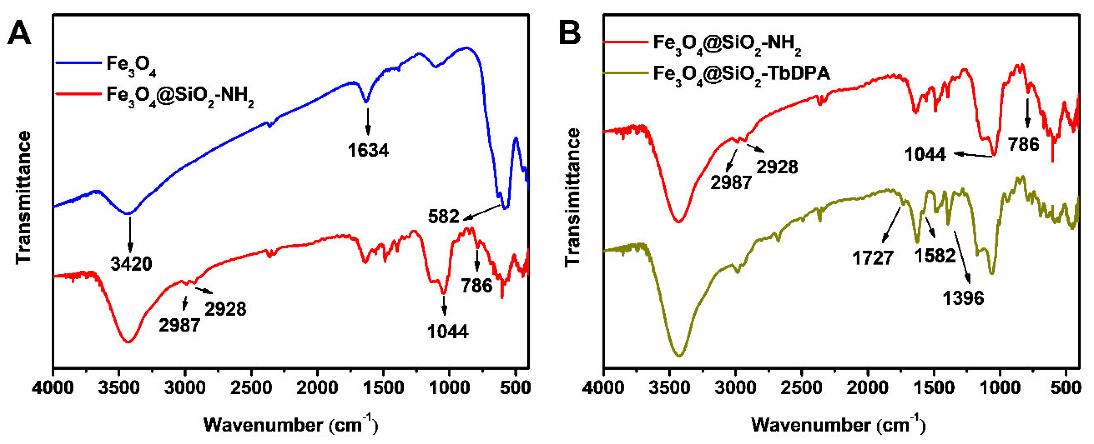
3.1. FT-IR Analysis

The surface functional groups of Fe3O4, Fe3O4@SiO2-NH2 and Fe3O4@SiO2-TbDPA nanospheres were studied using the FT-IR technique. As shown in Figure 2A, the strong, broad peak at about 582 cm−1 was attributed to the stretching vibration of the Fe-O bond, indicating the formation of magnetic Fe3O4 nanoparticles [34,35,36]. The broad bands at 1634 cm−1 and 3420 cm−1 were attributed to the O-H bending and stretching vibrations of water molecules [37].
After surface modification, Fe3O4@SiO2-NH2 nanospheres possessed absorption bands caused by symmetric vibration of Si-O-Si (786 cm−1) and asymmetric vibration of Si-O-Si (1044 cm−1) [26]. The emerging absorption bands at about 2928 cm−1 and 2987 cm−1 were attributed to the stretching vibrations of –CH2– groups from APTES units. The results supported that the SiO2-NH2 layer has covered the surface of the Fe3O4 nanoparticle. The appearance of two bands of 1727 and 1396 cm−1 in Fe3O4@SiO2-TbDPA corresponded to C=O-NH and the stretching vibration of C=O [38]. The weak absorption peak at 1582 cm−1 was assigned to the pyridine ring. The collected results indicated that the DPA molecule was successfully grafted onto the outer surface of Fe3O4@SiO2-NH2 and coordinated with a terbium ion to form Fe3O4@SiO2-DPA hybrid materials (Figure 2B).

3.2. UV-Vis Analysis

To further evaluate the structural information of Fe3O4@SiO2-TbDPA nanospheres, analyses of the UV-Vis spectra of Fe3O4, Fe3O4@SiO2-NH2 and Fe3O4@SiO2-TbDPA were carried out (Figure S1). Both Fe3O4 and Fe3O4@SiO2-NH2 suspension displayed a weak absorption at 383 nm, while Fe3O4@SiO2-TbDPA in water gave rise to not only the band at 383 nm but also the new signals at 270 and 279 nm. The achieved bands were derived from 2,6-pyridinedicarboxylic acid chloride. These results verified the successful modification of the organic ligands 2,6-pyridinedicarboxylic acid chloride onto the Fe3O4@SiO2-NH2 surface.

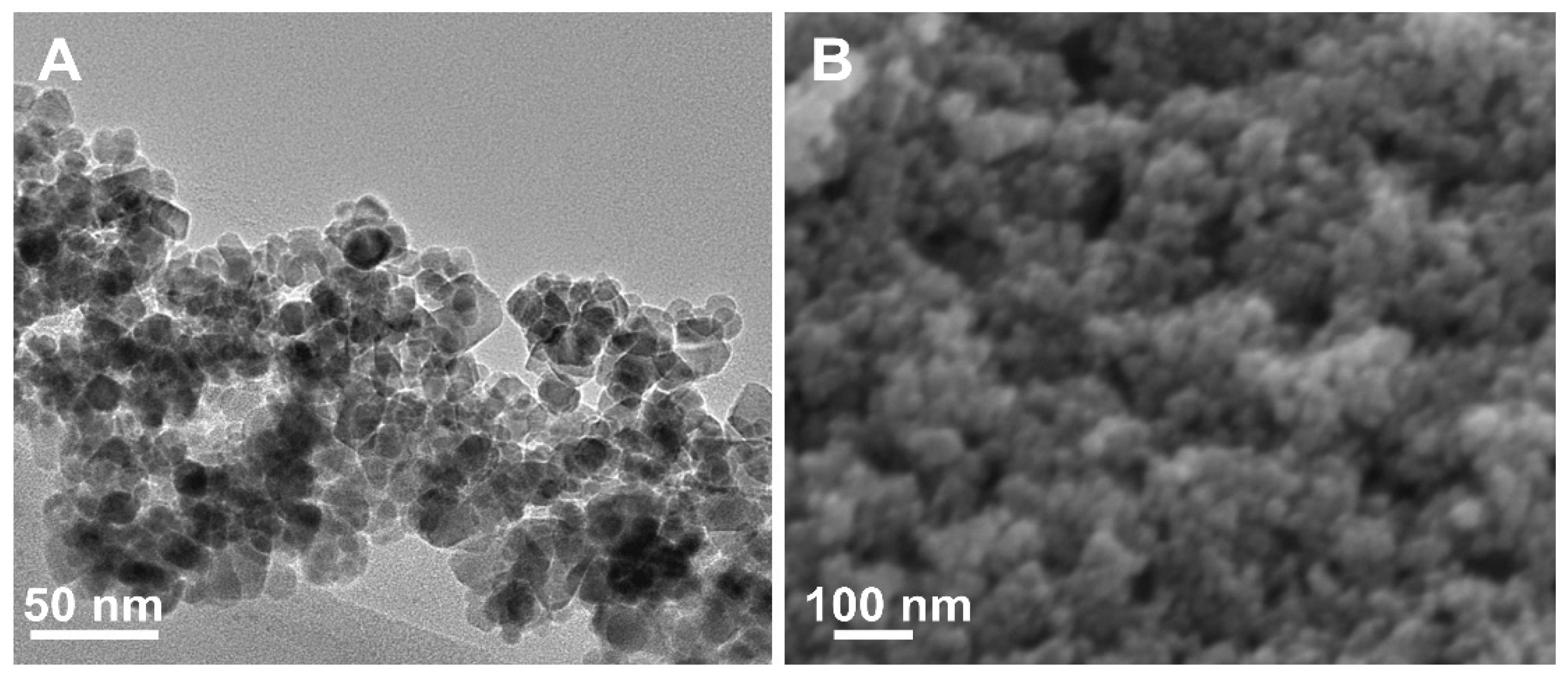
3.3. Morphological Analysis

The morphology of the as-prepared Fe3O4@SiO2-TbDPA hybrid material was investigated by transmission electron microscopy (TEM) and scanning electron microscopy (SEM) simultaneously. The TEM image showed that iron oxide nanoparticles were well encapsulated in the SiO2 layer (Figure 3A). The SEM graph supported that Fe3O4@SiO2-TbDPA hybrid materials were almost uniform, and regular spheres were found (Figure 3B). According to the analysis, the homogenous Fe3O4@SiO2-TbDPA nanospheres were established.

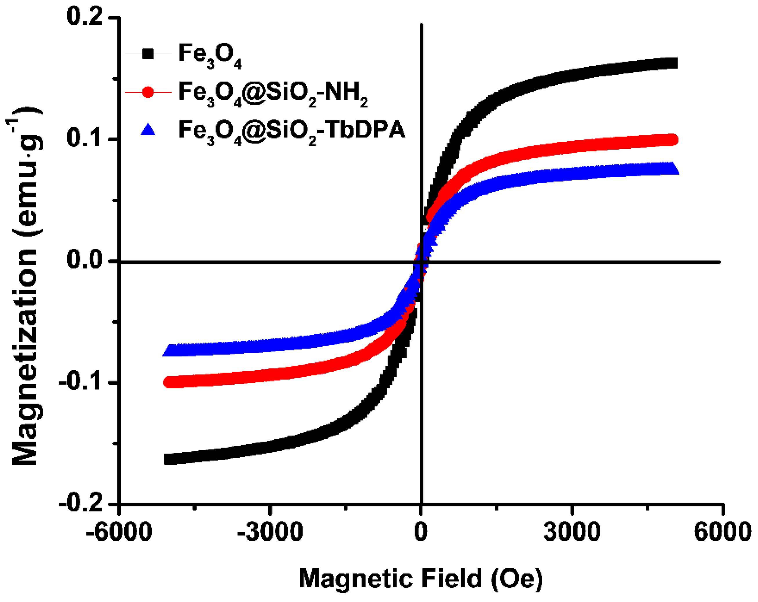
3.4. Magnetic Properties

Magnetic properties of Fe3O4, Fe3O4@SiO2-NH2 and Fe3O4@SiO2-TbDPA nanospheres were investigated at room temperature by vibrating sample magnetometer (VSM) in the field range from −5000 to 5000 Oe (Figure 4). The magnetization curves represent the soft magnetic behavior of the ferrite samples, which is beneficial for improving EM wave absorption [39]. The values of remanent magnetization (Mr), saturation magnetization (Ms) and coercivity (Hc) at room temperature were provided in Table S1. Compared with free Fe3O4 NPs (0.163 emu·g−1), the functionalized magnetic Fe3O4@SiO2-NH2 nanospheres were lower and had a magnetization saturation value of 0.099 emu·g−1. Such reduction in magnetism could be mainly attributed to the non-magnetic SiO2 layer coating on the Fe3O4 nanoparticles’ surface. Similarly, the saturation magnetization of the Fe3O4@SiO2-TbDPA magnetic nanospheres was found to be 0.075 emu g−1. After grafting DPA, the magnetic properties of Fe3O4@SiO2-NH2 were further decreased. In addition, the Hc values of the Fe3O4, Fe3O4@SiO2-NH2 and Fe3O4@SiO2-TbDPA nanocomposites were 33.856, 26.332 and 18.809 Oe, respectively. The low coercivity could be ascribed to the low resonance frequency [40,41]. Fortunately, the Fe3O4@SiO2-TbDPA nanospheres were easily separable under exposure to an external magnetic field, which proved that these magnetic nanomaterials possessed excellent magnetic properties and could be used for potential applications.

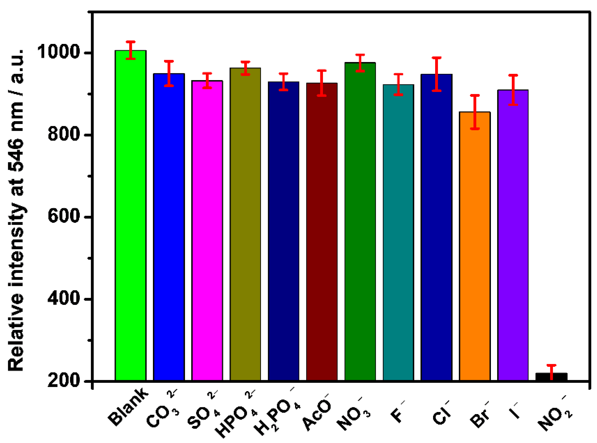
3.5. Selective and Sensitive Detecting NO2

To verify the fluorescence properties of Fe3O4@SiO2-TbDPA, the excitation and emission spectra were recorded. The excitation spectrum is dominated by the peaks centered at 249 nm and 290 nm, which were identified by monitoring the emission of Tb(III) ions at 546 nm (Figure S2). In its emission spectrum, the Tb(III) ion signal was evident from the appearance of linear emission bands at 495, 546, 586, and 624 nm, respectively, corresponding to the deactivation of the Tb(III) excited states 5D47F6, 5D47F5, 5D47F4, and 5D47F3 (excited wavelength at 290 nm). Under the irradiation at 254 nm UV light, its characteristic green emission was observed with the naked eye (insert photo in Figure 5). Upon the addition of various concentrations NO2 (0 μM to 100 μM), the fluorescence intensity of Fe3O4@SiO2-TbDPA gradually decreased and eventually almost disappeared (Figure 5). The fluorescence intensity variation of Fe3O4@SiO2-TbDPA versus the concentration of NO2 followed the excellent linear equation Y = 0.928 + 0.028X (R2 = 0.996) (Figure 6). The detection of limit (DL) was determined to be 1.03 μM according to the equation DL = 3 × SD/slope, where SD was the standard deviation of the blank sample. The calculated DL is much lower than the MCL of NO2 ions in drinking water permitted by WHO and EPA. The detection limit in our proposed method has been compared with various published literature (Table S2). It is believed that the magnetic Fe3O4@SiO2-TbDPA nanoprobes provide acceptable values in terms of detection limits and allow assays in 100% aqueous solutions [5,7,32,42,43,44,45,46,47]. Moreover, the magnetic Fe3O4@SiO2-TbDPA nanoprobes can be separated from the aqueous solution by taking advantage of their magnetic properties (Figure S3). The proposed method has the unique advantages of simple operation, high selectivity, high sensitivity and low cost.
Selectivity is an important index for evaluating fluorescent probes [48,49,50]. To explore the selectivity performance of Fe3O4@SiO2-TbDPA, we performed analogous experiments upon the addition of 100 μM of CO32−, SO42−, HPO42−, H2PO4, AcO, NO3, F, Cl, Br and I. No obvious changes were detected except NO2 (Figure 7). NO2 is a selective quencher for Tb3+ luminescence. This is due to the interaction between NO2 and Tb3+, and the fluorescence quenching is attributed to the energy transfer from Tb3+ to NO2. These results supported the selectivity of the Fe3O4@SiO2-TbDPA nanoprobe for the effective recognition of NO2 in aqueous solutions. Overall, this magnetic nanoprobe exhibited great potential in the recognition of NO2.

3.6. Detection of Nitrite Ions in Tap Water Sample

To investigate the practical applicability of the nanoprobe Fe3O4@SiO2-TbDPA, we have measured the emission intensity of tap water with varied NO2 concentrations. Appropriate amounts of NO2 (5, 10, 30 and 50 μM) were added to the tap water, and the final NO2 content was measured in all samples (4.75, 10.9, 28.9 and 50.9 μM) (Table S3). The average recoveries of nitrite for all spiked samples were in the range of 96–108%, and low relative standard deviations (2.2–3.1%) were obtained, which would be sufficient for practical use. These results substantiated that the proposed determination strategy could be valuable in real samples, suggesting its possibility in the sensing field and analytical assays.

4. Conclusions

In conclusion, we have successfully designed and synthesized a novel water-dispersible Fe3O4@SiO2-TbDPA inorganic-organic hybrid nanoprobe for the rapid and sensitive detection of NO2 in 100% aqueous solutions. The as-prepared nanoprobe exhibited a good linear response for NO2 concentrations from 5 μM to 80 μM with a lower limit of detection (1.03 μM). After detection, the magnetically sensitive nanoprobe Fe3O4@SiO2-TbDPA could be effectively separated from the aqueous solution using its magnetic properties. Therefore, this novel nanoprobe Fe3O4@SiO2-TbDPA can provide a promising way for NO2 measurements under practical conditions.

Supplementary Materials

The following supporting information can be downloaded at: https://www.mdpi.com/article/10.3390/molecules27144431/s1, Figure S1: UV-Vis spectra of Fe3O4, Fe3O4@SiO2-NH2 and Fe3O4@SiO2-TbDPA in aqueous solution; Figure S2: Emission spectra of Fe3O4@SiO2-TbDPA (0.1 mg/mL) in aqueous solution; Figure S3: Photograph of a magnet attracting Fe3O4@SiO2-TbDPA in aqueous solution; Table S1: Magnetic parameters of Fe3O4, Fe3O4@SiO2-NH2 and Fe3O4@SiO2-TbDPA nanocomposite; Table S2: Comparison between the current method and the reported literatures for the detection of nitrite; Table S3: Results of determination of nitrite in tap water (n = 3).

Author Contributions

X.L.; writing—original draft preparation, Q.W. (Qin Wen); methodology, J.C.; investigation, W.S.; data curation, Y.Z.; software, C.L.; supervision, Q.W. (Qianming Wang); funding acquisition and writing—review and editing. All authors have read and agreed to the published version of the manuscript.

Funding

X.Q. thanks the funding support from PhD Research Startup Foundation (PXY-BSQD-2022032). Q.M. appreciates the Science and Technology Plan of Guangdong Province (No. 2020A0505100055), Natural Science Foundation of Guangdong Province, China (No. 2021A1515010324) and Guangzhou Science and Technology Plan (No. 202002030325). Q.M. and C.G. thanks the innovation and entrepreneurship team project of Shunde district in Foshan city (2022), Guangdong province. Y.H. is grateful for the support from plan for excellent professors from the overseas Guangdong Province (C8304807).

Institutional Review Board Statement

Not application.

Informed Consent Statement

Not application.

Data Availability Statement

The data that support the findings of this study are available from the corresponding author, upon reasonable request.

Acknowledgments

The authors thank for the useful discussion by Cheng Cheng Zhang from the University of Texas Southwestern Medical Center at Dallas.

Conflicts of Interest

The authors declare no conflict of interest.

Sample Availability

Samples of the compounds are not available from the authors.

References

  1. Li, W.; Shi, Y.; Hu, X.; Li, Z.; Huang, X.; Holmes, M.; Gong, Y.; Shi, J.; Zou, X. Visual detection of nitrite in sausage based on a ratiometric fluorescent system. Food Control 2019, 106, 106704. [Google Scholar] [CrossRef]
  2. Feng, Z.; Li, Z.; Zhang, X.; Shi, Y.; Zhou, N. Nitrogen-doped carbon quantum dots as fluorescent probes for sensitive and selective detection of nitrite. Molecules 2017, 22, 2061. [Google Scholar] [CrossRef] [PubMed] [Green Version]
  3. Min, H.; Han, Z.; Wang, M.; Li, Y.; Zhou, T.; Shi, W.; Cheng, P. A water-stable terbium metal–organic framework as a highly sensitive fluorescent sensor for nitrite. Inorg. Chem. Front. 2020, 7, 3379–3385. [Google Scholar] [CrossRef]
  4. Ma, Z.; Li, J.; Hu, X.; Cai, Z.; Dou, X. Ultrasensitive, specific, and rapid fluorescence turn-on nitrite sensor enabled by precisely modulated fluorophore binding. Adv. Sci. 2020, 7, 2002991. [Google Scholar] [CrossRef] [PubMed]
  5. Chen, J.; Pang, S.; He, L.; Nugen, S.R. Highly sensitive and selective detection of nitrite ions using Fe3O4@SiO2/Au magnetic nanoparticles by surface-enhanced Raman spectroscopy. Biosens. Bioelectron. 2016, 85, 726–733. [Google Scholar] [CrossRef] [Green Version]
  6. Jayawardane, B.M.; Wei, S.; McKelvie, I.D.; Kolev, S.D. Microfluidic paper-based analytical device for the determination of nitrite and nitrate. Anal. Chem. 2014, 86, 7274–7279. [Google Scholar] [CrossRef]
  7. Rahim, A.; Santos, L.S.S.; Barros, S.B.A.; Kubota, L.T.; Landers, R.; Gushikem, Y. Electrochemical detection of nitrite in meat and water samples using a mesoporous carbon ceramic SiO2/C electrode modified with in situ generated manganese(II) phthalocyanine. Electroanalysis 2014, 26, 541–547. [Google Scholar] [CrossRef]
  8. Matteo, V.D.; Esposito, E. Methods for the determination of nitrite by high-performance liquid chromatography with electrochemical detection. J. Chromatogr. A 1997, 789, 213–219. [Google Scholar] [CrossRef]
  9. Wang, B.; Zheng, S.; Huang, Y.; Wang, Y.; Zhu, Z.; Ma, R.; Zhao, Y.; Yin, X.; Su, J.; Xiong, J.; et al. Novel GC/Py/GC/IRMS-based method for isotope measurements of nitrate and nitrite. I: Converting nitrate to benzyl nitrate for delta(18)O analysis. Anal. Chem. 2020, 92, 12216–12225. [Google Scholar] [CrossRef]
  10. Moravský, L.; Troška, P.; Klas, M.; Masár, M.; Matejčík, Š. Determination of nitrites and nitrates in plasma-activated deionized water by microchip capillary electrophoresis. Contrib. Plasm. Phys. 2020, 60, e202000014. [Google Scholar] [CrossRef]
  11. Vilian, A.T.E.; Umapathi, R.; Hwang, S.K.; Huh, Y.S.; Han, Y.K. Pd-Cu nanospheres supported on Mo2C for the electrochemical sensing of nitrites. J. Hazard. Mater. 2021, 408, 124914. [Google Scholar] [CrossRef] [PubMed]
  12. Wu, H.; Tong, C. Dual-emission fluorescent probe for the simultaneous detection of nitrite and mercury(II) in environmental water samples based on the Tb3+-modified carbon quantum dot/3-aminophenylboronic acid hybrid. Anal. Chem. 2020, 92, 8859–8866. [Google Scholar] [CrossRef]
  13. Zheng, X.J.; Liang, R.P.; Li, Z.J.; Zhang, L.; Qiu, J.D. One-step, stabilizer-free and green synthesis of Cu nanoclusters as fluorescent probes for sensitive and selective detection of nitrite ions. Sens. Actuators B Chem. 2016, 230, 314–319. [Google Scholar] [CrossRef]
  14. Wang, L.; Jana, J.; Chung, J.S.; Choi, W.M.; Hur, S.H. Designing an intriguingly fluorescent N, B-doped carbon dots based fluorescent probe for selective detection of NO2(−) ions. Spectrochim. Acta. Part A Molecul. Biomol. Spectrosc. 2022, 268, 120657. [Google Scholar] [CrossRef]
  15. Gu, J.; Zhang, F.; Zheng, Z.; Li, X.; Deng, R.; Zhou, Z.; Ma, L.; Liu, W.; Wang, Q. Establishment of a new molecular model for mercury determination verified by single crystal X-ray diffraction, spectroscopic analysis and biological potentials. Chin. Chem. Lett. 2021, 32, 87–91. [Google Scholar] [CrossRef]
  16. He, W.M.; Zhou, Z.; Han, Z.; Li, S.; Zhou, Z.; Ma, L.F.; Zang, S.Q. Ultrafast size expansion and turn-on luminescence of atomically precise silver clusters by hydrogen sulfide. Angew. Chem. Int. Ed. 2021, 60, 8505–8509. [Google Scholar] [CrossRef] [PubMed]
  17. Wu, C.Q.; Sun, W.J.; Wang, Q.M. Exploration of sulfur-containing nanoparticles: Synthesis, microstructure analysis, and sensing potential. Inorg. Chem. 2022, 61, 4159–4170. [Google Scholar] [CrossRef]
  18. Paderni, D.; Giorgi, L.; Fusi, V.; Formica, M.; Ambrosi, G.; Micheloni, M. Chemical sensors for rare earth metal ions. Coordin. Chem. Rev. 2021, 429, 213639. [Google Scholar] [CrossRef]
  19. Na, M.; Zhang, S.; Liu, J.; Ma, S.; Han, Y.; Wang, Y.; He, Y.; Chen, H.; Chen, X. Determination of pathogenic bacteria-Bacillus anthrax spores in environmental samples by ratiometric fluorescence and test paper based on dual-emission fluorescent silicon nanoparticles. J. Hazard. Mater. 2020, 386, 121956. [Google Scholar] [CrossRef]
  20. Aleem, A.R.; Liu, J.; Wang, J.; Wang, J.; Zhao, Y.; Wang, Y.; Wang, Y.; Wang, W.; Rehman, F.U.; Kipper, M.J.; et al. Selective sensing of Cu2+ and Fe3+ ions with vis-excitation using fluorescent Eu3+-induced aggregates of polysaccharides (EIAP) in mammalian cells and aqueous systems. J. Hazard. Mater. 2020, 399, 122991. [Google Scholar] [CrossRef]
  21. Wang, J.; Li, D.; Ye, Y.; Qiu, Y.; Liu, J.; Huang, L.; Liang, B.; Chen, B. A Fluorescent metal-organic framework for food real-time visual monitoring. Adv. Mater. 2021, 33, e2008020. [Google Scholar] [CrossRef] [PubMed]
  22. Zhang, X.; Fang, L.; Jiang, K.; He, H.; Yang, Y.; Cui, Y.; Li, B.; Qian, G. Nanoscale fluorescent metal-organic framework composites as a logic platform for potential diagnosis of asthma. Biosens. Bioelectron. 2019, 130, 65–72. [Google Scholar] [CrossRef] [PubMed]
  23. Yan, B. Lanthanide-functionalized metal-organic framework hybrid systems to create multiple luminescent centers for chemical sensing. Acc. Chem. Res. 2017, 50, 2789–2798. [Google Scholar] [CrossRef] [PubMed]
  24. Zhang, R.; Yuan, J. Responsive metal complex probes for time-gated luminescence biosensing and imaging. Acc. Chem. Res. 2020, 53, 1316–1329. [Google Scholar] [CrossRef]
  25. Ren, J.; Niu, Z.; Ye, Y.; Tsai, C.; Liu, S.; Liu, Q.; Huang, X.; Nafady, A.; Ma, S. Second-sphere interaction promoted turn-on fluorescence for selective sensing of organic amines in a Tb(III) framework. Angew. Chem. Int. Ed. 2021, 60, 23705–23712. [Google Scholar] [CrossRef]
  26. Sun, Z.; Huang, H.; Zhang, R.; Yang, X.; Yang, H.; Li, C.; Zhang, Y.; Wang, Q. Activatable rare earth near-infrared-II fluorescence ratiometric nanoprobes. Nano Lett. 2021, 21, 6576–6583. [Google Scholar] [CrossRef]
  27. Koo, T.M.; Ko, M.J.; Park, B.C.; Kim, M.S.; Kim, Y.K. Fluorescent detection of dipicolinic acid as a biomarker in bacterial spores employing terbium ion-coordinated magnetite nanoparticles. J. Hazard. Mater. 2021, 408, 124870. [Google Scholar] [CrossRef]
  28. Wang, Q.; Tan, C.; Chen, H.; Tamiaki, H. A new fluoride luminescence quencher based on a nanostructured covalently bonded terbium hybrid material. J. Phys. Chem. C 2010, 114, 13879–13883. [Google Scholar] [CrossRef]
  29. Tan, C.; Wang, Q. Reversible terbium luminescent polyelectrolyte hydrogels for detection of H2PO4 and HSO4 in water. Inorg. Chem. 2011, 50, 2953–2956. [Google Scholar] [CrossRef]
  30. Li, X.; Gu, J.; Zhou, Z.; Ma, L.; Tang, Y.; Gao, J.; Wang, Q. New lanthanide ternary complex system in electrospun nanofibers: Assembly, physico-chemical property and sensor application. Chem. Eng. J. 2019, 358, 67–73. [Google Scholar] [CrossRef]
  31. Zhou, Z.; Li, X.; Tang, Y.; Zhang, C.C.; Fu, H.; Wu, N.; Ma, L.; Gao, J.; Wang, Q. Oxidative deoximation reaction induced recognition of hypochlorite based on a new fluorescent lanthanide-organic framework. Chem. Eng. J. 2018, 351, 364–370. [Google Scholar] [CrossRef]
  32. Zhu, C.; Zhang, M.; Qiao, Y.; Xiao, G.; Zhang, G.F.; Chen, Y. Fe3O4/TiO2 core/shell nanotubes: Synthesis and magnetic and electromagnetic wave absorption characteristics. J. Phys. Chem. C 2010, 114, 16229–16235. [Google Scholar] [CrossRef]
  33. Chen, M.; Zheng, Y.; Gao, J.; Li, C.; Yu, C.; Wang, Q. Fluorometric determination of dopamine by using a terbium (III) inorganic-organic network. Microchim. Acta 2017, 184, 2275–2280. [Google Scholar] [CrossRef]
  34. Zhang, J.L.; Srivastava, R.S.; Misra, R.D.K. Core-shell magnetite nanoparticles surface encapsulated with smart stimuli-responsive polymer: Synthesis, characterization, and LCST of viable drug-targeting delivery system. Langmuir 2007, 23, 6342–6351. [Google Scholar] [CrossRef]
  35. Sundrarajan, M.; Ramalakshmi, M. Novel cubic magnetite nanoparticle synthesis using room temperature ionic liquid. E-J. Chem. 2012, 9, 1070–1076. [Google Scholar] [CrossRef]
  36. Du, N.; Xu, Y.; Zhang, H.; Zhai, C.; Yang, D. Selective synthesis of Fe2O3 and Fe3O4 nanowires via a single precursor: A general method for metal oxide nanowires. Nanoscale Res. Lett. 2010, 5, 1295–1300. [Google Scholar] [CrossRef] [PubMed] [Green Version]
  37. Wu, N.; Tang, Y.; Zeng, M.; Gao, J.; Lu, X.; Zheng, Y. Design of hybrid inorganic-organic nanosensor based on Fe3O4 as the core and recovery features. J. Lumin. 2018, 202, 502–507. [Google Scholar] [CrossRef]
  38. Zhao, X.; He, X.; Hou, A.; Cheng, C.; Wang, X.; Yue, Y.; Wu, Z.; Wu, H.; Liu, B.; Li, H.; et al. Growth of Cu2O nanoparticles on two-dimensional Zr-ferrocene-metal-organic framework nanosheets for photothermally enhanced chemodynamic antibacterial therapy. Inorg. Chem. 2022, 61, 9328–9338. [Google Scholar] [CrossRef] [PubMed]
  39. Akbarzadeh, A.; Samiei, M.; Joo, S.; Anzaby, M.; Hanifehpour, Y.; Nasrabadi, H.; Davaran, S. Synthesis, characterization and in vitro studies of doxorubicin-loaded magnetic nanoparticles grafted to smart copolymers on A549 lung cancer cell line. J. NanoBiotechnol. 2012, 10, 46. [Google Scholar] [CrossRef] [Green Version]
  40. Chen, Y.; Gao, P.; Wang, R.; Zhu, C.; Wang, L.; Cao, M.; Jin, H. Porous Fe3O4/SnO2 core/shell nanorods: Synthesis and electromagnetic properties. J. Phys. Chem. C 2009, 113, 10061–10064. [Google Scholar] [CrossRef]
  41. Bai, R.B.; Wang, J.F.; Wang, D.G.; Cui, J.J.; Zhang, Y.Q. Recovery of lithium from high Mg/Li ratio salt-lake brines using ion-exchange with NaNTf2 and TBP. Hydrometallurgy 2022, 213, 105914. [Google Scholar] [CrossRef]
  42. Liu, Z.; Mao, H. Towards fluorescent magnetic core shell composites for nitrite optical sensing. J. Lumin. 2017, 190, 179–187. [Google Scholar] [CrossRef]
  43. Zhang, Y.; Su, Z.; Li, B.; Zhang, L.; Fan, D.; Ma, H. Recyclable magnetic mesoporous nanocomposite with improved sensing performance toward nitrite. ACS Appl. Mater. Interfaces 2016, 8, 12344–12351. [Google Scholar] [CrossRef] [PubMed]
  44. Jia, J.; Lu, W.; Li, L.; Jiao, Y.; Gao, Y.F.; Shuang, S. Orange luminescent carbon dots as fluorescent probe for detection of nitrite. Chin. J. Anal. Chem. 2019, 47, 560–566. [Google Scholar] [CrossRef]
  45. Jin, L.; Wang, Y.; Liu, F.; Yu, S.; Gao, Y.; Zhang, J. The determination of nitrite by a graphene quantum dot fluorescence quenching method without sample pretreatment. Luminescence 2018, 33, 289–296. [Google Scholar] [CrossRef]
  46. Deng, Y.; Qian, J.; Zhou, Y.; Niu, Y. Preparation of N/S doped carbon dots and their application in nitrite detection. RSC Adv. 2021, 11, 10922–10928. [Google Scholar] [CrossRef]
  47. Hao, X.; Liang, Y.; Zhen, H.; Sun, X.; Liu, X.; Li, M.; Shen, A.; Yang, Y. Fast and sensitive fluorescent detection of nitrite based on an amino-functionalized MOFs of UiO-66-NH2. J. Solid State Chem. 2020, 287, 121323. [Google Scholar] [CrossRef]
  48. Qiao, G.X.; Liu, L.; Hao, X.X.; Zheng, J.K.; Liu, W.Q.; Gao, J.W.; Zhang, C.C.; Wang, Q.M. Signal transduction from small particles: Sulfur nanodots featuring mercury sensing, cell entry mechanism and in vitro tracking performance. Chem. Eng. J. 2020, 382, 122907. [Google Scholar] [CrossRef]
  49. Wu, N.T.; Wen, Q.; Wang, Q. Single optical sensor to multiple functions: Ratiometric sensing for SO32− and dual signal determination for copper (II). Spectrochim. Acta A Mol. Biomol. Spectrosc. 2021, 249, 119219. [Google Scholar] [CrossRef]
  50. Wen, Q.; Zheng, Y.H.; Liu, W.Q.; Wang, Q.M. Stepwise assembly protocols for the rational design of lanthanide functionalized carbon dots-hydrogel and its sensing evaluation. J. Fluores. 2021, 31, 695–702. [Google Scholar] [CrossRef]
Figure 1. Schematic diagram of Fe3O4@SiO2-TbDPA nanoprobe for NO2 detection.
Molecules 27 04431 g001
Figure 2. FT-IR of Fe3O4, Fe3O4@SiO2-NH2 (A) and Fe3O4@SiO2-TbDPA (B).
Molecules 27 04431 g002
Figure 3. (A) TEM and (B) SEM images of Fe3O4@SiO2-TbDPA.
Molecules 27 04431 g003
Figure 4. Magnetic hysteresis loops of the Fe3O4, Fe3O4@SiO2-NH2 and Fe3O4@SiO2-TbDPA.
Molecules 27 04431 g004
Figure 5. Emission spectra of DPA-Fe3O4@SiO2-Tb (0.1 mg/mL) aqueous solution upon the addition of NO2 (0–100 μM) under 290 nm excitation. (Inset: photographs of Fe3O4@SiO2-TbDPA dispersions taken before (left) and after (right) the addition of 100 µM NO2 under 254 nm UV lamp).
Molecules 27 04431 g005
Figure 6. Linearity of fluorescence intensity ratio (F0/F) of Fe3O4@SiO2-TbDPA versus NO2 ion concentration (F0 and F represent the fluorescence intensities at 546 nm in the absence and presence of nitrite, respectively).
Molecules 27 04431 g006
Figure 7. Fluorescence intensity of Fe3O4@SiO2-TbDPA (0.1 mg/mL) aqueous solution at 546 nm in the presence of 100 μM (CO32−, SO42−, HPO42−, H2PO4, AcO, NO3, F, Cl, Br, I and NO2), respectively.
Molecules 27 04431 g007
Publisher’s Note: MDPI stays neutral with regard to jurisdictional claims in published maps and institutional affiliations.

