Review

Marine Bacterial Dextranases: Fundamentals and Applications

1
Department of Marine Biology, Faculty of Marine Science and Technology, University of Hormozgan, Bandar Abbas 74576, Iran
2
Institute of Living Systems, Immanuel Kant Baltic Federal University, 236016 Kaliningrad, Russia
3
Department of Paraclinical Sciences, Faculty of Veterinary Medicine, Norwegian University of Life Sciences (NMBU), 1432 Ås, Norway
4
Persian Gulf and Oman Sea Ecology Research Center, Iranian Fisheries Sciences Research Institute, Agricultural Research Education and Extension Organization (AREEO), Bandar Abbas 14578, Iran
5
Department of Microbiology, University of Karachi, Karachi 75270, Pakistan
*
Author to whom correspondence should be addressed.
Molecules 2022, 27(17), 5533; https://doi.org/10.3390/molecules27175533
Submission received: 1 July 2022 / Revised: 18 August 2022 / Accepted: 22 August 2022 / Published: 28 August 2022

Abstract

:
Dextran, a renewable hydrophilic polysaccharide, is nontoxic, highly stable but intrinsically biodegradable. The α-1, 6 glycosidic bonds in dextran are attacked by dextranase (E.C. 3.2.1.11) which is an inducible enzyme. Dextranase finds many applications such as, in sugar industry, in the production of human plasma substitutes, and for the treatment and prevention of dental plaque. Currently, dextranases are obtained from terrestrial fungi which have longer duration for production but not very tolerant to environmental conditions and have safety concerns. Marine bacteria have been proposed as an alternative source of these enzymes and can provide prospects to overcome these issues. Indeed, marine bacterial dextranases are reportedly more effective and suitable for dental caries prevention and treatment. Here, we focused on properties of dextran, properties of dextran—hydrolyzing enzymes, particularly from marine sources and the biochemical features of these enzymes. Lastly the potential use of these marine bacterial dextranase to remove dental plaque has been discussed. The review covers dextranase-producing bacteria isolated from shrimp, fish, algae, sea slit, and sea water, as well as from macro- and micro fungi and other microorganisms. It is common knowledge that dextranase is used in the sugar industry; produced as a result of hydrolysis by dextranase and have prebiotic properties which influence the consistency and texture of food products. In medicine, dextranases are used to make blood substitutes. In addition, dextranase is used to produce low molecular weight dextran and cytotoxic dextran. Furthermore, dextranase is used to enhance antibiotic activity in endocarditis. It has been established that dextranase from marine bacteria is the most preferable for removing plaque, as it has a high enzymatic activity. This study lays the groundwork for the future design and development of different oral care products, based on enzymes derived from marine bacteria.

1. Introduction

Dextran is a natural polymer (Figure 1) with a main chain of molecules linked by α-1,6 bond, and side branches linked by α-1,3 bonds. Dextran is renewable and biodegradable chemical resource with the desirable properties of nontoxicity, stability and water solubility [1]. Chemically modified dextran can act as a potential drug carrier, as it is degraded by dextranase and releases the drug of choice at the target site [2,3].
Dextranases (α-1,6-D-glucan-6-glucanohydrolase; EC 3.2.1.11) randomly hydrolyze α-1,6-linkages of dextran and release isomaltooligosaccharides (IGs) of various sizes [4]. It is extremely rare that the product of hydrolysis is glucose. Large dextran compounds break down more easily than small ones due to the increased contact of dextran with dextranase molecules. IGs are applied in many processes including in sugar industry, in medicine making, and in dentistry. Sugar industries employ dextranase to degrade any residual dextran in sugar juice, which in turn reduces the viscosity of the juice and improves the yield of sucrose [5,6,7]. In medicine, dextranases are used to convert high molecular weight dextran into low molecular weight compounds; the dextran of specific molecular weight has an antithrombotic effect and hence can substitute blood under emergent conditions [8,9]. In dentistry, dextranases are applied to degrade dental plaque glucans and to prevent glucan formation [10]. Lately, the attention towards dextranases increased due to its recent advancement in prebiotics synthesis [11].
Active centers (Figure 1) are α-1,6, α-1,3 and α-1,4 bonds [12].
The effectiveness of dextranase depends on the acidity, solids content, temperature, contact time, agitation, dextran concentration and the source, activity and dosage of the dextranase used. The optimal acidity range for dextranase activity is 5.0–6.0. Moreover, lower values in this range are preferred. In addition, the effectiveness of dextranase depends on the temperature of the medium.
Historically, dextranase was discovered in Cellvibrio fulva around 80 years ago [13]. Currently various dextranase-producing microorganisms have been described including bacteria, filamentous fungus and yeasts [9,12,14]. The bacterial genera, Bacillus [15,16,17,18], Shewanella [19], Thermoanaerobacter [20], Arthrobacter [21], Cytophaga [22], Paenibacillus [23,24,25] and Streptococcus [26] have been reported to produce dextranases in good yield. Among fungal species, Paecilomyces lilacinus [27] and the species of Penicillium [28,29], Hypocrea [30], Pochonia [31], Chaetomium [32], Talaromyces [3], and Aspergillus [33] are reportedly promising producers of dextranases. The microbial sources of dextranases from marine environment have also been described, from bacteria [34,35,36,37] and from fungi [38]. The concerns raised by the US Food and Drug Administration (FDA) over the use of fungal dextranase [39,40] have diverted the attention of researchers to explore alternative or new sources of dextranase.
The marine environment is considered as a source of novel microorganisms with particular trait of cold-adaptation and salt and alkali tolerance can serve as a unique reservoir for dextranase [41]. Cold-adapted (cold-active) enzymes are already in demand in various commercial sections, particularly for formulation of improved detergents, for the development of environmental biosensors, and as a carrier matrix for bioremediation [42]. Moreover, cold-adapted dextranases are also uniquely suited for the synthesis of novel medicines. Likewise, marine alkaline dextranases are compatible with alkaline tooth-rinse products in the treatment of oral dental plaques [43]. Indeed, the attributes of high salt tolerance, thermostability, and low optimum temperature of marine dextranases render them suited for the human oral environment.
Marine bacterial dextranases have also exhibited their ability to prevent biofilm formation by Streptococcus mutans. Therefore, the prospects of utilizing more marine bacterial dextranases for the development of oral care products are quite considerable and envisage further research in this direction.
In addition to their applications in research, dextranases are also applied to prepare dextran and its derivative. The enzyme is also applied in molasses and beverage processing in food industries [44].
It is used to study the structure of other polysaccharides and to obtain oligosaccharides. Dextranase-cleaved polysaccharides are used in the cosmetic and medical industries as cryoprotectants, and in the food industry as stabilizers. Medical sterile blood substitutes (dextran of a certain molecular weight) are also produced using dextranase. The newest use of dextranase is for labeling in cancer therapy. In the sugar industry, this enzyme is used to remove sucrose sludge [44,45,46,47].
This research aimed to investigate the classification and properties of dextran hydrolyzing enzymes, as well as the methods for obtaining, biological properties, and practical applications of marine dextranases. This study compared dextranases from marine bacteria with dextranases from other microorganisms along with the effect of marine dextranase on plaque removal is described. Dextranases have a wide spectrum of bactericidal and bacteriostatic activity. Dextranases are classified based on the types of catalyzed reactions and the specificity of the resulting product. This review precisely presents dextranase-producing bacteria isolated from various marine sources: shrimp, fish, seaweed, sea silt and sea water and other alternative sources of dextranases (macro- and microfungi, bacteria).
The novelty of this study is that it is the first time that dextranases produced by alternative sources like marine bacteria have been analyzed, dextranases produced by micro- and macrofungi were compared, and the advantages and disadvantages of various dextranases have been discussed. In addition, the impact of marine dextranases on plaque removal has been summarized for the first time. This information will be used to design and develop oral care (biofilm removal) products using products and enzymes derived from marine bacteria.

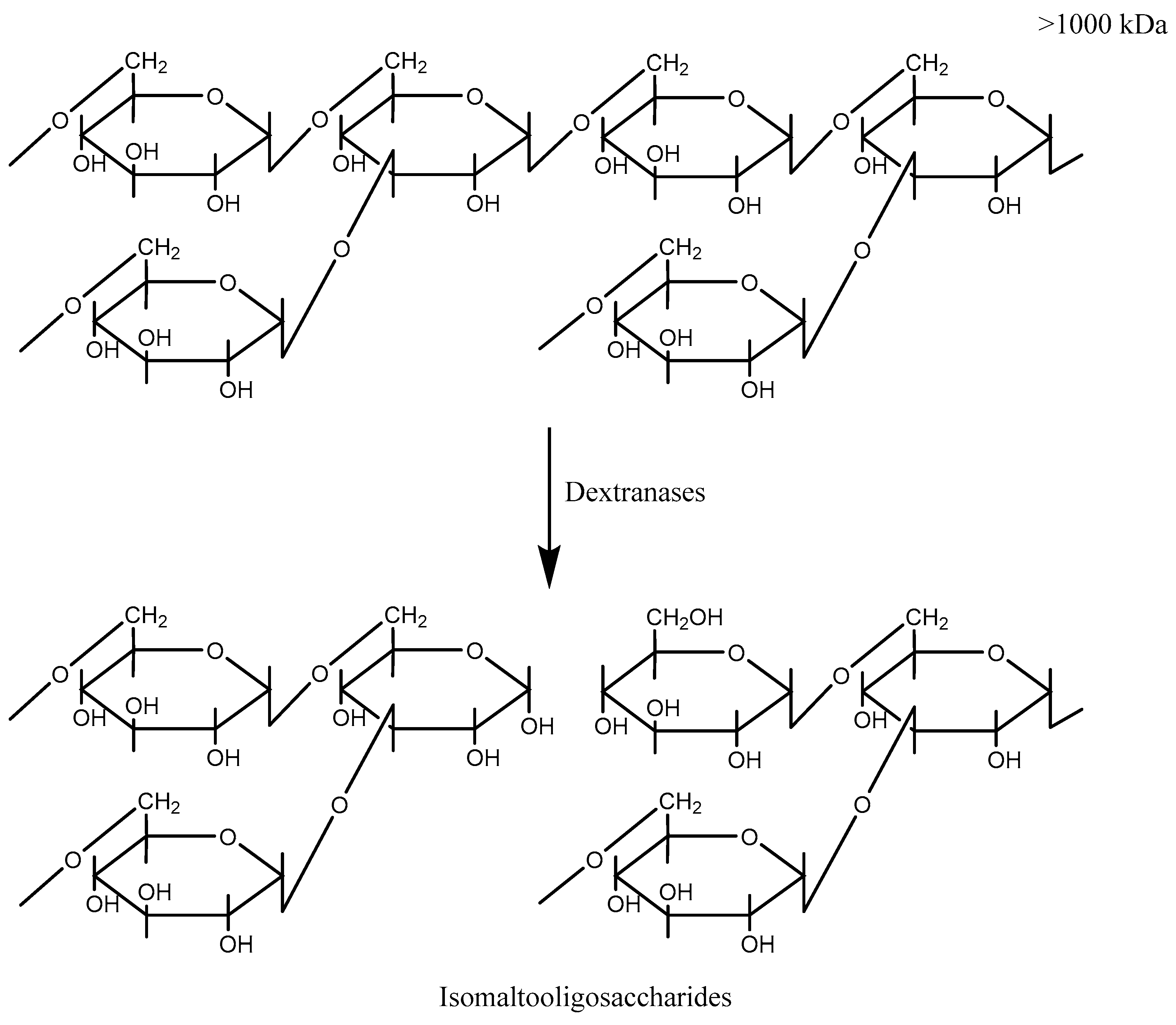
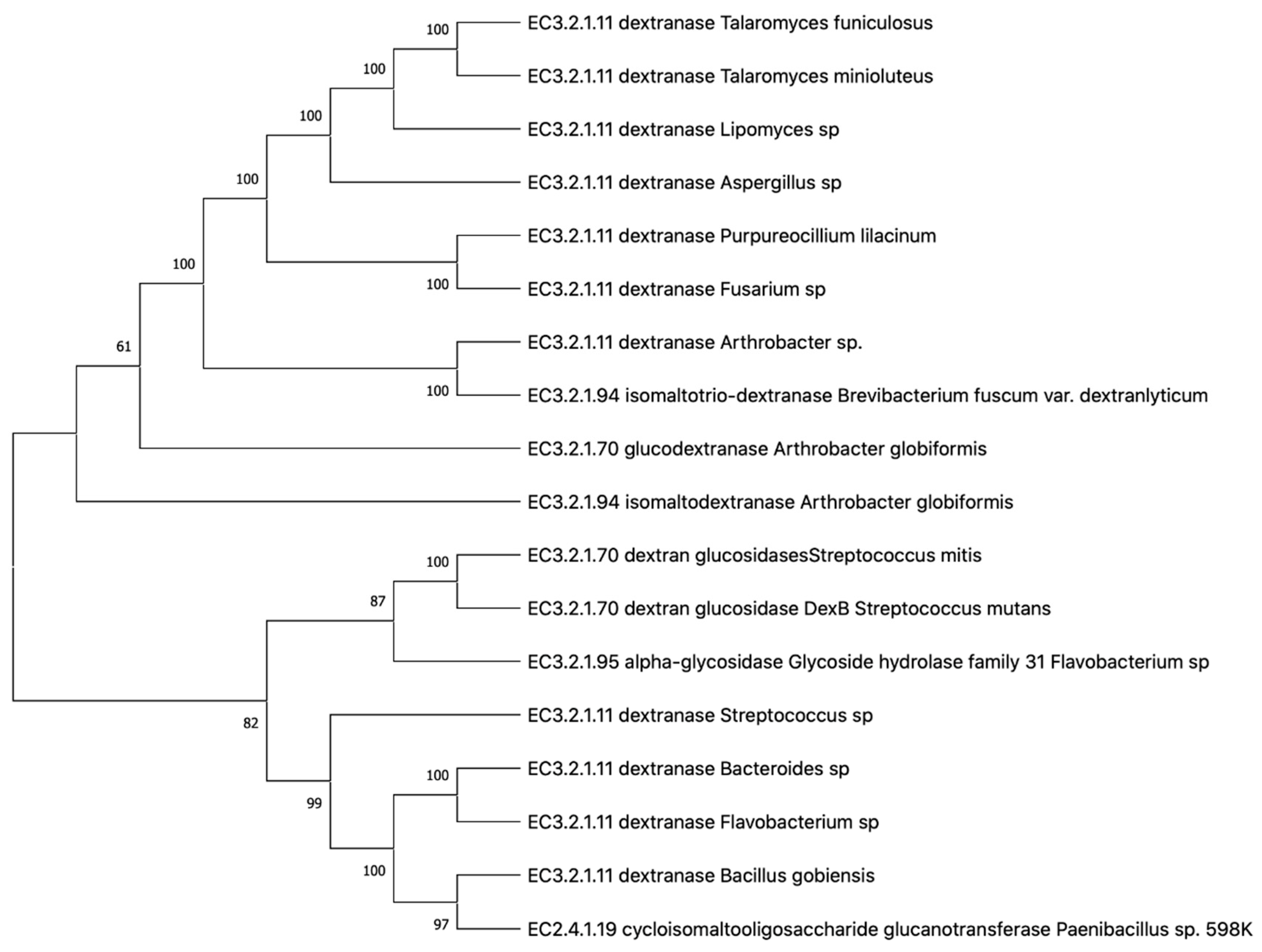
2. Classification and Properties of Dextran—Hydrolyzing Enzymes

The search for organisms that produce a large amount of the enzyme needed to degrade dextran began in the 19th century. Organisms producing trace amounts of both intracellular and extracellular dextranase were isolated in the late 1940s, but it was not until the 1950s that Penicillium species producing large amounts of extracellular dextranase were reported.
In the early 1970s, the first attempts were made to purify and characterize Penicillium dextranase. In the late 1970s, research began on the use of dextranase for the treatment of caries [48].
In the 1990s, the Penicillium dextranase gene was cloned and expressed from Pichia pastoris. An early classification system for dextranase and other dextran hydrolyzing enzymes was also developed using sequence-analysis software (GCG, Version 8.0.1, Madison, WI, USA) [49].
The crystal structure of Penicillium minioluteum was revealed by Larsson et al. in 2003 [50]. In this study, it was suggested that the reaction mechanism proceeds by pure inversion (rather than retention) of the anomeric carbon.
Extracellular dextranase is encoded by the dex gene. Post-translational modification includes signal peptide cleavage and N-glycosylation.
Dextranase enzymes belongs to two families of glycoside hydrolases, either 49 or 66, which do not share significant sequence similarity. Penicillium and Arthrobacter dextranases belong to family 49, while Streptococcus dextranases belong to family 66 [4].
Penicillium dectranase and related enzymes contain two domains. The first domain resembles the folding of an immunoglobulin and consists of 200 amino acid residues forming 13 β-chains. The β-sandwich is formed by nine of these strands, with all strands except 5 and 13 being antiparallel. The second domain contains a right-sided parallel β-helical fold containing 3 parallel β-sheets [50]. These two domains are connected by a large interface that contains 29 amino acids, completely conserved in the family of glycoside hydrolases 49. Disulfide bridges are formed by four of the six cysteines present in the protein, none of which is conserved in the family of glycoside hydrolases 49 [50].
Dextran-degrading enzymes belong to different groups of carbohydrases and transferases. Based on their mode of action, these can be categorized as endo- and exo-dextranases [51,52]. There are known endodextranases isolated from molds Penicillium luteum ATCC 9644, Penicillium funiculosum, Penicillium funiculosum NRRL, Penicillium lilacinum NRRL, Penicillium notatum, Penicillium acualeatum, Aspergillus carneus, Chatomium gracile, Fusarium sp., Sporotrix schencki, which have extracellular localization. There are also known endodextranases isolated from the yeast Lipomyces starkeyi ATCC 20825, Lipomyces starkeyi KSM 22, Lipomyces starkeyi IGC 4047. Endodextranases are also obtained from bacteria Bacteroides oralis Ig4a, Flavobacterium sp. M-73, Pseudomonas sp. All of them have extracellular localization [4].
There are known exodextranases from bacteria Bacteroides oralis IG 4a, Arthrobacter globiforms I-42, Pseudomonas sp., Streptococcus mitis ATCC 903, from yeast Lipomyces lipofer IGC 4042. All exodextranases are extracellular, except for exodextranases derived from Pseudomonas spp. and Streptococcus mitis ATCC 903. These exodextranases are intracellular [4].
Furthermore, the Nomenclature Committee of the International Union of Biochemistry and Molecular Biology (IUBMB) considers the types of reactions catalyzed by dextranases and the product specificity to classify these enzymes as simply dextranase (EC3.2.1.11), glucan-1,6-α-D-glucosidases (EC3.2.1.70), glucan-1,6-α-isomaltosidases (EC3.2.1.94), dextran 1,6-α-isomaltotriosidases (EC3.2.1.95), and branched-dextran exo-1,2-α-glucosidases (EC3.2.1.115) [53]. The reaction catalyzed by Cycloisomaltooligosaccharide glucanotransferase (CITase) also yields hydrolyzed dextran as a byproduct (Table 1) [54]. Interestingly, α-glucosidase (EC3.2.1.20) that is considered as an unrelated enzyme to dextran synthesis also catalyzes the reaction similar to those of exo dextranases (EC3.2.1.70) [55]. Based on the similarities in the amino acid sequences, glycosylhydrolases and glycosyltransferases have been divided into different families [56,57] (http://www.cazy.org, accessed on 1 July 2021) in the Carbohydrate Active Enzymes (CAZy) database. CAZy database was developed with the aim to classify the enzyme families with structurally related domains for catalysis of glycosidic bonds and for carbohydrate-binding (functional domains). An analogous classification system for dextran-hydrolyzing enzymes has also been proposed to divide the enzymes into four families [58] but the system did not gain much attention and it’s details are beyond the scope of this review. In addition, CAZy database has been developed to integrate structural properties of the enzymes with its mechanical features; therefore, the enzymes with different substrate specificities have been placed in the same family, while the enzymes with the ability to catalyze the same substrate are sometimes placed in different families [59]. In this database, dextran-glucosidases (EC3.2.1.70) have been included in glycosylhydrolase families 13 and 15 (http://www.cazy.org, accessed on 1 July 2021). The families 27 and 49 contains structurally different enzymes of isomaltodextranase (EC3.2.1.94) and isomaltotriosidase (EC3.2.1.95). While the families 49 and 66 contain endodextranases, the two families, however, do not harbour similarities in the sequences. Moreover, CITase from Bacillus circulans is classified in family glycosylhydrolase 66 and shares a high homology with endodextranases. The enzyme (α-1,6-D-glucan 6-glucanohydrolase; EC 3.2.1.11) that cleave dextran chain at α-1,6 glucosidic bond and release oligosaccharides are included in the GH families 49 and 66 based on amino acid sequence homology [4].
Figure 2 demonstrates the phylogenetic tree of dextranases from various sources.

3. Marine Dextranase

Oceans are the largest ecosystem and make this planet ‘blue’ by covering almost two third of the planet’s surface, and they are the most ancient habitat of living organisms [60]. The difficulties in sampling impede complete exploration of the marine organisms; therefore, it is believed that the habitat can still provide new novel species [61,62,63]. Salinity and generally cold and alkaline environments are peculiar features of oceans that influence the life in that habitat which is known for harboring unique catalysts [64,65,66,67,68,69,70,71,72,73,74,75]. Therefore, potential applications of marine-derived catalysts can easily be conceived. Although not enough research has been devoted to the study of marine dextranases, yet there are few reports on Catenovulum sp. [76,77,78,79], Arthrobacter sp. [36,37,80,81,82,83], Cellulosimicrobium sp. [35], and Bacillus sp. [84] and marine fungi, Aspergillus sp. [38] indicate about dextranase production potential of marine microorganisms. Interestingly, marine yeasts have not been reported for the production dextranases.
Marine ecosystems in China, such as Gaogong island seacoast in Jiangsu [77,79], the lower part of the intertidal zone of Lianyungang port [34], and Haizhou Bay in Jiangsu a [76,84] have been explored for the isolation of dextranase producing marine bacteria. Marine bacterial dextran producers have been isolated from shrimps [85], sea water [76,79], beach mud, fishes, and seaweeds [34], and sea mud, seaweed, and seawater [76].
Moreover, dextranase from marine bacteria has also been subjected to purification and characterization (Table 1). For that purpose, routine protein purification protocols, employing concentration by using methods (NH4)2SO4 precipitation [83] or combination of Alcohol and (NH4)2SO4 precipitation [77] or ultrafiltration [34,76,79,84] were carried out and the concentrated fractions were subjected to different chromatographic techniques. In the Table 1, a list of the purification strategies used for purifying these dextranases is depicted. Table 1 represents dextranases from microorganisms that use Dextran T500 as a nutrient medium which hydrolyze α-1.6 bonds [29].
Table 1. Dextranase producing bacteria isolated from different marine sources and their isolation and purification details.
SpeciesEnzyme Isolated fromConcentration MethodPurification MethodMw (kDa)Purification (-Fold)Specific Activity (U/mg)Ref
Arthrobacter sp.Dex410Beach mud, fishes, and seaweeds UltrafilterationDEAE-Sepharose64-11.9[34]
Catenovulum agarivorans MNH15-Sea mud, seaweed, and seawaterUltrafilteration-110--[76]
Catenovulum sp. DP03CadexSea water Alcohol and (NH4)2SO4 precipitateIon exchange chromatography7529.62309[77]
Catenovulum sp. DP03Cadex2870Sea waterUltrafilterationNi-NTA resin 29.946.3 [79]
Arthrobacter oxydans KQ11--(NH4)2SO4 precipitateIon-exchange chromatography on Q Sepharose Fast Flow66.243.0036.38[83]
Bacillus aquimaris S5BaDexShrimps caughtUltrafilterationMagnetic bead (His-tag protein purification beads)70--[84]
For the purification of dextranases, Ren et al. [74] used the following methods in comparison: ultrafiltration, ethanol precipitation, ammonium sulfate precipitation, thin layer and ion exchange chromatography [34]. The crude Cadex dextranase activity was 77.9 U/mg, and after successive purification by ultrafiltration, ethanol precipitation, and ammonium sulfate precipitation, 163.5 U/mg, 223.3 U/mg, and 341.6 U/mg, respectively. When ion exchange chromatography was used for purification, Cadex was purified 29.6 times with a specific activity of 2309 U/mg of protein and a yield of 16.9%. After ultrafiltration, about 5.4% of enzymatic activity was lost, and the specific activity of the enzyme increased by 48.7%. Ethanol precipitation and ammonium sulfate precipitation were used to remove polysaccharides and proteins [34]. A study [86] purified and characterized dextranase from Arthrobacter oxydans G6-4B. In addition, using anion exchange chromatography, dextranase was successfully purified by 32.25 times with a specific activity of 288.62 U/mg of protein and a molecular weight of 71.12 kDa.
In addition, the physicochemical properties of marine bacterial dextranases were presented (Table 2).
Generally, mold fungi dextranases have optimal pH 4.5–5.5, and temperature 50–60 °C; yeast dextranases have optimal pH 5.0 and temperature 55 °C; bacteria dextranases have optimal pH 5.0–7.5, and temperature 40–60 °C.
Dextranase (Dex410) from marine Arthrobacter sp. was isolated and described in a study [34]. Dex410 was a 64 kDa endoglycosidase. The specific activity of Dex410 was 11.9 U/mg at optimal pH values of 5.5 and 45 °C. The main end products of Dex410 were isomaltotriose, isomaltotheraosis, and isomaltopentaosis from dextran hydrolysis [34]. High thermal stability is one of the conditions for the use of dextranase. Previous results [76,87] have shown that the optimum temperature for the dextranase reaction is generally in the range of 25 °C to 60 °C, possibly related to the physiological characteristics and survival conditions of the various strains. Generally, the rate of a chemical reaction increases with increasing temperature. However, at high temperatures, the enzyme protein, like all proteins, becomes irreversibly deformed, causing the enzyme to lose its catalytic activity. Similarly, within a certain pH range, the enzyme exhibits catalytic activity, but outside the pH range, the activity of the enzyme is weakened or even lost. Deng. et al. [79] reported that expression of marine bacterial dextranase in E. coli was inactive at 50 °C for 1 h. The pH values of the medium were varied from acidic (pH 5.5–6.0) to neutral or slightly alkaline (pH 7.0–8.0) in a relatively wide range. At the stage of purification of dextranase, the temperature usually reached 65 °C and above. Compared to dextranase from most marine bacterial strains, dextranase from A. oxydans G6-4B significantly improved heat tolerance; such characteristics were more suitable for the application of high temperature dextran hydrolysis processes. Nevertheless, the pH and stability of metal ions were unsatisfactory [88,89]. Relatively high activity of the enzyme could be maintained only in a medium from neutral to slightly alkaline, in contrast to dextranase from Streptomyces sp. NK458 and Chaetomium gracile [90], which showed high enzymatic activity in the pH range of 5.0 to 10.0 (>50% activity). Thus, studies on thermal and acid stability need to be further continued and improved in order to fully utilize dextranase from marine bacteria in industry.
Dextranase from G6-4B exhibited relatively higher activity at 40–60 °C. A temperature of 55 °C was the optimal temperature for further experiments. At temperatures above 70 °C, almost 66% of enzyme activity (190.49 U/mg) was lost compared to the optimum temperature. The thermostability chart showed that dextranase activity remained at least 93% (268.42 U/mg) for 3 h at 50 °C and almost 60% (173.17 U/mg) was maintained after storage at 60 °C for 5 h. It was shown that the highest activity was at pH 7.5, and a relatively high level of activity remained in the pH range of 7.0–9.0, which is consistent with an increase in the pH of the medium above 7.0. However, under acidic conditions (at pH 3.0–6.0), more than 50% of the enzyme activity was lost. In general, dextranases from strain G6-4B can maintain a relatively high level of activity in a neutral or slightly alkaline environment. The optimal reaction conditions were 55 °C and pH 7.5, and it remained relatively stable in the pH range of 7.0–9.0 and below 60 °C, while being significantly inhibited by metal ions such as Ni +, Cu 2+, Zn 2+, Fe 3+, and Co 2+. Notably, unlike in previous studies, the dextran hydrolysates were mostly isomaltoiose (more than 73%), with no glucose, and the hydrolysates were relatively stable after 30 min; dextranase activity had a large effect on the hydrolyzate [86].
The ionization of the enzyme is the primary reason for the influence of the medium pH on enzymatic activity. A change in the environment induces a change in the degree of dissociation and protonation of various chemical groups of the enzyme protein molecule, as well as its total charge, resulting in a change in the polypeptide chain’s conformation. This affects the ability of the enzyme to attach the substrate. This process is strongly influenced by the change in the charge of chemical groups located in the active center of the enzyme [85]. The most long-term use of dextranases is observed at stable temperature and pH parameters [84,85]. Table 2 includes dextranases from marine bacteria the activity of which is influenced by metal ions. There are certain metal ions that affect dextranase activity, such as Fe 2+, Li + [91]. For instance, the presence of Co 2+, Mn 2+, Ca 2+ accelerated the activity of dextranase from A. allahabadii X26 [91].
Lai et al. [76] studied the effect of metal ions on dextranase activity from marine bacterial strain MNH15, Catenovulum agarivorans. The presence of Sr2+ had a positive effect on dextranase (Cadex) activity, it was enhanced to 128.71%, while Ni2+, Cd2+, Fe3+, Li+, Cu2+, and Co2+ had a strong inhibitory effect [76]. Interestingly, when dextranase from another species (DP03) of the same genus, Catenovulum, was investigated, very little effect of SrCl2 was observed (Table 3) indicating about the structural diversity in the enzymes from closely related species. In addition to that, positive impact of Mn2+ and inhibitory effect of Cu2+, Fe3+, Zn2+, Cd2+, Ni2+, and Co2+ was also described [77]. In a study by Wang et al. [81], the positive effect of Ca2+ and negative influence of Ni2+ and Fe3+ on dextranase activity was reported. Additionally, the presence of 10 mM Co2+, 0.02% xylitol and 1% alcohol enhanced dextranase activity by 196%, 132.25%, and 110.37%, respectively [83] (Table 2).
In addition, there are few recombinant dextranases from marine sources were investigated by selected anonymous authors for their characterization and possible applications. Table 3 represents dextranases from marine bacteria that heterologically converted dextran to the desired functional product with desired plaque-breaking properties.
The researchers at the Medical School of the University of Florida heterologously expressed dextranase from Streptococcus salivaris in Escherichia coli [85] that was followed by many similar studies. Japanese researchers adopted a different strategy to express dextranase from Arthrobacter sp. CB-8 in an oral bacterium, Streptococcus gordinii, to observe any effect in the prevention of dental caries [85].
Dong et al. [84] studied dextranase BaDex from the marine bacterium Bacillus aquimaris S5. The BaDex gene was 1788 bp long and encoded 573 amino acids. Using bioinformatics to predict and analyze the amino acid sequence of BaDex, it was found that the isoelectric point and the coefficient of instability were 4.55 and 29.22, respectively. The average hydrophilicity was 0.662 [84].
A study [79] cloned and expressed cold-adapted dextranase from the marine bacteria Catenovulum sp. DP03. Recombinant dextranase (Cadex2870) contained an intact open read length of 2511 bp and encoded 836 amino acids. The expression condition for the recombinant strain was 0.1 mM isopropylthiogalactoside and the reduced temperature was 16 °C [79].

4. Comparison of Dextranases from Marine Bacteria with Those from Other Sources of Microorganisms

Dextranase is found in plants, mammalian tissues, fungi, including yeast, and bacteria; amongst them fungi are a rich source of this enzyme [87]. Dextranase is used in many industries, but it is of limited use due to its low stability in aggressive environments, which negatively affects dextranase activity [76]. There are numerous causes of dextranase instability in oral care products, including temperature, acidity, alkalinity, surfactants, and other oral care product components [92]. Fungal dextranase is widely used in the food industry as it is safe and easy to obtain after harvesting mushrooms [93].
The study [94] compared the effect of extracts from various fungi (Lentinula edodes (shiitake), Pleurotus eryngii, Hypsizigus marmoreus, and Pholiota nameko) on the biofilm resistance of S. mutans XC and S. sobrinus ATCC 33485. It was found that extracts of shiitake, P. eryngii, and H. marmoreus could reduce biofilms formed by S. sobrinus only when using high concentrations of dextranases contained in extracts of the mycothallus of fungi. In addition, after the action of fungal extracts on biofilms ceased, S. sobrinus biofilms quickly recovered [94].
The study [95] demonstrated that when Penicillium sp. dextranase is used, activity-dependent degradation of higher oligosaccharides can be observed. Therefore, the isomaltose units are released from the non-reducing ends of the higher oligosaccharides. Interestingly, the higher dextranase activity of the Chaetomium sp. fungus also led to some further hydrolysis of the biofilm (although linear oligosaccharides are usually completely hydrolyzed at this level of activity). As it can be seen from the chromatograms, this compound (and most likely other branched oligosaccharides eluting later) was degraded to oligosaccharides. However, active Chaetomium sp. dextranase can remove isomaltosyl units from the non-reducing end of branched oligosaccharides. It is noteworthy that the proportion of oligosaccharides also increased with increasing enzyme activity. Most likely, this is the result of the hydrolysis of higher oligosaccharides with dimeric side chains.
The two bacterial dextranases showed distinctly different product characteristics compared to the dextranases from Chaetomium sp. and Penicillium sp. It is worth noting that oligosaccharides (O 6-linked glucose unit) were not detected in significant amounts in hydrolysates obtained with both bacterial dextranases, indicating that they (unlike fungal dextranases) can cleave the 1,6-bond in the 1,4, 6-linked glucose unit. Thus, bacterial dextranases showed a clearly higher activity against branched chain dextrans than fungal enzymes, as also indicated by the usually high intensity of the peaks and the high content of linear isomaltooligosaccharides [95]. Furthermore, some bacterial producers have the ability to simultaneously biosynthesize several α-glucosidases, which differ in localization and mechanism of action.
Table 4 compares the characteristics of sources for producing dextranase based on the results of literature source analysis [16,96,97].
Thus, based on the table data, it can be concluded that dextranase from marine bacteria is the most preferable to use and has a high enzymatic activity.
However, it was noted that dextranase should be chosen carefully, particularly for each case, and for structural analysis or technological purposes [95]. Optimal conditions for the use of plant, fungal, and bacterial dextranases, depending on the application, can be used to selectively produce high yields of certain oligosaccharides.
Studies [98] demonstrated the diversity of structures of dextranases of various origins. With regard to incubation conditions, high activity of all enzymes at about 40 °C and slightly acidic pH are in most cases the preferred growth conditions for the initial dextranase-producing microorganisms. Hydrolysis of linear dextrans showed that dextranases from B. thetaiotaomicron, S. salivarius, and Chaetomium sp., which have different structures, ultimately lead to the formation of isomaltose. However, isomaltose and isomaltotetraose have been found as end products of hydrolysis by dextranase from Penicillium sp. This unique spectrum of products is the result of a disproportionation reaction in which isomaltothriose is converted to isomaltose and isomaltotetraose. For O 3 -branched dextrans, the product patterns obtained using fungal and bacterial dextranases of different structures were comparable within the two groups but differed between them. Namely, dextranases from Chaetomium sp. and Penicillium sp. were able to hydrolyze O 3-branched dextrans to a greater extent. However, the same conclusions about the structures of dextranases can be drawn from the profiles of oligosaccharides. In addition, bacterial dextranases also hydrolyzed O.4-branched dextrans to a lesser extent than fungal dextranases, resulting in higher proportions of longer branched oligosaccharides. Notably, dextranase from Penicillium sp. was able to hydrolyze this type of dextran to a clearly higher degree than the dextranase from Chaetomium sp. Therefore, the structural analysis of the enzyme, which is necessary for technological purposes, is of great importance. However, differences between enzyme structures can also be used to selectively obtain certain oligosaccharides in higher yields [98].
In a study [99], the crystal structure of the non-liganded dextranase Flavobacterium johnsoniae (FjDex31 A) was determined with a resolution of 2.0 Å. The crystal belongs to space group P1 and four molecules, Mol-A, Mol-B, Mol-C and Mol-D, are present in the asymmetric unit. The structures of the four molecules are almost identical and show a continuous electron density for almost all amino acid residues from Glu25 to Asn836. Notably, the Leu308-Gln309 residue pair adopts the cis configuration. FjDex31A consists of four domains: an N-terminal β-sandwich domain (residues 25–242), a catalytic domain (243–600), a proximal C-terminal β-sandwich domain (601–681), and a distal C-sandwich domain and terminal β-sandwich domain (682–836). The catalytic domain is located at the end of the third and at the end of the fourth β-chain.
The search for structural similarity was carried out with Cellvibrio japonicus (CjAgd31B). CjAgd31B carries one glucosyl residue from α-1,4-glucans in transglucosylation reactions. FjDex31A is also highly homologous to Listeria monocytogenes GH31 enzymes 3-α-isomaltosyltransferase, which catalyzes transglycosylation to form α-1,3 bonds in cycloalternane (L. monocytogenes cycloalternane-forming enzyme and Blautia obeum α-glucosidase, which efficiently hydrolyzes α-1,6 bond of isomaltose. FjDex31A is also homologous to some GH31 enzymes, such as E. coli α-xylosidase and Pseudopedobacter saltans α-galactosidase [99].

5. Effect of Marine Dextranase on Dental Plaque Removal

The effect of fluorine is on the process of enamel dissolution of particular interest. Penetrating into the crystal lattice, it contributes to the formation of hydroxyfluoropapatite, a compound that is characterized by greater acid resistance and lower solubility [100]. In biopsy specimens of the enamel of permanent teeth with fluorosis, a decrease in calcium was noted with a normal content of phosphorus [101].
One of the main properties that determine the resistance of enamel is microhardness. Previous studies have determined the microhardness of hard tissues of animal and human teeth in normal conditions and in various pathological processes [102,103], in thyroid pathology [104,105], the influence of various biogeochemical factors [106] and prophylactic agents on the level of microhardness [107,108,109].
There are a large number of microorganisms in the oral cavity of animals [110]. Dental plaque biofilm is composed of many microbial floras that adheres to the surface of teeth, which has an important impact on oral health. Many oral diseases are related to the formation of dental plaque [111,112,113]. Current knowledge suggests that Streptococcus mutans plays a significant role in the forming of dental caries. Therefore, S. mutans is often used as a model strain in research in constructing dental plaque biofilms [114]. The formation of dental plaque biofilm changes dynamically with time. Kolenbrander [115] found that the initial stage of dental plaque biofilm is mainly carried out by Streptococcus, while in the later stage Bacillus spp., Filamentous bacteria and Actinomycetes play important roles. S. mutans, Streptococcus sobrinus and Streptococcus sanguis are the early strains of dental plaque biofilm formation and play a key role in the formation of dental caries [114,116]. Dextranase has represented excellent effect value in the prevention and removal of dental plaque biofilm [114]; therefore, many workers have exploited dextranase to remove and defend dental plaque biofilm. In 1968, Fitzgerald et al. for the first-time proposed dextranase as a therapeutic agent for dental caries. In vitro experiments supported their hypothesis as the hydrolysis of α-1,6-glycosidic bonds in dental plaque by dextranase was evident [117]. Later on the experiments in animal model confirmed that dextranase could effectively inhibit hamster’s dental caries [118].
Ren and colleagues [77] designed Arthrobacter oxydans KQ11 dextranase containing mouthwash and evaluated the efficacy of these mouth washes against biofilm formation by Streptococcus mutans using Response Surface methodology approach. The study provided the optimal levels of the factors as 2.16 g L−1 ZnSO4, 14 g L−1 lysozyme, 4.5 g L−1 citric acid and 5 g L−1 chitosan that can be used to improve the formulation of marine Arthrobacter oxydans KQ11 supplemented dextranase mouthwash. Therefore, this study provided a foundation for the future design and development of oral care products using marine derived enzymes [36].
The dextranase from the psychrotolerant isolate Catenovulum sp. DP03 [78] found to at least partially prevent S. mutans from forming biofilms on glass coverslips. The dextranase was found to prevent extensive biofilm formation by S. mutans with a minimum biofilm reduction concentration (MBRC) MBRC50 of 20–40 U mL−1 [78]. Likewise, dextranase by Arthrobacter oxydans KQ11 exhibited an excellent effect to prevent dental plaque with an MBRC50 5 U mL−1, MBIC50 (minimum biofilm inhibitory concentration) and MBIC90 were 2 and 6 U mL−1, respectively [81].
In a study carried out by Ning and coworkers [35], the effect of dextranase produced by the strain PX02 from Cellulosimicrobium sp. on preventing dental plaque biofilm was studied that exhibited the MBRC50 and MBRC90 as 9 and 15 U mL−1, respectively [35]. A novel dextranase (BaDex) from Bacillus aquimaris S5 could effectively remove 80% of dental plaque at 8 U mL−1 in MBRC experiment [84].
In addition to plaque removal, the dextranase enzyme has an important industrial application in medicine, where dextranases are used to partially hydrolyze native dextran in the production of blood substitutes. In addition, the enzyme is used to produce low molecular weight dextran and a cytotoxic dextran conjugate, and dextranase has been shown to enhance antibiotic activity in endocarditis. The use of dextranase in sugar factories not only improves productivity, but also the quality of the sugar, because polysaccharides interfere with the sugar production process, resulting in large losses for sugar factories; thus, the removal of dextran is required to improve the characteristics of sugar in sugar factories. Excessive crystal elongation and viscosity of syrups and molasses can also be reduced by enzymatic degradation of dextran [38].

6. Conclusions

The classification and characteristics of dextran hydrolyzing enzymes, as well as the production processes and functional characteristics of marine dextranases, were surveyed and discussed in this study based on the analysis of published literary sources. This review compared dextranases from marine bacteria with dextranases from other microorganisms. The review presented various sources of dextranase-producing microorganisms like bacteria and fungi isolated from various marine sources: shrimp, fish, seaweed, sea slit, and sea water. The effect of marine dextranase on plaque removal was also described in brief. The study has enabled to explore novel applications of marine bacterial dextranase in present and future course of research era. Since dextrans have enormous prebiotic properties, their effect on food texture and their reliability must be carefully evaluated. In addition, dextranase is used to produce low molecular weight dextran and a cytotoxic dextran conjugate, and it has been shown to enhance antibiotic activity in endocarditis.
Hence, the studies of marine bacterial dextranases for several use including oral hygiene products require more research and breakthrough technologies for their final application.

Author Contributions

Conceptualization, writing and original draft preparation, N.B.; editing, R.D., S.T.J. and M.S.; funding acquisition, O.B. and S.S. All authors have read and agreed to the published version of the manuscript.

Funding

The research was carried out with the financial support of the Russian Foundation for Basic Research (Agreement No. 19-416-390001), as well as with the support of the Government of the Kaliningrad Region (Agreement No. 14-s/2021).

Conflicts of Interest

The authors declare no conflict of interest.

References

  1. Du Boil, P.M.; Wienese, S. Enzymic Reduction of Dextran in Process-Laboratory Evaluation of Dextranases; Citeseer: State College, PA, USA, 2002. [Google Scholar]
  2. Kaewprapan, K.; Inprakhon, P.; Marie, E.; Durand, A. Enzymatically degradable nanoparticles of dextran esters as potential drug delivery systems. Carbohydr. Polym. 2012, 88, 875–881. [Google Scholar] [CrossRef]
  3. Zhang, Y.-Q.; Li, R.-H.; Zhang, H.-B.; Wu, M.; Hu, X.-Q. Purification, characterization, and application of a thermostable dextranase from Talaromyces pinophilus. J. Ind. Microbiol. Biotechnol. 2017, 44, 317–327. [Google Scholar] [CrossRef] [PubMed]
  4. Khalikova, E.; Susi, P.; Korpela, T. Microbial dextran-hydrolyzing enzymes: Fundamentals and applications. Microbiol. Mol. Biol. Rev. 2005, 69, 306–325. [Google Scholar] [CrossRef] [PubMed]
  5. Salassi, M.E.; Garcia, M.; Breaux, J.B.; No, S.C. Impact of sugarcane delivery schedule on product value at raw sugar factories. J. Agribus. 2004, 22, 61–75. [Google Scholar]
  6. Brown, C.F.; Inkerman, P.A. Specific method for quantitative measurement of the total dextran content of raw sugar. J. Agric. Food Chem. 1992, 40, 227–233. [Google Scholar] [CrossRef]
  7. Eggleston, G.; Monge, A.; Montes, B.; Stewart, D. Application of dextranases in sugarcane factory: Overcoming practical problems. Sugar Tech 2009, 11, 135–141. [Google Scholar] [CrossRef]
  8. Eggleston, G.; Dilks, A.; Blowers, M.; Winters, K. Successful Application of Dextranase in Sugar Beet Factories. In Proceedings of the American Society of Sugarbeet Technologists Meeting, Albuquerque, NM, USA, 2–5 March 2011; pp. 1–16. [Google Scholar]
  9. Erhardt, F.A.; Jördening, H.-J. Immobilization of dextranase from Chaetomium erraticum. J. Biotechnol. 2007, 131, 440–447. [Google Scholar] [CrossRef]
  10. Marotta, M.; Martino, A.; De Rosa, A.; Farina, E.; Cartenı, M.; De Rosa, M. Degradation of dental plaque glucans and prevention of glucan formation using commercial enzymes. Process Biochem. 2002, 38, 101–108. [Google Scholar] [CrossRef]
  11. Patel, S.; Goyal, A. Functional oligosaccharides: Production, properties and applications. World J. Microbiol. Biotechnol. 2011, 27, 1119–1128. [Google Scholar] [CrossRef]
  12. Bhatia, S.; Bhakri, G.; Arora, M.; Uppal, S.; Batta, S. Dextranase production from Paecilomyces lilacinus and its application for dextran removal from sugarcane juice. Sugar Tech 2010, 12, 133–138. [Google Scholar] [CrossRef]
  13. Igarashi, T.; Morisaki, H.; Goto, N. Molecular characterization of dextranase from Streptococcus rattus. Microbiol. Immunol. 2004, 48, 155–162. [Google Scholar] [CrossRef] [PubMed]
  14. Abdel-Naby, M.A.; Ismail, A.-M.S.; Abdel-Fattah, A.M.; Abdel-Fattah, A.F. Preparation and some properties of immobilized Penicillium funiculosum 258 dextranase. Process Biochem. 1999, 34, 391–398. [Google Scholar] [CrossRef]
  15. Esawy, M.A.; Mansour, S.H.; Ahmed, E.F.; Hassanein, N.M.; El Enshasy, H.A. Characterization of extracellular dextranase from a novel halophilic Bacillus subtilis NRC-B233b a mutagenic honey isolate under solid state fermentation. J. Chem. 2012, 9, 1494–1510. [Google Scholar]
  16. Khalikova, E.; Susi, P.; Usanov, N.; Korpela, T. Purification and properties of extracellular dextranase from a Bacillus sp. J. Chromatogr. B 2003, 796, 315–326. [Google Scholar] [CrossRef]
  17. Shahid, F.; Aman, A.; Pervez, S.; Ul Qader, S.A. Degradation of long chain polymer (Dextran) using thermostable dextranase from hydrothermal spring isolate (Bacillus megaterium). Geomicrobiol. J. 2019, 36, 683–693. [Google Scholar] [CrossRef]
  18. Zohra, R.R.; Aman, A.; Ansari, A.; Haider, M.S.; Qader, S.A.U. Purification, characterization and end product analysis of dextran degrading endodextranase from Bacillus licheniformis KIBGE-IB25. Int. J. Biol. Macromol. 2015, 78, 243–248. [Google Scholar] [CrossRef] [PubMed]
  19. Liu, X.; Deng, T.; Liu, X.; Lai, X.; Feng, Y.; Lyu, M.; Wang, S. Isomalto-oligosaccharides produced by endodextranase Shewanella sp. GZ-7 from sugarcane plants. Nat. Prod. Commun. 2020, 15, 1934578X20953286. [Google Scholar]
  20. Suzuki, N.; Kishine, N.; Fujimoto, Z.; Sakurai, M.; Momma, M.; Ko, J.-A.; Nam, S.-H.; Kimura, A.; Kim, Y.-M. Crystal structure of thermophilic dextranase from Thermoanaerobacter pseudethanolicus. J. Biochem. 2016, 159, 331–339. [Google Scholar]
  21. Lee, J.H.; Nam, S.H.; Park, H.J.; Kim, Y.-M.; Kim, N.; Kim, G.; Seo, E.-S.; Kang, S.-S.; Kim, D. Biochemical characterization of dextranase from Arthrobacter oxydans and its cloning and expression in Escherichia coli. Food Sci. Biotechnol. 2010, 19, 757–762. [Google Scholar] [CrossRef]
  22. Janson, J. Studies on dextran-degrading enzymes. Isolation and identification of a dextranase-producing strain of Cytophaga johnsonii and studies on the formation of the surface-bound enzyme. Microbiology 1975, 88, 205–208. [Google Scholar] [CrossRef]
  23. Kim, Y.-M.; Kiso, Y.; Muraki, T.; Kang, M.-S.; Nakai, H.; Saburi, W.; Lang, W.; Kang, H.-K.; Okuyama, M.; Mori, H. Novel dextranase catalyzing cycloisomaltooligosaccharide formation and identification of catalytic amino acids and their functions using chemical rescue approach. J. Biol. Chem. 2012, 287, 19927–19935. [Google Scholar] [CrossRef] [PubMed]
  24. Mizushima, D.; Miyazaki, T.; Shiwa, Y.; Kimura, K.; Suzuki, S.; Fujita, N.; Yoshikawa, H.; Kimura, A.; Kitamura, S.; Hara, H. A novel intracellular dextranase derived from Paenibacillus sp. 598K with an ability to degrade cycloisomaltooligosaccharides. Appl. Microbiol. Biotechnol. 2019, 103, 6581–6592. [Google Scholar] [CrossRef] [PubMed]
  25. Hild, E.; Brumbley, S.M.; O’Shea, M.G.; Nevalainen, H.; Bergquist, P.L. A Paenibacillus sp. dextranase mutant pool with improved thermostability and activity. Appl. Microbiol. Biotechnol. 2007, 75, 1071–1078. [Google Scholar] [CrossRef] [PubMed]
  26. Pulkownik, A.; Walker, G.J. Purification and substrate specificity of an endo-dextranase of Streptococcus mutans K1-R. Carbohydr. Res. 1977, 54, 237–251. [Google Scholar] [CrossRef]
  27. Bhatia, S.; Bhakri, G.; Arora, M.; Batta, S.; Uppal, S. Kinetic and thermodynamic properties of partially purified dextranase from Paecilomyces lilacinus and its application in dextran removal from cane juice. Sugar Tech 2016, 18, 204–213. [Google Scholar] [CrossRef]
  28. Mahmoud, K.; Gibriel, A.; Amin, A.A.; Nessrien, M.; Yassien, N.; El Banna, H.A. Microbial production and characterization of dextranase. Int. J. Curr. Microbiol. App. Sci. 2014, 3, 1095–1113. [Google Scholar]
  29. Huang, R.; Zhong, L.; Xie, F.; Wei, L.; Gan, L.; Wang, X.; Liao, A. Purification, characterization and degradation performance of a novel dextranase from Penicillium cyclopium CICC-4022. Int. J. Mol. Sci. 2019, 20, 1360. [Google Scholar] [CrossRef]
  30. Wu, D.-T.; Zhang, H.-B.; Huang, L.-J.; Hu, X.-Q. Purification and characterization of extracellular dextranase from a novel producer, Hypocrea lixii F1002, and its use in oligodextran production. Process Biochem. 2011, 46, 1942–1950. [Google Scholar] [CrossRef]
  31. Sufiate, B.L.; Soares, F.E.; Gouveia, A.S.; Moreira, S.S.; Cardoso, E.F.; Tavares, G.P.; Braga, F.R.; Araújo, J.V.; Queiroz, J.H. Statistical tools application on dextranase production from Pochonia chlamydosporia (VC4) and its application on dextran removal from sugarcane juice. An. Da Acad. Bras. De Ciênc. 2018, 90, 461–470. [Google Scholar] [CrossRef]
  32. Virgen-Ortíz, J.; Ibarra-Junquera, V.; Escalante-Minakata, P.; Ornelas-Paz, J.D.J.; Osuna-Castro, J.; González-Potes, A. Kinetics and thermodynamic of the purified dextranase from Chaetomium erraticum. J. Mol. Catal. B Enzym. 2015, 122, 80–86. [Google Scholar] [CrossRef]
  33. Netsopa, S.; Niamsanit, S.; Araki, T.; Kongkeitkajorn, M.B.; Milintawisamai, N. Purification and characterization including dextran hydrolysis of dextranase from Aspergillus allahabadii X26. Sugar Tech 2019, 21, 329–340. [Google Scholar] [CrossRef]
  34. Jiao, Y.-L.; Wang, S.-J.; Lv, M.-S.; Jiao, B.-H.; Li, W.-J.; Fang, Y.-W.; Liu, S. Characterization of a marine-derived dextranase and its application to the prevention of dental caries. J. Ind. Microbiol. Biotechnol. 2014, 41, 17–26. [Google Scholar] [CrossRef] [PubMed]
  35. Ning, Z.; Dong, D.; Tian, X.; Zu, H.; Tian, X.; Lyu, M.; Wang, S. Alkalic dextranase produced by marine bacterium Cellulosimicrobium sp. PX02 and its application. J. Basic Microbiol. 2021, 61, 1002–1015. [Google Scholar] [CrossRef] [PubMed]
  36. Ren, W.; Wang, S.; Lü, M.; Wang, X.; Fang, Y.; Jiao, Y.; Hu, J. Optimization of four types of antimicrobial agents to increase the inhibitory ability of marine Arthrobacter oxydans KQ11 dextranase mouthwash. Chin. J. Oceanol. Limnol. 2016, 34, 354–366. [Google Scholar] [CrossRef]
  37. Ren, W.; Liu, L.; Gu, L.; Yan, W.; Feng, Y.L.; Dong, D.; Wang, S.; Lyu, M.; Wang, C. Crystal structure of GH49 dextranase from Arthrobacter oxidans KQ11: Identification of catalytic base and improvement of thermostability using semirational design based on B-factors. J. Agric. Food Chem. 2019, 67, 4355–4366. [Google Scholar] [CrossRef]
  38. Abdelwahed, N.A.; Ahmed, E.F.; El-Gammal, E.W.; Hawas, U.W. Application of statistical design for the optimization of dextranase production by a novel fungus isolated from Red Sea sponge. 3 Biotech 2014, 4, 533–544. [Google Scholar] [CrossRef]
  39. Kim, Y.-M.; Kim, D. Characterization of novel thermostable dextranase from Thermotoga lettingae TMO. Appl. Microbiol. Biotechnol. 2010, 85, 581–587. [Google Scholar] [CrossRef]
  40. Park, T.-S.; Jeong, H.-J.; Ko, J.-A.; Ryu, Y.-B.; Park, S.-J.; Kim, D.-M.; Kim, Y.-M.; Lee, W.-S. Biochemical characterization of thermophilic dextranase from a thermophilic bacterium, Thermoanaerobacter pseudethanolicus. J. Microbiol. Biotechnol. 2012, 22, 637–641. [Google Scholar] [CrossRef]
  41. Papaleo, E.; Tiberti, M.; Invernizzi, G.; Pasi, M.; Ranzani, V. Molecular determinants of enzyme cold adaptation: Comparative structural and computational studies of cold-and warm-adapted enzymes. Curr. Protein Pept. Sci. 2011, 12, 657–683. [Google Scholar] [CrossRef]
  42. Cieśliński, H.; Kur, J.; Białkowska, A.; Baran, I.; Makowski, K.; Turkiewicz, M. Cloning, expression, and purification of a recombinant cold-adapted β-galactosidase from Antarctic bacterium Pseudoalteromonas sp. 22b. Protein Expr. Purif. 2005, 39, 27–34. [Google Scholar] [CrossRef]
  43. Majeed, A.; Grobler, S.R.; Moola, M.H. The pH of various tooth-whitening products on the South African market: Scientific. S. Afr. Dent. J. 2011, 66, 278–281. [Google Scholar]
  44. Thitaram, S.; Chung, C.-H.; Day, D.; Hinton, A., Jr.; Bailey, J.; Siragusa, G. Isomaltooligosaccharide increases cecal Bifidobacterium population in young broiler chickens. Poult. Sci. 2005, 84, 998–1003. [Google Scholar] [CrossRef] [PubMed]
  45. Larsson, A.M.; Andersson, R.; Ståhlberg, J.; Kenne, L.; Jones, T.A. Dextranase from Penicillium minioluteum: Reaction course, crystal structure, and product complex. Structure 2003, 11, 1111–1121. [Google Scholar] [CrossRef]
  46. Hayacibara, M.F.; Koo, H.; Smith, A.M.V.; Kopec, L.K.; Scott-Anne, K.; Cury, J.A.; Bowen, W.H. The influence of mutanase and dextranase on the production and structure of glucans synthesized by streptococcal glucosyltransferases. Carbohydr. Res. 2004, 339, 2127–2137. [Google Scholar] [CrossRef]
  47. Kim, D.; Day, D.F. A new process for the production of clinical dextran by mixed-culture fermentation of Lipomyces starkeyi and Leuconostoc mesenteroides. Enzym. Microb. Technol. 1994, 16, 844–848. [Google Scholar] [CrossRef]
  48. Simonson, M.R. Designing Instruction for Attitudinal Outcomes. J. Instr. Dev. 1979, 2, 15–19. [Google Scholar] [CrossRef]
  49. Aoki, H.; Yopi, S.Y. Molecular cloning and heterologous expression of the isopullulanase gene from Aspergillus niger A.T.C.C. 9642. Biochem. J. 1997, 323, 757–764. [Google Scholar] [CrossRef]
  50. Larsson, C.L.; Rönnlund, U.; Johansson, G.; Dahlgren, L. Veganism as status passage: The process of becoming a vegan among youths in Sweden. Appetite 2003, 41, 61–67. [Google Scholar] [CrossRef]
  51. García, B.; Rodríguez, E. Carbon source regulation of a dextranase gene from the filamentous fungus Penicillium minioluteum. Curr. Genet. 2000, 37, 396–402. [Google Scholar] [CrossRef]
  52. García, B.; Castellanos, A.; Menéndez, J.; Pons, T. Molecular cloning of an α-glucosidase-like gene from Penicillium minioluteum and structure prediction of its gene product. Biochem. Biophys. Res. Commun. 2001, 281, 151–158. [Google Scholar] [CrossRef]
  53. Nomenclature Committee of the International Union of Biochemistry and Molecular Biology (NC-IUBMB). Enzyme Nomenclature: Recommendations of the Nomenclature Committee of the International Union of Biochemistry and Molecular Biology on the Nomenclature and Classification of Enzymes; Academic Press: Cambridge, MA, USA, 1965. [Google Scholar]
  54. Oguma, T.; Kawamoto, H. Production of cyclodextran and its application. Trends Glycosci. Glycotechnol. 2003, 15, 91–99. [Google Scholar] [CrossRef]
  55. Jensen, B.; Olsen, J. Extracellular α-glucosidase with dextran-hydrolyzing activity from the thermophilic fungus, Thermomyces lanuginosus. Curr. Microbiol. 1996, 33, 152–155. [Google Scholar] [CrossRef] [PubMed]
  56. Henrissat, B. A classification of glycosyl hydrolases based on amino acid sequence similarities. Biochem. J. 1991, 280, 309–316. [Google Scholar] [CrossRef] [PubMed]
  57. Henrissat, B.; Davies, G.J. Glycoside hydrolases and glycosyltransferases. Families, modules, and implications for genomics. Plant Physiol. 2000, 124, 1515–1519. [Google Scholar] [CrossRef]
  58. Aoki, H.; Sakano, Y. A classification of dextran-hydrolysing enzymes based on amino-acid-sequence similarities. Biochem. J. 1997, 323, 859. [Google Scholar] [CrossRef] [PubMed]
  59. Bourne, Y.; Henrissat, B. Glycoside hydrolases and glycosyltransferases: Families and functional modules. Curr. Opin. Struct. Biol. 2001, 11, 593–600. [Google Scholar] [CrossRef]
  60. Lovelock, J.E.; Rapley, C.G. Ocean pipes could help the Earth to cure itself. Nature 2007, 449, 403. [Google Scholar] [CrossRef]
  61. Haefner, B. Drugs from the deep: Marine natural products as drug candidates. Drug Discov. Today 2003, 8, 536–544. [Google Scholar] [CrossRef]
  62. Barzkar, N.; Fariman, G.A.; Taheri, A. Proximate composition and mineral contents in the body wall of two species of sea cucumber from Oman Sea. Environ. Sci. Pollut. Res. 2017, 24, 18907–18911. [Google Scholar] [CrossRef]
  63. Jahromi, S.T.; Pourmozaffar, S.; Jahanbakhshi, A.; Rameshi, H.; Gozari, M.; Khodadadi, M.; Sohrabipour, J.; Behzadi, S.; Barzkar, N.; Nahavandi, R. Corrigendum to “Effect of different levels of dietary Sargassum cristaefolium on growth performance, hematological parameters, histological structure of hepatopancreas and intestinal microbiota of Litopenaeus vannamei”. Aquaculture 2021, 535, 736376. [Google Scholar] [CrossRef]
  64. Barzkar, N. Marine microbial alkaline protease: An efficient and essential tool for various industrial applications. Int. J. Biol. Macromol. 2020, 161, 1216–1229. [Google Scholar] [CrossRef] [PubMed]
  65. Barzkar, N.; Homaei, A.; Hemmati, R.; Patel, S. Thermostable marine microbial proteases for industrial applications: Scopes and risks. Extremophiles 2018, 22, 335–346. [Google Scholar] [CrossRef] [PubMed]
  66. Barzkar, N.; Jahromi, S.T.; Poorsaheli, H.B.; Vianello, F. Metabolites from marine microorganisms, micro, and macroalgae: Immense scope for pharmacology. Mar. Drugs 2019, 17, 464. [Google Scholar] [CrossRef]
  67. Barzkar, N.; Jahromi, S.T.; Vianello, F. Marine Microbial Fibrinolytic Enzymes: An Overview of Source, Production, Biochemical Properties and Thrombolytic Activity. Mar. Drugs 2022, 20, 46. [Google Scholar] [CrossRef] [PubMed]
  68. Barzkar, N.; Khan, Z.; Jahromi, S.T.; Pourmozaffar, S.; Gozari, M.; Nahavandi, R. A critical review on marine serine protease and its inhibitors: A new wave of drugs? Int. J. Biol. Macromol. 2021, 170, 674–687. [Google Scholar] [CrossRef] [PubMed]
  69. Barzkar, N.; Sohail, M. An overview on marine cellulolytic enzymes and their potential applications. Appl. Microbiol. Biotechnol. 2020, 104, 6873–6892. [Google Scholar] [CrossRef]
  70. Barzkar, N.; Sohail, M.; Jahromi, S.T.; Gozari, M.; Poormozaffar, S.; Nahavandi, R.; Hafezieh, M. Marine bacterial esterases: Emerging biocatalysts for industrial applications. Appl. Biochem. Biotechnol. 2021, 193, 1187–1214. [Google Scholar] [CrossRef]
  71. Barzkar, N.; Sohail, M.; Jahromi, S.T.; Nahavandi, R.; Khodadadi, M. Marine microbial L-glutaminase: From pharmaceutical to food industry. Appl. Microbiol. Biotechnol. 2021, 105, 4453–4466. [Google Scholar] [CrossRef] [PubMed]
  72. Bayer, E.A.; Belaich, J.-P.; Shoham, Y.; Lamed, R. The cellulosomes: Multienzyme machines for degradation of plant cell wall polysaccharides. Annu. Rev. Microbiol. 2004, 58, 521–554. [Google Scholar] [CrossRef]
  73. Jahromi, S.T.; Barzkar, N. Marine bacterial chitinase as sources of energy, eco-friendly agent, and industrial biocatalyst. Int. J. Biol. Macromol. 2018, 120, 2147–2154. [Google Scholar] [CrossRef]
  74. Jahromi, S.T.; Barzkar, N. Future direction in marine bacterial agarases for industrial applications. Appl. Microbiol. Biotechnol. 2018, 102, 6847–6863. [Google Scholar] [CrossRef] [PubMed]
  75. Barzkar, N.; Sheng, R.; Sohail, M.; Jahromi, S.T.; Babich, O.; Sukhikh, S.; Nahavandi, R. Alginate Lyases from Marine Bacteria: An Enzyme Ocean for Sustainable Future. Molecules 2022, 27, 3375. [Google Scholar] [CrossRef] [PubMed]
  76. Lai, X.; Liu, X.; Liu, X.; Deng, T.; Feng, Y.; Tian, X.; Lyu, M.; Wang, S. The marine Catenovulum agarivorans MNH15 and dextranase: Removing dental plaque. Mar. Drugs 2019, 17, 592. [Google Scholar] [CrossRef] [PubMed]
  77. Ren, W.; Cai, R.; Yan, W.; Lyu, M.; Fang, Y.; Wang, S. Purification and characterization of a biofilm-degradable dextranase from a marine bacterium. Mar. Drugs 2018, 16, 51. [Google Scholar] [CrossRef] [PubMed]
  78. Cai, R.; Lu, M.; Fang, Y.; Jiao, Y.; Zhu, Q.; Liu, Z.; Wang, S. Screening, production, and characterization of dextranase from Catenovulum sp. Ann. Microbiol. 2014, 64, 147–155. [Google Scholar] [CrossRef]
  79. Deng, T.; Feng, Y.; Xu, L.; Tian, X.; Lai, X.; Lyu, M.; Wang, S. Expression, purification and characterization of a cold-adapted dextranase from marine bacteria and its ability to remove dental plaque. Protein Expr. Purif. 2020, 174, 105678. [Google Scholar] [CrossRef] [PubMed]
  80. Liu, H.; Ren, W.; Ly, M.; Li, H.; Wang, S. Characterization of an alkaline GH49 dextranase from marine bacterium Arthrobacter oxydans KQ11 and its application in the preparation of isomalto-oligosaccharide. Mar. Drugs 2019, 17, 479. [Google Scholar] [CrossRef]
  81. Wang, X.; Cheng, H.; Lu, M.; Fang, Y.; Jiao, Y.; Li, W.; Zhao, G.; Wang, S. Dextranase from Arthrobacter oxydans KQ11-1 inhibits biofilm formation by polysaccharide hydrolysis. Biofouling 2016, 32, 1223–1233. [Google Scholar] [CrossRef]
  82. Wang, X.; Lu, M.; Wang, S.; Fang, Y.; Wang, D.; Ren, W.; Zhao, G. The atmospheric and room-temperature plasma (ARTP) method on the dextranase activity and structure. Int. J. Biol. Macromol. 2014, 70, 284–291. [Google Scholar] [CrossRef]
  83. Wang, D.; Lu, M.; Wang, S.; Jiao, Y.; Li, W.; Zhu, Q.; Liu, Z. Purification and characterization of a novel marine Arthrobacter oxydans KQ11 dextranase. Carbohydr. Polym. 2014, 106, 71–76. [Google Scholar] [CrossRef]
  84. Dong, D.; Wang, X.; Deng, T.; Ning, Z.; Tian, X.; Zu, H.; Ding, Y.; Wang, C.; Wang, S.; Lyu, M. A novel dextranase gene from the marine bacterium Bacillus aquimaris S5 and its expression and characteristics. FEMS Microbiol. Lett. 2021, 368, fnab007. [Google Scholar] [CrossRef] [PubMed]
  85. Lawman, P.; Bleiweis, A. Molecular cloning of the extracellular endodextranase of Streptococcus salivarius. J. Bacteriol. 1991, 173, 7423–7428. [Google Scholar] [CrossRef] [PubMed]
  86. Liu, N.; Li, P.; Dong, X.; Lan, Y.; Xu, L.; Wei, Z.; Wang, S. Purification, Characterization, and Hydrolysate Analysis of Dextranase From Arthrobacter oxydans G6-4B. Front. Bioeng. Biotechnol. 2022, 9, 813079. [Google Scholar] [CrossRef] [PubMed]
  87. Sufiate, B.L.; Soares, F.E.D.F.; Moreira, S.S.; Gouveia, A.D.S.; Cardoso, E.F.; Braga, F.R. In Vitro and In Silico Characterization of a Novel Dextranase from Pochonia Chlamydosporia. Biotech 2018, 8, 167. [Google Scholar] [CrossRef]
  88. Lee, S.Y.; Khoiroh, I.; Ooi, C.W.; Ling, T.C.; Show, P.L. Recent Advances in Protein Extraction Using Ionic Liquid-Based Aqueous Two-Phase Systems. Sep. Purif. Rev. 2017, 46, 291–304. [Google Scholar] [CrossRef]
  89. Phong, W.N.; Show, P.L.; Le, C.F.; Tao, Y.; Chang, J.-S.; Ling, T.C. Improving Cell Disruption Efficiency to Facilitate Protein Release from Microalgae Using Chemical and Mechanical Integrated Method. Biochem. Eng. J. 2018, 135, 83–90. [Google Scholar] [CrossRef]
  90. Zhao, J.; Wang, L.; Wei, X.; Li, K.; Liu, J. Food-Grade Expression and Characterization of a Dextranase from Chaetomium Gracile Suitable for Sugarcane Juice Clarification. Chem. Biodivers. 2021, 18, e2000797. [Google Scholar] [CrossRef]
  91. Huang, S.-X.; Hou, D.-Z.; Qi, P.-X.; Wang, Q.; Chen, H.-L.; Ci, L.-Y. Enzymatic Synthesis of Non-Digestible Oligosaccharide Catalyzed by Dextransucrase and Dextranase from Maltose Acceptor Reaction. Biochem. Biophys. Res. Commun. 2020, 523, 651–657. [Google Scholar] [CrossRef]
  92. Kubo, S.; Kubota, H.; Ohnishi, Y.; Morita, T.; Matsuya, T.; Matsushiro, A. Expression and secretion of an Arthrobacter dextranase in the oral bacterium Streptococcus gordonii. Infect. Immun. 1993, 61, 4375–4381. [Google Scholar] [CrossRef]
  93. Juntarachot, N.; Kantachote, D.; Peerajan, S.; Sirilun, S.; Chaiyasut, C. Optimization of fungal dextranase production and its antibiofilm activity, encapsulation and stability in toothpaste. Molecules 2020, 25, 4784. [Google Scholar] [CrossRef]
  94. Wang, Y.; Wang, Q.; Song, X.; Cai, J. Improving the stability and reusability of dextranase by immobilization on polyethylenimine modified magnetic particles. New J. Chem. 2018, 42, 8391–8399. [Google Scholar] [CrossRef]
  95. Mateo, C.; Palomo, J.M.; Fernandez-Lorente, G.; Guisan, J.M.; Fernandez-Lafuente, R. Improvement of enzyme activity, stability and selectivity via immobilization techniques. Enzym. Microb. Technol. 2007, 40, 1451–1463. [Google Scholar] [CrossRef]
  96. Abd AL-MAnhEL, A.J. Production of exopolysaccharide from local fungal isolate. Curr. Res. Nutr. Food Sci. J. 2017, 5, 338–346. [Google Scholar] [CrossRef]
  97. Yano, A.; Kikuchi, S.; Yamashita, Y.; Sakamoto, Y.; Nakagawa, Y.; Yoshida, Y. The inhibitory effects of mushroom extracts on sucrose-dependent oral biofilm formation. Appl. Microbiol. Biotechnol. 2010, 86, 615–623. [Google Scholar] [CrossRef] [PubMed]
  98. Pittrof, S.L.; Kaufhold, L.; Fischer, A.; Wefers, D. Products Released from Structurally Different Dextrans by Bacterial and Fungal Dextranases. Foods 2021, 10, 244. [Google Scholar] [CrossRef]
  99. Tsutsumi, K.; Gozu, Y.; Nishikawa, A.; Tonozuka, T. Structural insights into polysaccharide recognition by Flavobacterium johnsoniae dextranase, a member of glycoside hydrolase family 31. FEBS J. 2020, 287, 1195–1207. [Google Scholar] [CrossRef]
  100. Karpova, N.; Mogilevskaya, I.; Semenova, O.Y.; Kolotova, O.; Suprunyuk, A.; Zibarov, E.; Kravchenko, T. Advantages of Using the Dextranase Enzyme Based on Saccharomyces Cerevisiae in Beet Sugar Production. Int. Res. J. 2021, 6, 33–38. (In Russion) [Google Scholar]
  101. Karpova, N.N.; Mogilevskaya, I.V. Selection of culture medium components for S. cerevisiae strain Y-1531-dextranase producer. In Proceedings of the Annual Youth Scientific Conference, South of Russia: Challenges of Time, Discoveries, Prospects: Materials of the Conf, Rostov-on-Don, Russia, 20 April 2021. [Google Scholar]
  102. Gres, N.A.; Skalny, A.V. Bioelemental Status of the Population of Belarus: Ecological, Physiological and Pathological Aspects; Harvest: Minsk, Belarus, 2011. [Google Scholar]
  103. Volgin, M.; Mayer-Lukel, H.A. Kalbassa, Etiology, pathogenesis and treatment methods. Dent. Art. 2006, 3, 59–63. [Google Scholar]
  104. Tsimbalistov, A.; Voytyatskaya, I. Pikhur OL P Increased abrasion of hard dental tissues. Clin. Pict. Morphol. Cryst. Struct. Klin. Stomatol. 2005, 2, 12–14. [Google Scholar]
  105. Ameen, F.; AlNadhari, S.; Al-Homaidan, A.A. Marine microorganisms as an untapped source of bioactive compounds. Saudi J. Biol. Sci. 2021, 28, 224–231. [Google Scholar] [CrossRef]
  106. Fedorov, Y.; Drojjina, V.; Matelo, S.; Tumanova, S. Klinicheskie vozmojnosti primeneniya sovremennyih remineralizuyuschih sostavov u vzroslyih [Clinical possibilities of application of modern remineralizing compositions in adults]. Clin. Stomatol. 2008, 3, 32–34. [Google Scholar]
  107. Kobiyasova, I.V. Objective assessment method enamel mineral maturity and efficiency the effect of the drug “Calcium-D3 Nycomed” on the rate of maturation of hard tissues of permanent teeth in adolescents. Dent. Forum 2005, 3, 37–42. [Google Scholar]
  108. Matviychuk, O.Y. Occlusal damage yak one of the primary reasons for vindication of non-carious cervical lesions. Bull. Dent. 2005, 1, 32–34. [Google Scholar]
  109. Ahmedbeyli, R.M. Modern data on the mineral composition, structure and properties of hard dental tissues. BioMed 2016, 2, 22–27. [Google Scholar]
  110. Gurenlian, J.R. The role of dental plaque biofilm in oral health. Am. Dent. Hyg. Assoc. 2007, 81, 116. [Google Scholar]
  111. Marsh, P.D. Microbiology of dental plaque biofilms and their role in oral health and caries. Dent. Clin. 2010, 54, 441–454. [Google Scholar] [CrossRef]
  112. Jenkinson, H.F.; Lamont, R.J. Oral microbial communities in sickness and in health. Trends Microbiol. 2005, 13, 589–595. [Google Scholar] [CrossRef]
  113. Beighton, D. The complex oral microflora of high-risk individuals and groups and its role in the caries process. Commun. Dent. Oral Epidemiol. 2005, 33, 248–255. [Google Scholar] [CrossRef]
  114. Kolenbrander, P.E.; Andersen, R.N.; Blehert, D.S.; Egland, P.G.; Foster, J.S.; Palmer, R.J., Jr. Communication among oral bacteria. Microbiol. Mol. Biol. Rev. 2002, 66, 486–505. [Google Scholar] [CrossRef]
  115. Salam, M.A.; Matsumoto, N.; Matin, K.; Tsuha, Y.; Nakao, R.; Hanada, N.; Senpuku, H. Establishment of an animal model using recombinant NOD. B10. D2 mice to study initial adhesion of oral streptococci. Clin. Vaccine Immunol. 2004, 11, 379–386. [Google Scholar] [CrossRef]
  116. Kang, H.K.; Kim, S.H.; Park, J.Y.; Jin, X.J.; Oh, D.K.; Soo Kang, S.; Kim, D. Cloning and characterization of a dextranase gene from Lipomyces starkeyi and its expression in Saccharomyces cerevisiae. Yeast 2005, 22, 1239–1248. [Google Scholar] [CrossRef] [PubMed]
  117. Fitzgerald, R.; Spinell, D.; Stoudt, T. Enzymatic removal of artificial plaques. Arch. Oral Biol. 1968, 13, 125–128. [Google Scholar] [CrossRef]
  118. Gibbons, R.; Keyes, P. Inhibition of insoluble dextran synthesis, plaque formation and dental caries in hamsters by low molecular weight dextran. Arch. Oral Biol. 1969, 14, 721–724. [Google Scholar] [CrossRef]
Figure 1. Cleavage of dextran by dextranase.
Molecules 27 05533 g001
Figure 2. Phylogenetic tree of dextranases from various sources. Phylogenetic tree performed with Neighbor-Joining method by MEGA X of various sources of dextranases, including those identified from bacteria, Fungi etc. The tree was drawn to scale, with branch lengths in the same units as those of the evolutionary distances used to infer the phylogenetic tree. The percentage of replicate trees in which the associated taxa clustered together in the bootstrap test (10,000 replicates) are shown next to the branches.
Molecules 27 05533 g002
Table 2. The physicochemical properties of marine bacterial dextranases.
SpeciesEnzyme Enzyme FamilyEnzymatic Digestion ProductspH (Opt)Temp (Opt)Stable at pHStable at TempActivatorInhibitorRef
Arthrobacter sp.Dex410Endoglycosidaseisomaltotriose, isomaltoteraose, and isomaltopentaose5.545----[34]
Cellulosimicrobium sp. PX02--isomaltotriose, maltopentaose, and isomaltooligosaccharide7.5407.0–9.0up to 45--[35]
Catenovulum agarivorans MNH15-α-1,6-glucosidic bondsglucose, maltose, and maltoheptaose8.0405.0–9.030 °CSr2+NH4+, Co2+, Cu2+, Li+[76]
Catenovulum sp.Cadexα-1,6 glycosidic bondIsomaltoogligosaccharides 8.0405.0- 11.0under 30 °CMn2+, Sr2+Cu2+, Fe3+, Zn2+, Cd2+, Ni2+, Co2+[77]
Catenovulum sp. DP03---8.0406.0–11.030--[78]
Catenovulum sp. DP03Cadex2870-maltose, maltotetraose, maltopentose, maltoheptaose and higher molecular weight maltooligosaccharides84560% activity at pH 5–9 for 1 h10% catalytic activity at 0 °C--[79]
Arthrobacter oxydans KQ11-1--glucose, maltose, maltotriose, and maltotetraose6.560----[81]
Arthrobacter oxydans KQ11---7.050->60% activity at 60 °C for 1 hCo2+, Ca2+, xylitol, alcoholNi2+, Fe3+, 0.05% SDS[83]
Bacillus aquimaris S5BaDex66-6.040-80% after incubation at 10–30 °C for 3 h --[84]
Table 3. Recombinant production of dextranase from marine bacterial origin.
Bacterial StrainDextranase GenePrimerHost CellVector PlasmidGeneBank
Accession
Number
Ref
Arthrobacter sp.Dex41027F (5′-AGAGTTTGATCCTGGCTCAG-3′) and 1492R (5′-GGTTACCTTGTTACGACTT-3′)--JX481352[34]
Arthrobacter oxidans KQ11Aodex-E. coli DH5α and E. coli BL21 (DE3)pCold III-KQKJ571608[37]
Catenovulum agarivorans MNH15-27F (5′-AGAGTTTGATCCTGGCTCAG-3′) and 1492R (5′-GGTTACCTTGTTACGACTT-3′)E. colipMD19-T-[76]
Catenovulum sp.Cadex--pMD19-T-[77]
Catenovulum sp. DP03Cadex2870F (5′-GAAGATCTGGGCTGCTCAAGCAGCAGCTCGT-3′) and R (5′-ATAAGAATGCGGCCGCAATTTCGA TTTTTGTAATTTGATA-3′)E. coli BL21(DE3)pET29a-[79]
Arthrobacter oxydans KQ11DexKQKQ-28aF: GGGAATTCCATATGAAGCATTACCTCCGTCTA; KQ-28aR: CCCAAGCTTCC-ACGCGTTCCAGTTATCCCAE. coli BL21(DE3)pET28aAHZ97853.1[80]
Arthrobacter oxydans KQ11-1-5′-CGCGGATCCCAGGAGCCCCGCTGCGACAGA-3′ (BamHI site is underlined) and (5′-CCCAAGCTTCCACGCGTTCCAGTTATCCA-3′ (HindIII site is underlined)E. coli DH5αpET-28a-(+)D00834.1[81]
Arthrobacter KQ11-5′-CGCGGATCCCAGGAGCCCCGCTGCGACAGA-3′ and 5′-CCCAAGCTTCCACGCGTTCCA TTATCCA-3′E. coli DH 5αPMD-19KJ571608[82]
Bacillus aquimaris S5BaDexF (5′-CGCGAGCTCATGGGGAAAAAGAA-3′) and R (5′-CCGCTCGAGTTTATAGTCGATCACGACC-3′)E. coli BL21(DE3)pET29a-[84]
Table 4. Comparative characteristics of sources for producing dextranase according to the results of the analysis of literature sources.
Dextranase SourceAdvantagesDisadvantages
Mold fungihigh enzymatic activitysome mold fungi can be poisonous; fungal spores are volatile and can contaminate production facilities
Bacteriafast cultivation;
high enzymatic activity
development of a complex isolation method is required
Yeastthere is no bacterial DNA in the preparations produced by cultivating yeastlong cultivation period
Publisher’s Note: MDPI stays neutral with regard to jurisdictional claims in published maps and institutional affiliations.

