


Evaluation of the In Vitro Wound-Healing Potential of Ayahuasca


Abstract
:1. Introduction
2. Results and Discussion
2.1. Evaluation of Cell Viability
2.2. Evaluation of the In Vitro Wound-Healing Activity
2.3. Evaluation of the Electrical Resistance of the Cell Transendothelial Membrane
2.4. Evaluation of Cell Monolayer Permeability
2.5. Characterisation of the Main Compounds after Cell Incubation
3. Materials and Methods
3.1. Chemicals and Materials
3.2. Sample and Work Solutions Preparation
3.3. Cell Culture
3.3.1. Cytotoxicity
3.3.2. Wound-Healing Assay
3.3.3. Parallel Artificial Membrane Permeability Assay
3.3.4. Transepithelial Electrical Resistance Assay
3.3.5. Lucifer Yellow Permeability Assay
3.4. Instrumental and Chromatographic Conditions
3.5. Statistical Analysis
4. Conclusions
Author Contributions
Funding
Institutional Review Board Statement
Informed Consent Statement
Data Availability Statement
Conflicts of Interest
Sample Availability
References
- Houle, S.K.D.; Evans, D.; Carter, C.A.; Schlagenhauf, P. Ayahuasca and the traveller: A scoping review of risks and possible benefits. Travel Med. Infect. Dis. 2021, 44, 102206. [Google Scholar] [CrossRef] [PubMed]
- Gonçalves, J.; Luís, Â.; Gradillas, A.; García, A.; Restolho, J.; Fernández, N.; Domingues, F.; Gallardo, E.; Duarte, A.P. Ayahuasca Beverages: Phytochemical Analysis and Biological Properties. Antibiotics 2020, 9, 731. [Google Scholar] [CrossRef] [PubMed]
- Gonçalves, J.; Luís, Â.; Gallardo, E.; Duarte, A.P. Psychoactive Substances of Natural Origin: Toxicological Aspects, Therapeutic Properties and Analysis in Biological Samples. Molecules 2021, 26, 1397. [Google Scholar] [CrossRef] [PubMed]
- Ruffell, S.G.D.; Netzband, N.; Tsang, W.; Davies, M.; Butler, M.; Rucker, J.J.H.; Tófoli, L.F.; Dempster, E.L.; Young, A.H.; Morgan, C.J.A. Ceremonial Ayahuasca in Amazonian Retreats-Mental Health and Epigenetic Outcomes from a Six-Month Naturalistic Study. Front. Psychiatry 2021, 12, 898. [Google Scholar] [CrossRef]
- Simão, A.Y.; Antunes, M.; Marques, H.; Rosado, T.; Soares, S.; Gonçalves, J.; Barroso, M.; Andraus, M.; Gallardo, E. Recent bionalytical methods for the determination of new psychoactive substances in biological specimens. Bioanalysis 2020, 12, 1557–1595. [Google Scholar] [CrossRef]
- Durante, Ĺ.; Dos Santos, R.G.; Bouso, J.C.; Hallak, J.E. Risk assessment of ayahuasca use in a religious context: Self-reported risk factors and adverse effects. Rev. Bras. Psiquiatr. 2021, 43, 362–369. [Google Scholar] [CrossRef]
- Malcolm, B.J.; Lee, K.C. Ayahuasca: An ancient sacrament for treatment of contemporary psychiatric illness? Ment. Health Clin. 2017, 7, 39–45. [Google Scholar] [CrossRef]
- Brabec de Mori, B. The Power of Social Attribution: Perspectives on the Healing Efficacy of Ayahuasca. Front. Psychol. 2021, 12, 748131. [Google Scholar] [CrossRef]
- Gonçalves, J.; Castilho, M.; Rosado, T.; Luís, Â.; Restolho, J.; Fernández, N.; Gallardo, E.; Duarte, A.P. In Vitro Study of the Bioavailability and Bioaccessibility of the Main Compounds Present in Ayahuasca Beverages. Molecules 2021, 26, 5555. [Google Scholar] [CrossRef]
- Mckenna, D.J. Clinical investigations of the therapeutic potential of ayahuasca: Rationale and regulatory challenges. Pharmacol. Ther. 2004, 102, 111–129. [Google Scholar] [CrossRef]
- Miller, M.J.; Albarracin-Jordan, J.; Moore, C.; Capriles, J.M. Chemical evidence for the use of multiple psychotropic plants in a 1,000-year-old ritual bundle from South America. Proc. Natl. Acad. Sci. USA 2019, 116, 11207–11212. [Google Scholar] [CrossRef] [PubMed]
- Da Silva, F.S.; Silva, E.A.S.; De Sousa, G.M.; Maia-De-oliveira, J.P.; De Soares-Rachetti, V.P.; De Araujo, D.B.; Sousa, M.B.C.; Lobão-Soares, B.; Hallak, J.; Galvão-Coelho, N.L. Acute effects of ayahuasca in a juvenile non-human primate model of depression. Braz. J. Psychiatry 2019, 41, 280. [Google Scholar] [CrossRef] [PubMed]
- Barbosa, P.C.R.; Cazorla, I.M.; Giglio, J.S.; Strassman, R.S. A six-month prospective evaluation of personality traits, psychiatric symptoms and quality of life in ayahuasca-naïve subjects. J. Psychoact. Drugs 2009, 41, 205–212. [Google Scholar] [CrossRef] [PubMed]
- Palhano-Fontes, F.; Barreto, D.; Onias, H.; Andrade, K.C.; Novaes, M.M.; Pessoa, J.A.; Mota-Rolim, S.A.; Osório, F.L.; Sanches, R.; Dos Santos, R.G.; et al. Rapid antidepressant effects of the psychedelic ayahuasca in treatment-resistant depression: A randomized placebo-controlled trial. Psychol. Med. 2019, 49, 655–663. [Google Scholar] [CrossRef] [PubMed]
- De Osório, F.L.; Sanches, R.F.; Macedo, L.R.; dos Santos, R.G.; Maia-De-Oliveira, J.P.; Wichert-Ana, L.; de Araujo, D.B.; Riba, J.; Crippa, J.A.; Hallak, J.E. Antidepressant effects of a single dose of ayahuasca in patients with recurrent depression: A preliminary report. Rev. Bras. Psiquiatr. 2015, 37, 13–20. [Google Scholar] [CrossRef] [PubMed]
- Barbosa, P.C.R.; Giglio, J.S.; Dalgalarrondo, P. Altered States of Consciousness and Short-Term Psychological After-Effects Induced by the First Time Ritual Use of Ayahuasca in an Urban Context in Brazil. J. Psychoact. Drugs 2011, 37, 193–201. [Google Scholar] [CrossRef] [PubMed]
- Da Silveira, D.X.; Grob, C.S.; de Rios, M.D.; Lopez, E.; Alonso, L.K.; Tacla, C.; Doering-Silveira, E. Ayahuasca in Adolescence: A Preliminary Psychiatric Assessment. J. Psychoact. Drugs 2011, 37, 129–133. [Google Scholar] [CrossRef]
- Afonso, A.M.; Gonçalves, J.; Luís, Â.; Gallardo, E.; Duarte, A.P. Evaluation of the In Vitro Wound-Healing Activity and Phytochemical Characterization of Propolis and Honey. Appl. Sci. 2020, 10, 1845. [Google Scholar] [CrossRef]
- Felician, F.F.; Yu, R.H.; Li, M.Z.; Li, C.J.; Chen, H.Q.; Jiang, Y.; Tang, T.; Qi, W.Y.; Xu, H.M. The wound healing potential of collagen peptides derived from the jellyfish Rhopilema esculentum. Chin. J. Traumatol. = Zhonghua chuang shang za zhi 2019, 22, 12–20. [Google Scholar] [CrossRef]
- Santos, E.S.; Luís, Â.; Gonçalves, J.; Rosado, T.; Pereira, L.; Gallardo, E.; Duarte, A.P. Julbernardia paniculata and Pterocarpus angolensis: From Ethnobotanical Surveys to Phytochemical Characterization and Bioactivities Evaluation. Molecules 2020, 25, 1828. [Google Scholar] [CrossRef]
- Da Silva, M.G.; Daros, G.C.; de Bitencourt, R.M. Anti-inflammatory activity of ayahuasca: Therapeutical implications in neurological and psychiatric diseases. Behav. Brain Res. 2021, 400, 113003. [Google Scholar] [CrossRef] [PubMed]
- Dakic, V.; Minardi Nascimento, J.; Costa Sartore, R.; maciel, R.D.M.; De Araujo, D.B.; Ribeiro, S.; Martins-De-Souza, D.; Rehen, S.K. Short term changes in the proteome of human cerebral organoids induced by 5-meo-DMT. Sci. Rep. 2017, 7, 1–13. [Google Scholar] [CrossRef]
- Kumar, P.; Nagarajan, A.; Uchil, P.D. Analysis of Cell Viability by the MTT Assay. Cold Spring Harb. Protoc. 2018, 2018, 469–471. [Google Scholar] [CrossRef] [PubMed]
- Samoylenko, V.; Rahman, M.M.; Tekwani, B.L.; Tripathi, L.M.; Wang, Y.H.; Khan, S.I.; Khan, I.A.; Miller, L.S.; Joshi, V.C.; Muhammad, I. Banisteriopsis caapi, a unique combination of MAO inhibitory and antioxidative constituents for the activities relevant to neurodegenerative disorders and Parkinson’s disease. J. Ethnopharmacol. 2010, 127, 357–367. [Google Scholar] [CrossRef] [PubMed]
- Katchborian-Neto, A.; Santos, W.T.; Nicácio, K.J.; Corrêa, J.O.A.; Murgu, M.; Martins, T.M.M.; Gomes, D.A.; Goes, A.M.; Soares, M.G.; Dias, D.F.; et al. Neuroprotective potential of Ayahuasca and untargeted metabolomics analyses: Applicability to Parkinson’s disease. J. Ethnopharmacol. 2020, 255, 112743. [Google Scholar] [CrossRef]
- Santos, B.W.L.; Moreira, D.C.; Borges, T.K.D.S.; Caldas, E.D. Components of Banisteriopsis caapi, a Plant Used in the Preparation of the Psychoactive Ayahuasca, Induce Anti-Inflammatory Effects in Microglial Cells. Molecules 2022, 27, 2500. [Google Scholar] [CrossRef]
- Simão, A.Y.; Gonçalves, J.; Gradillas, A.; García, A.; Restolho, J.; Fernández, N.; Rodilla, J.M.; Barroso, M.; Duarte, A.P.; Cristóvão, A.C.; et al. Evaluation of the Cytotoxicity of Ayahuasca Beverages. Molecules 2020, 25, 5594. [Google Scholar] [CrossRef]
- Talekar, Y.P.; Apte, K.G.; Paygude, S.V.; Tondare, P.R.; Parab, P.B. Studies on wound healing potential of polyherbal formulation using in vitro and in vivo assays. J. Ayurveda Integr. Med. 2017, 8, 73–81. [Google Scholar] [CrossRef]
- Elbrecht, D.H.; Long, C.J.; Hickman, J.J. Transepithelial/endothelial Electrical Resistance (TEER) theory and ap-plications for microfluidic body-on-a-chip devices Keywords TEER Body-on-a-chip Barrier tissue Blood-brain barrier Organ Endothelial cells Epithelial cells Human-on-a-chip. J. Rare Dis Res. Treat. 2016, 1, 46–52. [Google Scholar]
- Colombini, A.; Perego, S.; Ardoino, I.; Marasco, E.; Lombardi, G.; Fiorilli, A.; Biganzoli, E.; Tettamanti, G.; Ferraretto, A. Evaluation of a possible direct effect by casein phosphopeptides on paracellular and vitamin D controlled transcellular calcium transport mechanisms in intestinal human HT-29 and Caco2 cell lines. Food Funct. 2013, 4, 1195–1203. [Google Scholar] [CrossRef]
- Satsu, H.; Yokoyama, T.; Ogawa, N.; Fujiwara-Hatano, Y.; Shimizu, M. The changes in the neuronal PC12 and the intestinal epithelial Caco-2 cells during the coculture. The functional analysis using an in vitro coculture system. Cytotechnology 2001, 35, 73–79. [Google Scholar] [CrossRef] [PubMed]
- Piccolino, M.; Neyton, J.; Gerschenfeld, H.M. Decrease of gap junction permeability induced by dopamine and cyclic adenosine 3′:5′-monophosphate in horizontal cells of turtle retina. J. Neurosci. 1984, 4, 2477–2488. [Google Scholar] [CrossRef] [PubMed]

| Samples | Cell Viability (%) | |
|---|---|---|
| 250 mg/L | 500 mg/L | |
| P. viridis | 150.24 ± 0.18 | 130.22 ± 0.08 |
| B. caapi | 91.72 ± 0.17 | 134.80 ± 0.09 |
| P. harmala | 38.07 ± 0.03 | 24.91 ± 0.001 |
| M. hostilis | 95.38 ± 0.11 | 121.79 ± 0.00 |
| Commercial mixture | 77.86 ± 0.11 | 98.35 ± 0.01 |
| P. viridis + B. caapi | 110.25 ± 0.08 | 229.85 ± 0.07 |
| P. viridis + P. harmala | 74.11 ± 0.06 | 174.54 ± 0.02 |
| M. hostilis + B. caapi | 111.89 ± 0.12 | 160.62 ± 0.03 |
| M. hostilis + P. harmala | 136.19 ± 0.03 | 132.42 ± 0.09 |
| Representative Image of the Cells at the Initial Moment (0 h) | ||||
|---|---|---|---|---|
 | ||||
| 2 h | 8 h | 12 h | 24 h | |
| Control |  | 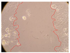 |  | 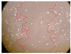 |
| P. viridis 250 mg/L | 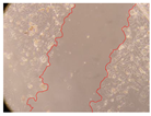 |  |  | 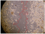 |
| P. viridis 500 mg/L | 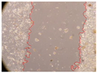 |  |  |  |
| B. caapi 250 mg/L |  |  |  |  |
| B. caapi 500 mg/L |  | 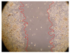 |  |  |
| M. hostilis 250 mg/L |  | 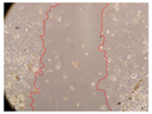 |  | 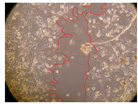 |
| M. hostilis 500 mg/L |  |  |  |  |
| Commercial mixture 250 mg/L |  |  |  | 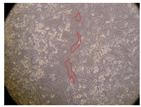 |
| Commercial mixture 500 mg/L | 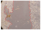 |  |  |  |
| P. viridis + B. caapi 250 mg/L | 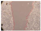 |  | 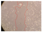 |  |
| P. viridis + B. caapi 500 mg/L |  | 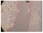 |  |  |
| P. viridis + P. harmala 250 mg/L |  |  | 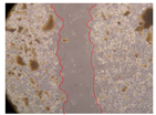 |  |
| P. viridis + P. harmala 500 mg/L | 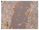 |  |  |  |
| M. hostilis + B. caapi 250 mg/L |  |  |  |  |
| M. hostilis + B. caapi 500 mg/L | 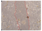 | 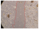 |  |  |
| M. hostilis + P. harmala 250 mg/L |  |  |  |  |
| M. hostilis + P. harmala 500 mg/L |  | 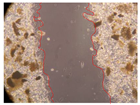 | 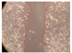 |  |
| Samples | 0 h | 2 h | p-Value | 8 h | p-Value | 12 h | p-Value | 24 h | p-Value |
|---|---|---|---|---|---|---|---|---|---|
| Control | 3.8 | 3.79 | - | 3.13 | - | 2.93 | - | 2.65 | - |
| P. viridis 250 mg/L | 2.36 | <0.001 | 2.06 | <0.001 | 1.5 | <0.001 | 0.53 | <0.001 | |
| P. viridis 500 mg/L | 2.97 | <0.001 | 2 | <0.001 | 1.64 | <0.001 | 0.54 | <0.001 | |
| B. caapi 250 mg/L | 3.23 | <0.001 | 1.71 | <0.001 | 1.42 | <0.001 | 0.75 | <0.001 | |
| B. caapi 500 mg/L | 3.08 | <0.001 | 2.24 | <0.001 | 1.42 | <0.001 | 1 | <0.001 | |
| M. hostilis 250 mg/L | 4.14 | <0.001 | 2.43 | <0.001 | 1.49 | <0.001 | 0.81 | <0.001 | |
| M. hostilis 500 mg/L | 2.43 | <0.001 | 2.07 | <0.001 | 1.76 | <0.001 | 0.4 | <0.001 | |
| Commercial mixture 250 mg/L | 3.79 | 1 | 2.05 | <0.001 | 1.39 | <0.001 | 0.49 | <0.001 | |
| Commercial mixture 500 mg/L | 3.14 | <0.001 | 1.85 | <0.001 | 1.11 | <0.001 | 0.69 | <0.001 | |
| P. viridis + B. caapi 250 mg/L | 2.10 | 0.001 | 1.38 | <0.001 | 1.23 | <0.001 | 0.25 | <0.001 | |
| P. viridis + B. caapi 500 mg/L | 2.73 | <0.001 | 1.5 | <0.001 | 1.29 | <0.001 | 0.63 | <0.001 | |
| P. viridis + P. harmala 250 mg/mL | 1.95 | <0.001 | 1.44 | <0.001 | 1.26 | <0.001 | 0.37 | <0.001 | |
| P. viridis + P. harmala 500 mg/L | 2.15 | <0.001 | 1.98 | <0.001 | 1.34 | <0.001 | 0.8 | <0.001 | |
| M. hostilis + B. caapi 250 mg/L | 2.08 | <0.001 | 1.65 | <0.001 | 1.38 | <0.001 | 0.47 | <0.001 | |
| M. hostilis + B. caapi 500 mg/L | 1.72 | <0.001 | 1.38 | <0.001 | 1.2 | <0.001 | 0.33 | <0.001 | |
| M. hostilis + P. harmala 250 mg/L | 2.83 | <0.001 | 1.71 | <0.001 | 1.07 | <0.001 | 0.3 | <0.001 | |
| M. hostilis + P. harmala 500 mg/L | 2.73 | <0.001 | 2 | <0.001 | 1.47 | <0.001 | 0.2 | <0.001 |
| Samples | TEER (Ω cm2) | ||
|---|---|---|---|
| Before | After | p-Value | |
| Control | 924 ± 124.45 | 1166 ± 217.79 | 0.306 |
| P. viridis 250 mg/L | 1166 ± 31.11 | 1056 ± 62.23 | 0.155 |
| P. viridis 500 mg/L | 1122 ± 155.56 | 1188 ± 248.90 | 0.781 |
| B. caapi 250 mg/L | 1309 ± 140.01 | 1100 ± 124.45 | 0.255 |
| B. caapi 500 mg/L | 1023 ± 202.23 | 1430 ± 93.34 | 0.123 |
| M. hostilis 250 mg/L | 1342 ± 93.34 | 1320 ± 124.45 | 0.860 |
| M. hostilis 500 mg/L | 869 ± 171.12 | 968 ± 186.68 | 0.636 |
| Commercial mixture 250 mg/L | 1265 ± 233.35 | 1056 ± 0.00 | 0.333 |
| Commercial mixture 500 mg/L | 836 ± 62.23 | 1144 ± 124.45 | 0.089 |
| P. viridis + B. caapi 250 mg/L | 858 ± 31.11 | 1254 ± 217.79 | 0.126 |
| P. viridis + B. caapi 500 mg/L | 1078 ± 217.79 | 1386 ± 31.11 | 0.186 |
| P. viridis + P. harmala 250 mg/L | 1254 ± 31.11 | 1298 ± 31.11 | 0.293 |
| P. viridis + P. harmala 500 mg/L | 1056 ± 62.23 | 1298 ± 31.11 | 0.039 |
| M. hostilis + B. caapi 250 mg/L | 979 ± 202.23 | 1342 ± 155.56 | 0.182 |
| M. hostilis + B. caapi 500 mg/L | 1331 ± 202.23 | 1276 ± 124.45 | 0.774 |
| M. hostilis + P. harmala 250 mg/L | 924 ± 186.68 | 1056 ± 0.00 | 0.403 |
| M. hostilis + P. harmala 500 mg/L | 968 ± 124.45 | 1210 ± 155.56 | 0.228 |
| Samples | Permeability (%) | p-Value |
|---|---|---|
| Control | 13.55 ± 0.51 | - |
| P. viridis 250 mg/L | 14.69 ± 3.04 | 0.652 |
| P. viridis 500 mg/L | 13.75 ± 2.49 | 0.922 |
| B. caapi 250 mg/L | 13.40 ± 2.65 | 0.947 |
| B. caapi 500 mg/L | 13.41 ± 2.76 | 0.951 |
| M. hostilis 250 mg/L | 15.46 ± 2.98 | 0.465 |
| M. hostilis 500 mg/L | 14.40 ± 2.74 | 0.709 |
| Commercial mixture 250 mg/L | 13.80 ± 2.74 | 0.910 |
| Commercial mixture 500 mg/L | 12.33 ± 0.73 | 0.193 |
| P. viridis + B. caapi 250 mg/L | 12.69 ± 0.29 | 0.174 |
| P. viridis + B. caapi 500 mg/L | 12.51 ± 2.46 | 0.617 |
| P. viridis + P. harmala 250 mg/L | 11.33 ± 1.29 | 0.151 |
| P. viridis + P. harmala 500 mg/L | 11.29 ± 0.38 | 0.037 |
| M. hostilis + B. caapi 250 mg/L | 12.51 ± 2.55 | 0.628 |
| M. hostilis + B. caapi 500 mg/L | 13.66 ± 0.05 | 0.789 |
| M. hostilis + P. harmala 250 mg/L | 14.52 ± 1.45 | 0.467 |
| M. hostilis + P. harmala 500 mg/L | 13.85 ± 0.12 | 0.501 |
Publisher’s Note: MDPI stays neutral with regard to jurisdictional claims in published maps and institutional affiliations. |
© 2022 by the authors. Licensee MDPI, Basel, Switzerland. This article is an open access article distributed under the terms and conditions of the Creative Commons Attribution (CC BY) license (https://creativecommons.org/licenses/by/4.0/).
Share and Cite
Gonçalves, J.; Luís, Â.; Gallardo, E.; Duarte, A.P. Evaluation of the In Vitro Wound-Healing Potential of Ayahuasca. Molecules 2022, 27, 5760. https://doi.org/10.3390/molecules27185760
Gonçalves J, Luís Â, Gallardo E, Duarte AP. Evaluation of the In Vitro Wound-Healing Potential of Ayahuasca. Molecules. 2022; 27(18):5760. https://doi.org/10.3390/molecules27185760
Chicago/Turabian StyleGonçalves, Joana, Ângelo Luís, Eugenia Gallardo, and Ana Paula Duarte. 2022. "Evaluation of the In Vitro Wound-Healing Potential of Ayahuasca" Molecules 27, no. 18: 5760. https://doi.org/10.3390/molecules27185760
APA StyleGonçalves, J., Luís, Â., Gallardo, E., & Duarte, A. P. (2022). Evaluation of the In Vitro Wound-Healing Potential of Ayahuasca. Molecules, 27(18), 5760. https://doi.org/10.3390/molecules27185760