Review

Discovery of E3 Ligase Ligands for Target Protein Degradation

1
Department of Chemistry, Kangwon National University, Chuncheon 24341, Korea
2
Department of Chemistry and Biochemistry, University of California, San Diego, CA 92093, USA
3
Kangwon Radiation Convergence Research Support Center, Kangwon National University, Chuncheon 24341, Korea
4
Kangwon Institute of Inclusive Technology, Kangwon National University, Chuncheon 24341, Korea
5
Department of Biomedical Science, Kangwon National University, Chuncheon 24341, Korea
*
Author to whom correspondence should be addressed.
These authors contributed equally to this work.
Molecules 2022, 27(19), 6515; https://doi.org/10.3390/molecules27196515
Submission received: 31 August 2022 / Revised: 22 September 2022 / Accepted: 30 September 2022 / Published: 2 October 2022
(This article belongs to the Special Issue Young Scholars’ Developments in Chemical Biology)

Abstract

:
Target protein degradation has emerged as a promising strategy for the discovery of novel therapeutics during the last decade. Proteolysis-targeting chimera (PROTAC) harnesses a cellular ubiquitin-dependent proteolysis system for the efficient degradation of a protein of interest. PROTAC consists of a target protein ligand and an E3 ligase ligand so that it enables the target protein degradation owing to the induced proximity with ubiquitin ligases. Although a great number of PROTACs has been developed so far using previously reported ligands of proteins for their degradation, E3 ligase ligands have been mostly limited to either CRBN or VHL ligands. Those PROTACs showed their limitation due to the cell type specific expression of E3 ligases and recently reported resistance toward PROTACs with CRBN ligands or VHL ligands. To overcome these hurdles, the discovery of various E3 ligase ligands has been spotlighted to improve the current PROTAC technology. This review focuses on currently reported E3 ligase ligands and their application in the development of PROTACs.

1. Introduction

Proteins are the basic machinery of the cellular system and execute genetically programmed behaviors for cellular survival. Around 20,000 proteins have been identified from human cells and their balance in the protein network is extremely important to maintain the healthy status of cells. The dysfunction or breakdown of a single protein could lead to the disease status of cells. Therefore, discovering the small molecules that are modulating the dysregulated protein is the key strategy for the drug discovery process. However, only a limited number of proteins has been reported as druggable proteins and traditional drug discovery has been focused on those druggable proteins. Considering the fact that a numerous number of proteins in cells are essential for cellular homeostasis, targeting undruggable proteins could be a solution for the treatment of incurable diseases.
Targeted Protein Degradation (TPD) is an emerging therapeutic strategy which is considered a solution to overcome the limitations of conventional drug discovery. TPD is a powerful chemical biology approach for inducing the degradation of proteins known as undruggable, and it has had a tremendous impact on the field of recent drug discovery. TPD manipulates the E3 ligase to selectively degrade the protein of interest (POI) via the Ubiquitin Proteasome System (UPS), an intracellular proteolysis mechanism. A targeted protein degrader consists of a ligand that binds to E3 ligases, a ligand that targets the POI, and a chemical linker between the ligands. These degraders are also called proteolysis-targeting chimeras (PROTACs), and the induced proximity between the target protein and the E3 ligase promotes the ubiquitination and proteasomal degradation of the target protein. Along with PROTAC, a protein degradation strategy using the protein tag has been developed to understand the inherent function of proteins. The additional aminoacidic signal sequences such as dTAG, AiD, and SMASh Tag induce proximity-based POI ubiquitination for its degradation via the ubiquitin–proteasome system. The development of tag-based strategies is well documented in the following references [1,2,3].
Even though there are over 600 types of E3 ligases in human cells, only a very limited number of E3 ligases have been used for PROTAC technology (CRBN, VHL, IAP, and MDM2). The majority of PROTACs developed so far have been restricted to CRBN or VHL, which are ubiquitously expressed in the human body. However, the recent emergence of drug resistance for CRBN- or VHL-based PROTACs strongly suggested that the discovery of various E3 ligase ligands is highly demanding to fully exploit the PROTAC strategy [4,5,6]. In addition, considering that a number of E3 ligases have been shown to overexpress in specific types of cells or tissues—for example, brain (FBXL16, KCTD8), pancreas (ASB9), skeletal muscle (KLHL40, KLHL41), testis (DCAF4L1), fallopian tube (DCAF8L1)—harnessing new E3 ligases may offer better opportunities for PROTACs having a higher selectivity and specificity for the efficient disease treatment. In this context, an overview of the PROTAC design strategies is highly necessary to develop efficient protein degraders. This review focuses on the currently reported E3 ligase ligands and their application in the development of PROTACs.

2. CRBN Ligands

In the 1950s, thalidomide (1) was first developed by Grünenthal as a sedative for morning sickness in pregnant women. However, thalidomide was withdrawn from the market due to its severely teratogenic effects in the early 1960s [7]. During the following decades, thalidomide was further studied extensively, redeveloped as a promising immunomodulatory imide drug (IMiD), and approved for the treatment of erythema nodosum leprosum (ENL) and multiple myeloma. The thalidomide derivatives, such as pomalidomide and lenalidomide, have shown an excellent efficacy in the treatment of multiple myeloma with their immunomodulatory activities (Figure 1) [8,9,10]. However, the mechanism of action of thalidomide had not been elucidated until Ito et al. revealed that the target protein of thalidomide is cereblon, a subunit of the E3 ubiquitin ligase CUL4-RBX1-DDB1-CRBN(CRL4CRBN), in 2010 [11]. They demonstrated a thalidomide–CRBN complex-induced teratogenic effect in in vivo models. In addition to thalidomide, the target protein of pomalidomide and lenalidomide was identified as CRBN by Zhe et al. in 2011 [12]. The antimultiple myeloma activity of pomalidomide (2) and lenalidomide (3) was CRBN expression dependent.
In 2014, Fischer et al. presented the cocrystal structure of the DDB1–CRBN complex bound to thalidomide, lenalidomide, and pomalidomide. This research showed that CRBN is the substrate receptor of CRL4CRBN and binds to thalidomide derivatives in an enantioselective manner. They further revealed that the thalidomide–CRBN complex recruits IKZF1 or IKZF3 and induces their degradations [13]. Thalidomide derivatives are composed of the phthalimide and glutarimide group (Figure 2A). The cocrystal structure (PDB code: 4CI1) of the thalidomide and CRBN complex showed that the glutarimide group of thalidomide derivatives plays an important role in CRBN binding via two major interactions: (1) the H-bond between carbonyl and amide groups of the glutarimide group and His380 and Trp382 of CRBN; and (2) van der Waals interactions between the glutarimide group and the hydrophobic pocket of CRBN composed of Phe404, Trp388, and Trp402 (Figure 2B). A carbonyl group of the phthalimide group also contributes a H2O-mediated hydrogen bonding with the His359 of CRBN. This cocrystal structure revealed that the solvent-exposed site of thalidomide is the benzene ring of the phthalimide group, which can be further conjugated for the PROTAC design without a loss of the binding affinity [14]. Based on this structural information, the development of thalidomide derivatives has been able to be accelerated.
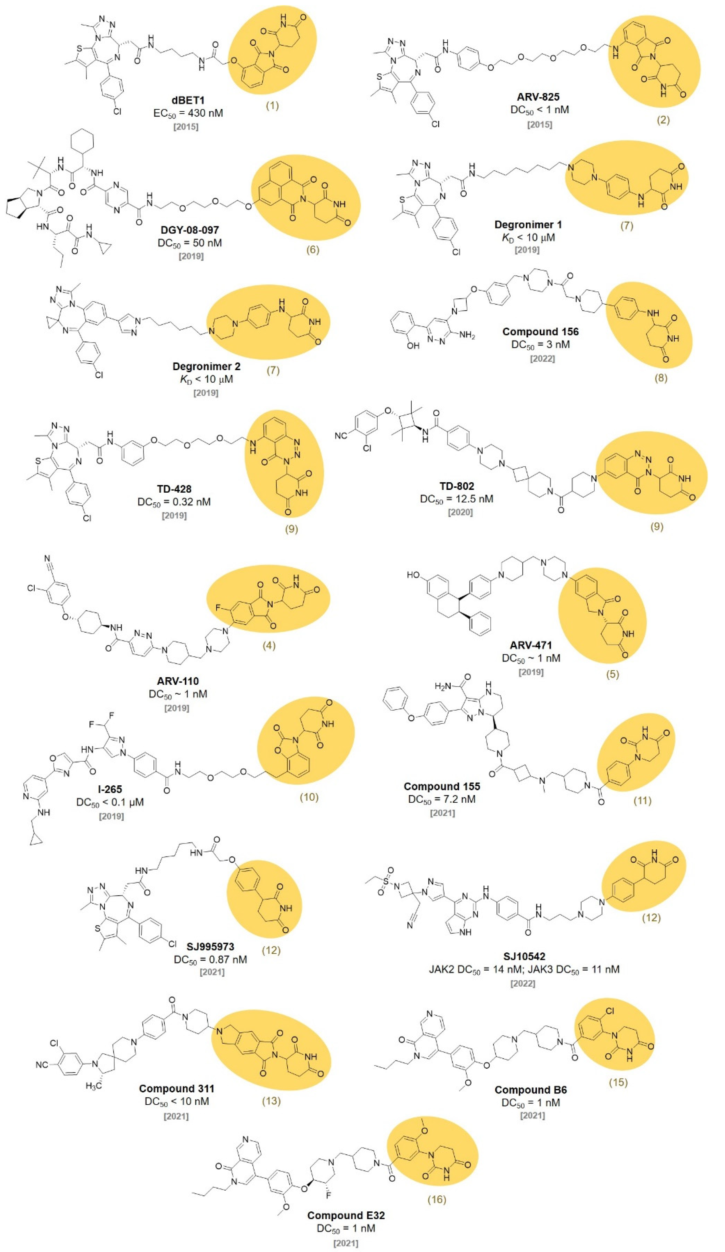
In 2015, the Crews and Bradner group reported that bromodomain-containing protein 4 (BRD4) targets PROTACs. ARV-825 (DC50 < 1 nM) [15] and dBET1 (EC50 = 430 nM) were synthesized by conjugating a BRD4 ligand, JQ1, with a thalidomide derivative for CRBN engagement (Figure 3). Both PROTACs effectively triggered the degradation of BRD4 in the cells and inhibited the cell proliferation. dBET1 showed an excellent antiacute myeloid leukemia (AML) efficacy in vitro and in vivo [16]. After dBET1 was reported as the first PROTAC in vivo, various thalidomide derivatives have been explored to improve the efficiency of the PROTAC technique.
In 2018, the Crew group reported 22 thalidomide analogs with a rapid one-pot synthesis without purification. They measured the binding affinities of newly synthesized thalidomide analogs to CRBN with a surface plasmon resonance analysis. In addition, the ability to induce the degradation of Aiolos and CK1α was investigated. Among them, three thalidomide analogs (4, 5, 6) showed improved pharmacological properties and good CRBN binding affinities (KD for 4, 5, and 6 were 55, 549, and 111 nM, respectively). They found that the chemical modification of the phthalimide part in thalidomide did not induce a significant deterioration in the CRBN binding affinities [17]. In 2019, the Yang group synthesized PROTAC, DGY-08-097, which induces hepatitis C virus (HCV) NS3/4A protease degradation by conjugating 6 with Telaprevir, an FDA-approved drug for the treatment of HCV [18] (IC50 = 247 nM, DC50 = 50 nM). DGY-08-097 showed antiHCV activity in the cellular infection model.
C4 Therapeutics also filed a patent for the synthesis of CRBN-targeting moieties (Degrons) that can be conjugated to target protein ligands. They reported various piperidine-2,6-dione derivatives by conjugating O- and N-linked heterocycles. A fluorescence polarization (FP) assay demonstrated that 7 was one of the most promising CRBN ligands. The degrons are capable of functioning as molecular glues and downregulating the levels of the Aiolos or Ikaros protein, which can lead to the treatment of leukemia, acute myeloid leukemia, chronic lymphoblastic leukemia, and multiple myeloma just like other IMiDs. They conjugated the developed E3 ligase ligand to JQ1 and named them Degronimers. The representative Degronimers 1 and 2 showed an excellent binding affinity to CRBN [19] (KD < 10 μM). They also reported various SMARCA2 degraders using the degrons. The N-linked degrons, 8, was developed as PROTAC (compound 156) for SMARCA2 degradation with a nanomolar efficacy through a HiBiT degradation assay (DC50 = 3 nM) [20].
The Hwang group designed aminobenzotriazino glutarimides as novel CRBN ligands and discovered TD-106 (9). The TD-106 (9) exhibited a better degradation efficiency of IKZF1/3 than that of pomalidomide in NCI-H929 cells. After the confirmation of TD-106 (9) as a direct CRBN binder through a thermal stability shift, an in vivo xenograft model study demonstrated that intraperitoneally injected TD-106 (9) showed antitumor activity after 14 days of administration [21]. They further synthesized a BRD4-targeting PROTAC, TD-428 (DC50 = 0.32 nM), by conjugating JQ1 and TD-106 (9). The successful BRD4 degradation in 22Rv1 cells induced by TD-428 was confirmed. They also reported an androgen receptor (AR) degrader, TD-802 (DC50 = 12.5 nM), for the treatment of metastatic castration-resistant prostate cancer using the TD-106 (9) ligand [22].
In 2019, Arvinas unveiled that the ARV-110 (DC50 ~ 1 nM) and ARV-471 (DC50 ~ 1 nM) are targeting AR and estrogen receptor (ER), respectively. Both degraders have been spotlighted since they are currently in phase two of clinical trials [23,24]. ARV-110 was synthesized via the conjugation of a thalidomide derivative (4) and AR ligand. ARV-471 was developed via the conjugation of a thalidomide derivative (5) and ER ligand. Both shared the same linker structure for the conjugation of CRBN and target protein ligands. The most interesting feature of these PROTACs is that both can be administered orally, which was not easily achievable in other PROTACs.
Kymera Therapeutics reported various CRBN ligands having two rings conjugated with diverse linkers in 2019. They measured their affinities toward CRBN through a time-resolved fluorescence resonance energy transfer (TR-FRET) assay [25]. They synthesized a PROTAC, I-265 (DC50 < 0.1 μM), by conjugating 10 and an interleukin-1 receptor activated kinase (IRAK) inhibitor. They showed that I-265 degraded the IRAK4 protein in human peripheral blood mononuclear cells (PBMCs) [26]. One of their ligands, SB572027 (11), was used for the synthesis of BTK-targeting PROTACs by the Chinese biotechnology company Beigene. Beigene synthesized a series of PROTACs that conjugated a BTK inhibitor with various thalidomide derivatives capable of recruiting the target protein to the E3 ubiquitin ligase. They performed an ELISA assay for BTK detection to evaluate the activity of a series of the PROTACs. Among them, compound 155 using SB57027 (11) as a CRBN ligand showed nanomolar degradation activity (DC50 = 7.2 nM) [27].
In 2021, the Rankovic group reported highly stable CRBN binders and their application for PROTACs. They replaced the hydrolysis-labile phthalimide moieties of thalidomide with phenyl groups to synthesize phenyl glutarimide (PG, 12). PG (12, IC50 = 2.191 μM) showed outstanding stability (t1/2 > 24 h) compared with thalidomide (t1/2 = 3.3 h) in cell media without affecting the binding affinity with CRBN (IC50 for thalidomide = 1.282 μM, lenamide = 0.699 μM, and pomalidomide = 0.4 μM). PG (12) also showed superior metabolic stability in mouse and human liver microsomes. A PG-based PROTAC (SJ995973, DC50 = 0.87 nM) showed potent antiproliferative efficacy in both MV4-11 (IC50 = 3 pM) and HD-MB03 cells (IC50 = 1.83 nM) [28]. In a follow-up study, the Rankovic group used PG (12) to synthesize Janus kinase (JAK)-targeting PROTACs. Through the structure activity relationship (SAR) study, they found a PG (12)-based PROTAC, SJ10542, with highly selective JAK degradation and reduced GSPT1 degradation compared with thalidomide-based PROTACs (JAK2 DC50 = 14 nM, JAK3 DC50 = 11 nM) [29].
In 2021, Wang et al. filed a patent about the synthesis of various CRBN ligands for PROTAC design [30]. Among their compounds, 13 and 14 were conjugated to spirocyclic AR ligands to synthesize AR-degrading PROTACs for the treatment of prostate cancer. They measured AR degradation in VCaP cells and compound 311 showed the efficient degradation of AR with a nanomolar concentration (DC50 < 10 nM). Moreover, they demonstrated the superior oral bioavailability of compound 311 [31].
In 2021, Novartis developed a BRD9 degrader for cancer treatment. BRD9 is a subunit of the SWI/SNF complex, which has been reported as a drug target for the treatment of synovial sarcoma and acute myeloid leukemia. A series of BRD9 PROTACs were synthesized via the conjugation of BRD9-targeting ligands and CRBN-targeting E3 ligase ligands with various types of short and rigid piperidine linkers. Among the synthesized PROTACs, compound B6 and compound E32, based on the CRBN binders 15 and 16, respectively, showed BRD9 DC50 at the nanomolar concentration [32] (DC50 = 1 nM for both compound B6 and E32).

3. VHL Ligands

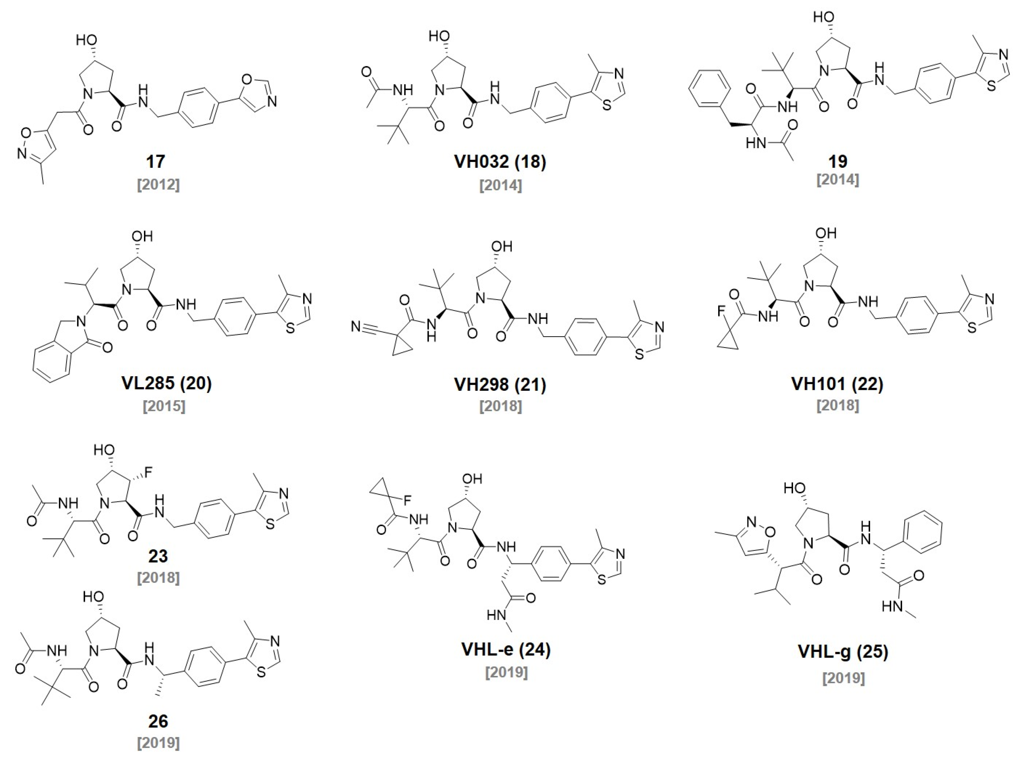
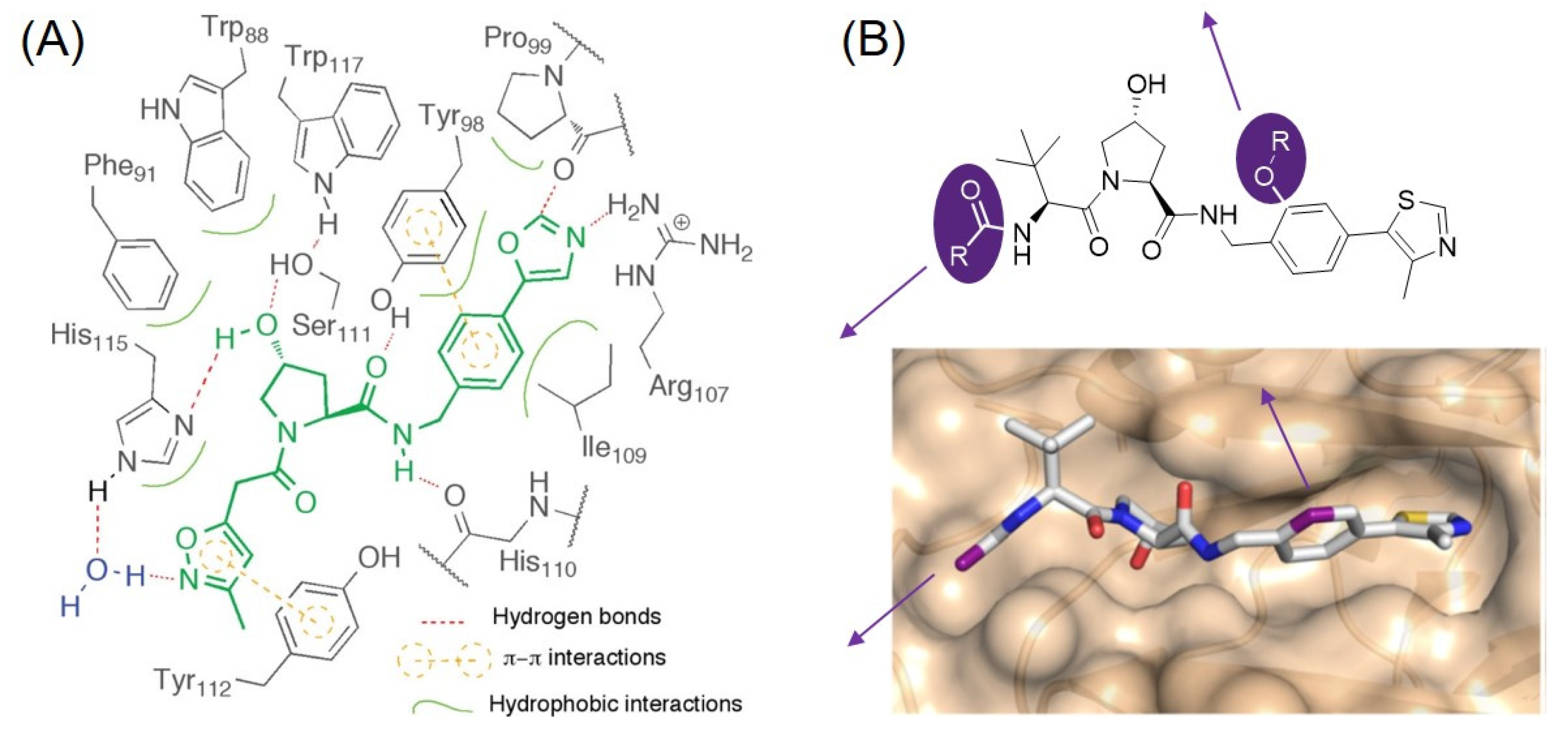
The von Hippel–Lindau (VHL) protein is the substrate receptor protein of the Cullin 2 E3 ligase. Hypoxia-inducible factor 1α (HIF-1α) is one of the substrate proteins of VHL. Considering the HIF-1α-mediated upregulation of proangiogenic factors, an inhibitor of VHL would increase erythropoietin and can be used for the treatment of chronic anemia and cancer chemotherapy. In 2012, the Ciulli and Crews group reported a series of small molecule inhibitors targeting VHL for the first time. They rationally designed inhibitors of the VHL/HIF-1α interaction in silico. After the discovery of their initial hit, they synthesized a focused library of hydroxy proline derivatives with solid phase synthesis. They found VHL ligand 17 with single-digit micromolar activity through an FP assay (Figure 4). The cocrystal structure of the ligand bound to VHL confirmed that the ligand mimics the binding mode of the HIF-1α to VHL [33]. They demonstrated that the major binding affinity of VHL ligands originates from the H-bond between the hydroxyl group of pyrrolidine and His115 and Ser111, as well as the interaction between the amide NH group and a carbonyl group of His110 (Figure 5A). In addition, the phenyl group, adjacent to the amide group, contributes to the high binding affinity of the VHL ligand via a π-π interaction with Tyr98.
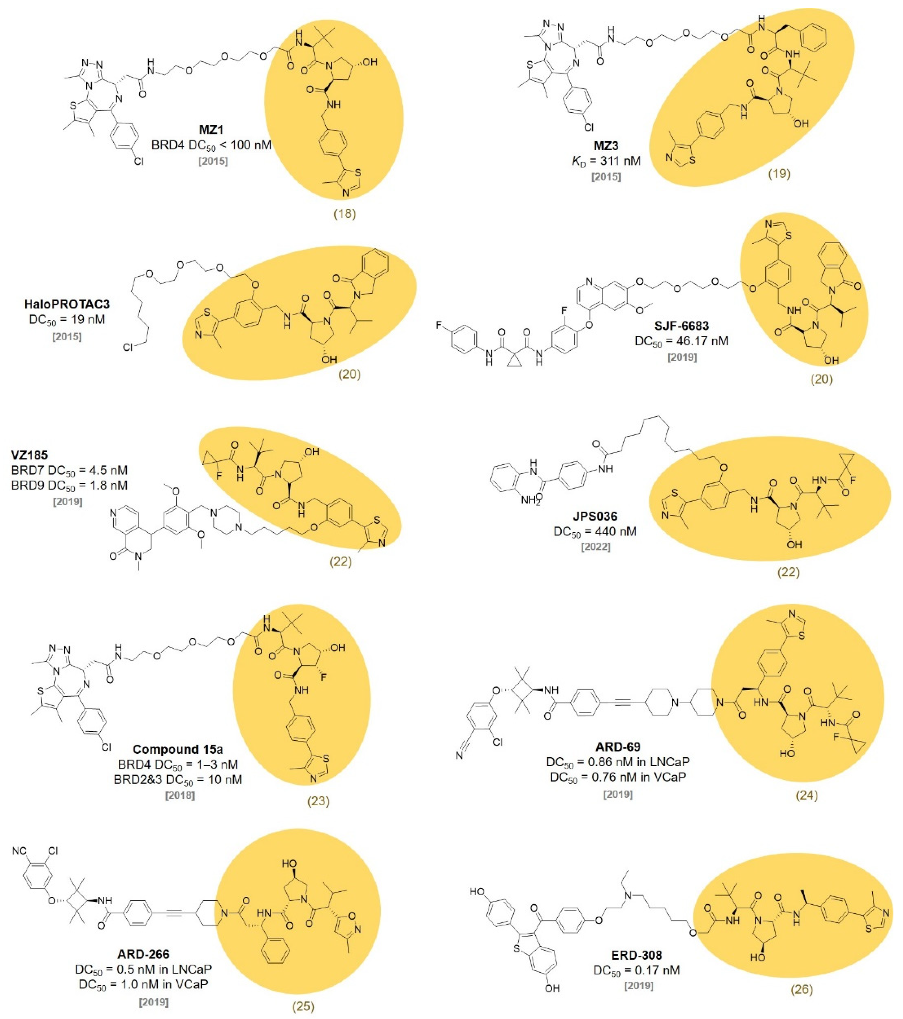
In a follow-up study, the Ciulli group designed and optimized the initial VHL ligands based on X-ray crystal structures and reported new VHL ligands, VH032 (18, KD = 185 nM) and 19 (KD = 291 nM), with nanomolar binding affinities, in 2014 [34]. Using VH032 (18) and 19, the Ciulli group reported bromodomain and extra-terminal domain (BET) protein-targeting PROTACs MZ1 (KD = 149 nM) and MZ3 (KD = 311 nM) (Figure 6) [35]. They observed the MZ1-mediated degradation of BRD2, BRD3, and BRD4 at the single digit micromolar concentration (BRD4 DC50 < 100 nM).
In 2015, the Crews group proposed HaloPROTAC using their VHL ligands. Based on their previously reported crystallographic evidence, they identified the possible linker positions by confirming the solvent-exposed sites of the VHL ligands. Not only the linker position but also a proper linker length were studied for VHL-mediated protein degradation. Finally, they successfully designed HaloPROTAC3 (DC50 = 19 nM) by conjugating a chloroalkane linker to the VHL ligand (20, IC50 = 0.34 μM) for the degradation of the HaloTag fusion protein [36]. The Crews group revealed that the linker position of the VHL ligand largely affects the substrate specificity of PROTAC. They synthesized PROTACs with the linkers on the left-handed amide side and right-handed phenyl side of the VHL ligand, respectively (Figure 5B). The PROTAC (SJF-6683) conjugated a p38 MAPK ligand, foretinib, to the right-hand phenyl side of the VHL ligand (20) selectively and strongly degraded p38δ, a specific p38 isoform (p38δ DC50 = 46.17 nM) [37].
Figure 4. Chemical structures of reported VHL ligands during the last decade.
Molecules 27 06515 g004
Figure 5. (A) Schematic illustrations showing the binding mode of the VHL ligand 17. Reprinted with permission from ref. [33] (Copyright 2012 American Chemical Society). (B) Chemical structure of a representative VHL ligand analogue having two different linker positions. Reprinted with permission from ref. [37] (Copyright 2019 Springer Nature).
Molecules 27 06515 g005
Figure 6. Reported PROTACs based on the VHL ligands.
Molecules 27 06515 g006
In 2018, Ciulli reported the structure-guided rational optimization of VH032 (18). Increasing the lipophilicity of the VHL ligands led to a higher cell permeability and higher binding affinity to the VHL protein. They discovered VH298 (21, KD = 52 nM) and VH101 (22, KD = 16 nM), which showed the effective protein–protein inhibition between the VHL and HIF-1α protein. [38]. Their SAR study provided novel VHL ligands, which can be used for VHL-based PROTACs. Note that this discovery expanded the landscape of PROTAC research to other E3 ligases, which was only restricted to CRBN. VH101 (22) was used in the SAR study to develop BRD7 and BRD9 degraders by the Ciulli group. A PROTAC (VZ185) was developed by coupling the BRD7/9 inhibitor, BI-7273, and VH101 (22). VZ185 efficiently degraded BRD7 and BRD9 simultaneously (BRD7 DC50 = 4.5 nM, BRD9 DC50 = 1.8 nM) [39].
The Hodgkinson group reported histone deacetylase (HDAC)-targeting PROTACs. They synthesized HDAC-degrading PROTACs with various linkers and VHL ligands and monitored their degradation activities. JPS036, a PROTAC composed of VH101 (22), was developed as a selective degrader of HDAC3 with a submicromolar activity (DC50 = 0.44 μM) [40].
In 2018, the Ciulli group reported VHL ligands with a different stereochemistry of fluoro-hydroxyproline (F-Hyp). They synthesized four diastereoisomers of 3-fluoro-4-hydroxyproline containing VHL ligands and found that VHL can stereoselectively recognize the (3R,4S) epimer of F-Hyp (23). A JQ1-based PROTAC (compound 15a) using the (3R,4S) epimer of F-Hyp selectively degraded BRD4 at nanomolar concentrations, despite its weak affinity for VHL (KD = 3.08 μM, BRD4 DC50 = 1~3 nM, BRD2 and BRD3 DC50 = 10 nM). This discovery was an important advance in expanding the chemical space of TPD toward low affinity molecules [41].
In 2019, the Wang group discovered new VHL ligands via the introduction of an (S)-methyl group on VH101 based on previous work [42,43]. Through a SAR study, they found that appending an amide group to the (S)-methyl group increased the potency of the VHL ligands. The FP-based binding assay showed that VHL-e (24) binds to VHL with a high affinity (IC50 = 190 nM). In addition, they confirmed that the introduced stereochemistry was crucial for their binding affinity to VHL. With VHL-e (24), an effective AR degrader, ARD-69, was discovered after the optimization of the linker length and linking site on the ligands. ARD-69 showed effective AR degradation activity in LNCaP (DC50 = 0.86 nM), VCap (DC50 = 0.76 nM), and 22Rv1 prostate cancer cell lines and in a VCaP xenograft mouse model [43]. The Wang group further optimized the AR-targeting PROTAC with a shorter linker length and a low-affinity VHL ligand. ARD-266 using a weak binding VHL ligand, VHL-g (25), showed a much higher AR degradation activity than other PROTACs with higher-affinity VHL ligands (DC50 = 0.5 nM in LNCaP cell line, DC50 = 1.0 nM in VCaP cell line). Along with compound 15a, this study demonstrated that a low-affinity E3 ligase ligand-based PROTAC could induce the successful formation of a ternary complex with the POI for efficient degradation [44]. Subsequently, the Wang group extensively studied the SAR of an ER degrader based on a VHL ligand (26) and FDA-approved ER modulator, Raloxifene. As a result, a very potent ER degrader (ERD-308) was developed with a subnanomolar activity (DC50 = 0.17 nM). ERD-308 degraded the ER and inhibited cell growth more than those of the FDA-approved selective ER degrader molecule Fulvestrant [45]. In this report, changing the VHL ligand to the CRBN ligand completely abolished the ER degradation activity of ERD-308.

4. IAP Ligands

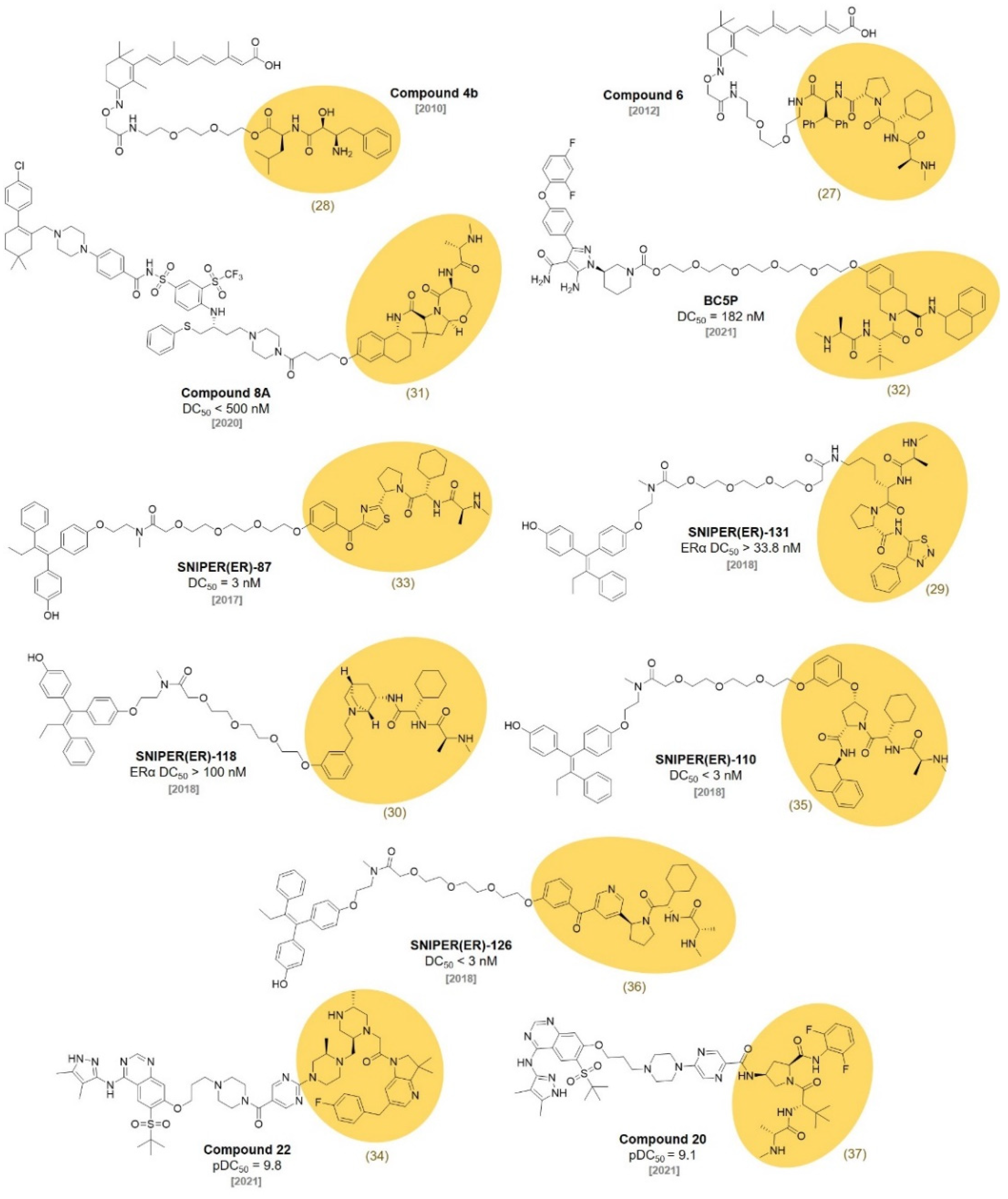
Inhibitors of apoptosis proteins (IAPs) are the regulators of cell death and they control apoptotic events triggered by diverse stimuli. In 2007, the Vucic group developed a cellular inhibitor of the apoptosis 1 and 2 (c-IAP1 and c-IAP2) antagonist (MV1, 27) that binds to the baculovirus IAP repeat (BIR) domains of IAP proteins, leading to the autoubiquitination and proteasomal degradation of c-IAPs (KD = 5.8 nM) (Figure 7). The degradation of c-IAPs by MV1 induced TNF signaling-pathway-dependent cell death [46]. Sekine et al. reported a different cIAP1 ligand, bestatin-methyl ester (ME-BS, 28), which binds to the BIR3 domain of cIAP1 and induces autoubiquitination followed by the proteasomal degradation of cIAP1 [47]. Itoh et al. developed bifunctional small molecules using the two ligands described above. One of the molecules (Compound 4b) was designed by the conjugation of ME-BS (28) and all-trans retinoic acid (ATRA) with a polyethylene glycol (PEG) linker (Figure 8). The other was developed via the conjugation of MV1 (27) and ATRA (Compound 6) [48,49]. These compounds were found to induce the degradation of both cIAP1 and cellular retinoic acid binding protein-II (CRABPII). They named this degrader the specific and nongenetic IAP-dependent protein eraser (SNIPER). It was later utilized on other targets, such as ER [50], BRD4 [51], and BCR-ABL [52].
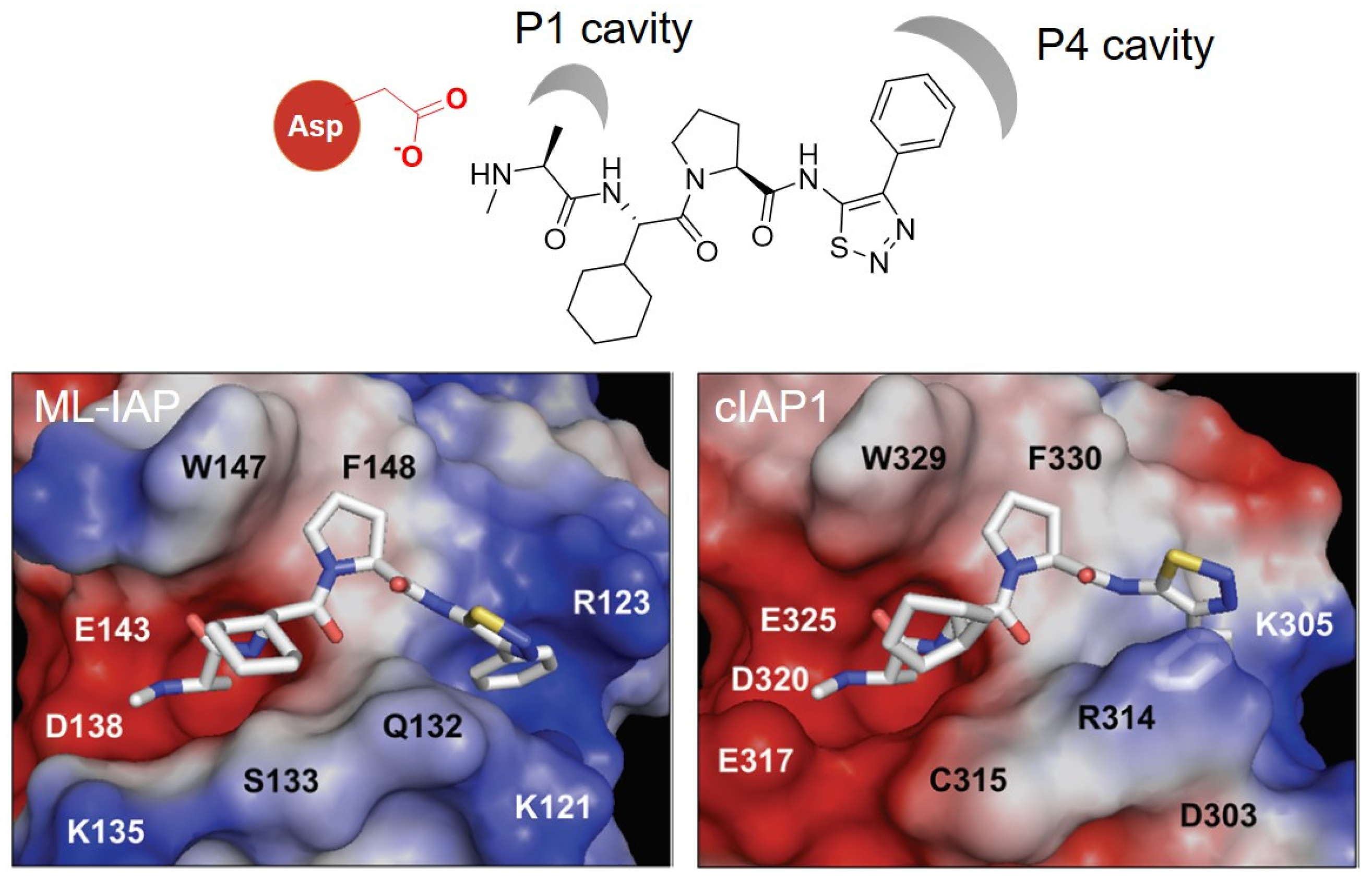
In 2012, Genentech discovered a potent antagonist of cIAP1/2, ML-IAP, and XIAP. The SAR study using the crystal structure led to the development of a broad spectrum IAP inhibitor, GDC-0152 (29) (Ki values for XIAP-BIR3 = 28 nM, MLXBIR3SG = 14 nM, cIAP1-BIR3 = 17 nM, and cIAP2-BIR3 = 43 nM) [53]. In cocrystal structures of GDC-0152 with ML-IAP or cIAP1, the critical interaction was the H-bond between the Asp (Asp138 for ML-IAL, Asp320 for cIAP1) and the amide group of the ligand (Figure 9). It allows for the proper positioning of the α-methyl group of the ligand to reside in the P1 cavity. In addition, another hydrophobic interaction was observed between the phenyl group of the ligand and P4 hydrophobic pocket in both crystal structures. Since most of the ligand structure is exposed to the solvent, the target protein ligands can be conjugated to diverse positions of the IAP ligands.
The Hennessy group discovered a series of aminopiperidine-based inhibitors of the IAP by mimicking the IAP binding residues of the second mitochondrial activator of caspases (Smac). They found that a bicyclic piperidine, 30 (KD for XIAP-BIR3 = 0.9 μM), fixed in a boat form was a potent inhibitor of cIAP1 and effectively induced cIAP1 degradation in MDA-MB231 cells (EC50 = 5 nM) [54].
In 2013, the Cosford group reported the synthesis of a potent IAP antagonist via a highly efficient application of the Ugi four-component reaction. Their optimized IAP antagonist (31) showed the best binding affinity to IAPs, especially against ML-IAP (Ki = 2 nM). 31 showed a powerful anticancer activity in breast, ovarian, and prostate cell lines and had no general toxicity to noncancerous human foreskin fibroblast (HFF) cells. They performed molecular modeling to reveal key interacting residues of IAP proteins with 31 [55]. The Zheng group used 31 to synthesize IAP-recruiting BCL-XL PROTACs, compound 8A (IC50 = 62 nM, DC50 < 500 nM). Compound 8A showed efficient BCL-XL degradation in the T-cell lymphoma cell line, while it had reduced the human platelet toxicity [56].
In 2014, Bristol-Myers Squibb reported that 32 embedded bivalent heterodimeric IAP antagonists showed a high affinity for the BIR2 domain and an excellent IAP inhibitory activity (IC50 up to 3.6 nM) [57]. Pfizer synthesized BC5P, a PROTAC that degrades BTK using 32 (DC50 = 182 nM). They confirmed that BC5P bound to only the BIR3 domain of IAP1 but not BIR1 or BIR2 by using biolayer interferometry (BLI). They utilized molecular modeling, solution NMR, and X-ray crystallography to elucidate the structural insights of the IAP-BC5P-BTK ternary complex [58].
In 2017, the Naito group developed an ER-targeting SNIPER, SNIPER(ER)-87, via the conjugation of an ER ligand, 4-hydroxytamoxifen, and an IAP ligand, LCL-161 (33) [50]. SNIPER(ER)-87 effectively reduced ERα protein levels at nanomolar concentrations in vitro (DC50 = 3 nM, IC50 = 15.6 nM in MCF-7 cell line, IC50 = 9.6 nM in T47D cell line). SNIPER(ER)-87 showed good metabolic stability in the serum. The intraperitoneal administration of SNIPER(ER)-87 reduced the growth of ER-positive human breast tumors in vivo. They also conjugated LCL-161 derivatives to JQ1, a PDE4 inhibitor, and dasatinib for SNIPERs targeting BRD4, PDE4, and BCR-ABL proteins to demonstrate the usefulness of LCL-161 derivatives for the development of various targeted protein degraders.
In 2018, the Naito group optimized their previously reported SNIPER(ER)-87 by incorporating various LCL-161-derivative IAP ligands (29, 30, 35, 36). With the improved IAP binding affinities of the LCL-161 derivatives, the optimized SNIPER(ER)s exhibited better binding affinities toward cIAP1, cIAP2, and XIAP. However, the E3 ligase binding affinities of SNIPER(ER)s were not exactly correlated to their target protein degradation efficiencies. The GDC-0152 (29)-based SNIPER(ER)-131 did not efficiently degrade ERα, despite its higher IAPs affinity than that of SNIPER(ER)-87 (SNIPER(ER)-131: ERα DC50 > 33.8 nM, IC50 = 80 nM, SNIPER(ER)-87: ERα DC50 < 3 nM, IC50 = 110 nM). 30-based SNIPER(ER)-118 had a low ERα degradation efficiency compared to the original compound SNIPER(ER)-87 (SNIPER(ER)-118: ERα DC50 > 100 nM, IC50 = 230 nM). SNIPER(ER)-110 and SNIPER(ER)-126 were the most potent ERα degraders with the lowest DC50 (< 3 nM) among the SNIPER(ER)s (SNIPER(ER)-110: ERα DC50 < 3 nM, IC50 = 120 nM, SNIPER(ER)-126: ERα DC50 < 3 nM, IC50 = 83 nM). SNIPER(ER)-110 showed the best ERα degradation efficiency and excellent antitumor activity in the in vivo tumor xenograft model [59].
Astex Pharmaceuticals successfully discovered a nonpeptidomimetic cIAP1 and XIAP inhibitor, AT-IAP (34), through a fragment-based drug discovery using structure information from X-ray crystallography, computational studies, and NMR solution structure analysis. AT-IAP (34) showed a strong dual antagonistic efficacy toward XIAP and cIAP1 (XIAP EC50 = 5.1 nM, cIAP1 EC50 = 0.32 nM). An oral administration of AT-IAP (34) in a mouse xenograft model effectively inhibited the tumor growth without affecting the body weight of the mouse [60].
In 2020, GlaxoSmithKline reported a palbociclib-based PROTAC with a CRBN ligand, a VHL ligand, and an IAP-binder (37) for the degradation of CDK4 and CDK6. They conjugated three different E3 ligase ligands with palbociclib, an FDA-approved anti-breast-cancer agent, using various linkers. With previously reported CRBN-based CDK4/6-targeting PROTACs [61,62], VHL- and IAP-based PROTACs showed an ability to effectively degrade CDK4/6, (DC50 < 10 nM) which could not be achieved with the previously reported VHL- and IAP-based PROTACs [63]. This work again emphasized the importance of linker structures in the development of PROTACs [64]. Moreover, all the Palbociclib-based PROTACs with three different E3 ligases showed the preferential degradation of CDK6 over CDK4 with a marginal degradation efficiency difference.
37 and AT-IAP (34) were also utilized in the development of a series of receptor-interacting serine/threonine protein kinase 2 (RIPK2) PROTACs including compound 20 and compound 22 by GlaxoSmithKline (compound 20: RIPK2 pDC50 = 9.1, compound 22: RIPK2 pDC50 = 9.8). RIPK2 plays a crucial role in the innate immune system. Therefore, the dysregulation of RIPK2 signaling pathways is highly related to various inflammatory diseases such as inflammatory bowel disease [65], severe pulmonary sarcoidosis [66], and multiple sclerosis [67]. Among the synthesized RIPK2 PROTACs, compound 20 showed the best profile with excellent solubility, a strong RIPK2 degradation ability, and TNFα inhibition [68].

5. MDM2 Ligands

The mouse double minute 2 homolog (MDM2) protein is an E3 ubiquitin ligase that regulates the ubiquitination of p53 and the subsequent proteasomal degradation of p53. In 2004, Roche reported a potent and selective small molecule inhibitor of the MDM2-p53 interaction. They screened a diverse library of synthetic compounds and identified Nutlin-3 (38) as a hit compound (Figure 10) [69]. Notably, two enantiomers of cis-imidazoline Nutlin-3 (38) possessed a highly different binding affinity to MDM2 (IC50 of enantiomer a = 13.6 μM, enantiomer b = 0.09 μM). In 2008, the Crews group first reported MDM2-based PROTAC 14 by conjugating Nutlin-3 (38) and a nonsteroidal AR ligand with a PEG linker (Figure 11) [70].
The Sheng group reported a Nutlin-3 (38)-based homo-PROTAC 11a that induces the self-degradation of MDM2 to inhibit the MDM2-p53 interaction (DC50 = 1.01 μM). Homo-PROTAC 11a induced effective MDM2 dimerization and triggered the proteasomal degradation of MDM2 in A549 non-small cell lung cancer cells. In addition, homo-PROTAC 11a-1, one of the stereoisomer of homo-PROTAC 11a, showed the highest antitumor activity in the A549 xenograft model (IC50 = 1.0 μM) [71].
In 2013, Roche optimized the original Nutlin-3 (38) compound based on the crystal structure of the p53-MDM2 complex and synthesized a new MDM2 inhibitor, RG7112 (39), with dimethyl substitution on the imidazoline ring and replacement of the methoxy group with a tert-butyl group (IC50 = 18 nM) [72]. RG7112 was the first orally available p53-MDM2 inhibitor under clinical trials. In a follow-up study, they discovered a new ligand with an improved affinity based on the crystal structure of MDM2. They replaced the imidazoline structure of RG7112 with a pyrrolidine moiety and introduced stereochemical configurations for the higher affinity. They reported a second-generation clinical MDM2 inhibitor, RG7388 (40), with an excellent efficacy and selectivity through a SAR study [73] (IC50 = 6 nM). In 2020, the Calabretta group reported that RG7112 (39)-based PROTAC YX-2-233 showed a strong degradation of CDK4 and CDK6 in Ph+ ALL cells and suppressed S-phase [74]. The Crews group reported that an RG7388-based PROTAC, A1874, showed a 98% degradation of the BRD4 protein in HCT116 cells with a submicromolar concentration. This was a substantial improvement in target potency compared with their first nutlin-based PROTAC 14 (PROTAC 14: DC50 = 10 μM vs. A1874: DC50 = 32 nM). In addition, A1874 increased p53 stability due to the RG7388 moiety. The dual mode of action, BRD4 degradation and p53 stabilization, by A1874 strongly suppressed cancer cell viability compared with VHL ligand-based PROTACs [75].

6. DCAF Ligands

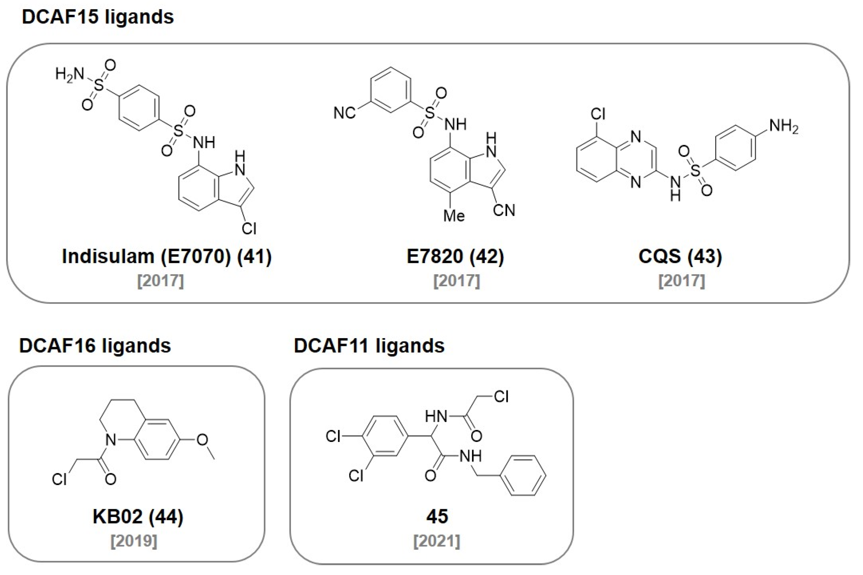
Sulfonamide derivatives have drawn attention due to their antibacterial, antifungal, antiviral, and anticancer activities. Recent studies reported that the sulfonamide derivatives indisulam (41), E7820 (42), and chloroquinoxaline sulfonamide (CQS, 43) function as a molecular glue that induces the protein–protein interaction between the E3 ligase and the target protein (Figure 12) [76,77].
In 2017, the Nijhawan group revealed that sulfonamide induces the proteasomal degradation of RNA-binding motif protein 39 (RBM39) by interacting with the DCAF15-DDB1-CUL4 complex [76]. In a follow-up study, they investigated how E7820 (42) recruits RBM39 to DCAF15 with kinetic analysis and crystal structures (KD = 22 μM) [77].
In an independent study, the Owa group reported that sulfonamide induces ubiquitination and the proteasomal degradation of CAPERα through the formation of CAPERα-sulfonamide-DCAF15 [78]. The Chen group reported an E7820 (42)-based PROTAC (DP1)-targeting BRD4 by employing JQ1 as a target protein ligand (Figure 13). DP1 showed an excellent degradation of BRD4 in SU-DHL-4 cells (DC50 = 10.84 ± 0.92 μM, Dmax = 98%) and inhibited tumor growth in a mouse model [79].
In 2019, the Cravatt group used a chemical proteomic approach that utilized a FKBP12 ligand, SLF, conjugated with the cysteine-directed electrophilic fragments for the discovery of a nuclear localized E3 ligase. After identifying the electrophilic fragments that induce FKBP12 degradation in the nucleus, they used pull-down-based proteomics and identified DCAF16 E3 ligase as their target protein. One of the electrophilic fragments, KB02 (44), were extensively studied after conjugation with the FKBP ligand (KB02-SLF) to monitor its FKBP12 degradation ability (DC50 < 2 μM). In addition, they also designed a BRD4-targeting PROTAC, KB02-JQ1 (DC50 ~ 20 μM), by coupling KB02 (44) to JQ1, and observed DCAF16 as a valuable nuclear localized E3 ligase [80]. With a similar screening strategy, the Cravatt group also developed electrophilic ligands (45) of E3 ligase DCAF11. Using the discovered ligand 45, they synthesized 21-ARL for AR degradation with a recruited DCAF11 [81].

7. RNF Ligands

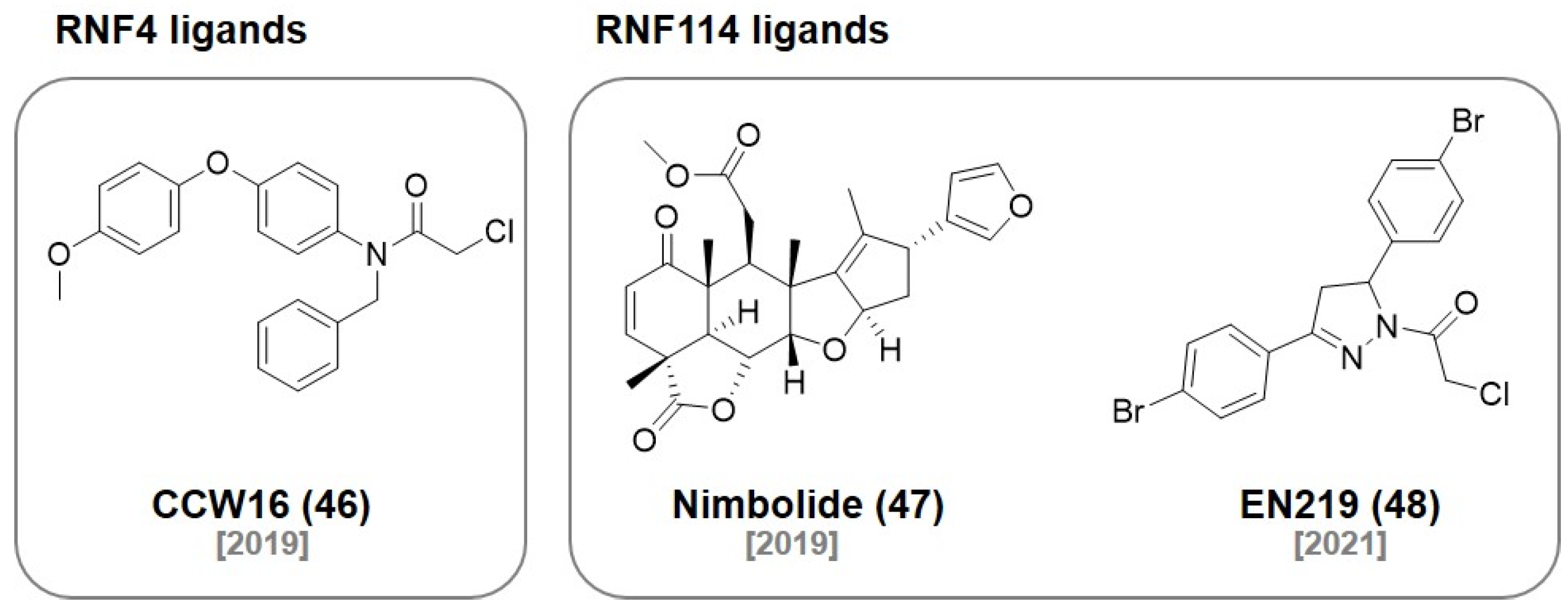
In 2019, the Nomura group reported a set of binders to the E3 ligase RNF4 using the ABPP-based covalent ligand screening approach. The optimized covalent ligand CCW16 (46) (Figure 14) reacted with one of two zinc-coordinated cysteines in the RING domain without affecting the zinc binding ability of RNF4 (IC50 1.8 μM). They demonstrated a covalent PROTAC (CCW 28-3) for BRD4 degradation (Figure 15) [82]. The same group also reported that Nimbolide (47), a natural product exhibiting anticancer activities, was identified as a covalent binder for the E3 ligase RNF114. They used activity-based protein profiling (ABPP) chemoproteomic platforms to discover that Nimbolide (47) binds to a cysteine residue of RNF114. Based on the structure of Nimbolide (47), they developed a covalent PROTAC, XH2, targeting BRD4 (IC50 = 240 nM) [83]. They also demonstrated that Nimbolide (47) can be used as a BCR-ABL-targeting PROTAC by recruiting RNF114. They synthesized the degrader BT1 by coupling the BCR-ABL inhibitor, dasatinib, with Nimbolide (47). They demonstrated that BT1 selectively degraded BCR-ABL rather than c-ABL, which was also observed in previously reported CRBN or VHL-based BCR-ABL PROTACs [84].
In 2021, they used the same ABPP-based approach to discover the fully synthetic covalent ligand EN219 (48), which targets RNF114 (IC50 = 470 nM). The mode of action study showed that EN219 (48) mimics the function of a complex natural product, Nimbolide (47). They developed a covalent PROTAC (ML 2-14) by conjugating EN219 (48) to the BET inhibitor JQ1 for BRD4 degradation [85] (BRD4 [short] DC50 = 14 nM, BRD4 [long] DC50 = 36 nM).

8. AhR Ligands

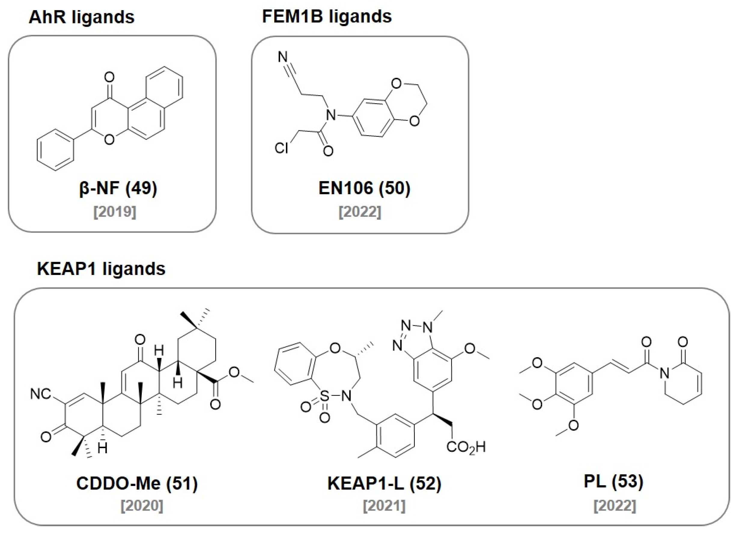
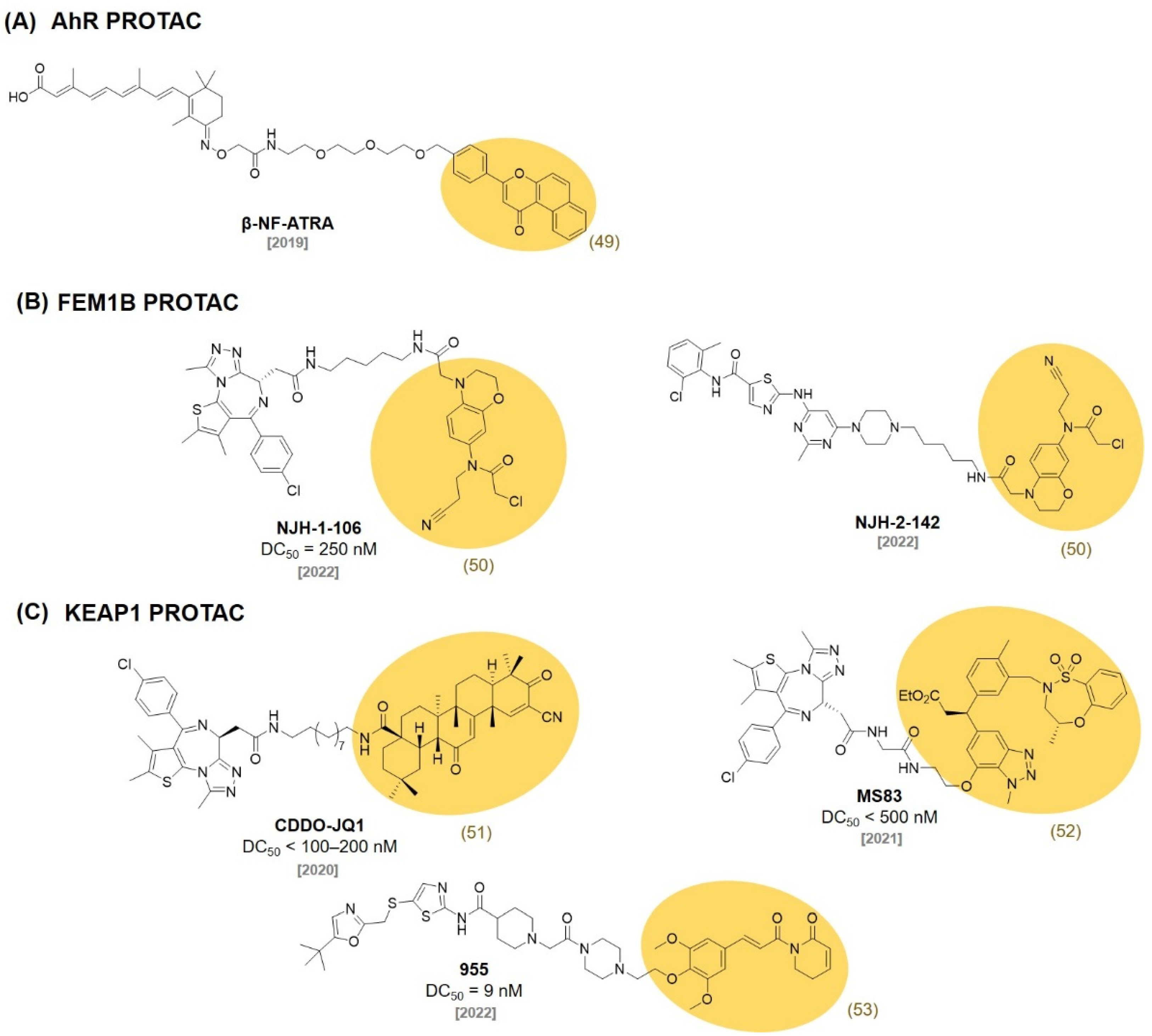
In 2019, the Naito group developed a novel PROTAC recruiting the aryl hydrocarbon receptor (AhR) E3 ligase complex. They conjugated an AhR ligand (β-NF, 49) (Figure 16) with ATRA resulting in a chimeric molecule β-NF-ATRA (Figure 17). β-NF-ATRA, a PROTAC recruiting CRABPs, induced the degradation of CRABPI and CRABPII in an AhR-dependent manner via the ubiquitin–proteasome pathway [86].

9. FEM1B Ligands

The CUL2 E3 ligase FEM1B was recently discovered as an important regulator of the cellular response to reductive stress. In 2022, the Nomura group discovered a chloroacetamide-based covalent ligand, EN106 (50) (Figure 16), as a FEM1B ligand (IC50 = 2.2 μM). They found the formation of a direct covalent bond between EN106 (50) and a cysteine residue on FEM1B by ABPP. They demonstrated that an EN106 (50)-based PROTAC can be used for the FEM1B recruitment toward target protein degradations. The conjugation of EN106 (50) to JQ1 and dasatinib generated NJH-1-106 (DC50 = 250 nM) and NJH-2-142 and showed the successful degradation of BRD4 and BCR-ABL (Figure 17) [87].

10. KEAP1 Ligands

Kelch-like ECH-associated protein-1 (KEAP1) has been known to interact with nuclear factor erythroid 2-related factor-2 (Nrf2) to regulate cellular protective proteins. Therefore, the discovery of the protein–protein interaction inhibitor of KEAP1-Nrf2 has attracted attentions for the treatment of stress-related diseases [88]. In 2020, the Nomura group reported a reversible covalent binding PROTAC using a known KEAP1 ligand bardoxolone methyl (CDDO-Me, 51) (Figure 16). They synthesized a bardoxolone-based PROTAC, CDDO-JQ1 (Figure 17) by conjugating bardoxolone to the BET inhibitor JQ1 (DC50 < 100–200 nM). They found that the deletion of a Michael acceptor in CDDO-JQ1 reduced the BRD4 degradation activity. This indicates that the formation of a reversible covalent bond between the cysteine of KEAP1 and CDDO-Me (51) is essential for the recruitment of BRD4 to the KEAP1 E3 ligase [89].
In 2021, the Jin group reported that the E3 ligase KEAP1 can be utilized to develop PROTACs using a highly selective and noncovalent KEAP1 ligand. They developed a KEAP1-recruiting PROTAC, MS83, using the previously reported KEAP1 small molecule ligand (52, KD = 1.3 nM) [90]. MS83 showed a more durable degradation of BRD3 and BRD4 than dBET1 in MDA-MB-468 cells (DC50 < 500 nM) [91].
In 2022, the Lv group discovered a nature product, Piperlongumine (PL, 53), as an E3 ligase ligand. They first confirmed that PL (53) was bound to multiple E3 ligases with a competitive ABPP assay. They synthesized PROTAC, 955, by coupling PL (53) with a CDK9 selective inhibitor (SNS-032) and observed the proteasomal degradation of CDK9 by 955 (DC50 = 9 nM). Interestingly, they identified that KEAP1 was the only E3 ligase protein recruited by 955 via a covalent attachment of PL using the TurboID-bait assay [92]. This observation suggested that E3 ligase selectivity could be changed after the conjugation of the E3 ligase ligand to a target protein ligand.

11. Conclusions

PROTAC has been developed as a new strategy for disease treatment in the chemical biology community over the past 20 years. PROTACs harness an intracellular proteolytic system for the degradation of a POI. This strategy has emerged as an alternative to overcome the limitations of conventional drug discovery by targeting undruggable proteins. For effective TPD, the selection of E3 ligase ligands and target protein ligands is critical for PROTAC design. Although a number of ligands for various proteins have been reported during the traditional drug discovery campaigns so far, only a few E3 ligase ligands are currently available for TPD. For those reasons, most of the PROTAC research has focused on the demonstration of the TPD concept for the various drug target proteins using CRBN or VHL ligands during the last decades. However, the desired target protein degradations often are compromised due to the cell type or tissue type dependent expression profiles of CRBN or VHL. Moreover, resistance to CRBN- or VHL-based PROTACs has been recently observed. Therefore, the discovery of ligands for new E3 ligases has drawn attention as they can be used for effective PROTAC development. In addition, there is still a lack of understanding of the previously developed PROTACs. For example, a given drug molecule connected to different E3 ligase ligands has shown to exhibit different PROTAC efficiencies [45,74], target selectivities [64], and drug resistance profiles in various cancer cells [4]. Although there is a report on the systematic approach taken to select the E3 ligase ligand in PROTAC designs, it is restricted to the kinase degraders and only three E3 ligase ligands were studied [93]. Their kinase-targeting PROTACs mostly prefer CRBN ligands over VHL or IAP ligands for the efficient protein degradation. Additional studies on other protein families are needed for a comprehensive understanding of E3 ligase ligand selection for efficient protein degradation. This suggests that the ternary complexes that are recruited by bifunctional PROTACs could be susceptible to ligand orientation, the ligand’s hydrophobicity, and the physicochemical properties of the chemical linkers. Further studies related to various ternary complexes should provide clearer pictures on future PROTAC designs.
Herein, we reviewed the discovery of various E3 ligase ligands and their applications for the design of PROTACs. Considering more than 600 types of E3 ligases, the development of ligands for various E3 ligases would involve the expansion of the toolbox in TPD to overcome the current limitation of PROTACs. Additionally, the cell or tissue type specific unexplored E3 ligases would be the basis for a new type of PROTAC, which could control a certain protein in a spatially specific way. The spatial protein degradation in lesions would treat diseases very effectively without toxicity or side effects in normal tissues. Therefore, the discovery of novel E3 ligase ligands would be an important goal to expand the landscape of PROTACs toward promising therapeutics in the future.

Author Contributions

Conceptualization, J.L., Y.L., and J.P.; resources, J.P.; writing—original draft preparation, J.L., Y.L., Y.M.J., J.H.P., H.S.Y., and J.P.; writing—review and editing, J.L., Y.L., Y.M.J., J.H.P., H.S.Y., and J.P.; visualization, J.L., Y.L., and J.P.; supervision, J.P.; funding acquisition, J.P. All authors have read and agreed to the published version of the manuscript.

Funding

This work was supported by the Basic Science Research Program (NRF-2019R1C1C1008792 and 2022R1F1A1076477) and the Basic Research Lab Program (NRF-2020R1A4A1016093) through the National Research Foundation of Korea funded by the Ministry of Science, ICT and Future Planning. This work was also supported by a 2019 Research Grant from Kangwon National University and by a Korea Basic Science Institute (KBSI) National Research Facilities and Equipment Center (NFEC) grant (2019R1A6C1010006) from the Ministry of Education, South Korea.

Institutional Review Board Statement

Not applicable.

Informed Consent Statement

Not applicable.

Data Availability Statement

Not applicable.

Conflicts of Interest

The authors declare no conflict of interest.

References

  1. Nabet, B.; Roberts, J.M.; Buckley, D.L.; Paulk, J.; Dastjerdi, S.; Yang, A.; Leggett, A.L.; Erb, M.A.; Lawlor, M.A.; Souza, A.; et al. The dTAG system for immediate and target-specific protein degradation. Nat. Chem. Biol. 2018, 14, 431–441. [Google Scholar] [CrossRef] [PubMed]
  2. Röth, S.; Fulcher, L.J.; Sapkota, G.P. Advances in targeted degradation of endogenous proteins. Cell Mol. Life Sci. 2019, 76, 2761–2777. [Google Scholar] [CrossRef] [Green Version]
  3. Prozzillo, Y.; Fattorini, G.; Santopietro, M.V.; Suglia, L.; Ruggiero, A.; Ferreri, D.; Messina, G. Targeted Protein Degradation Tools: Overview and Future Perspectives. Biology 2020, 9, 421. [Google Scholar] [CrossRef] [PubMed]
  4. Ottis, P.; Palladino, C.; Thienger, P.; Britschgi, A.; Heichinger, C.; Berrera, M.; Julien-Laferriere, A.; Roudnicky, F.; Kam-Thong, T.; Bischoff, J.R.; et al. Cellular Resistance Mechanisms to Targeted Protein Degradation Converge Toward Impairment of the Engaged Ubiquitin Transfer Pathway. ACS Chem. Biol. 2019, 14, 2215–2223. [Google Scholar] [CrossRef] [PubMed]
  5. Zhang, L.; Riley-Gillis, B.; Vijay, P.; Shen, Y. Acquired Resistance to BET-PROTACs (Proteolysis-Targeting Chimeras) Caused by Genomic Alterations in Core Components of E3 Ligase Complexes. Mol. Cancer Ther. 2019, 18, 1302–1311. [Google Scholar] [CrossRef] [PubMed] [Green Version]
  6. Alabi, S.B.; Crews, C.M. Major advances in targeted protein degradation: PROTACs, LYTACs, and MADTACs. J. Biol. Chem. 2021, 296, 100647. [Google Scholar] [CrossRef] [PubMed]
  7. Kumar, V.; Harjai, K.; Chhibber, S. Thalidomide treatment modulates macrophage pro-inflammatory function and cytokine levels in Klebsiella pneumoniae B5055 induced pneumonia in BALB/c mice. Int. Immunopharmacol. 2010, 10, 777–783. [Google Scholar] [CrossRef]
  8. Singhal, S.; Mehta, J.; Desikan, R.; Ayers, D.; Roberson, P.; Eddlemon, P.; Munshi, N.; Anaissie, E.; Wilson, C.; Dhodapkar, M. Antitumor activity of thalidomide in refractory multiple myeloma. N. Engl. J. Med. 1999, 341, 1565–1571. [Google Scholar] [CrossRef] [Green Version]
  9. Krönke, J.; Udeshi, N.D.; Narla, A.; Grauman, P.; Hurst, S.N.; McConkey, M.; Svinkina, T.; Heckl, D.; Comer, E.; Li, X.; et al. Lenalidomide causes selective degradation of IKZF1 and IKZF3 in multiple myeloma cells. Science 2014, 343, 301–305. [Google Scholar] [CrossRef] [Green Version]
  10. Krönke, J.; Fink, E.C.; Hollenbach, P.W.; MacBeth, K.J.; Hurst, S.N.; Udeshi, N.D.; Chamberlain, P.P.; Mani, D.R.; Man, H.W.; Gandhi, A.K.; et al. Lenalidomide induces ubiquitination and degradation of CK1α in del(5q) MDS. Nature 2015, 523, 183–188. [Google Scholar] [CrossRef]
  11. Ito, T.; Ando, H.; Suzuki, T.; Ogura, T.; Hotta, K.; Imamura, Y.; Yamaguchi, Y.; Handa, H. Identification of a Primary Target of Thalidomide Teratogenicity. Science 2010, 327, 1345–1350. [Google Scholar] [CrossRef] [PubMed] [Green Version]
  12. Zhu, Y.X.; Braggio, E.; Shi, C.-X.; Bruins, L.A.; Schmidt, J.E.; Van Wier, S.; Chang, X.-B.; Bjorklund, C.C.; Fonseca, R.; Bergsagel, P.L.; et al. Cereblon expression is required for the antimyeloma activity of lenalidomide and pomalidomide. Blood 2011, 118, 4771–4779. [Google Scholar] [CrossRef] [PubMed]
  13. Fischer, E.S.; Böhm, K.; Lydeard, J.R.; Yang, H.; Stadler, M.B.; Cavadini, S.; Nagel, J.; Serluca, F.; Acker, V.; Lingaraju, G.M.; et al. Structure of the DDB1–CRBN E3 ubiquitin ligase in complex with thalidomide. Nature 2014, 512, 49–53. [Google Scholar] [CrossRef] [PubMed] [Green Version]
  14. Dong, G.; Ding, Y.; He, S.; Sheng, C. Molecular Glues for Targeted Protein Degradation: From Serendipity to Rational Discovery. J. Med. Chem. 2021, 64, 10606–10620. [Google Scholar] [CrossRef] [PubMed]
  15. Lu, J.; Qian, Y.; Altieri, M.; Dong, H.; Wang, J.; Raina, K.; Hines, J.; Winkler, J.D.; Crew, A.P.; Coleman, K.; et al. Hijacking the E3 Ubiquitin Ligase Cereblon to Efficiently Target BRD4. Chem. Biol. 2015, 22, 755–763. [Google Scholar] [CrossRef] [Green Version]
  16. Winter, G.E.; Buckley, D.L.; Paulk, J.; Roberts, J.M.; Souza, A.; Dhe-Paganon, S.; Bradner, J.E. Phthalimide conjugation as a strategy for in vivo target protein degradation. Science 2015, 348, 1376–1381. [Google Scholar] [CrossRef] [Green Version]
  17. Burslem, G.M.; Ottis, P.; Jaime-Figueroa, S.; Morgan, A.; Cromm, P.M.; Toure, M.; Crews, C.M. Efficient Synthesis of Immunomodulatory Drug Analogues Enables Exploration of Structure-Degradation Relationships. ChemMedChem 2018, 13, 1508–1512. [Google Scholar] [CrossRef]
  18. De Wispelaere, M.; Du, G.; Donovan, K.A.; Zhang, T.; Eleuteri, N.A.; Yuan, J.C.; Kalabathula, J.; Nowak, R.P.; Fischer, E.S.; Gray, N.S.; et al. Small molecule degraders of the hepatitis C virus protease reduce susceptibility to resistance mutations. Nat. Commun. 2019, 10, 3468. [Google Scholar] [CrossRef] [Green Version]
  19. Kargbo, R.B. PROTAC Molecules for the Treatment of Autoimmune Disorders. ACS Med. Chem. Lett. 2019, 10, 276–277. [Google Scholar] [CrossRef] [Green Version]
  20. Sabnis, R.W. Bifunctional Compounds as SMARCA2 Degraders for Treating Cancer. ACS Med. Chem. Lett. 2022, 13, 15–16. [Google Scholar] [CrossRef]
  21. Kim, S.A.; Go, A.; Jo, S.-H.; Park, S.J.; Jeon, Y.U.; Kim, J.E.; Lee, H.K.; Park, C.H.; Lee, C.-O.; Park, S.G.; et al. A novel cereblon modulator for targeted protein degradation. Eur. J. Med. Chem. 2019, 166, 65–74. [Google Scholar] [CrossRef] [PubMed]
  22. Takwale, A.D.; Jo, S.-H.; Jeon, Y.U.; Kim, H.S.; Shin, C.H.; Lee, H.K.; Ahn, S.; Lee, C.O.; Ha, J.D.; Kim, J.-H.; et al. Design and characterization of cereblon-mediated androgen receptor proteolysis-targeting chimeras. Eur. J. Med. Chem. 2020, 208, 112769. [Google Scholar] [CrossRef] [PubMed]
  23. Gao, X.; Burris III, H.A.; Vuky, J.; Dreicer, R.; Sartor, A.O.; Sternberg, C.N.; Percent, I.J.; Hussain, M.H.A.; Kalebasty, A.R.; Shen, J.; et al. Phase 1/2 study of ARV-110, an androgen receptor (AR) PROTAC degrader, in metastatic castration-resistant prostate cancer (mCRPC). J. Clin. Oncol. 2022, 40, 17. [Google Scholar] [CrossRef]
  24. Hamilton, E.P.; Schott, A.F.; Nanda, R.; Lu, H.; Keung, C.F.; Gedrich, R.; Parameswaran, J.; Han, H.S.; Hurvitz, S.A. ARV-471, an estrogen receptor (ER) PROTACdegrader, combined with palbociclib in advanced ER+/human epidermal growth factor receptor 2–negative (HER2-) breast cancer: Phase 1b cohort (part C) of a phase 1/2 study. J. Clin. Oncol. 2022, 40, TPS1120. [Google Scholar] [CrossRef]
  25. Mainolfi, N.; JI, N.; Kluge, A.F. CRBN Ligands and Uses Thereof. International Patent WO 2019140387 A1, 18 July 2019. [Google Scholar]
  26. Kargbo, R.B. PROTAC Degradation of IRAK4 for the Treatment of Cancer. ACS Med. Chem. Lett. 2019, 10, 1370–1371. [Google Scholar] [CrossRef] [Green Version]
  27. Kargbo, R.B. PROTAC-Mediated Degradation of Bruton’s Tyrosine Kinase as a Therapeutic Strategy for Cancer. ACS Med. Chem. Lett. 2021, 12, 688–689. [Google Scholar] [CrossRef]
  28. Min, J.; Mayasundari, A.; Keramatnia, F.; Jonchere, B.; Yang, S.W.; Jarusiewicz, J.; Actis, M.; Das, S.; Young, B.; Slavish, J.; et al. Phenyl-Glutarimides: Alternative Cereblon Binders for the Design of PROTACs. Angew. Chem. Int. Ed. 2021, 60, 26663–26670. [Google Scholar] [CrossRef]
  29. Alcock, L.J.; Chang, Y.; Jarusiewicz, J.A.; Actis, M.; Nithianantham, S.; Mayasundari, A.; Min, J.; Maxwell, D.; Hunt, J.; Smart, B.; et al. Development of Potent and Selective Janus Kinase 2/3 Directing PG–PROTACs. ACS Med. Chem. Lett. 2022, 13, 475–482. [Google Scholar] [CrossRef]
  30. Wang, S.; Xu, T.; Wang, M.; Hu, J.; Han, X.; Xiang, W.; REJ, R. Cereblon E3 Ligase Inhibitors. International Patent WO 2021041664 A1, 4 March 2021. [Google Scholar]
  31. Kargbo, R.B. Discovery of Spirocyclic Androgen Receptor Protein Degraders for the Treatment of Prostate Cancer. ACS Med. Chem. Lett. 2021, 12, 1635–1636. [Google Scholar] [CrossRef]
  32. Sabnis, R.W. BRD9 Bifunctional Degraders for Treating Cancer. ACS Med. Chem. Lett. 2021, 12, 1879–1880. [Google Scholar] [CrossRef]
  33. Buckley, D.L.; Van Molle, I.; Gareiss, P.C.; Tae, H.S.; Michel, J.; Noblin, D.J.; Jorgensen, W.L.; Ciulli, A.; Crews, C.M. Targeting the von Hippel–Lindau E3 Ubiquitin Ligase Using Small Molecules To Disrupt the VHL/HIF-1α Interaction. J. Am. Chem. Soc. 2012, 134, 4465–4468. [Google Scholar] [CrossRef]
  34. Galdeano, C.; Gadd, M.S.; Soares, P.; Scaffidi, S.; Van Molle, I.; Birced, I.; Hewitt, S.; Dias, D.M.; Ciulli, A. Structure-Guided Design and Optimization of Small Molecules Targeting the Protein–Protein Interaction between the von Hippel–Lindau (VHL) E3 Ubiquitin Ligase and the Hypoxia Inducible Factor (HIF) Alpha Subunit with in Vitro Nanomolar Affinities. J. Med. Chem. 2014, 57, 8657–8663. [Google Scholar] [CrossRef] [PubMed]
  35. Zengerle, M.; Chan, K.-H.; Ciulli, A. Selective Small Molecule Induced Degradation of the BET Bromodomain Protein BRD4. ACS Chem. Biol. 2015, 10, 1770–1777. [Google Scholar] [CrossRef] [PubMed] [Green Version]
  36. Buckley, D.L.; Raina, K.; Darricarrere, N.; Hines, J.; Gustafson, J.L.; Smith, I.E.; Miah, A.H.; Harling, J.D.; Crews, C.M. HaloPROTACS: Use of Small Molecule PROTACs to Induce Degradation of HaloTag Fusion Proteins. ACS Chem. Biol. 2015, 10, 1831–1837. [Google Scholar] [CrossRef] [PubMed] [Green Version]
  37. Smith, B.E.; Wang, S.L.; Jaime-Figueroa, S.; Harbin, A.; Wang, J.; Hamman, B.D.; Crews, C.M. Differential PROTAC substrate specificity dictated by orientation of recruited E3 ligase. Nat. Commun. 2019, 10, 131. [Google Scholar] [CrossRef] [Green Version]
  38. Soares, P.; Gadd, M.S.; Frost, J.; Galdeano, C.; Ellis, L.; Epemolu, O.; Rocha, S.; Read, K.D.; Ciulli, A. Group-Based Optimization of Potent and Cell-Active Inhibitors of the von Hippel–Lindau (VHL) E3 Ubiquitin Ligase: Structure–Activity Relationships Leading to the Chemical Probe (2S,4R)-1-((S)-2-(1-Cyanocyclopropanecarboxamido)-3,3-dimethylbutanoyl)-4-hydroxy-N-(4-(4-methylthiazol-5-yl)benzyl)pyrrolidine-2-carboxamide (VH298). J. Med. Chem. 2018, 61, 599–618. [Google Scholar]
  39. Zoppi, V.; Hughes, S.J.; Maniaci, C.; Testa, A.; Gmaschitz, T.; Wieshofer, C.; Koegl, M.; Riching, K.M.; Daniels, D.L.; Spallarossa, A.; et al. Iterative Design and Optimization of Initially Inactive Proteolysis Targeting Chimeras (PROTACs) Identify VZ185 as a Potent, Fast, and Selective von Hippel–Lindau (VHL) Based Dual Degrader Probe of BRD9 and BRD7. J. Med. Chem. 2019, 62, 699–726. [Google Scholar] [CrossRef]
  40. Smalley, J.P.; Baker, I.M.; Pytel, W.A.; Lin, L.-Y.; Bowman, K.J.; Schwabe, J.W.R.; Cowley, S.M.; Hodgkinson, J.T. Optimization of Class I Histone Deacetylase PROTACs Reveals that HDAC1/2 Degradation is Critical to Induce Apoptosis and Cell Arrest in Cancer Cells. J. Med. Chem. 2022, 65, 5642–5659. [Google Scholar] [CrossRef]
  41. Testa, A.; Lucas, X.; Castro, G.V.; Chan, K.-H.; Wright, J.E.; Runcie, A.C.; Gadd, M.S.; Harrison, W.T.A.; Ko, E.-J.; Fletcher, D.; et al. 3-Fluoro-4-hydroxyprolines: Synthesis, Conformational Analysis, and Stereoselective Recognition by the VHL E3 Ubiquitin Ligase for Targeted Protein Degradation. J. Am. Chem. Soc. 2018, 140, 9299–9313. [Google Scholar] [CrossRef] [Green Version]
  42. Raina, K.; Lu, J.; Qian, Y.; Altieri, M.; Gordon, D.; Rossi, A.M.K.; Wang, J.; Chen, X.; Dong, H.; Siu, K.; et al. PROTAC-induced BET protein degradation as a therapy for castration-resistant prostate cancer. Proc. Natl. Acad. Sci. USA 2016, 113, 7124–7129. [Google Scholar] [CrossRef] [Green Version]
  43. Han, X.; Wang, C.; Qin, C.; Xiang, W.; Fernandez-Salas, E.; Yang, C.-Y.; Wang, M.; Zhao, L.; Xu, T.; Chinnaswamy, K.; et al. Discovery of ARD-69 as a Highly Potent Proteolysis Targeting Chimera (PROTAC) Degrader of Androgen Receptor (AR) for the Treatment of Prostate Cancer. J. Med. Chem. 2019, 62, 941–964. [Google Scholar] [CrossRef] [PubMed]
  44. Han, X.; Zhao, L.; Xiang, W.; Qin, C.; Miao, B.; Xu, T.; Wang, M.; Yang, C.-Y.; Chinnaswamy, K.; Stuckey, J.; et al. Discovery of Highly Potent and Efficient PROTAC Degraders of Androgen Receptor (AR) by Employing Weak Binding Affinity VHL E3 Ligase Ligands. J. Med. Chem. 2019, 62, 11218–11231. [Google Scholar] [CrossRef] [PubMed]
  45. Hu, J.; Hu, B.; Wang, M.; Xu, F.; Miao, B.; Yang, C.-Y.; Wang, M.; Liu, Z.; Hayes, D.F.; Chinnaswamy, K.; et al. Discovery of ERD-308 as a Highly Potent Proteolysis Targeting Chimera (PROTAC) Degrader of Estrogen Receptor (ER). J. Med. Chem. 2019, 62, 1420–1442. [Google Scholar] [CrossRef] [PubMed]
  46. Varfolomeev, E.; Blankenship, J.W.; Wayson, S.M.; Fedorova, A.V.; Kayagaki, N.; Garg, P.; Zobel, K.; Dynek, J.N.; Elliott, L.O.; Wallweber, H.J.; et al. IAP Antagonists Induce Autoubiquitination of c-IAPs, NF-kB Activation, and TNFa-Dependent Apoptosis. Cell 2007, 131, 669–681. [Google Scholar] [CrossRef] [PubMed] [Green Version]
  47. Sekine, K.; Takubo, K.; Kikuchi, R.; Nishimoto, M.; Kitagawa, M.; Abe, F.; Nishikawa, K.; Tsuruo, T.; Naito, M. Small Molecules Destabilize cIAP1 by Activating Auto-ubiquitylation. J. Biol. Chem. 2008, 283, 8961–8968. [Google Scholar] [CrossRef] [PubMed] [Green Version]
  48. Itoh, Y.; Ishikawa, M.; Naito, M.; Hashimoto, Y. Protein Knockdown Using Methyl Bestatin−Ligand Hybrid Molecules: Design and Synthesis of Inducers of Ubiquitination-Mediated Degradation of Cellular Retinoic Acid-Binding Proteins. J. Am. Chem. Soc. 2010, 132, 5820–5826. [Google Scholar] [CrossRef]
  49. Itoh, Y.; Ishikawa, M.; Kitaguchi, R.; Okuhira, K.; Naito, M.; Hashimoto, Y. Double protein knockdown of cIAP1 and CRABP-II using a hybrid molecule consisting of ATRA and IAPs antagonist. Bioorg. Med. Chem. Lett. 2012, 22, 4453–4457. [Google Scholar] [CrossRef]
  50. Ohoka, N.; Okuhira, K.; Ito, M.; Nagai, K.; Shibata, N.; Hattori, T.; Ujikawa, O.; Shimokawa, K.; Sano, O.; Koyama, R.; et al. In Vivo Knockdown of Pathogenic Proteins via Specific and Nongenetic Inhibitor of Apoptosis Protein (IAP)-dependent Protein Erasers (SNIPERs). J. Biol. Chem. 2017, 292, 4556–4570. [Google Scholar] [CrossRef] [Green Version]
  51. Ohoka, N.; Ujikawa, O.; Shimokawa, K.; Sameshima, T.; Shibata, N.; Hattori, T.; Nara, H.; Cho, N.; Naito, M. Different Degradation Mechanisms of Inhibitor of Apoptosis Proteins (IAPs) by the Specific and Nongenetic IAP-Dependent Protein Eraser (SNIPER). Chem. Pharm. Bull. 2019, 67, 203–209. [Google Scholar] [CrossRef] [Green Version]
  52. Shimokawa, K.; Shibata, N.; Sameshima, T.; Miyamoto, N.; Ujikawa, O.; Nara, H.; Ohoka, N.; Hattori, T.; Cho, N.; Naito, M. Targeting the Allosteric Site of Oncoprotein BCR-ABL as an Alternative Strategy for Effective Target Protein Degradation. ACS Med. Chem. Lett. 2017, 8, 1042–1047. [Google Scholar] [CrossRef]
  53. Flygare, J.A.; Beresini, M.; Budha, N.; Chan, H.; Chan, I.T.; Cheeti, S.; Cohen, F.; Deshayes, K.; Doerner, K.; Eckhardt, S.G.; et al. Discovery of a Potent Small-Molecule Antagonist of Inhibitor of Apoptosis (IAP) Proteins and Clinical Candidate for the Treatment of Cancer (GDC-0152). J. Med. Chem. 2012, 55, 4101–4113. [Google Scholar] [CrossRef] [PubMed]
  54. Hennessy, E.J.; Saeh, J.C.; Sha, L.; MacIntyre, T.; Wang, H.; Larsen, N.A.; Aquila, B.M.; Ferguson, A.D.; Laing, N.M.; Omer, C.A. Discovery of aminopiperidine-based Smac mimetics as IAP antagonists. Bioorg. Med. Chem. Lett. 2012, 22, 1690–1694. [Google Scholar] [CrossRef] [PubMed]
  55. Vamos, M.; Welsh, K.; Finlay, D.; Lee, P.S.; Mace, P.D.; Snipas, S.J.; Gonzalez, M.L.; Ganji, S.R.; Ardecky, R.J.; Riedl, S.J.; et al. Expedient Synthesis of Highly Potent Antagonists of Inhibitor of Apoptosis Proteins (IAPs) with Unique Selectivity for ML-IAP. ACS Chem. Biol. 2013, 8, 725–732. [Google Scholar] [CrossRef] [PubMed] [Green Version]
  56. Zhang, X.; He, Y.; Zhang, P.; Budamagunta, V.; Lv, D.; Thummuri, D.; Yang, Y.; Pei, J.; Yuan, Y.; Zhou, D.; et al. Discovery of IAP-recruiting BCL-XL PROTACs as potent degraders across multiple cancer cell lines. Eur. J. Med. Chem. 2020, 199, 112397. [Google Scholar] [CrossRef]
  57. Kim, K.S.; Zhang, L.; Williams, D.; Perez, H.L.; Stang, E.; Borzilleri, R.M.; Posy, S.; Lei, M.; Chaudhry, C.; Emanuel, S.; et al. Discovery of tetrahydroisoquinoline-based bivalent heterodimeric IAP antagonists. Bioorg. Med. Chem. Lett. 2014, 24, 5022–5029. [Google Scholar] [CrossRef]
  58. Schiemer, J.; Horst, R.; Meng, Y.; Montgomery, J.I.; Xu, Y.; Feng, X.; Borzilleri, K.; Uccello, D.P.; Leverett, C.; Brown, S.; et al. Snapshots and ensembles of BTK and cIAP1 protein degrader ternary complexes. Nat. Chem. Biol. 2021, 17, 152–160. [Google Scholar] [CrossRef]
  59. Ohoka, N.; Morita, Y.; Nagai, K.; Shimokawa, K.; Ujikawa, O.; Fujimori, I.; Ito, M.; Hayase, Y.; Okuhira, K.; Shibata, N.; et al. Derivatization of inhibitor of apoptosis protein (IAP) ligands yields improved inducers of estrogen receptor alpha degradation. J. Biol. Chem. 2018, 293, 6776–6790. [Google Scholar] [CrossRef] [Green Version]
  60. Tamanini, E.; Buck, I.M.; Chessari, G.; Chiarparin, E.; Day, J.E.H.; Frederickson, M.; Griffiths-Jones, C.M.; Hearn, K.; Heightman, T.D.; Iqbal, A.; et al. Discovery of a Potent Nonpeptidomimetic, Small-Molecule Antagonist of Cellular Inhibitor of Apoptosis Protein 1 (cIAP1) and X-Linked Inhibitor of Apoptosis Protein (XIAP). J. Med. Chem. 2017, 60, 4611–4625. [Google Scholar] [CrossRef]
  61. Jiang, B.; Wang, E.S.; Donovan, K.A.; Liang, Y.; Fischer, E.S.; Zhang, T.; Gray, N.S. Development of Dual and Selective Degraders of Cyclin-Dependent Kinases 4 and 6. Angew. Chem. Int. Ed. 2019, 58, 6321–6326. [Google Scholar] [CrossRef]
  62. Rana, S.; Bendjennat, M.; Kour, S.; King, H.M.; Kizhake, S.; Zahid, M.; Natarajan, A. Selective degradation of CDK6 by a palbociclib based PROTAC. Bioorg. Med. Chem. Lett. 2019, 29, 1375–1379. [Google Scholar] [CrossRef]
  63. Su, S.; Yang, Z.; Gao, H.; Yang, H.; Zhu, S.; An, Z.; Wang, J.; Li, Q.; Chandarlapaty, S.; Deng, H.; et al. Potent and Preferential Degradation of CDK6 via Proteolysis Targeting Chimera Degraders. J. Med. Chem. 2019, 62, 7575–7582. [Google Scholar] [CrossRef] [PubMed]
  64. Anderson, N.A.; Cryan, J.; Ahmed, A.; Dai, H.; McGonagle, G.A.; Rozier, C.; Benowitz, A.B. Selective CDK6 degradation mediated by cereblon, VHL, and novel IAP-recruiting PROTACs. Bioorg. Med. Chem. Lett. 2020, 30, 127106. [Google Scholar] [CrossRef] [PubMed]
  65. Biswas, A.; Liu, Y.-J.; Hao, L.; Mizoguchi, A.; Salzman, N.H.; Bevins, C.L.; Kobayashi, K.S. Induction and rescue of Nod2-dependent Th1-driven granulomatous inflammation of the ileum. Proc. Natl. Acad. Sci. USA 2010, 107, 14739–14744. [Google Scholar] [CrossRef] [Green Version]
  66. Sato, H.; Williams, H.R.T.; Spagnolo, P.; Abdallah, A.; Ahmad, T.; Orchard, T.R.; Copley, S.J.; Desai, S.R.; Wells, A.U.; Du Bois, R.M.; et al. CARD15/NOD2 polymorphisms are associated with severe pulmonary sarcoidosis. Eur. Respir. J. 2010, 35, 324–330. [Google Scholar] [CrossRef] [PubMed]
  67. Shaw, P.J.; Barr, M.J.; Lukens, J.R.; McGargill, M.A.; Chi, H.; Mak, T.W.; Kanneganti, T.-D. Signaling via the RIP2 Adaptor Protein in Central Nervous System-Infiltrating Dendritic Cells Promotes Inflammation and Autoimmunity. Immunity 2011, 34, 75–84. [Google Scholar] [CrossRef] [PubMed] [Green Version]
  68. Miah, A.H.; Smith, I.E.D.; Rackham, M.; Mares, A.; Thawani, A.R.; Nagilla, R.; Haile, P.A.; Votta, B.J.; Gordon, L.J.; Watt, G.; et al. Optimization of a Series of RIPK2 PROTACs. J. Med. Chem. 2021, 64, 12978–13003. [Google Scholar] [CrossRef] [PubMed]
  69. Vassilev, L.T.; Vu, B.T.; Graves, B.; Carvajal, D.; Podlaski, F.; Filipovic, Z.; Kong, N.; Kammlott, U.; Lukacs, C.; Klein, C.; et al. In Vivo Activation of the p53 Pathway by Small-Molecule Antagonists of MDM2. Science 2004, 303, 844–848. [Google Scholar] [CrossRef] [Green Version]
  70. Schneekloth, A.R.; Pucheault, M.; Tae, H.S.; Crews, C.M. Targeted intracellular protein degradation induced by a small molecule: En route to chemical proteomics. Bioorg. Med. Chem. Lett. 2008, 18, 5904–5908. [Google Scholar] [CrossRef] [Green Version]
  71. He, S.; Ma, J.; Fang, Y.; Liu, Y.; Wu, S.; Dong, G.; Wang, W.; Sheng, C. Homo-PROTAC mediated suicide of MDM2 to treat non-small cell lung cancer. Acta Pharm. Sin. B. 2021, 11, 1617–1628. [Google Scholar] [CrossRef]
  72. Vu, B.; Wovkulich, P.; Pizzolato, G.; Lovey, A.; Ding, Q.; Jiang, N.; Liu, J.-J.; Zhao, C.; Glenn, K.; Wen, Y.; et al. Discovery of RG7112: A Small-Molecule MDM2 Inhibitor in Clinical Development. ACS Med. Chem. Lett. 2013, 4, 466–469. [Google Scholar] [CrossRef] [Green Version]
  73. Ding, Q.; Zhang, Z.; Liu, J.-J.; Jiang, N.; Zhang, J.; Ross, T.M.; Chu, X.-J.; Bartkovitz, D.; Podlaski, F.; Janson, C.; et al. Discovery of RG7388, a Potent and Selective p53–MDM2 Inhibitor in Clinical Development. J. Med. Chem. 2013, 56, 5979–5983. [Google Scholar] [CrossRef] [PubMed]
  74. De Dominici, M.; Porazzi, P.; Xiao, Y.; Chao, A.; Tang, H.-Y.; Kumar, G.; Fortina, P.; Spinelli, O.; Rambaldi, A.; Peterson, L.F.; et al. Selective inhibition of Ph-positive ALL cell growth through kinase-dependent and -independent effects by CDK6-specific PROTACs. Blood 2020, 135, 1560–1573. [Google Scholar] [CrossRef] [PubMed]
  75. Hines, J.; Lartigue, S.; Dong, H.; Qian, Y.; Crews, C.M. MDM2-Recruiting PROTAC Offers Superior, Synergistic Antiproliferative Activity via Simultaneous Degradation of BRD4 and Stabilization of p53. Cancer Res. 2019, 79, 251–262. [Google Scholar] [CrossRef] [PubMed] [Green Version]
  76. Han, T.; Goralski, M.; Gaskill, N.; Capota, E.; Kim, J.; Ting, T.C.; Xie, Y.; Williams, N.S.; Nijhawan, D. Anticancer sulfonamides target splicing by inducing RBM39 degradation via recruitment to DCAF15. Science 2017, 356, eaal3755. [Google Scholar] [CrossRef]
  77. Du, X.; Volkov, O.A.; Czerwinski, R.M.; Tan, H.; Huerta, C.; Morton, E.R.; Rizzi, J.P.; Wehn, P.M.; Xu, R.; Nijhawan, D.; et al. Structural Basis and Kinetic Pathway of RBM39 Recruitment to DCAF15 by a Sulfonamide Molecular Glue E7820. Structure 2019, 27, 1625–1633. [Google Scholar] [CrossRef]
  78. Uehara, T.; Minoshima, Y.; Sagane, K.; Sugi, N.H.; Mitsuhashi, K.O.; Yamamoto, N.; Kamiyama, H.; Takahashi, K.; Kotake, Y.; Uesugi, M.; et al. Selective degradation of splicing factor CAPERα by anticancer sulfonamides. Nat. Chem. Biol. 2017, 13, 675–680. [Google Scholar] [CrossRef]
  79. Li, L.; Mi, D.; Pei, H.; Duan, Q.; Wang, X.; Zhou, W.; Jin, J.; Li, D.; Liu, M.; Chen, Y. In vivo target protein degradation induced by PROTACs based on E3 ligase DCAF15. Signal Transduct. Target. Ther. 2020, 5, 129. [Google Scholar] [CrossRef]
  80. Zhang, X.; Crowley, V.M.; Wucherpfennig, T.G.; Dix, M.M.; Cravatt, B.F. Electrophilic PROTACs that degrade nuclear proteins by engaging DCAF16. Nat. Chem. Biol. 2019, 15, 737–746. [Google Scholar] [CrossRef]
  81. Zhang, X.; Luukkonen, L.M.; Eissler, C.L.; Crowley, V.M.; Yamashita, Y.; Schafroth, M.A.; Kikuchi, S.; Weinstein, D.S.; Symons, K.T.; Nordin, B.E.; et al. DCAF11 Supports Targeted Protein Degradation by Electrophilic Proteolysis-Targeting Chimeras. J. Am. Chem. Soc. 2021, 143, 5141–5149. [Google Scholar] [CrossRef]
  82. Ward, C.C.; Kleinman, J.I.; Brittain, S.M.; Lee, P.S.; Chung, C.Y.S.; Kim, K.; Petri, Y.; Thomas, J.R.; Tallarico, J.A.; McKenna, J.M.; et al. Covalent Ligand Screening Uncovers a RNF4 E3 Ligase Recruiter for Targeted Protein Degradation Applications. ACS Chem. Biol. 2019, 14, 2430–2440. [Google Scholar] [CrossRef]
  83. Spradlin, J.N.; Hu, X.; Ward, C.C.; Brittain, S.M.; Jones, M.D.; Ou, L.; To, M.; Proudfoot, A.; Ornelas, E.; Woldegiorgis, M.; et al. Harnessing the anti-cancer natural product nimbolide for targeted protein degradation. Nat. Chem. Biol. 2019, 15, 747–755. [Google Scholar] [CrossRef] [PubMed]
  84. Tong, B.; Spradlin, J.N.; Novaes, L.F.T.; Zhang, E.; Hu, X.; Moeller, M.; Brittain, S.M.; McGregor, L.M.; McKenna, J.M.; Tallarico, J.A.; et al. A Nimbolide-Based Kinase Degrader Preferentially Degrades Oncogenic BCR-ABL. ACS Chem. Biol. 2020, 15, 1788–1794. [Google Scholar] [CrossRef] [PubMed]
  85. Luo, M.; Spradlin, J.N.; Boike, L.; Tong, B.; Brittain, S.M.; McKenna, J.M.; Tallarico, J.A.; Schirle, M.; Maimone, T.J.; Nomura, D.K. Chemoproteomics-enabled discovery of covalent RNF114-based degraders that mimic natural product function. Cell Chem. Biol. 2021, 28, 559–566. [Google Scholar] [CrossRef]
  86. Ohoka, N.; Tsuji, G.; Shoda, T.; Fujisato, T.; Kurihara, M.; Demizu, Y.; Naito, M. Development of Small Molecule Chimeras That Recruit AhR E3 Ligase to Target Proteins. ACS Chem. Biol. 2019, 14, 2822–2832. [Google Scholar] [CrossRef] [PubMed]
  87. Henning, N.J.; Manford, A.G.; Spradlin, J.N.; Brittain, S.M.; Zhang, E.; McKenna, J.M.; Tallarico, J.A.; Schirle, M.; Rape, M.; Nomura, D.K. Discovery of a Covalent FEM1B Recruiter for Targeted Protein Degradation Applications. J. Am. Chem. Soc. 2022, 144, 701–708. [Google Scholar] [CrossRef] [PubMed]
  88. Lee, S.; Hu, L. Nrf2 activation through the inhibition of Keap1–Nrf2 protein–protein interaction. Med. Chem. Res. 2020, 29, 846–867. [Google Scholar] [CrossRef]
  89. Tong, B.; Luo, M.; Xie, Y.; Spradlin, J.N.; Tallarico, J.A.; McKenna, J.M.; Schirle, M.; Maimone, T.J.; Nomura, D.K. Bardoxolone conjugation enables targeted protein degradation of BRD4. Sci. Rep. 2020, 10, 15543. [Google Scholar] [CrossRef]
  90. Davies, T.G.; Wixted, W.E.; Coyle, J.E.; Griffiths-Jones, C.; Hearn, K.; McMenamin, R.; Norton, D.; Rich, S.J.; Richardson, C.; Saxty, G.; et al. Monoacidic Inhibitors of the Kelch-like ECH-Associated Protein 1: Nuclear Factor Erythroid 2-Related Factor 2 (KEAP1:NRF2) Protein–Protein Interaction with High Cell Potency Identified by Fragment-Based Discovery. J. Med. Chem. 2016, 59, 3991–4006. [Google Scholar] [CrossRef]
  91. Wei, J.; Meng, F.; Park, K.-S.; Yim, H.; Velez, J.; Kumar, P.; Wang, L.; Xie, L.; Chen, H.; Shen, Y.; et al. Harnessing the E3 Ligase KEAP1 for Targeted Protein Degradation. J. Am. Chem. Soc. 2021, 143, 15073–15083. [Google Scholar] [CrossRef]
  92. Pei, J.; Xiao, Y.; Liu, X.; Hu, W.; Sobh, A.; Yuan, Y.; Zhou, S.; Hua, N.; Mackintosh, S.G.; Zhang, X.; et al. Identification of Piperlongumine (PL) as a new E3 ligase ligand to induce targeted protein degradation. bioRxiv 2022. [Google Scholar] [CrossRef]
  93. Donovan, K.A.; Ferguson, F.M.; Bushman, J.W.; Eleuteri, N.A.; Bhunia, D.; Ryu, S.; Tan, L.; Shi, K.; Yue, H.; Liu, X.; et al. Mapping the Degradable Kinome Provides a Resource for Expedited Degrader Development. Cell 2020, 183, 1714–1731. [Google Scholar] [CrossRef] [PubMed]
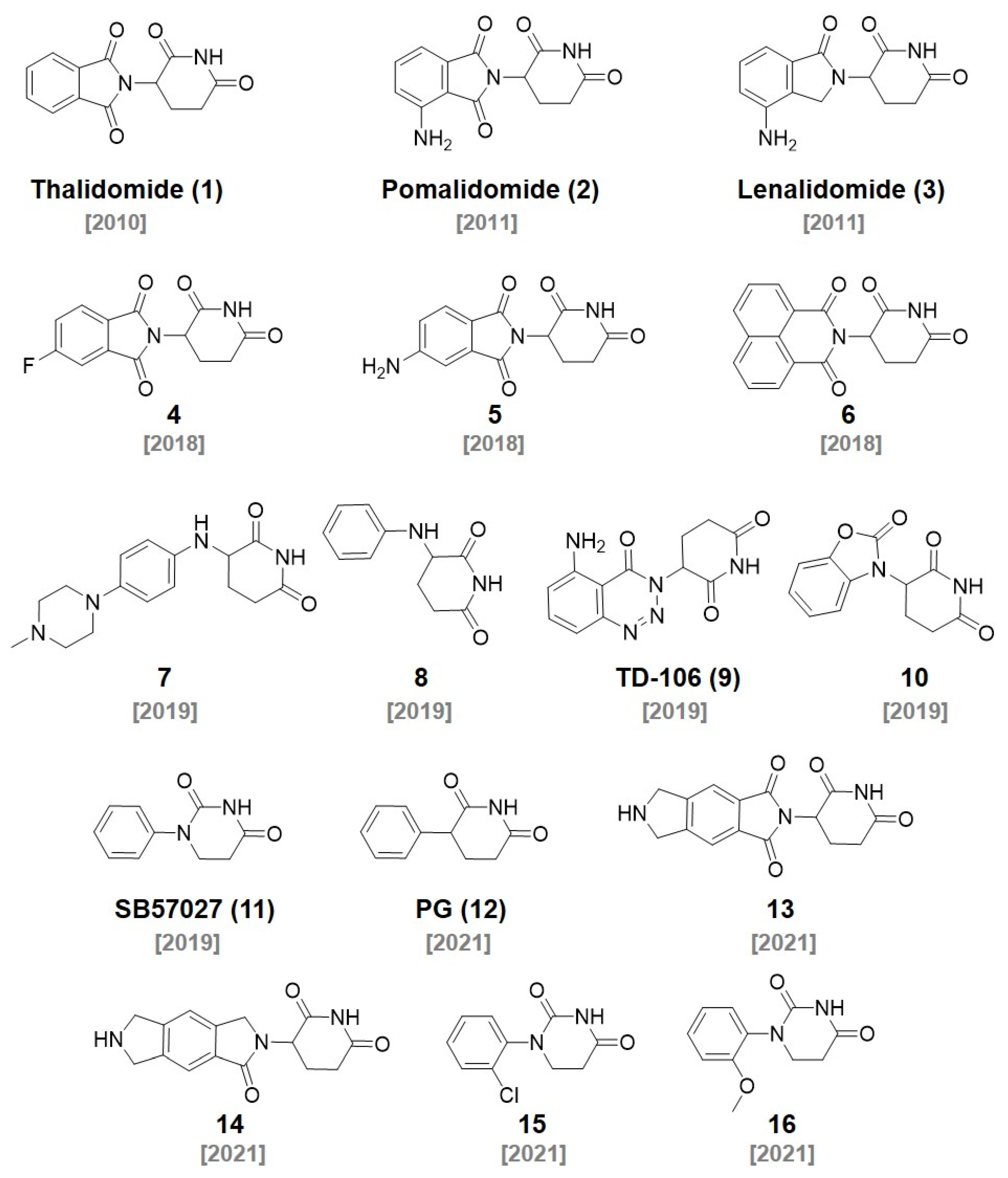
Figure 1. Chemical structures of CRBN ligands and their derivatives.
Molecules 27 06515 g001
Figure 2. (A) Chemical structure of thalidomide composed of phthalimide and glutarimide groups. (B) Schematic illustrations of thalidomide binding mode to CRBN (PDB: 4CI1). Reprinted with permission from ref. [14] (Copyright 2021 American Chemical Society).
Molecules 27 06515 g002
Figure 3. Examples of CRBN ligand-based PROTACs.
Molecules 27 06515 g003
Figure 7. Representative examples of IAP ligands.
Molecules 27 06515 g007
Figure 8. Selected examples of IAP ligand-based PROTACs.
Molecules 27 06515 g008
Figure 9. Chemical structure of GDC-0152 and its key binding modes to ML-IAP (left) and cIAP1 (right). Reprinted with permission from ref. [53] (Copyright 2012 American Chemical Society).
Molecules 27 06515 g009
Figure 10. Chemical structures of reported MDM2 ligands.
Molecules 27 06515 g010
Figure 11. Representative examples MDM2 ligand-based PROTACs.
Molecules 27 06515 g011
Figure 12. Chemical structures of recently reported DCAF ligands.
Molecules 27 06515 g012
Figure 13. Recently developed PROTACs utilizing various DCAF ligands.
Molecules 27 06515 g013
Figure 14. Recent examples of RNF ligands.
Molecules 27 06515 g014
Figure 15. RNF ligand-based PROTACs targeting RNF4 and RNF114.
Molecules 27 06515 g015
Figure 16. Recently developed AhR, FEM1B, and KEAP1 ligands.
Molecules 27 06515 g016
Figure 17. Chemical structures of PROTACs using (A) an AhR ligand, (B) FEM1B ligands, and (C) KEAP1 ligands.
Molecules 27 06515 g017
Publisher’s Note: MDPI stays neutral with regard to jurisdictional claims in published maps and institutional affiliations.