Article

First Evidence of Anti-Steatotic Action of Macrotympanain A1, an Amphibian Skin Peptide from Odorrana macrotympana

1
Department of Earth, Environmental, and Life Sciences (DISTAV), University of Genoa, Corso Europa 26, 16132 Genoa, Italy
2
Inter-University Center for the Promotion of the 3Rs Principles in Teaching & Research (Centro 3R), 56122 Pisa, Italy
3
Department of Experimental Medicine (DIMES), Section of Biochemistry, University of Genoa, Viale Benedetto XV 1, 16132 Genoa, Italy
4
Angel Consulting, Via San Senatore 14, 20122 Milano, Italy
5
Department of Experimental Medicine (DIMES), Cellular Electron Microscopy Laboratory, University of Genoa, Via Antonio de Toni 14, 16132 Genoa, Italy
*
Authors to whom correspondence should be addressed.
Molecules 2022, 27(21), 7417; https://doi.org/10.3390/molecules27217417
Submission received: 8 September 2022 / Revised: 10 October 2022 / Accepted: 27 October 2022 / Published: 1 November 2022

Abstract

:
Many different amphibian skin peptides have been characterized and proven to exert various biological actions, such as wound-healing, immunomodulatory, anti-oxidant, anti-inflammatory and anti-diabetic effects. In this work, the possible anti-steatotic effect of macrotympanain A1 (MA1) (FLPGLECVW), a skin peptide isolated from the Chinese odorous frog Odorrana macrotympana, was investigated. We used a well-established in vitro model of hepatic steatosis, consisting of lipid-loaded rat hepatoma FaO cells. In this model, a 24 h treatment with 10 µg/mL MA1 exerted a significant anti-steatotic action, being able to reduce intracellular triglyceride content. Accordingly, the number and diameter of cytosolic lipid droplets (LDs) were reduced by peptide treatment. The expression of key genes of hepatic lipid metabolism, such as PPARs and PLINs, was measured by real-time qPCR. MA1 counteracted the fatty acid-induced upregulation of PPARγ expression and increased PLIN3 expression, suggesting a role in promoting lipophagy. The present data demonstrate for the first time a direct anti-steatotic effect of a peptide from amphibian skin secretion and pave the way to further studies on the use of amphibian peptides for beneficial actions against metabolic diseases.

1. Introduction

The amphibian skin is a complex organ able to exert a multitude of vital functions related to respiration, osmoregulation, thermoregulation and defense against physical and biological environmental threats [1,2]. The importance of this organ is underlined by the presence of several glands (mucous and granular) able to maintain the integrity and humidity of the skin. Among these glands, granular glands can secrete several proteins, such as immunoglobulins and lysozymes, and several types of peptides, with the aim of protecting the skin from any kind of insult [3,4,5]. The secretion of granular gland content occurs through the contraction of surrounding myoepithelial cells, triggered by the release into the bloodstream of epinephrine and/or norepinephrine stimulated by any kind of stress [6]. Thus, amphibian skin peptides promote wound healing and display antimicrobial activity against bacterial, viral and fungal infections, which are causing a worldwide species decline and even local extinctions [2,7]. To render each species able to occupy a specific niche and thus increase its fitness in a given environment, each amphibian species produces its own specific set of peptides with well-defined sequences. To date, more than 2000 peptides from amphibian skin have been characterized [8], showing multiple biological activities besides anti-microbial activity, including anti-oxidant, anti-inflammatory, immunomodulatory, anti-tumor and anti-diabetic activities [8,9]. Such a wide array of bioactive molecules represents a huge opportunity to disclose new therapeutic tools for human health [10]. Particularly, anti-oxidant and anti-inflammatory actions could be exploited against the global burden of metabolic diseases, such as metabolic syndrome and liver diseases. In the present work, we consider the skin peptide macrotympanain A1 (MA1), which is secreted by the Chinese odorous frog Odorrana macrotympana [11]. The genus Odorrana (Amphibia, Anura, Ranidae) is endemic to East and Southeast Asia and includes approximately 53 species [12], each producing its own specific set of peptides. Yang et al. [11] purified 80 skin peptides from three different Chinese odorous frogs; among them, MA1 caught our attention due to its high scavenging activity against free radicals, coupled with a strong anti-inflammatory action.
Oxidative stress and inflammation are among the “multiple hits” involved in the pathogenesis of nonalcoholic fatty liver disease (NAFLD) [13]. This liver disease is a global health burden affecting 25% of the population worldwide, becoming more and more prevalent also in children [14]. Despite being reversible in its early stages, NAFLD can worsen to steatohepatitis, fibrosis, cirrhosis and hepatocellular carcinoma. As no pharmacological treatment has yet been approved for NAFLD, there is an urgent need to find new preventive and therapeutic strategies, namely safe and effective drugs to be integrated with lifestyle modifications, the latter being essential for optimal metabolic homeostasis [15]. As a matter of fact, metabolic dysfunction-associated fatty liver disease (MAFLD) has recently been proposed as a novel term that better overarches the current knowledge of fatty liver diseases associated with metabolic dysfunction [16]. Indeed, insulin resistance (IR) is crucial for the development of fatty liver, due to the reduced insulin sensitivity, particularly in the hepatic and adipose tissue, which leads to enhanced free fatty acid delivery to the liver and increased stimulation of anabolic processes [17]. Of interest, among the most intriguing properties of amphibian skin peptides, there are their multiple anti-diabetic actions [18].
In this scenario, we employed a well-established in vitro model of fatty liver consisting in fat-enriched FaO rat hepatoma cells that can be treated with potential anti-steatotic compounds [19,20]. By using this model, we have been able to demonstrate for the first time a significant anti-steatotic effect of an amphibian peptide such as MA1.

2. Results

2.1. MA1 Does Not Affect FaO Cell Viability

FaO rat hepatoma cells were cultured as described in Section 4. Briefly, steatosis was achieved by cell exposure to a mixture of oleate/palmitate (OP, 0.75 mM) for 3 h [20,21]. Thereafter, cells were incubated for 24 h either in a control medium (OP steatotic cells) or in the presence of MA1 at different concentrations (0.1, 1 and 10 μg/mL, corresponding to 0.9–9 μM). The concentration range was chosen on the basis of previous studies that investigated the insulinotropic properties of amphibian peptides using in vitro models. These studies demonstrated the ability of several amphibian peptides to stimulate insulin release at concentrations ranging from 0.1 to 10 μM without affecting pancreatic β-cell viability [22,23,24].
Preliminary experiments were performed to check the effects of MA1 on cell proliferation and viability. FaO cells were treated for 24 h with 0.1, 1 and 10 µg/mL MA1, in the absence or in the presence of OP. Cell viability was measured by the MTT assay as described in Section 4. The results reported in Figure 1 demonstrated that MA1 did not affect the viability of FaO cells under any of the experimental conditions tested.
Having been shown to be safe for cell viability, the concentration of 10 μg/mL MA1 and the time interval of 24 h were selected for further experiments. Twenty-four hours was also the optimal incubation interval for investigating the anti-steatotic effects of compounds in this in vitro model [19,20,25].

2.2. MA1 Exerts Anti-Steatotic Effects

The anti-steatotic action of MA1 was assessed by measuring triglyceride (TG) accumulation in FaO cells, and the results are reported in Figure 2. Figure 2A shows that cell exposure to 0.75 mM OP for 3 h resulted in a significant intracellular TG accumulation (+110%; % p ≤ 0.001 vs. control), as previously demonstrated [21]. However, the subsequent exposure of steatotic FaO cells to 10 μg/mL MA1 significantly decreased intracellular TG content to the control value (p ≤ 0.001 vs. OP). These results were confirmed by fluorescence microscopy visualization of intracellular lipid droplets (LDs) stained in green with BODIPY 493/503 (Figure 2C). As reported in Figure 2B, a few small LDs (about 0.37 μm in size) were dispersed in the cytosol of control cells. In OP-exposed cells, together with small LDs, numerous larger droplets (about 0.92 µm in size; p ≤ 0.001 vs. C) were also detectable. After exposure to 10 μg/mL MA1, significant decreases in the number and diameter (about 0.79 μm; p ≤ 0.05 vs. OP) of LDs were observed with respect to steatotic cells. TG content in the cell culture medium was also measured as an indication of TG secretion by FaO cells. Figure 2D shows that TG secretion was significantly increased in OP cells with respect to control (+60%, p ≤ 0.01), while subsequent MA1 treatment decreased this value to the control level.

2.3. MA1 Regulates the Hepatic Expression of PLINs

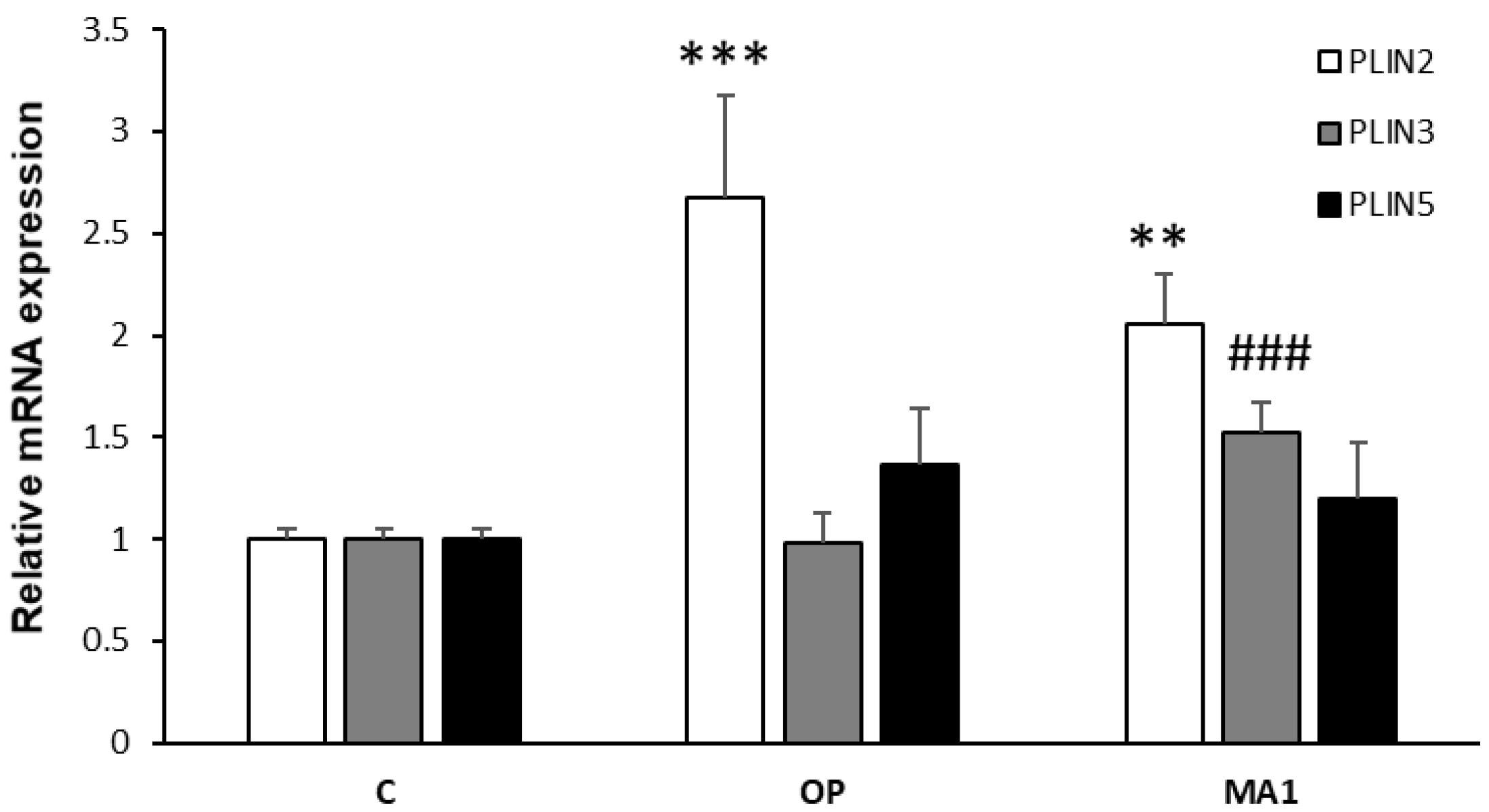
The above results indicate an effect of MA1 on LD trafficking in steatotic FaO cells. Therefore, we decided to use qPCR to investigate the expression of genes encoding LD-associated proteins. The perilipin family (PLINs) is the most documented group of such proteins [26], including five members with different functions, of which PLIN2 is the most abundant isoform in the liver. PLIN2 expression is enhanced in NAFLD and correlates with LD abundance [27]. Accordingly, Figure 3 shows that in our model PLIN2 expression was increased in OP cells (2.7-fold increase; p ≤ 0.001 vs. C); however, in presence of MA1 we could only observe a decreasing tendency for PLIN2, which did not reach statistical significance. On the other hand, MA1 was capable of enhancing PLIN3 expression in steatotic cells (1.5-fold increase; p ≤ 0.001 vs. OP), whereas no significant variations could be appreciated for PLIN5 expression, neither in absence nor in presence of MA1.

2.4. MA1 Regulates the Hepatic Expression of PPARs

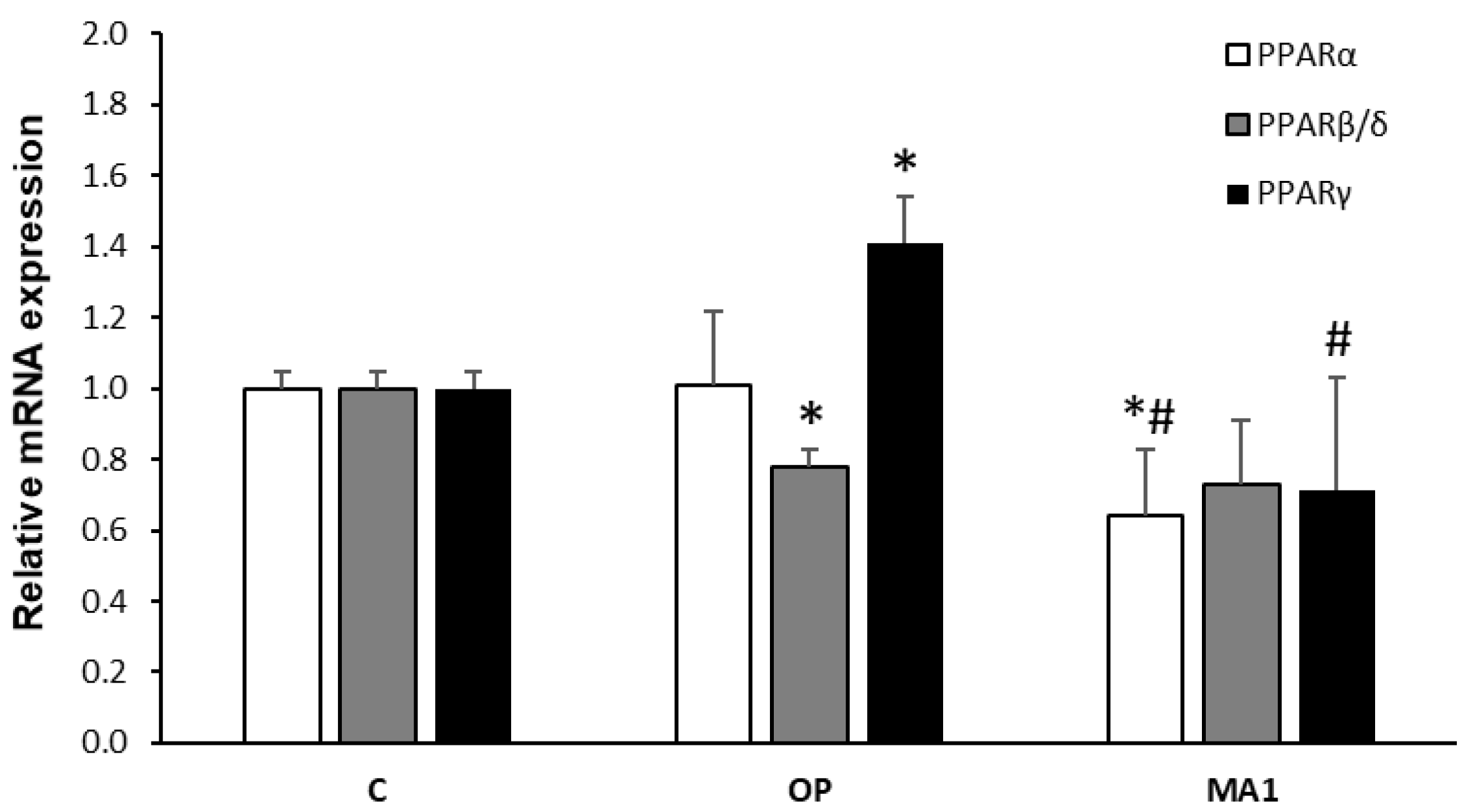
As for most of the key genes of hepatic lipid metabolism, PLIN expression is controlled by peroxisome proliferator-activated receptors (PPARs), a family of nuclear transcription factors comprising three members [28], of which PPARα and PPARγ are the most abundantly expressed in FaO cells [29]. Figure 4 shows that in steatotic FaO cells, PPARα expression did not differ significantly from control cells; however, 10 μg/mL MA1 reduced PPARα expression (1.6-fold decrease; p ≤ 0.05 vs. C and OP). The mRNA levels of PPARβ/δ were downregulated by OP treatment (1.3-fold decrease; p ≤ 0.05 vs. C), but subsequent MA1 administration was ineffective. PPARγ mRNA levels were upregulated in steatotic FaO cells (1.37-fold increase; p ≤ 0.05 vs. C), but the subsequent exposure to 10 μg/mL MA1 reduced the expression (2-fold decrease; p ≤ 0.05 vs. OP).

3. Discussion

Due to their wide range of biological activities, amphibian peptides represent a new fascinating possibility of finding novel compounds useful for human health. Promising results are achieved in different fields [22,30,31], but their modulating actions on metabolic pathways are of particular interest when considering the global health burden that metabolic diseases represent in Western lifestyle societies. NAFLD is recognized as the leading cause of more severe liver diseases and is linked to other metabolic dysfunctions, such as oxidative stress, endoplasmic reticulum and mitochondria impairments and IR [32] Altogether, these multiple hits [13] lead to the hepatic accumulation of lipids into cytosolic LDs, consisting of a central core of neutral lipids (mainly TGs) surrounded by a monolayer of phospholipids and associated proteins. The LD proteome allows the dynamicity of these organelles, which are considered at the crossroad of lipid trafficking inside the cells [33]. The expression of LD-associated proteins, as well as that of the key genes of hepatic lipid homeostasis, is controlled by specific transcription factors, such as PPARs, and thus different patterns of PPAR expression can be associated with dysregulations of hepatic lipid metabolism in clinical and experimental in vivo and in vitro models [20,34,35]. The anti-oxidant, anti-inflammatory and anti-diabetic properties establish amphibian skin peptides as potential beneficial compounds to be used against fatty liver. We tested this possibility by using a well-established in vitro model of NAFLD, in which we already described PLIN and PPAR expression levels [20,25,36]. In fact, the mRNA levels of both PPARs and PLINs are considered as specific markers in NAFLD models, particularly PPARγ and PLIN2 [20,37].
Our present results demonstrate a strong anti-steatotic effect of MA1, a skin peptide with anti-oxidant and anti-inflammatory properties, which was isolated from the Chinese odorous frog O. macrotympana [11]. The anti-steatotic effect of MA1 was evidenced by the decrease in intracellular TG content measured in lipid-overloaded FaO cells. To our knowledge, this is the first time that such a direct anti-steatotic effect has been demonstrated by an amphibian skin peptide. Several amphibian peptides have been evaluated in animal models of high-fat feeding (for a review, see [18]). Some of them showed the ability to significantly reduce body weight, total body fat and circulating triglyceride levels. It is worth noting that our results are in line with this lipid-lowering action, even if no data were reported on the hepatic fat content in these models. Moreover, an insulinotropic activity has been demonstrated for amphibian peptides [18], potentially counteracting fatty liver-associated IR. In addition, anti-oxidant and anti-inflammatory properties have been reported [11]. Should a direct anti-steatotic effect of some amphibian peptides be confirmed by further investigations on experimental in vitro and in vivo models of NAFLD, promising perspectives about the clinical use of these compounds would open. Indeed, the potential ability of amphibian peptides to simultaneously influence several metabolic dysfunctions associated with NAFLD is intriguing, considering that a specific pharmacological treatment against NALFD has not been approved yet.
In accordance with the diminished TG intracellular content, the number and diameter of LDs, where TGs are stored, were decreased by MA1 treatment, indicating some changes in LD remodeling and trafficking. The increase in PLIN2 expression in OP cells is in accordance with previous findings, demonstrating a correlation between PLIN2 and LD abundance. Consequently, we could expect a decrease in PLIN2 mRNA levels following MA1 treatment. We actually saw a tendency toward downregulation, which deserves further investigation, since it did not reach statistical significance, at least in our hands. The expression of PLIN5 was not significantly affected by OP or MA1 treatments. PLIN5 is known to alleviate palmitic acid-induced inflammatory responses in hepatocytes [38]. However, adding oleate to palmitate-treated cells can fully prevent palmitate lipotoxicity [20,39]. Therefore, a lack of PLIN5 regulation in our experiments is not completely surprising. On the other hand, a novel result is that MA1 was able to increase PLIN3 expression in steatotic FaO cells. PLIN3 (previously known as tail-interacting protein of 47 kDa (TIP47)) is co-expressed with PLIN2 in many tissues, where they often exert overlapping actions on TAG synthesis and LD biogenesis [40]. However, a distinct role of PLIN3 has not been fully clarified yet [41]. A recent paper discovered a new pathway linking PLIN3 and mTOR, in which PLIN3 was essential to activate lipophagy and protect against fatty liver worsening to steatohepatitis [42]. Although our data cannot directly demonstrate that MA1 activates this pathway in steatotic FaO cells, the observed upregulation of PLIN3 by MA1 is in line with this possibility, which should become an interesting line of investigation in future studies. On the contrary, MA1 did not increase extracellular TG content, as measured in cell culture media, indicating that a stimulation of TG secretion by MA1 as a mechanism for intracellular lipid-lowering is unlikely.
Our results also demonstrate a decrease in PPARα and PPARγ expression by MA1. The regulation of PPARs is of great interest because they may serve as potential therapeutic targets for treating NAFLD and metabolic dysfunctions [43]. PPARα is considered the master regulator of hepatic lipid metabolism, particularly during fasting, mainly involved in the regulation of oxidative pathways [44,45]. In vivo, both increased and decreased levels of PPARα expression have been reported in response to high-fat diets [46,47,48]. By using in vitro models, no significant modulations of PPARα expression were observed [20]. These discrepancies can be ascribed to the different experimental conditions utilized to induce hepatic steatosis in vivo and in vitro. This being the case, the downregulation of PPARα expression by MA1 might be associated with a decrease in the activity of lipid oxidative pathways, but the significance of such regulation in steatotic FaO cells is uncertain. On the contrary, PPARγ is a well-recognized marker of liver steatosis that allows energy storage through the activation of lipogenic genes [37,49]. PPARγ expression is increased in lipid-overloaded FaO cells, as previously reported [20]. Accordingly, the anti-steatotic effect of MA1 is coupled with a significant downregulation of PPARγ expression. PPARγ is also a major regulator of PLIN2 expression [50], so the observed tendency of a decrease in PLIN2 expression in response to MA1 could be related to the downregulation of PPARγ mRNA in MA1-treated cells.
We are aware of the need for further studies able to investigate the levels of proteins actually present in steatotic cells and to gain insights into the mechanism of action of MA1. Hopefully, our promising results will pave the way for the application of MA1 as an anti-steatotic compound.

4. Materials and Methods

4.1. Materials

Unless otherwise indicated, all materials and chemicals were supplied by Sigma-Aldrich Corp. (Milan, Italy). All the reagents were of analytical or cell culture grade (≥95% conforming to ACS specifications).

4.2. MA1 Synthesis

MA1 (FLPGLECVW) peptide was manually synthesized using the standard method of solid-phase peptide synthesis according to the 9-fluorenylmethoxycarbonyl (Fmoc) strategy with some modifications [51]. Briefly, Wang-Trp (Boc) deprotected resin was treated with a coupling reaction mixture of 5 equivalents (eq.) of the appropriate Fmoc-amino acid, 4.5 eq. of O-benzotriazol-N,N,N′,N′-tetramethyluroniumhexafluorophosphate (HBTU) and 5 eq. of N,N-diisopropilethylamine (DIPEA) at 0.2 M amino acid final concentration in anhydrous N-methylpyrrolidone (NMP). A solution of 20% (v/v) piperidine in DMF was used to remove the Fmoc group. The resin was incubated with a cocktail of acetic anhydride, sym-collidine and DMF (0.5:0.5:9 ratio (v/v)) to block the remaining amino groups on the resin.
The resin was rinsed with dichloromethane and dried. The final cleavage of the peptide from the solid support and removal of all protecting groups were carried out with a trifluoroacetic acid (TFA): tri-isopropylsilane:H2O:DODT (90:2.5:2.5:5) mixture. The peptide was precipitated in ice-cold diethyl ether and then purified by preparative reverse phase high-performance liquid chromatography (RP-HPLC) on an Agilent 1260 Infinity preparative HPLC instrument equipped with a Kinetex C18 column (21.20 mm × 150 mm). The separation was obtained with a gradient starting with 5% solvent B for 5 min, linearly increasing to 70% solvent B in 30 min and up to 100% B in 10 min. The solvents used were 0.1% formic acid in water (A) and 0.1% formic acid in acetonitrile (B). The peaks of interest were then collected and evaporated under a vacuum, before being lyophilized and stored at 4 °C. The identity and purity of the MA1 peptide were verified by RP-HPLC, and the collected peak was analyzed in off-line mass spectrometry analysis using an Agilent 1100 series LC/MSD ion trap instrument. The chromatogram and MS spectra are available as Supplementary Materials (Figure S1).

4.3. Cell Culture and Treatments

FaO rat hepatoma cell line (European Collection of Authenticated Cell Cultures, ECACC, Salisbury, Wiltshire, UK) was grown in Coon’s modified Ham’s F-12 medium supplemented with L-glutamine and 10% fetal bovine serum (FBS).
Cells were incubated in a humidified atmosphere with 5% CO2 at 37 °C. For treatments, cells were grown until 80% confluence and then incubated overnight in serum-free medium with 0.25% bovine serum albumin. To induce intracellular lipid accumulation, cells were treated for 3 h with a mixture of oleate/palmitate (OP) at a final concentration of 0.75 mM (2:1 molar ratio) [20]. Thereafter, cells were incubated for 24 h either in control medium (referred to as OP steatotic cells) or in the presence of MA1 at different concentrations. For cell treatment, lyophilized MA1 was dissolved in DMSO at 10 mg/mL and then diluted at 1 mg/mL in serum-free culture medium (stock solution). Serial working dilutions were performed in culture medium. Untreated cells were referred to as controls.

4.4. MTT Assay for Determination of Cell Viability

Cell viability was checked by performing the MTT (3-(4,5-dimethylthiazol-2-yl)-2,5-diphenyltetrazolium bromide) assay. MTT was dissolved in PBS at a concentration of 5 mg/mL and filtered through 0.22 µm pores. The working solution was diluted in culture medium at the final concentration of 0.5 mg/mL. The cells were then incubated for 3 h at 37 °C. Precipitated formazan was then dissolved in acid–alcohol (0.04 N HCl in 2-propanol) solution and read at 570 nm in a Varian Cary-50Bio spectrophotometer (Agilent, Milan, Italy) [21,36].

4.5. Quantification of Triglycerides (TGs)

Intracellular TG content was measured in FaO cells using the “Triglycerides liquid” kit (Sentinel, Milan, Italy), as previously described [25,36]. Absorbance was recorded at 546 nm in a Varian Cary-50Bio spectrophotometer (Agilent, Milan, Italy). For the measurement of extracellular TG content, the culture media were processed according to the same method. Values were normalized to protein content as measured by Bradford assay [52], and data are expressed as percent TG content relative to controls.

4.6. Fluorescence Microscopy

For intracellular lipid staining, cells grown directly on collagen-coated coverslips were rinsed with PBS and fixed with 4% paraformaldehyde for 20 min at room temperature and then quenched with 30 mM NH4Cl for 10 min and stained with 250 μg mL−1 BODIPY 493/503 (Molecular Probes, Life Technologies, Monza, Italy) for 20 min without permeabilization. After washing, nuclei were stained with 4′,6-diamidino-2-phenylindole (DAPI, 5 μg/mL, (ProLong Gold medium with DAPI; Invitrogen). Images were captured under oil using an Olympus IX70 inverted microscope with a 63x plan apochromatic objective and processed with Adobe Photoshop CS5 [53].

4.7. RNA Extraction and Quantitative Real-Time PCR

Total RNA was extracted by using Trizol Reagent (Sigma-Aldrich, Oakville, ON, Canada, and St. Louis, MO, USA) according to the manufacturer’s instructions. One microgram of cDNA was synthesized using RevertAid H-Minus M-MuLV Reverse Transcriptase (Fermentas, Hannover, MD, USA) as previously explained [54]. Real-time quantitative (qPCR) reactions were performed in triplicate in a final volume of 25 μL using 1× SybrGreen SuperMix and Chromo4TM System apparatus (Biorad, Monza, Italy) as previously described [55]. Primer pairs for the genes under analysis (Table 1) were designed ad hoc and synthesized by TibMolBiol custom oligo synthesis service (Genova, Italy). Amplification conditions were as follows: 3 min at 95 °C, followed by 5 s at 95 °C and 1 min at 60 °C for 40 cycles. A melting curve of qPCR products (65–94 °C) was also performed to ensure the absence of artifacts. The relative quantity of target mRNA was calculated by the comparative Cq method using glyceraldehyde 3-phosphate dehydrogenase (GAPDH) as a housekeeping gene and expressed as fold change with respect to controls [56].

4.8. Statistical Analysis

Data are means ± S.D. of at least three independent experiments. Statistical analysis was performed using ANOVA with Tukey’s post hoc test (GraphPad Software, Inc., San Diego, CA, USA).

5. Conclusions

For the first time, the anti-steatotic action of an amphibian skin peptide, namely macrotympanain A1 (MA1- FLPGLECVW), has been demonstrated in a well-established in vitro model of hepatic steatosis. MA1 is able to decrease intracellular TG content, which results in a reduction in LD number and size. The effect is associated with a downregulation of the expression of PPARγ, which is a marker of hepatic steatosis, and the upregulation of PLIN3, which is known to be involved in the activation of lipophagy. Together with literature evidence documenting anti-oxidant, anti-inflammatory and anti-diabetic effects, our results disclose the possibility of further investigating amphibian skin peptides as a new class of compounds able to reduce hepatic steatosis.

Supplementary Materials

The following supporting information can be downloaded at: https://www.mdpi.com/article/10.3390/molecules27217417/s1. Figure S1: Analysis of purified MA1 peptide.

Author Contributions

Conceptualization, I.D. and E.G.; methodology, I.D., E.M., K.C. and E.G.; validation, I.D., Z.E.R., A.S., K.C., A.C., G.R. and E.G.; formal analysis, Z.E.R., K.C. and E.G.; investigation, Z.E.R., E.M., A.P. and A.S.; data curation, S.S. and E.G.; writing—original draft preparation, I.D. and E.G.; writing—review and editing, I.D., E.M., S.S., K.C., G.D.N.A., M.Z.R. and E.G.; visualization, I.D. and E.G.; supervision, S.S. and E.G.; funding acquisition, S.S. and E.G. All authors have read and agreed to the published version of the manuscript.

Funding

This work was funded by the University of Genoa (FRA 2019 and FRA 2020 to S.S. and E.G.) and from Fondazione Compagnia San Paolo under the initiative ‘Call for the promotion of European project 2020’-BIPE 2020.

Institutional Review Board Statement

Not applicable.

Informed Consent Statement

Not applicable.

Data Availability Statement

The data presented in this study are available in this article.

Conflicts of Interest

The authors declare no conflict of interest.

Sample Availability

Samples of synthesized MA1 are available from the authors.

References

  1. Duellman, W.E.; Trueb, L. Biology of Amphibians, 2nd ed.; The Johns Hopkins University Press: Baltimore, MD, USA, 1994. [Google Scholar]
  2. Demori, I.; Rashed, Z.E.; Corradino, V.; Catalano, A.; Rovegno, L.; Queirolo, L.; Salvidio, S.; Biggi, E.; Zanotti-Russo, M.; Canesi, L.; et al. Peptides for Skin Protection and Healing in Amphibians. Molecules 2019, 24, 347. [Google Scholar] [CrossRef] [PubMed] [Green Version]
  3. Ramsey, J.P.; Reinert, L.K.; Harper, L.K.; Woodhams, D.C.; Rollins-Smith, L.A. Immune Defenses against Batracho-chytrium Dendrobatidis, a Fungus Linked to Global Amphibian Declines, in the South African Clawed Frog, Xenopus Laevis. Infect. Immun. 2010, 78, 3981–3992. [Google Scholar] [CrossRef] [PubMed] [Green Version]
  4. Rollins-Smith, L.A.; Conlon, J.M. Antimicrobial Peptide Defenses against Chytridiomycosis, an Emerging Infec-tious Disease of Amphibian Populations. Dev. Comp. Immunol. 2005, 29, 589–598. [Google Scholar] [CrossRef]
  5. Rollins-Smith, L.A.; Ramsey, J.P.; Pask, J.D.; Reinert, L.K.; Woodhams, D.C. Amphibian Immune Defenses against Chytridiomycosis: Impacts of Changing Environments. Integr. Comp. Biol. 2011, 51, 552–562. [Google Scholar] [CrossRef] [Green Version]
  6. Gammill, W.M.; Scott Fites, J.; Rollins-Smith, L.A. Norepinephrine Depletion of Antimicrobial Peptides from the Skin Glands of Xenopus laevis. Dev. Comp. Immunol. 2012, 37, 19–27. [Google Scholar] [CrossRef]
  7. Crump, M.L. Why Are Some Species in Decline but Others Not? In Amphibian Declines; Lannoo, M., Ed.; University of California Press: Berkeley, CA, USA, 2005; pp. 7–9. [Google Scholar]
  8. Xu, X.; Lai, R. The Chemistry and Biological Activities of Peptides from Amphibian Skin Secretions. Chem. Rev. 2015, 115, 1760–1846. [Google Scholar] [CrossRef] [PubMed]
  9. Bevins, C.L.; Zasloff, M. Peptides from Frog Skin. Annu. Rev. Biochem. 1990, 59, 395–414. [Google Scholar] [CrossRef]
  10. Conlon, J.M.; Mechkarska, M.; Leprince, J. Peptidomic Analysis in the Discovery of Therapeutically Valuable Peptides in Amphibian Skin Secretions. Expert Rev. Proteom. 2019, 16, 897–908. [Google Scholar] [CrossRef]
  11. Yang, X.; Lee, W.-H.; Zhang, Y. Extremely Abundant Antimicrobial Peptides Existed in the Skins of Nine Kinds of Chinese Odorous Frogs. J. Proteome Res. 2012, 11, 306–319. [Google Scholar] [CrossRef]
  12. Chen, X.; Chen, Z.; Jiang, J.; Qiao, L.; Lu, Y.; Zhou, K.; Zheng, G.; Zhai, X.; Liu, J. Molecular Phylogeny and Diversi-fication of the Genus Odorrana (Amphibia, Anura, Ranidae) Inferred from Two Mitochondrial Genes. Mol. Phylogenetics Evol. 2013, 69, 1196–1202. [Google Scholar] [CrossRef]
  13. Buzzetti, E.; Pinzani, M.; Tsochatzis, E.A. The Multiple-Hit Pathogenesis of Non-Alcoholic Fatty Liver Disease (NAFLD). Metabolism 2016, 65, 1038–1048. [Google Scholar] [CrossRef] [PubMed]
  14. Younossi, Z.M.; Koenig, A.B.; Abdelatif, D.; Fazel, Y.; Henry, L.; Wymer, M. Global Epidemiology of Nonalcoholic Fatty Liver Disease-Meta-Analytic Assessment of Prevalence, Incidence, and Outcomes. Hepatology 2016, 64, 73–84. [Google Scholar] [CrossRef] [PubMed] [Green Version]
  15. Neuschwander-Tetri, B.A. Therapeutic Landscape for NAFLD in 2020. Gastroenterology 2020, 158, 1984–1998.e3. [Google Scholar] [CrossRef] [PubMed]
  16. Eslam, M.; Sanyal, A.J.; George, J.; Sanyal, A.; Neuschwander-Tetri, B.; Tiribelli, C.; Kleiner, D.E.; Brunt, E.; Bugiane-si, E.; Yki-Järvinen, H.; et al. MAFLD: A Consensus-Driven Proposed Nomenclature for Metabolic Associated Fatty Liver Disease. Gastroenterology 2020, 158, 1999–2014.e1. [Google Scholar] [CrossRef]
  17. Palma, R.; Pronio, A.; Romeo, M.; Scognamiglio, F.; Ventriglia, L.; Ormando, V.M.; Lamazza, A.; Pontone, S.; Fed-erico, A.; Dallio, M. The Role of Insulin Resistance in Fueling NAFLD Pathogenesis: From Molecular Mechanisms to Clinical Implications. J. Clin. Med. 2022, 11, 3649. [Google Scholar] [CrossRef]
  18. Conlon, J.M.; Mechkarska, M.; Abdel-Wahab, Y.H.; Flatt, P.R. Peptides from Frog Skin with Potential for Develop-ment into Agents for Type 2 Diabetes Therapy. Peptides 2018, 100, 275–281. [Google Scholar] [CrossRef]
  19. El Rashed, Z.; Lupidi, G.; Grasselli, E.; Canesi, L.; Khalifeh, H.; Demori, I. Antioxidant and Antisteatotic Activities of Fucoidan Fractions from Marine and Terrestrial Sources. Molecules 2021, 26, 4467. [Google Scholar] [CrossRef]
  20. Grasselli, E.; Canesi, L.; Portincasa, P.; Voci, A.; Vergani, L.; Demori, I. Models of Non-Alcoholic Fatty Liver Disease and Potential Translational Value: The Effects of 3,5-L-Diiodothyronine. Ann. Hepatol. 2017, 16, 707–719. [Google Scholar] [CrossRef]
  21. Grasselli, E.; Baldini, F.; Vecchione, G.; Oliveira, P.; Sardāo, V.; Voci, A.; Portincasa, P.; Vergani, L. Excess Fructose and Fatty Acids Trigger a Model of Non-alcoholic Fatty Liver Disease Progression in Vitro: Protective Effect of the Flavonoid Silybin. Int. J. Mol. Med. 2019, 44, 705–712. [Google Scholar] [CrossRef]
  22. Soltaninejad, H.; Zare-Zardini, H.; Ordooei, M.; Ghelmani, Y.; Ghadiri-Anari, A.; Mojahedi, S.; Hamidieh, A.A. Antimicrobial Peptides from Amphibian Innate Immune System as Potent Antidiabetic Agents: A Literature Review and Bioinformatics Analysis. J. Diabetes Res. 2021, 2021, 2894722. [Google Scholar] [CrossRef]
  23. Srinivasan, D.K.; Ojo, O.O.; Owolabi, B.O.; Conlon, J.M.; Flatt, P.R.; Abdel-Wahab, Y.H.A. [I10W]Tigerinin-1R En-hances Both Insulin Sensitivity and Pancreatic Beta Cell Function and Decreases Adiposity and Plasma Triglycerides in High-Fat Mice. Acta Diabetol. 2016, 53, 303–315. [Google Scholar] [CrossRef] [PubMed]
  24. Conlon, J.M.; Power, G.J.; Abdel-Wahab, Y.H.A.; Flatt, P.R.; Jiansheng, H.; Coquet, L.; Leprince, J.; Jouenne, T.; Vaudry, H. A Potent, Non-Toxic Insulin-Releasing Peptide Isolated from an Extract of the Skin of the Asian Frog, Hylarana Guntheri (Anura:Ranidae). Regul. Pept. 2008, 151, 153–159. [Google Scholar] [CrossRef]
  25. El Rashed, Z.; Lupidi, G.; Kanaan, H.; Grasselli, E.; Canesi, L.; Khalifeh, H.; Demori, I. Antioxidant and Antisteatotic Activities of a New Fucoidan Extracted from Ferula Hermonis Roots Harvested on Lebanese Mountains. Molecules 2021, 26, 1161. [Google Scholar] [CrossRef] [PubMed]
  26. Kimmel, A.R.; Brasaemle, D.L.; McAndrews-Hill, M.; Sztalryd, C.; Londos, C. Adoption of PERILIPIN as a Unifying Nomenclature for the Mammalian PAT-Family of Intracellular Lipid Storage Droplet Proteins. J. Lipid Res. 2010, 51, 468–471. [Google Scholar] [CrossRef] [PubMed] [Green Version]
  27. Fujii, H.; Ikura, Y.; Arimoto, J.; Sugioka, K.; Iezzoni, J.C.; Park, S.H.; Naruko, T.; Itabe, H.; Kawada, N.; Caldwell, S.H.; et al. Expression of Perilipin and Adipophilin in Nonalcoholic Fatty Liver Disease; Relevance to Oxidative Injury and Hepatocyte Ballooning. J. Atheroscler. Thromb. 2009, 16, 893–901. [Google Scholar] [CrossRef] [Green Version]
  28. Souza-Mello, V. Peroxisome Proliferator-Activated Receptors as Targets to Treat Non-Alcoholic Fatty Liver Disease. World J. Hepatol. 2015, 7, 1012. [Google Scholar] [CrossRef]
  29. Remington, G.J.; Teo, C.; Wilson, V.; Chintoh, A.; Guenette, M.; Ahsan, Z.; Giacca, A.; Hahn, M. Metformin Attenuates Olanzapineinduced Hepatic, but Not Peripheral Insulin Resistance. J. Endocrinol. 2015, 277, 71–81. [Google Scholar] [CrossRef] [Green Version]
  30. Conlon, J.M.; Mechkarska, M.; Lukic, M.L.; Flatt, P.R. Potential Therapeutic Applications of Multifunctional Host-Defense Peptides from Frog Skin as Anti-Cancer, Anti-Viral, Immunomodulatory, and Anti-Diabetic Agents. Peptides 2014, 57, 67–77. [Google Scholar] [CrossRef]
  31. Mechkarska, M.; Attoub, S.; Sulaiman, S.; Pantic, J.; Lukic, M.L.; Michael Conlon, J. Anti-Cancer, Immunoregulatory, and Antimicrobial Activities of the Frog Skin Host-Defense Peptides Pseudhymenochirin-1Pb and Pseudh-ymenochirin-2Pa. Regul. Pept. 2014, 194–195, 69–76. [Google Scholar] [CrossRef]
  32. Byrne, C.D.; Targher, G. NAFLD: A Multisystem Disease. J. Hepatol. 2015, 62, S47–S64. [Google Scholar] [CrossRef]
  33. Thiam, A.R.; Farese, R.V., Jr.; Walther, T.C. The Biophysics and Cell Biology of Lipid Droplets. Nat. Rev. Mol. Cell Biol. 2013, 14, 775–786. [Google Scholar] [CrossRef] [Green Version]
  34. De la Rosa Rodriguez, M.A.; Kersten, S. Regulation of Lipid Droplet-Associated Proteins by Peroxisome Prolifera-tor-Activated Receptors. Biochim. Biophys. Acta (BBA) Mol. Cell Biol. Lipids 2017, 1862, 1212–1220. [Google Scholar] [CrossRef]
  35. Wahli, W.; Michalik, L. PPARs at the Crossroads of Lipid Signaling and Inflammation. Trends Endocrinol. Metab. 2012, 23, 351–363. [Google Scholar] [CrossRef] [PubMed]
  36. Lambruschini, C.; Demori, I.; El Rashed, Z.; Rovegno, L.; Canessa, E.; Cortese, K.; Grasselli, E.; Moni, L. Synthesis, Photoisomerization, Antioxidant Activity, and Lipid-Lowering Effect of Ferulic Acid and Feruloyl Amides. Molecules 2020, 26, 89. [Google Scholar] [CrossRef] [PubMed]
  37. Rahimian, R.; Masih-Khan, E.; Lo, M.; van Breemen, C.; McManus, B.M.; Dubé, G.P. Hepatic Over-Expression of Peroxisome Proliferator Activated Receptor Γ2 in the Ob/Ob Mouse Model of Non-insulin Dependent Diabetes Mellitus. Mol. Cell. Biochem. 2001, 224, 29–37. [Google Scholar] [CrossRef] [PubMed]
  38. Zhang, E.; Cui, W.; Lopresti, M.; Mashek, M.T.; Najt, C.P.; Hu, H.; Mashek, D.G. Hepatic PLIN5 Signals via SIRT1 to Promote Autophagy and Prevent Inflammation during Fasting. J. Lipid Res. 2020, 61, 338–350. [Google Scholar] [CrossRef]
  39. Leamy, A.K.; Egnatchik, R.A.; Shiota, M.; Ivanova, P.T.; Myers, D.S.; Brown, H.A.; Young, J.D. Enhanced Synthesis of Saturated Phospholipids Is Associated with ER Stress and Lipotoxicity in Palmitate Treated Hepatic Cells. J. Lipid Res. 2014, 55, 1478–1488. [Google Scholar] [CrossRef] [Green Version]
  40. Sztalryd, C.; Bell, M.; Lu, X.; Mertz, P.; Hickenbottom, S.; Chang, B.H.-J.; Chan, L.; Kimmel, A.R.; Londos, C. Func-tional Compensation for Adipose Differentiation-Related Protein (ADFP) by Tip47 in an ADFP Null Embryonic Cell Line. J. Biol. Chem. 2006, 281, 34341–34348. [Google Scholar] [CrossRef] [Green Version]
  41. Minehira, K.; Gual, P. Role of Lipid Droplet Proteins in the Development of NAFLD and Hepatic Insulin Resistance. In Non-Alcoholic Fatty Liver Disease—Molecular Bases, Prevention and Treatment; Valenzuela, R., Ed.; InTech Open: London, UK, 2018; ISBN 978-953-51-3923-2. [Google Scholar]
  42. Garcia-Macia, M.; Santos-Ledo, A.; Leslie, J.; Paish, H.L.; Collins, A.L.; Scott, R.S.; Watson, A.; Burgoyne, R.A.; White, S.; French, J.; et al. A Mammalian Target of Rapamycin-Perilipin 3 (MTORC1-Plin3) Pathway Is Essential to Activate Lipophagy and Protects Against Hepatosteatosis. Hepatology 2021, 74, 3441–3459. [Google Scholar] [CrossRef]
  43. Fougerat, A.; Montagner, A.; Loiseau, N.; Guillou, H.; Wahli, W. Peroxisome Proliferator-Activated Receptors and Their Novel Ligands as Candidates for the Treatment of Non-Alcoholic Fatty Liver Disease. Cells 2020, 9, 1638. [Google Scholar] [CrossRef]
  44. Kersten, S. Integrated Physiology and Systems Biology of PPARα. Mol. Metab. 2014, 3, 354–371. [Google Scholar] [CrossRef] [PubMed]
  45. Pyper, S.R.; Viswakarma, N.; Yu, S.; Reddy, J.K. PPARα: Energy Combustion, Hypolipidemia, Inflammation and Cancer. Nucl. Recept. Signal 2010, 8, nrs.08002. [Google Scholar] [CrossRef] [PubMed] [Green Version]
  46. Grasselli, E.; Canesi, L.; Voci, A.; De Matteis, R.; Demori, I.; Fugassa, E.; Vergani, L. Effects of 3,5-Diiodo-L-Thyronine Administration on the Liver of High Fat Diet-Fed Rats. Exp. Biol. Med. 2008, 233, 549–557. [Google Scholar] [CrossRef] [PubMed]
  47. Redonnet, A.; Groubet, R.; No[euml ]l-Suberville, C.; Bonilla, S.; Martinez, A.; Higueret, P. Exposure to an Obesity-Inducing Diet Early Affects the Pattern of Expression of Peroxisome Proliferator, Retinoic Acid, and Triiodothyronine Nuclear Receptors in the Rat. Metabolism 2001, 50, 1161–1167. [Google Scholar] [CrossRef]
  48. Patsouris, D.; Reddy, J.K.; Müller, M.; Kersten, S. Peroxisome Proliferator-Activated Receptor α Mediates the Effects of High-Fat Diet on Hepatic Gene Expression. Endocrinology 2006, 147, 1508–1516. [Google Scholar] [CrossRef] [Green Version]
  49. Pettinelli, P.; Videla, L.A. Up-Regulation of PPAR-γ MRNA Expression in the Liver of Obese Patients: An Additional Reinforcing Lipogenic Mechanism to SREBP-1c Induction. J. Clin. Endocrinol. Metab. 2011, 96, 1424–1430. [Google Scholar] [CrossRef]
  50. Schadinger, S.E.; Bucher, N.L.R.; Schreiber, B.M.; Farmer, S.R. PPARγ2 Regulates Lipogenesis and Lipid Accumu-lation in Steatotic Hepatocytes. Am. J. Physiol.-Endocrinol. Metab. 2005, 288, E1195–E1205. [Google Scholar] [CrossRef]
  51. Cardinali, B.; Lunardi, G.; Millo, E.; Armirotti, A.; Damonte, G.; Profumo, A.; Gori, S.; Iacono, G.; Levaggi, A.; Del Mastro, L. Trastuzumab Quantification in Serum: A New, Rapid, Robust ELISA Assay Based on a Mimetic Peptide That Specifically Recognizes Trastuzumab. Anal. Bioanal. Chem. 2014, 406, 4557–4561. [Google Scholar] [CrossRef]
  52. Bradford, M.M. A Rapid and Sensitive Method for the Quantitation of Microgram Quantities of Protein Utilizing the Principle of Protein-Dye Binding. Anal. Biochem. 1976, 72, 248–254. [Google Scholar] [CrossRef]
  53. D’Alesio, C.; Bellese, G.; Gagliani, M.C.; Lechiara, A.; Dameri, M.; Grasselli, E.; Lanfrancone, L.; Cortese, K.; Castag-nola, P. The Chromodomain Helicase CHD4 Regulates ERBB2 Signaling Pathway and Autophagy in ERBB2+ Breast Cancer Cells. Biol. Open 2019, 8, bio.038323. [Google Scholar] [CrossRef]
  54. Grasselli, E.; Voci, A.; Demori, I.; Vecchione, G.; Compalati, A.D.; Gallo, G.; Goglia, F.; De Matteis, R.; Silvestri, E.; Vergani, L. Triglyceride Mobilization from Lipid Droplets Sustains the Anti-Steatotic Action of Iodothyronines in Cultured Rat Hepatocytes. Front. Physiol. 2016, 6, 418. [Google Scholar] [CrossRef]
  55. Grasselli, E.; Voci, A.; Canesi, L.; Goglia, F.; Ravera, S.; Panfoli, I.; Gallo, G.; Vergani, L. Non-receptor-mediated actions are responsible for the lipid-lowering effects of iodothyronines in FaO rat hepatoma cells. J. Endocrinol. 2011, 210, 59–69. [Google Scholar] [CrossRef]
  56. Pfaffl, M.W. A New Mathematical Model for Relative Quantification in Real-Time RT-PCR. Nucleic Acids Res. 2001, 29, e45. [Google Scholar] [CrossRef]
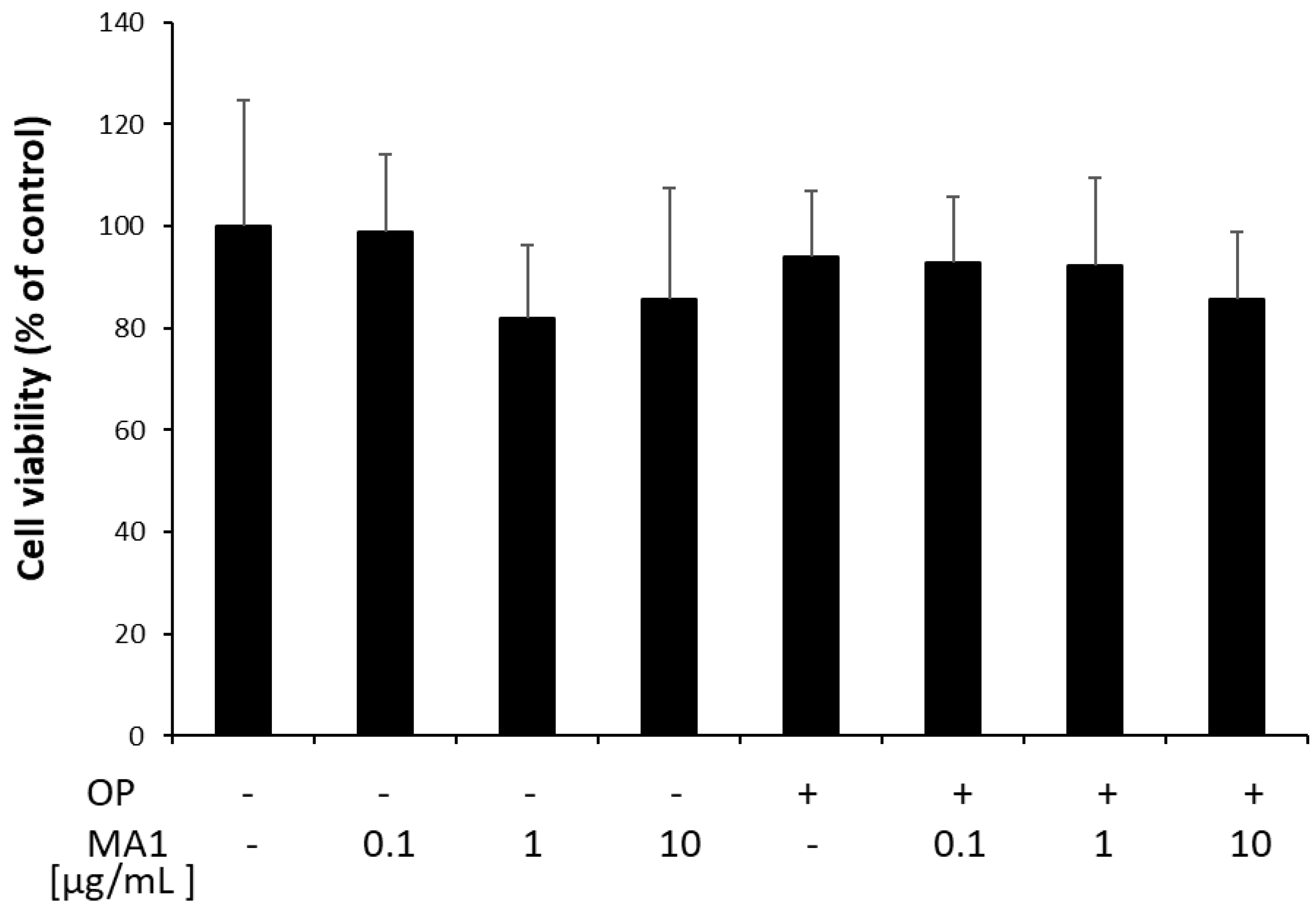
Figure 1. Viability of FaO cells treated for 24 h with 0.1, 1 and 10 µg/mL MA1, in the absence or in the presence of 0.75 mM OP. Data are expressed as percentage of control. Values are mean ± SD from three independent experiments.
Molecules 27 07417 g001
Figure 2. Effects of MA1 on lipid accumulation in steatotic FaO cells. (A) Intracellular TG content in control (C) and steatotic cells incubated in the absence (OP) or in the presence of 10 μg/mL MA1 for 24 h. Data are mean ± SD of three independent experiments and are expressed with respect to controls. Significant differences are denoted by symbols on bars (*** p ≤ 0.001 vs. C; ### p ≤ 0.001 vs. OP). (B) Measurements of LD diameter and number in 40 cells for each treatment. The measures of diameters of all counted LDs were plotted as a box-and-whisker plot, showing the interquartile range and the median as a horizontal bar. The whiskers are the minimum and maximum values. The mean number of LDs is reported in the inset table. Significant differences are denoted by symbols (*** p ≤ 0.001 vs. C; # p ≤ 0.05 vs. OP). (C) Representative images of 3 independent experiments showing BODIPY (green)/DAPI (blue) staining of FaO cells to visualize cytosolic LDs (magnification 63×; scale bar: 10 µm). (D) Extracellular TG content as measured in the culture medium of the same cells as in (A). Data are mean ± SD of three independent experiments and are expressed with respect to controls. Significant differences are denoted by symbols on bars (** p ≤ 0.01 vs. C; # p ≤ 0.05 vs. OP).
Molecules 27 07417 g002
Figure 3. Effects of MA1 on PLIN expression in steatotic FaO cells. Relative expression of PLINs as evaluated by real-time PCR. Steatotic FaO cells (OP) were then incubated for 24 h in the presence of 10 μg/mL MA1. Data are mean ± SD of three independent experiments and are expressed with respect to controls. Significant differences are denoted by symbols on bars (** p ≤ 0.01, *** p ≤ 0.001 vs. control; ### p ≤ 0.001 vs. OP).
Molecules 27 07417 g003
Figure 4. Effects of MA1 on PPAR expression in steatotic FaO cells. Relative expression of PPAR isoforms as evaluated by real-time PCR. Steatotic FaO cells (OP) were then incubated for 24 h in the presence of 10 μg/mL MA1. Data are mean ± SD of three independent experiments and are expressed with respect to controls. Significant differences are denoted by symbols on bars (* p ≤ 0.05 vs. control; # p ≤ 0.05 vs. OP).
Molecules 27 07417 g004
Table 1. Primer pairs used for RT-qPCR analysis.
Primer NamePrimer Sequence (5′→3′)Annealing
T (°C)
Product Length (bp)Accession ID
GAPDH FwdGACCCCTTCATTGACCTCAAC60136DQ403053
GAPDH RevCGCTCCTGGAAGATGGTGATGGG
PPARα FwdCCCCACTTGAAGCAGATGACC60139NM_013196
PPARα RevCCCTAAGTACTGGTAGTCCGC
PPARβ/δ FwdAATGCCTACCTGAAAAACTTCAAC6096AJ306400.1
PPARβ/δ RevTGCCTGCCACAGCGTCTCAAT
PPARγ FwdCGGAGTCCTCCCAGCTGTTCGCC60116Y12882
PPARγ RevGGCTCATATCTGTCTCCGTCTTC
PLIN2 FwdCCGAGCGTGGTGACGAGGG60148AAH85861
PLIN2 RevGAGGTCACGGTCCTCACTCCC
PLIN3 FwdGGAACTGGTGTCATCAACAG60108NW_047865.1
PLIN3 RevGGTCACATCCACTGCTCCTG
PLIN5 FwdGGATGTCCGGTGATCAGAC6096XM_576698
PLIN5 RevGTGCACGTGGCCCTGACCAG
Publisher’s Note: MDPI stays neutral with regard to jurisdictional claims in published maps and institutional affiliations.