Article

Aminoquinolones and Their Benzoquinone Dimer Hybrids as Modulators of Prion Protein Conversion

by
Amanda Rodrigues Pinto Costa
1,
Marcelly Muxfeldt
2,
Fernanda da Costa Santos Boechat
1,
Maria Cecília Bastos Vieira de Souza
1,
Jerson Lima Silva
3,
Marcela Cristina de Moraes
1,
Luciana Pereira Rangel
2,
Tuane Cristine Ramos Gonçalves Vieira
3 and
Pedro Netto Batalha
1,*
1
Instituto de Química, Universidade Federal Fluminense, Niterói 24020-141, RJ, Brazil
2
Faculdade de Farmácia, Universidade Federal do Rio de Janeiro, Rio de Janeiro 21941-902, RJ, Brazil
3
Instituto de Bioquímica Médica Leopoldo de Meis, Instituto Nacional de Ciência e Tecnologia de Biologia Estrutural e Bioimagem, Universidade Federal do Rio de Janeiro, Rio de Janeiro 21941-902, RJ, Brazil
*
Author to whom correspondence should be addressed.
Molecules 2022, 27(22), 7935; https://doi.org/10.3390/molecules27227935
Submission received: 28 September 2022 / Revised: 21 October 2022 / Accepted: 10 November 2022 / Published: 16 November 2022
(This article belongs to the Section Bioorganic Chemistry)

Abstract

:
Prion Diseases or Transmissible Spongiform Encephalopathies are neurodegenerative conditions associated with a long incubation period and progressive clinical evolution, leading to death. Their pathogenesis is characterized by conformational changes of the cellular prion protein—PrPC—in its infectious isoform—PrPSc—which can form polymeric aggregates that precipitate in brain tissues. Currently, there are no effective treatments for these diseases. The 2,5-diamino-1,4-benzoquinone structure is associated with an anti-prion profile and, considering the biodynamic properties associated with 4-quinolones, in this work, 6-amino-4-quinolones derivatives and their respective benzoquinone dimeric hybrids were synthesized and had their bioactive profile evaluated through their ability to prevent prion conversion. Two hybrids, namely, 2,5-dichloro-3,6-bis((3-carboxy-1-pentyl-4-quinolone-6-yl)amino)-1,4-benzoquinone (8e) and 2,5-dichloro-3,6-bis((1-benzyl-3-carboxy-4-quinolone-6-yl)amino)-1,4-benzoquinone (8f), stood out for their prion conversion inhibition ability, affecting the fibrillation process in both the kinetics—with a shortening of the lag phase—and thermodynamics and their ability to inhibit the formation of protein aggregates without significant cytotoxicity at ten micromolar.

Graphical Abstract

1. Introduction

Prions are proteins capable of undergoing conformational changes from their cellular isoform (PrPC) with a soluble and predominantly α-helical structure into their infectious isoform (PrPSc) with an insoluble, β-sheet rich, and protease-resistant structure. PrPC is encoded by a single gene known as PRNP and is expressed in different tissues, with a higher concentration in the central nervous system in the postsynaptic membranes of neurons [1].
There are two main models explaining the mechanisms of prion conformational change. One is the nucleation model, in which PrPC converts, through a spontaneous conformational equilibrium, to the infecting PrPSc. Although the conversion thermodynamics favors PrPC conformation, the equilibrium can be shifted due to still unknown factors, leading to the formation of insoluble polymeric protein aggregates analogous to crystal nucleation. The second model is the refolding model, which proposes that once PrPSc is generated, it may serve as a template and induce further conformational changes in other PrPC molecules, leading to protein aggregation. In both models, prion aggregates levels slowly increase, and they are deposited in neuronal tissues, causing the emergence of neurodegenerative symptoms and neuron death by apoptosis [2,3].
Prion Diseases, also known as Transmissible Spongiform Encephalopathies (TSEs), can affect diverse animals, including humans, and have no cure, being invariably fatal. In addition, they are infectious and can be transmitted, for example, through infected food ingestion or surgical instruments. Such diseases’ social and economic impacts make developing chemotherapeutic agents for treating TSEs urgent. Such drugs could, at first, act through different mechanisms, such as by modulating PrPC expression and biosynthesis, stabilizing PrPC through direct interaction with the protein, thus inhibiting the formation of PrPSc, or decreasing the levels of prion aggregates directly [4,5,6,7,8,9].
Studies focusing on the development of new anti-prion agents or the identification of hits from virtual screening have grown in recent years, which can be justified by the development of more robust and sensitive methods to detect the modulatory effect of small molecules on prion conversion, using in silico, in vitro, and in vivo models [10,11].
The 2,5-diamino-1,4-benzoquinone structure is a promising scaffold for the design of new substances with anti-prion profiles. In this context, two examples can be highlighted. Derivatives 1 and 2, shown in Figure 1, containing a 4-aminoquinoline [12] and an α-aminoester [13] moiety, respectively, have presented excellent anti-PrPSc activity in vitro.
On the other hand, nitrogen-containing heterocycles are present in various bioactive structures. Such substances are associated with interesting physicochemical properties, including a high molecular dipole moment, susceptibility to ionization in a physiological medium, and the ability to perform polar and hydrogen bond interactions. Such characteristics make them structures of great importance in the Medicinal-Chemistry field and are continuously studied by the scientific community [14,15]. The development of new versatile synthetic methods for the construction of these molecular systems [16,17,18,19], the discovery of new substances with broad bioactive profiles [18,20], and the evaluation of their interaction mechanisms with different pharmacological receptors [20] are examples of research that has been developed over the last decades in this context.
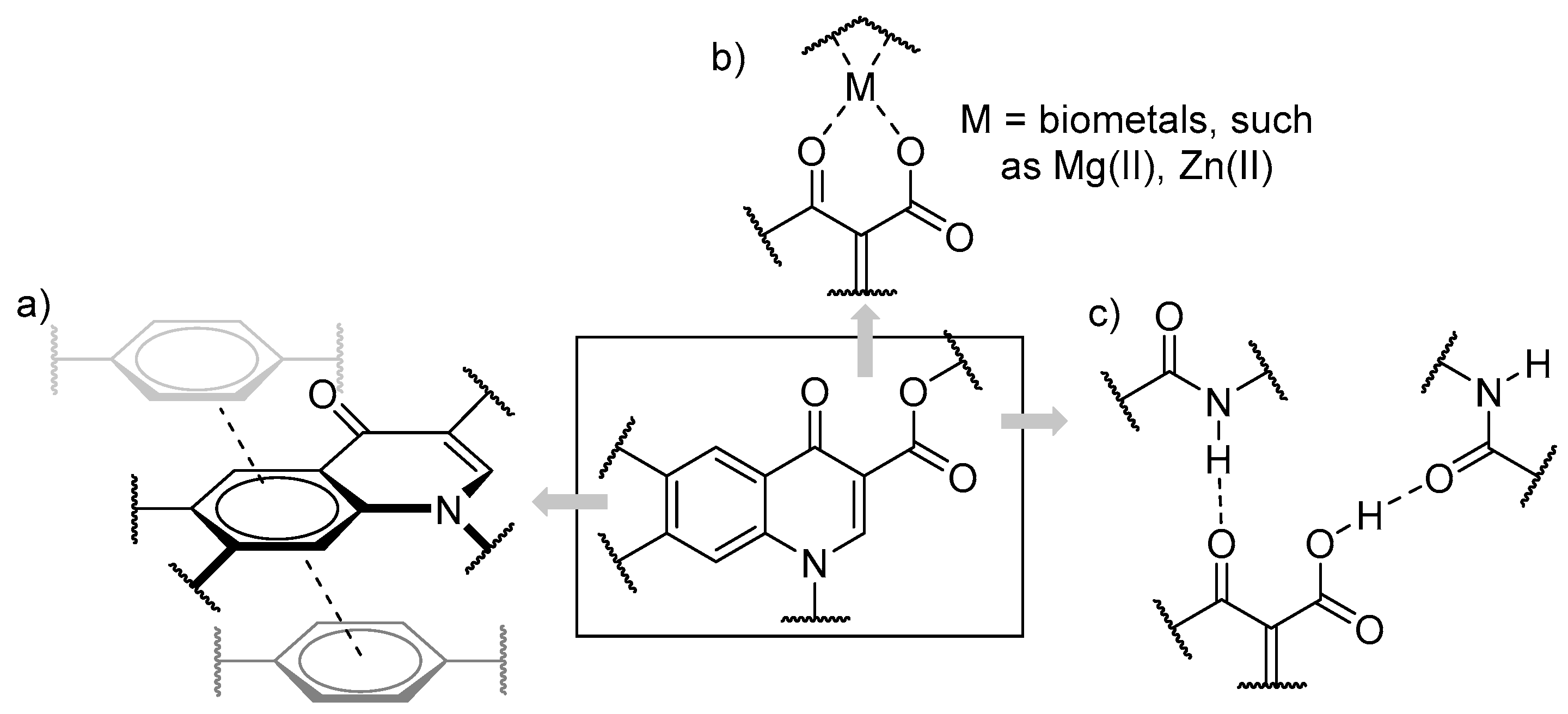
Despite their most common clinical application as antibacterials [21], 4-quinolone derivatives have also been described in numerous studies that demonstrate non-classical bioactivities, including anticancer [22,23], antiviral [24,25], antiparasitic [26,27], and antifungal [28]. These are biologically versatile substances due to their unique and easily functionalized structure. They may interact with different biological targets through intermolecular interactions, including hydrogen bonds, biometal chelation, and π-π stacking interactions (Figure 2) [29]. The biodynamic properties of these substances prompt us to consider their application as anti-prion agents.
Thus, contributing to the search for substances with chemotherapeutic potential against TSEs, we report the synthesis and anti-prion activity of 6-amino-4-quinolone derivatives and their 1,4-benzoquinone dimer hybrids and demonstrate through RT-QuIC, using in vitro-produced fibrils, their ability to inhibit prion aggregation.

2. Results and Discussion

2.1. Synthesis

Compounds 3, 4a–d [27], 5a–d, and 6a [30] were synthesized as described in previous works. Additionally, 6-amino-4-quinolone-3-carboxylic acids 6bd were prepared in the same way as described for 6a, as shown in Scheme 1.
6-Amino-4-quinolones 5ac and 6bd were successfully applied to the dimerization reaction via condensation with chloranil (7) in a stoichiometric ratio of 2:1, respectively (Scheme 2), following the protocol already described for the preparation of different 2,5-diamino-1,4-benzoquinone systems [12,13].
Substances 4ad, 5ad, and 6a had their structures properly characterized in previously published reports [27,30]. Substances 6bd and 8af had their structures confirmed by analyzing their IR, 1H NMR, and 13C-APT NMR spectra. The analysis of their HRMS spectra also confirmed their molecular formulae. These data are described in the experimental section. The spectral data of these compounds are available in the Supplementary Materials File.

2.2. Biological Assays

PrP aggregation is associated with the pathology of prion diseases in which we can find fibrillar or amorphous aggregates. PrPSc can induce PrPC structure conversion leading to the formation of auto-propagative aggregates [3]. This characteristic is involved in the infectious nature of prion diseases, but it is also observed in other conditions related to protein misfolding [31,32]. In vitro approaches can be used to investigate this auto-propagative ability, and the RT-QuIC assay is one of the most reliable for TSE diagnosis and therapeutic molecule screening [11,33].
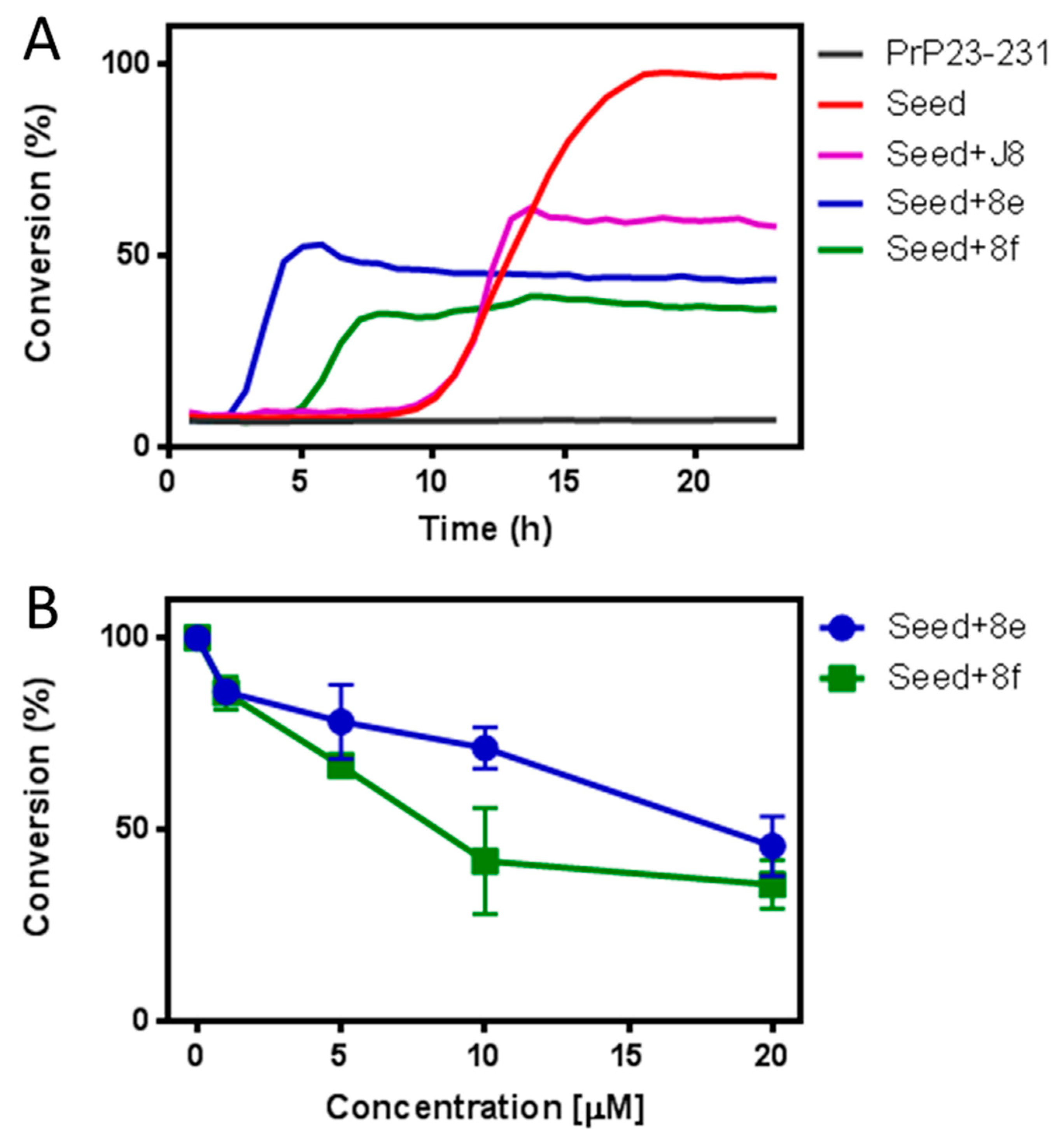
Here, we performed the RT-QuIC assay to investigate the inhibitory effect of the synthesized compounds. In vitro fibrils produced under denaturing conditions induced PrP23-231 conversion and seeded its aggregation [26,34]. Previously, J8, a trimethoxychalcone, exhibited an inhibitory effect of prion conversion through this assay and inhibited approximately 50% of recombinant PrP conversion at 1 μM [33]. Table 1 shows the residual percentages of PrP conversion after treatment with compounds 5a–d, 6a–d, and 8a–f.
Among all the evaluated compounds, 8e and 8f were able to reduce PrP conversion with a conversion percentage of 44 ± 10% and 27 ± 3%, respectively. In Figure 3, the seeding kinetics of PrP in the presence of 8e and 8f is shown to be inhibited. Moreover, the shortening of the lag phase of the curve is observed for both compounds when compared to the control and J8 curve (Figure 3A). The reduction was concentration-dependent: 8e achieved a more than 50% decrease in PrP conversion at 10 μM, while 8f exhibited the same potency at 20 μM (Figure 3B). 8f was more efficient than 8e, mainly at low concentrations (Figure 4B).
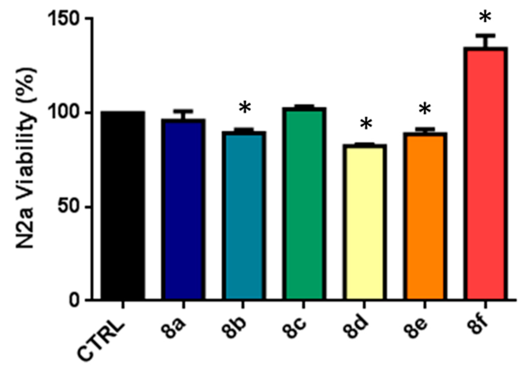
The aggregates produced in the presence of the compounds in the RT-QuIC experiment presented different conversion kinetics with varying curve profiles, including shortening in their lag phases. This could alter the final aggregates formed in terms of structure and toxicity. For this reason, these aggregates were compared in a cell viability assay using N2a cells. Although the compounds were shown to accelerate the lag phase and the exponential phase constant rate, their toxicity to N2a cells was comparable to the control (see Supporting Information File).
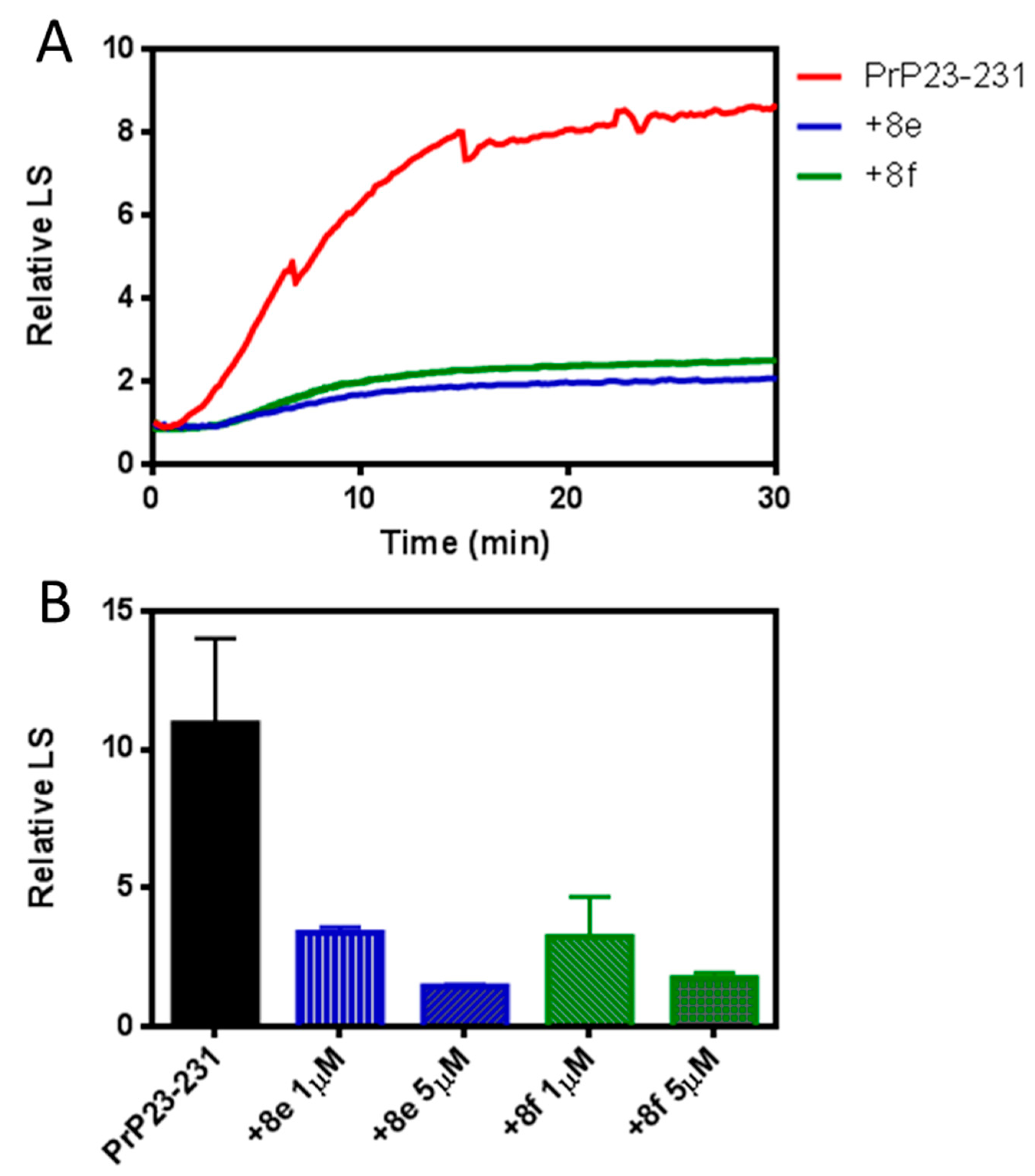
Protein aggregation can occur via different routes and form highly polymorphic species with other secondary structures and stability [35]. The environmental conditions, pH, temperature, agitation, ionic strength, and denaturant concentration are vital factors in generating variability [36,37]. An increase in temperature leads to PrP thermal denaturation, with solvent-exposed nonpolar residues, and consequently, aggregation [38,39]. Here, we chose to observe PrP aggregation at 60 °C (Figure 4) to investigate the effect of the compounds 8e and 8f on aggregates formed under conditions different than those presented above.
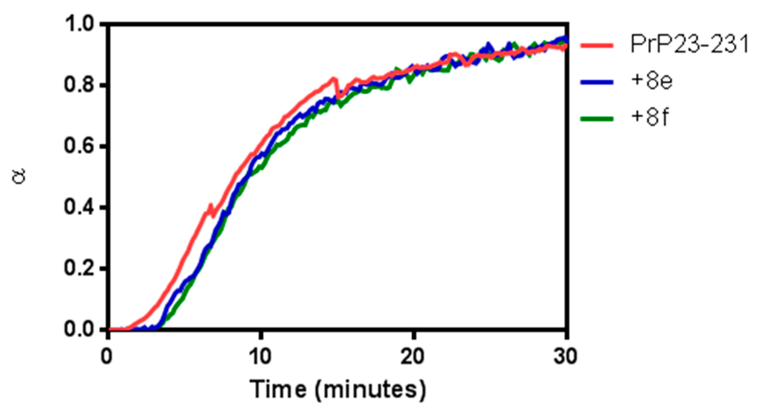
The temperature chosen for this assay induced the formation of aggregates with less ThT binding compared to lower temperatures (35–45 °C) [38]. PrP23-231 light scattering increased at 60 °C, reaching a plateau within 30 min, confirming PrP23-231 aggregation under this condition (Figure 4A). The addition of 8e and 8f decreased aggregation (Figure 4A) without changing its kinetics (better visualized in Figure 5). This effect is concentration-dependent, reaching a maximum effect at a 1:5 (PrP: compound) molar ratio (Figure 4B), as observed in the propagation experiments. The data suggest that 8e and 8f can modulate the aggregation of varied types of PrP aggregates.
The compounds must present low toxicity to neural cells as a first-order requisite of PrP conversion inhibitors to treat PrP diseases. Thus, our assay was designed to evaluate the cytotoxicity of compounds 8e and 8f on the N2a cell line, a neural cell line frequently used in PrP conversion studies. Figure 6 shows that none of the compounds used in this assay negatively affected cell viability at the concentration of 10 µM. More specifically, 8e demonstrated very little inhibition of N2a cell line viability among the compounds with more prominent effects, and 8f appeared to induce cell proliferation in the concentration used.

3. Conclusions

In conclusion, eight 1-alkyl-6-amino-4-quinolone derivatives (5a–d and 6a–d) were synthesized and used in the preparation of six 2,5-dichloro-3,6-bis((1-alkyl-4-quinolone-6-yl)amino)-1,4-benzoquinone derivatives (8af), in a regioselective way. These compounds were evaluated regarding their prion conversion inhibition ability, fibrillation modulatory effect, and anti-aggregative profile. The results shown in this work indicate that compounds 8e and 8f are promising new candidates to be used as lead compounds for developing new PrP inhibitors. Further analyses are required to understand the mechanisms of PrP conversion inhibition and confirm their in vivo effects.

4. Materials and Methods

4.1. Synthesis

4.1.1. General

All reagents and solvents were purchased from Merck & Co and used without further purification. Melting points were measured with a Fisher-Johns apparatus. NMR spectra were recorded on a Varian spectrometer operating at 500.00 MHz or 300.00 MHz (1H) and 125.00 MHz or 75.00 MHz (13C) using DMSO-d6 or CDCl3 as a solvent. Chemical shifts were reported in parts per million (ppm) relative to the solvent. Hydrogen and carbon NMR spectra were typically obtained at room temperature. The two-dimensional experiments were conducted using standard Varian Associates automated data acquisition and processing programs. Multiplicities were abbreviated as follows: s for singlets, d for doublets, t for triplets, q for quartets, quin for quintets, sxt for sextets, dd for doublet of doublets, m for multiplets, tt for a triplet of triplets, and br for broad signals. IR spectra were recorded on an ABB FTLA2000 spectrophotometer (KBr pellets) in the range of 4000–400 cm−1. The synthetic methods for preparing substances 4ad, 5ad [27,30], and 6a [30], as well as their structural characterization, have already been reported.

4.1.2. Ester Hydrolysis: General Procedure

First, 1 mmol of the corresponding ethyl 1-alkyl-6-amino-4-oxo-1,4-dihydroquinoline-3-carboxylate (4b–d), 12.5 mmol (500 mg) of sodium hydroxide, and 15 mL of ethanol were added to a round-bottom flask and kept under stirring at room temperature for 24 h, upon which chromatographic analysis showed complete consumption of the starting material (CH2Cl2/MeOH 20:1 as eluent). The solvent was evaporated, and the resulting residue was dissolved in 15 mL of distilled water. Neutralization with a dropwise addition of concentrated hydrochloric acid until pH 5–6 provided the product as a precipitated solid, which was then filtrated, washed with water, and recrystallized from ethanol.
6-amino-4-oxo-1-propyl-1,4-dihydroquinoline-3-carboxylic acid (6b). 45% yield; yellow solid; m.p. 242–244 °C. IV νmax (cm−1, KBr): 3436, 3334, 1706, 1611, 1420. 1H NMR (500.00 MHz, DMSO-d6-δ 2.50 ppm), δ in ppm: 8.73 (s, 1H, H-2), 7.74 (d, 1H, J = 9.2 Hz, H-8), 7.46 (d, 1H, J = 2.7 Hz, H-5), 7.24 (dd, 1H, J = 9.2, 2.7 Hz, H-7), 5.72 (s, 2H, NH2), 4.43 (t, 2H, J = 7.4 Hz, H-1′), 1.75 (sxt, 2H, J = 7.4 Hz, H-2′), 0.91 (t, 3H, J = 7.4 Hz, H-3′). 13C-APT NMR (125.00 MHz, DMSO-d6-δ 39.51 ppm), δ in ppm: 176.74 (C-4); 166.74 (CO2H), 147.47 (C-6 or C-4a), 145.31 (C-2), 130.53 (C-8a), 127.19 (C-6 or C-4a), 122.51 (C-7), 118.87 (C-8), 105.82 (C-3), 105.51 (C-5), 54.98 (C-1′), 22.04 (C-2′), 10.34 (C-3′). HRMS (ESI) m/z calcd for C13H15N2O3+ [M + H]+: 247.1077, found 247.1077.
6-amino-4-oxo-1-pentyl-1,4-dihydroquinoline-3-carboxylic acid (6c). 51% yield; yellow solid; m.p. 214–216 °C. IR νmax (cm−1, KBr): 3435, 3340, 1708, 1604, 1419. 1H NMR (500.00 MHz, DMSO-d6-δ 2.50 ppm), δ in ppm: 8.73 (s, 1H, H-2), 7.73 (d, 1H, J = 9.2 Hz, H-8), 7.46 (d, 1H, J = 2.7 Hz, H-5), 7.24 (dd, 1H, J = 9.2, 2.7 Hz, H-7), 5.72 (s, 2H, NH2), 4.46 (t, 2H, J = 7.4 Hz, H-1′), 1.80 (sxt, 2H, J = 7.2 Hz, H-2′), 1.35–1.28 (m, 4H, H-3′ e H-4′), 0.86 (t, 3H, J = 7.2 Hz, H-5′). 13C-APT NMR (75.00 MHz, DMSO-d6-δ 39.51 ppm), δ in ppm: 176.64 (C-4); 166.56 (CO2H), 147.40 (C-6 or C-4a), 145.18 (C-2), 130.39 (C-8a), 127.16 (C-6 or C-4a), 122.38 (C-7), 118.73 (C-8), 105.83 (C-3), 105.44 (C-5), 53.48 (C-1′), 28.32 (C-2′), 27.72 (C-3′ or C-4′), 21.38 (C-3′ or C-4′), 10.34 (C-5′). HRMS (ESI) m/z calcd for C15H19N2O3+ [M + H]+: 275.1390, found 275.1390.
6-amino-1-benzyl-4-oxo-1,4-dihydroquinoline-3-carboxylic acid (6d). 49% yield; yellow solid; m. p. 275–278 °C. IV νmax (cm−1, KBr): 3445, 3357, 1695, 1605, 1432. 1H NMR (300.00 MHz, DMSO-d6-δ 2.50 ppm), δ in ppm: 8.93 (s, 1H, H-2), 7.60 (d, 1H, J = 9.2 Hz, H-8), 7.45 (d, 1H, J = 2.7 Hz, H-5), 7.39–7.29 (m, 3H, H-3″/5″ and H-4″), 7.24 (dd, 2H, J = 8.1 and 1.6 Hz, H-2″/6″), 5.75 (s, 2H, H-1′), 5.69 (s, 1H, NH2). 13C-APT NMR (75.00 MHz, DMSO-d6-δ 39.51 ppm), δ in ppm: 177.27 (C-4); 167.12 (CO2H), 147.72 (C-6 or C-4a), 146.30 (C-2), 135.46 (C-1″), 131.05 (C-8a), 129.10 (C-3″/5″), 128.29 (C-4″), 127.53 (C-6 or C-4a), 126.77 (C-2″/6″), 122.73 (C-7), 119.65 (C-8), 106.44 (C-3), 105.80 (C-5), 56.85 (C-1′). HRMS (ESI) m/z calcd for C17H15N2O3+ [M + H]+: 295.1077, found 295.1077.

4.1.3. Dimerization through Benzoquinone Amination: General Procedure

In a round-bottom flask containing 0.163 mmol (40.0 mg) of p-chloranil (7) solubilized in 5.0 mL of ethanol, a previously prepared solution of 0.325 mmol of the respective 6-amino-4-quinolone in 5.0 mL of ethanol was added dropwise at room temperature under constant stirring. The mixture was kept under stirring at 60 °C for 24 h when another 0.163 mmol (40.0 mg) of p-chloranil (7) was added to the reaction flask, which was kept at this temperature for another 24 h. At the end of the reaction, the precipitated solid was isolated by filtration and washed with 30.0 mL of heated ethanol. The product was dried in a vacuum-sealed desiccator.
2,5-dichloro-3,6-bis((3-(ethoxycarbonyl)-1-ethyl-4-oxo-1,4-dihydroquinolin-6-yl)amino)-1,4-benzoquinone (8a): 83% yield, dark brown solid, m.p. > 300 °C. IR νmax (cm−1, KBr): 3222, 1724, 1588, 1509, 804. 1H NMR (500.00 MHz, DMSO-d6-δ 2.50 ppm), δ in ppm: 9.78 (s, 1H, NH), 8.64 (s, 1H, H-2), 7.94 (d, 1H, J = 2.6 Hz, H-5), 7.80 (d, 1H, J = 9.1 Hz, H-8), 7.64 (dd, 1H, J = 9.1 and 2.6 Hz, H-7), 4.42 (q, 2H, J = 7.1 Hz, H-1′), 4.24 (q, 2H, J = 7.1 Hz, CO2CH2CH3), 1.41 (t, 3H, J = 7.1 Hz, H-2′), 1.30 (t, 3H, J = 7.1 Hz, CO2CH2CH3). 13C NMR (75.00 MHz, with 4 eq. of K2CO3 in DMSO-d6-δ 39.51 ppm –) δ in ppm: 172.46 (C-1″/4″), 172.22 (both C-4), 164.54 (both CO2Et), 147.09 (both C-2), 145.95, 145.52, 143.74, 128.36, 120.44, 117.43, 116.36, 109.23 (both C-3), 107.59 (C-2″/5″ or C-3″/6″), 59.26 (both CO2CH2CH3), 47.68 (both C-1′), 14.03 (both C-2′ or both CO2CH2CH3), 13.95 (both CO2CH2CH3 or both C-2′). HRMS (ESI) m/z calcd for C34H30Cl2N4NaO8+ [M + Na]+: 715.1333, found 715.1331 and calcd for C34H31Cl2N4O8+ [M + H]+: 693.1513, found 693.1511.
2,5-dichloro-3,6-bis((3-(ethoxycarbonyl)-4-oxo-1-propyl-1,4-dihydroquinolin-6-yl)amino)-1,4-benzoquinone (8b): 56% yield, dark brown solid, m.p. > 300 °C. IR νmax (cm−1, KBr): 3228, 2979, 1729, 1589, 1505, 805. 1H NMR (500.00 MHz, DMSO-d6-δ 2.50 ppm), δ in ppm: 9.78 (s, 2H, both NH), 8.63 (s, 2H, both H-2), 7.94 (d, 2H, J = 2.7 Hz, both H-5), 7.80 (d, 2H, J = 9.0 Hz, both H-8), 7.61 (dd, 2H, J = 9.0 and 2.7 Hz, both H-7), 4.34 (t, 4H, J = 7.3 Hz, both H-1′ sets), 4.24 (q, 4H, J = 7.1 Hz, both CO2CH2CH3 sets), 1.82 (sxt, 4H, J = 7.3 Hz, both H-2′ sets), 1.30 (t, 3H, J = 7.1 Hz, both CO2CH2CH3 sets), 0.94 (t, 6H, J = 7.3 Hz, both H-3′ sets). 13C-APT NMR (75.00 MHz, DMSO-d6—δ 39.51 ppm) δ in ppm: 173.75 (C-1″/4″), 172.26 (both C-4), 164.51 (both CO2Et), 148.65 (both C-2), 142.53, 135.88 (both C-8a), 134.99 (both C-4a or both C-6), 128.96 (both C-7), 127.88, 120.68 (both C-5), 116.97 (both C-8), 109.54 (both C-3), 105.94 (C-2″/5″ or C-3″/6″), 59.55 (both CO2CH2CH3), 54.05 (both C-1′), 21.78 (both C-2′), 14.16 (both CO2CH2CH3), 10.43 (both C-3′). HRMS (ESI) m/z calcd for C36H34Cl2N4NaO8+ [M + Na]+: 743.1646, found 743.1643 and calcd for C36H35Cl2N4O8+ [M + H]+: 721.1826, found 721.1823.
2,5-dichloro-3,6-bis((3-(ethoxycarbonyl)-4-oxo-1-pentyl-1,4-dihydroquinolin-6-yl)amino)-1,4-benzoquinone (8c): 41% yield, dark brown solid, m. p. > 300 °C. IR νmax (cm−1, KBr): 3241, 2957, 1721, 1590, 1503, 805. 1H NMR (500.00 MHz, DMSO-d6-δ 2.50 ppm), δ in ppm: 9.77 (s, 2H, both NH), 8.62 (s, 2H, both H-2), 7.94 (d, 2H, J = 2.9 Hz, both H-5), 7.78 (d, 2H, J = 9.3 Hz, both H-8), 7.63 (dd, 2H, J = 9.3 and 2.9 Hz, both H-7), 4.36 (q, 4H, J = 7.1 Hz, both H-1′ sets), 4.24 (q, 4H, J = 7.3 Hz, both CO2CH2CH3 sets), 1.79 (quin, 4H, J = 7.1 Hz, both H-2′ sets), 1.37–1.31 (m, 8H, both H-3′ and H-4′ sets), 1.30 (t, 3H, J = 7.3 Hz, both CO2CH2CH3 sets), 0.87 (t, 6H, J = 7.3 Hz, both H-5′ sets). 13C-APT NMR (75.00 MHz, DMSO-d6-δ 39.51 ppm) δ in ppm: 173.58 (C-1″/4″), 172.19 (both C-4), 164.40 (both CO2Et), 148.33 (both C-2), 142.50, 135.90 (both C-8a), 134.95 (both C-4a or both C-6), 128.96 (both C-7), 127.88, 120.77 (both C-5), 116.80 (both C-8), 109.71 (both C-3), 105.96 (C-2″/5″ or C-3″/6″), 59.46 (both CO2CH2CH3), 52.61 (both C-1′), 27.99 (both C-2′), 27.79 (both C-3′ or both C-4′), 21.37 (both C-3′ or both C-4′), 14.04 (both CO2CH2CH3), 13.43 (both C-5′). HRMS (ESI) m/z calcd for C40H42Cl2N4NaO8+ [M + Na]+: 799.2272, found 799.2269.
2,5-dichloro-3,6-bis((3-carboxy-4-oxo-1-propyl-1,4-dihydroquinolin-6-yl)amino)-1,4-benzoquinone (8d): 57% yield, dark brown solid, m.p. 250 °C with decomposition. IR νmax (cm−1, KBr): 3262, 1724, 1653, 1584, 821. 1H NMR (500.00 MHz, DMSO-d6-δ 2.50 ppm), δ in ppm: 9.79 (s, 2H, both NH), 8.96 (s, 2H, both H-2), 8.04 (d, 2H, J = 2.7 Hz, both H-5), 8.02 (d, 2H, J = 9.3 Hz, both H-8), 7.80 (dd, 2H, J = 9.3 and 2.7 Hz, both H-7), 4.53 (t, 4H, J = 7.7 Hz, both H-1′ sets), 1.88 (sxt, 4H, J = 7.7 Hz, both H-2′ sets), 0.96 (t, 6H, J = 7.7 Hz, both H-3′ sets). 13C-APT NMR (125.00 MHz, DMSO-d6-δ 39.51 ppm) δ in ppm: 177.12 (both C-4), 173.89 (C-1″/4″), 165.69 (both CO2H), 148.42 (both C-2), 142.16 (C-2″/5″ or C-3″/6″), 136.33 (both C-4a or both C-6), 136.04 (both C-8a), 130.02 (both C-7), 125.28 (both C-4a or both C-6), 118.98 (both C-5), 117.81 (both C-8), 107.68 (C-2″/5″ or C-3″/6″), 107.24 (both C-3), 54.94 (both C-1′), 21.91 (both C-2′), 10.25 (both C-3′). HRMS (ESI) m/z calcd for C32H27Cl2N4O8+ [M + H]+: 665.1200, found 665.1201.
2,5-dichloro-3,6-bis((3-carboxy-4-oxo-1-pentyl-1,4-dihydroquinolin-6-yl)amino)-1,4-benzoquinone (8e): 45% yield, dark brown solid, m. p. > 300 °C. IR νmax (cm−1, KBr): 3257, 1722, 1654, 1583, 820. 1H NMR (500.00 MHz, DMSO-d6-δ 2.50 ppm), δ in ppm: 9.79 (s, 2H, both NH), 8.97 (s, 2H, both H-2), 8.05 (d, 2H, J = 2.7 Hz, both H-5), 8.01 (d, 2H, J = 9.3 Hz, both H-8), 7.80 (dd, 2H, J = 9.3 and 2.7 Hz, both H-7), 4.56 (t, 4H, J = 6.8 Hz, both H-1′ sets), 1.85 (quin, 4H, J = 6.8 Hz, both H-2′ sets), 1.39–1.31 (m, 8H, both H-3′ and H-4′ sets), 0.88 (t, 6H, J = 6.8 Hz, both H-5′ sets). 13C-APT NMR (75.00 MHz, DMSO-d6-δ 39.51 ppm) δ in ppm: 177.12 (both C-4), 173.88 (C-1″/4″), 165.70 (both CO2H), 148.29 (both C-2), 142.18 (C-2″/5″ or C-3″/6″), 136.32 (both C-4a or both C-6), 136.03 (both C-8a), 130.05 (both C-7), 125.30 (both C-4a or both C-6), 119.04 (both C-5), 117.78 (both C-8), 107.68 (C-2″/5″ or C-3″/6″), 107.31 (both C-3), 53.62 (both C-1′), 28.23 (both C-2′), 27.69 (both C-3′ or both C-4′), 21.36 (both C-3′ or both C-4′), 13.41 (both C-5′). HRMS (ESI) m/z calcd for C36H35Cl2N4O8+ [M + H]+: 721.1826, found 721.1826.
2,5-dichloro-3,6-bis((1-benzyl-3-carboxy-4-oxo-1,4-dihydroquinolin-6-yl)amino)-1,4-benzoquinone (8f): 66% yield, dark brown solid, m.p. > 300 °C. IR νmax (cm−1, KBr): 3263, 1717, 1655, 1585, 819. 1H NMR (500.00 MHz, DMSO-d6-δ 2.50 ppm), δ in ppm: 9.89 (s, 2H, both NH), 9.22 (s, 2H, both H-2), 7.98 (d, 2H, J = 2.7 Hz, both H-5), 7.85 (d, 2H, J = 9.4 Hz, both H-8), 7.67 (dd, 2H, J = 9.4 and 2.7 Hz, both H-7), 7.40–7.28 (m, 10H, both H-2′/6′, H-3′/5′ and H-4′ sets), 5.86 (s, 4H, both CH2 sets). 13C-APT NMR (125.00 MHz, DMSO-d6-δ 39.51 ppm) δ in ppm: 178.05 (both C-4), 174.52 (C-1″/4″), 166.30 (both CO2H), 149.71 (both C-2), 142.78 (C-2″/5″ or C-3″/6″), 137.12 (both C-4a or both C-6), 136.92 (both C-8a), 135.66 (both C-1′), 130.55 (both C-7), 129.42 (both C-3′/5′), 128.60 (C-4′), 127.32 (both C-2′/6′), 126.14 (both C-4a or both C-6), 119.58 (both C-5), 118.94 (both C-8), 108.55 (C-2″/5″ or C-3″/6″), 108.34 (both C-3), 57.12 (both NCH2). HRMS (ESI) m/z calcd for C40H27Cl2N4O8+ [M + H]+: 761.1200, found 761.1199.

4.2. Biological Assays

4.2.1. Prion Protein Expression and Purification

The murine recombinant full-length prion protein (PrP23-231) was expressed in Escherichia coli and purified by high-affinity chromatography according to the protocol described in [11].

4.2.2. In Vitro-Produced Fibril Preparation and Amplification

PrP23-231 fibrils were prepared in denaturing conditions according to previous works [11,33]. Briefly, PrP23-231 was lyophilized after purification and resuspended in 6 M GdnHCl. This sample was diluted in a 10 mM phosphate buffer at pH 6.8 containing 1 M GdnHCl, 3 M urea, and 150 mM NaCl to a final concentration of 10 μM. After that, it was incubated for 70 h at 37 °C with continuous shaking at 600 rpm in conical plastic tubes in a 0.75 mL reaction volume. This procedure leads to the formation of amyloid fibrils. These in vitro-produced fibrils were used as seeds to amplify recombinant PrP23-231 as substrate in a modified RT-QuIC protocol [11,33]. Four percent of seeds were incubated with 100 μM of 10 mM phosphate buffer at pH 7.4 containing 300 mM NaCl, 10 μM thioflavin T (ThT), and 4 μM PrP23-231 substrate. The sample was loaded into a black 96-well plate with four replicates for each sample and then sealed and transferred to a FluoStar OMEGA microplate reader (BMG LabTech) with double orbital shaking (1 min shaking at 700 rpm double orbital/1 min resting) at 42 °C for ~40 h. ThT fluorescence was measured with excitation at 450 nm and emission at 480 nm every ~15 min. Compounds were at 1 μM, 5 μM, 10 μM, and 20 μM, and the vehicle’s final concentration corresponded to 0.02% of DMSO. The exact amount of DMSO was added to PrP23-231 and PrP23-231 + seed control samples. Data are represented as the percentage of fibril conversion, using the condition PrP23-231 + seed as 100% conversion.

4.2.3. Spectroscopic Measurements

PrP light scattering was acquired using a PC1 spectrofluorometer (ISS, Champaign, IL, USA) in an “L” geometry (at 90° relative to the excitation light). The samples were illuminated at 320 nm with data acquisition at 320 nm for kinetic experiments and from 300 to 340 nm to collect light-scattering spectra. PrP23-231 aggregation was induced by the temperature at 60 °C and evaluated for 30 min after reaching equilibrium. PrP23-231 was diluted to a 4 mM final concentration in a 50 mM Tris buffer containing 100 mM NaCl at pH 7.4. Compounds were 1 μM for kinetic measurements and 1 μM and 5 μM for steady-state measurements. Once the compound vehicle was DMSO, the same amount of DMSO (0.02%) was added to the PrP23-231 sample. Data were represented related to PrP23-231 or PrP23-231 + compound light scattering at 25 °C. The fraction aggregated/non-aggregated was also shown for kinetic comparison.

4.2.4. MTT Assays

Ninety-six-well plates were used, and cells were grown to a confluence between 80 and 90% at the time of the assay. After 24 h, the medium was changed, the compounds or aggregates were added, and the cells were treated for 48 h. Then, 3-(4,5- dimethylthiazol-2-yl)-2,5-diphenyltetrazolium bromide (MTT) at 0.5 mg/mL in PBS was added to the cells for 3 h. The resulting crystals were solubilized in DMSO, and the plate was analyzed using a SpectraMax Paradigm Multi-mode Microplate Reader (Molecular Devices) at 570 nm and 650nm. The values corresponding to the difference between the absorption at 650 nm and 570 nm were used to calculate cell viability.

Supplementary Materials

The following supporting information can be downloaded at: https://www.mdpi.com/article/10.3390/molecules27227935/s1, Figures S1–S51: Spectroscopic data.

Author Contributions

A.R.P.C.: Formal analysis, investigation (synthesis), writing—original draft; M.M.: Formal analysis, investigation (biological assays).; F.d.C.S.B.: Methodology (synthesis), review and editing, supervision (synthesis).; M.C.B.V.d.S.: Conceptualization, review and editing, supervision (synthesis), funding acquisition.; J.L.S.: Funding acquisition, methodology (biological assays), supervision.; M.C.d.M.: Methodology (biological assays), investigation (biological assays), data curation, formal analysis, writing—original draft.; L.P.R.: Investigation (biological assays), data curation, formal analysis, writing—original draft.; T.C.R.G.V.: Methodology (biological assays), investigation (biological assays), data curation, formal analysis, writing—original draft.; P.N.B.: Conceptualization, methodology (synthesis), writing—original draft, review and editing, supervision, project administration (synthesis). All authors have read and agreed to the published version of the manuscript.

Funding

The authors gratefully acknowledge Coordenação de Aperfeiçoamento de Pessoal de Nível Superior—Brazil (CAPES)—Financial Code 001 and are thankful for the financial support of the Brazilian agencies Conselho Nacional de Desenvolvimento Científico e Tecnológico (CNPq)—grant number CNPq: 465395/2014-7—and Fundação de Amparo à Pesquisa do Estado do Rio de Janeiro (FAPERJ)—grant numbers E-26/203.170/2017, E-26/010.002128/2019, E-26/202.909/2019, E-26/200.419/2021 and E-26/201.416/2022.

Institutional Review Board Statement

Not applicable.

Informed Consent Statement

Not applicable.

Data Availability Statement

Not applicable.

Conflicts of Interest

The authors declare no conflict of interest.

Sample Availability

Samples of compounds 5, 6, and 8 are available from the authors.

References

  1. Riesner, D. Biochemistry and structure of PrPC and PrPSc. Br. Med. Bull. 2003, 66, 21. [Google Scholar] [CrossRef] [PubMed] [Green Version]
  2. Weissmann, C.; Enari, M.; Klöhn, P.-C.; Rossi, D.; Flechsig, E. Molecular biology of prions. Acta Neurobiol. Exp. 2002, 62, 153–166. [Google Scholar]
  3. Prusiner, S.B. Prions. Proc. Natl. Acad. Sci. USA 1998, 95, 13363. [Google Scholar] [CrossRef] [Green Version]
  4. Silber, B.M.; Gever, J.R.; Li, Z.; Gallardo-Godoy, A.; Renslo, A.R.; Widjaja, K.; Irwin, J.J.; Rao, S.; Jacobson, M.P.; Ghaemmaghami, S.; et al. Antiprion compounds that reduce PrPSc levels in dividing and stationary-phase cells. Bioorg. Med. Chem. 2013, 21, 7999. [Google Scholar] [CrossRef] [PubMed] [Green Version]
  5. Skinner, P.J.; Kim, H.O.; Bryant, D.; Kinzel, N.J.; Reilly, C.; Priola, S.A.; Ward, A.E.; Goodman, P.A.; Olson, K.; Seelig, D.M. Treatment of Prion Disease with Heterologous Prion Proteins. PLoS ONE 2015, 10, e0131993. [Google Scholar] [CrossRef] [PubMed] [Green Version]
  6. Zattoni, M.; Legname, G. Tackling prion diseases: A review of the patent landscape. Expert Opin. Ther. Pat. 2021, 31, 1097. [Google Scholar] [CrossRef] [PubMed]
  7. Spagnolli, G.; Massignan, T.; Astolfi, A.; Biggi, S.; Rigoli, M.; Brunelli, P.; Libergoli, M.; Ianeselli, A.; Orioli, S.; Boldrini, A.; et al. Pharmacological inactivation of the prion protein by targeting a folding intermediate. Commun. Biol. 2021, 4, 62. [Google Scholar] [CrossRef] [PubMed]
  8. Aguzzi, A.; Lakkaraju, A.K.K.; Frontzek, K. Toward Therapy of Human Prion Diseases. Annu. Rev. Pharmacol. Toxicol. 2018, 58, 331. [Google Scholar] [CrossRef] [PubMed] [Green Version]
  9. Barreca, M.; Iraci, N.; Biggi, S.; Cecchetti, V.; Biasini, E. Pharmacological Agents Targeting the Cellular Prion Protein. Pathogens 2018, 7, 27. [Google Scholar] [CrossRef] [Green Version]
  10. de Moraes, M.C.; Santos, J.B.; dos Anjos, D.M.; Rangel, L.P.; Vieira, T.C.R.G.; Moaddel, R.; da Silva, J.L. Prion protein-coated magnetic beads: Synthesis, characterization and development of a new ligands screening method. J. Chromatogr. A 2015, 1379, 1. [Google Scholar] [CrossRef] [PubMed] [Green Version]
  11. Vieira, T.C.R.G.; Silva, J.L. In Vitro Prion Amplification Methodology for Inhibitor Screening. In Protein Misfolding Diseases; Humana Press: New York, NY, USA, 2019; pp. 305–316. [Google Scholar] [CrossRef]
  12. Bongarzone, S.; Tran, H.N.A.; Cavalli, A.; Roberti, M.; Carloni, P.; Legname, G.; Bolognesi, M.L. Parallel Synthesis, Evaluation, and Preliminary Structure−Activity Relationship of 2,5-Diamino-1,4-benzoquinones as a Novel Class of Bivalent Anti-Prion Compound. J. Med. Chem. 2010, 53, 8197. [Google Scholar] [CrossRef] [PubMed]
  13. Tran, H.N.A.; Bongarzone, S.; Carloni, P.; Legname, G.; Bolognesi, M.L. Synthesis and evaluation of a library of 2,5-bisdiamino-benzoquinone derivatives as probes to modulate protein–protein interactions in prions. Bioorg. Med. Chem. Lett. 2010, 20, 1866. [Google Scholar] [CrossRef] [PubMed]
  14. Jangir, N.; Poonam, D.S.; Jangid, D.K. Recent advances in the synthesis of five- and six-membered heterocycles as bioactive skeleton: A concise overview. ChemistrySelect 2022, 7, e202103139. [Google Scholar] [CrossRef]
  15. Jampilek, J. Heterocycles in Medicinal Chemistry. Molecules 2019, 24, 3839. [Google Scholar] [CrossRef] [Green Version]
  16. Ilkin, V.G.; Filimonov, V.O.; Seliverstova, E.A.; Novikov, M.S.; Beryozkina, T.V.; Gagarin, A.A.; Belskaya, N.P.; Muthipeedika, N.J.; Bakulev, V.A.; Dehaen, W. Thioisomünchnones versus Acrylamides via Copper-Catalyzed Reaction of Thioamides with Diazocarbonyl Compounds. J. Org Chem. 2022, 87, 12196–12213. [Google Scholar] [CrossRef]
  17. Babu, A.; Joy, M.N.; Sunil, K.; Sajith, A.M.; Santra, S.; Zyryanov, G.V.; Konovalova, O.A.; Butorin, I.I.; Muniraju, K. Towards novel tacrine analogues: Pd(dppf)Cl2·CH2Cl2 catalyzed improved synthesis, in silico docking and hepatotoxicity studies. RSC Adv. 2022, 12, 22476–22491. [Google Scholar] [CrossRef]
  18. Nemallapudi, B.R.; Zyryanov, G.V.; Avula, B.; Guda, M.R.; Cirandur, S.R.; Venkataramaiah, C.; Rajendra, W.; Gundala, S. Meglumine as a green, efficient and reusable catalyst for synthesis and molecular docking studies of bis(indolyl)methanes as antioxidant agents. Bioorg. Chem. 2019, 87, 465–473. [Google Scholar] [CrossRef] [PubMed]
  19. Krinochkin, A.P.; Starnovskaya, E.S.; Valieva, M.I.; Kopchuk, D.S.; Santra, S.; Slepukhin, P.A.; Zyryanov, G.V.; Majee, A.; Chupakhin, O.N. One-pot synthesis of cyclopentane-fused 5’-aryl-4-cycloalkylamino-2,2’-bipyridines via the aza-Diels–Alder/SNipso reactions. Mendeleev Commun. 2022, 32, 449–451. [Google Scholar] [CrossRef]
  20. Veligeti, R.; Madhu, R.B.; Anireddy, J.; Pasupuleti, V.R.; Avula, V.K.R.; Ethiraj, K.S.; Uppalanchi, S.; Kasturi, S.; Perumal, Y.; Anantaraju, H.S.; et al. Synthesis of novel cytotoxic tetracyclic acridone derivatives and study of their molecular docking, ADMET, QSAR, bioactivity and protein binding properties. Sci. Rep. 2020, 10, 20720. [Google Scholar] [CrossRef] [PubMed]
  21. Millanao, A.R.; Mora, A.Y.; Villagra, N.A.; Bucarey, S.A.; Hidalgo, A.A. Biological Effects of Quinolones: A Family of Broad-Spectrum Antimicrobial Agents. Molecules 2021, 26, 7153. [Google Scholar] [CrossRef] [PubMed]
  22. Advani, R.H.; Hurwitz, H.I.; Gordon, M.S.; Ebbinghaus, S.W.; Mendelson, D.S.; Wakelee, H.A.; Hoch, U.; Silverman, J.A.; Havrilla, N.A.; Berman, C.J.; et al. Voreloxin, a First-in-Class Anticancer Quinolone Derivative, in Relapsed/Refractory Solid Tumors: A Report on Two Dosing Schedules. Clin. Cancer Res. 2010, 16, 2167. [Google Scholar] [CrossRef] [Green Version]
  23. Batalha, P.; Vieira de Souza, M.C.; Peña-Cabrera, E.; Cruz, D.; Santos Boechat, F.D. Quinolones in the Search for New Anticancer Agents. Curr. Pharm. Des. 2016, 22, 6009. [Google Scholar] [CrossRef]
  24. Daneshtalab, M.; Ahmed, A. Nonclassical Biological Activities of Quinolone Derivatives. J. Pharm. Pharm. Sci. 2011, 15, 52. [Google Scholar] [CrossRef] [Green Version]
  25. Batalha, P.N.; da Forezi, S.M.L.; Tolentino, N.M.d.C.; Sagrillo, F.S.; de Oliveira, V.G.; de Souza, M.C.B.V.; da Boechat, C.S.F. 4-Oxoquinoline Derivatives as Antivirals: A Ten Years Overview. Curr. Top. Med. Chem. 2020, 20, 244. [Google Scholar] [CrossRef]
  26. Martins, D.A.; Gouvea, L.R.; Muniz, G.S.V.; Louro, S.R.W.; Batista, D.d.G.J.; Soeiro, M.d.N.C.; Teixeira, L.R. Norfloxacin and N-Donor Mixed-Ligand Copper(II) Complexes: Synthesis, Albumin Interaction, and Anti- Trypanosoma cruzi Activity. Bioinorg. Chem. Appl. 2016, 2016, 1. [Google Scholar] [CrossRef] [Green Version]
  27. Oliveira, V.G.; dos Santos Faiões, V.; Gonçalves, G.B.R.; Lima, M.F.O.; Boechat, F.C.S.; Cunha, A.C.; de Andrade-Neto, V.V.; de C.d.Silva, F.; Torres-Santos, E.C.; de Souza, M.C.B.V. Design, Synthesis and Antileishmanial Activity of Naphthotriazolyl-4- Oxoquinolines. Curr. Top. Med. Chem. 2018, 18, 1454. [Google Scholar] [CrossRef]
  28. Zhang, B. Quinolone derivatives and their antifungal activities: An overview. Arch. Pharm. 2019, 352, 1800382. [Google Scholar] [CrossRef]
  29. Sharma, V.; Das, R.; Kumar Mehta, D.; Gupta, S.; Venugopala, K.N.; Mailavaram, R.; Nair, A.B.; Shakya, A.K.; Kishore Deb, P. Recent insight into the biological activities and SAR of quinolone derivatives as multifunctional scaffold. Bioorg. Med. Chem. 2022, 59, 116674. [Google Scholar] [CrossRef] [PubMed]
  30. Costa, A.R.P.; de Andrade, K.N.; Moreira, M.L.S.; Oliveira, V.G.; Carneiro, J.W.M.; da CS Boechat, S.; de Souza, M.C.B.V.; Fiorot, R.G.; Teixeira, R.I.; de Lucas, N.C.; et al. Unveiling the photophysical properties of 3-acyl-6-amino-4-quinolones and their use as proton probes. Dyes Pigment. 2022, 207, 110692. [Google Scholar] [CrossRef]
  31. Willbold, D.; Strodel, B.; Schröder, G.F.; Hoyer, W.; Heise, H. Amyloid-type Protein Aggregation and Prion-like Properties of Amyloids. Chem. Rev. 2021, 121, 8285. [Google Scholar] [CrossRef] [PubMed]
  32. Costa, D.C.F.; de Oliveira, G.A.P.; Cino, E.A.; Soares, I.N.; Rangel, L.P.; Silva, J.L. Aggregation and Prion-Like Properties of Misfolded Tumor Suppressors: Is Cancer a Prion Disease? Cold Spring Harb. Perspect. Biol. 2016, 8, a023614. [Google Scholar] [CrossRef] [PubMed]
  33. Ferreira, N.C.; Ascari, L.M.; Hughson, A.G.; Cavalheiro, G.R.; Góes, C.F.; Fernandes, P.N.; Hollister, J.R.; da Conceição, R.A.; Silva, D.S.; Souza, A.M.T.; et al. A Promising Antiprion Trimethoxychalcone Binds to the Globular Domain of the Cellular Prion Protein and Changes Its Cellular Location. Antimicrob. Agents Chemother. 2018, 62, e01441-17. [Google Scholar] [CrossRef] [PubMed] [Green Version]
  34. Vieira, T.C.R.G.; Cordeiro, Y.; Caughey, B.; Silva, J.L. Heparin binding confers prion stability and impairs its aggregation. FASEB J. 2014, 28, 2667. [Google Scholar] [CrossRef] [PubMed] [Green Version]
  35. Tycko, R. Physical and structural basis for polymorphism in amyloid fibrils. Protein Sci. 2014, 23, 1528. [Google Scholar] [CrossRef] [PubMed] [Green Version]
  36. Ziaunys, M.; Sakalauskas, A.; Mikalauskaite, K.; Snieckute, R.; Smirnovas, V. Temperature-Dependent Structural Variability of Prion Protein Amyloid Fibrils. Int. J. Mol. Sci. 2021, 22, 5075. [Google Scholar] [CrossRef] [PubMed]
  37. Yoshimura, Y.; Lin, Y.; Yagi, H.; Lee, Y.-H.; Kitayama, H.; Sakurai, K.; So, M.; Ogi, H.; Naiki, H.; Goto, Y. Distinguishing crystal-like amyloid fibrils and glass-like amorphous aggregates from their kinetics of formation. Proc. Natl. Acad. Sci. USA 2012, 109, 14446. [Google Scholar] [CrossRef] [Green Version]
  38. Chamachi, N.G.; Chakrabarty, S. Temperature-Induced Misfolding in Prion Protein: Evidence of Multiple Partially Disordered States Stabilized by Non-Native Hydrogen Bonds. Biochemistry 2017, 56, 833. [Google Scholar] [CrossRef]
  39. Cordeiro, Y.; Kraineva, J.; Ravindra, R.; Lima, L.M.T.R.; Gomes, M.P.B.; Foguel, D.; Winter, R.; Silva, J.L. Hydration and Packing Effects on Prion Folding and β-Sheet Conversion. J. Biol. Chem. 2004, 279, 32354. [Google Scholar] [CrossRef]
Figure 1. 2,5-Diamino-1,4-benzoquinone derivatives, containing aminoquinoline (1) and phenylalanine (2) moieties, with anti-prion activity [12,13].
Molecules 27 07935 g001
Figure 2. 4-Quinolone motif and possible intermolecular interactions with biomolecules. (a) π-π Stacking interactions. (b) Chelating property with biometals. (c) Hydrogen bonds.
Molecules 27 07935 g002
Scheme 1. The synthetic route toward aminoquinolones 5ad and 6ad. i: (1) K2CO3, DMF, r.t. (2) alkyl halide (bromoethane, 1-bromopropane, 1-bromopentane or benzyl chloride), DMF, 80 °C, 24 h; ii: H2, Pd/C, EtOH; iii: Fe, NH4Cl aq., reflux; iv: (1) NaOH, EtOH, r.t., 24 h. (2) HCl aq. pH = 5–6.
Molecules 27 07935 sch001
Scheme 2. Synthetic strategy for the preparation of dimers 8af. i: EtOH, 60 °C, 24 h.
Molecules 27 07935 sch002
Figure 3. 8e and 8f decrease PrP seeding conversion and aggregation into fibrils. (A) RT-QuIC reactions with recombinant PrP23-231 (4 μM) as substrate in the absence (black line, negative control) or presence of PrP23-231 fibril seed (4%) (red line, positive control). The conversion was normalized to the values for the positive control (100%) and the negative control (0%). All compounds were incubated in the starting reaction at 20 μM. All conditions were performed in quadruplicate. Representative data of three independent experiments. (B) Conversion after RT-QuIC reactions of recombinant PrP23-231 with different compound concentrations. Data are represented as the mean of three independent experiments, and error bars represent the standard deviation.
Molecules 27 07935 g003
Figure 4. 8e and 8f decrease PrP aggregation induced by high temperature. (A) Recombinant PrP23-231 (1 μM) was incubated at 60 °C in the absence (red line) or the presence of compounds (1 μM), and light scattering (LS) was monitored over time. LS was represented relative to initial LS values at 25 °C. Representative data of three independent experiments. (B) Comparison of PrP23-231 steady-state aggregation at 60 °C (red bar) and in the presence of compounds at 1:1 (1 μM) and 1:5 (5 μM) molar ratios. Data are represented as the mean of three independent experiments, and error bars represent the standard deviation.
Molecules 27 07935 g004
Figure 5. Representation of the fraction aggregated/non-aggregated (α) of PrP23-231 samples. PrP23-231 (1 μM) was incubated at 60 °C in the absence (red line) or presence of compounds (1 μM), and light scattering (LS) was monitored over time. LS increase was analyzed relative to initial LS values at 25 °C and normalized to the final values of each curve. Representative data of three independent experiments.
Molecules 27 07935 g005
Figure 6. N2a cell line viability in the presence of 10 µM of compounds 8a–f. Cells were treated with the compounds for 48 h, and the cell viability was measured through the MTT method. This set of compounds does not promote an inhibitory effect on N2a cell viability at 10 µM. * p > 0.005; n = 3.
Molecules 27 07935 g006
Table 1. Compound screening according to RT-QuIC percentage of PrP conversion ± Standard error.
Compound% Conversion
5a109 ± 22
5b118 ± 13
5c90 ± 14
5d100 ± 15
6a102 ± 3
6b73 ± 24
6c94 ± 5
6d83 ± 9
8a97 ± 12
8b78 ± 7
8c113 ± 10
8d62 ± 4
8e44 ± 10
8f27 ± 3
Publisher’s Note: MDPI stays neutral with regard to jurisdictional claims in published maps and institutional affiliations.