Article

Callus Culture of Scorzonera radiata as a New, Highly Productive and Stable Source of Caffeoylquinic Acids

by
Olga V. Grishchenko
,
Valeria P. Grigorchuk
,
Galina K. Tchernoded
,
Olga G. Koren
and
Victor P. Bulgakov
*
Federal Scientific Center of the East Asia Terrestrial Biodiversity, Far Eastern Branch of Russian Academy of Sciences, 690022 Vladivostok, Russia
*
Author to whom correspondence should be addressed.
Molecules 2022, 27(22), 7989; https://doi.org/10.3390/molecules27227989
Submission received: 18 October 2022 / Revised: 14 November 2022 / Accepted: 15 November 2022 / Published: 17 November 2022

Abstract

:
During our ongoing efforts to investigate biotechnological sources of caffeoylquinic acid (CQA) metabolites, we discovered the plant Scorzonera radiata Fisch. (Asteraceae), which is able to produce callus cultures with high yield and extremely high stability. An actively growing callus line, designated as Sr-L1, retained the ability to produce 11 CQAs during long-term cultivation (more than 20 years). A total of 29 polyphenolic compounds were identified in the leaves and Sr-L1 callus culture of S. radiata, including CQAs, lignol derivatives, flavonoids, and dihydrostilbenes. The composition of CQAs in the Sr-L1 culture was identical to that in the S. radiata leaves. Sr-L1 calli did not produce flavonoids and dihydrostilbenes, but produced lignol derivatives, which were absent in leaves. The HPLC-UV-HRMS determination showed the presence of monoacyl derivatives of CQAs such as 5-CQA, 4-CQA, cis-5-CQA, and 5-O-p-coumaroylquinic acid in the Sr-L1 culture. Among diacyl derivatives, 3,4-diCQA, 3,5-diCQA, cis-3,5-diCQA, 4,5-diCQA, 3-O-p-coumaroyl-5-O-CQA, and 3-O-caffeoyl-5-O-p-coumaroylquinic acid were found. The content of 5-CQA reached 7.54 mg/g dry weight and the content of 3,5-diCQA was as high as 18.52 mg/g dry weight. 3,5-diCQA has been reported to be of high nutritional and pharmacological value, as it alleviates inflammatory pain, reverses memory impairment by preventing neuronal apoptosis, and counteracts excessive adipose tissue expansion, serving as an attractive treatment option for obesity. The high content of 3,5-diCQA and the exceptional stability of biosynthesis make callus cultures of S. radiata a promising source for the development of drugs and nutraceuticals.

1. Introduction

The genus Scorzonera L. (Asteraceae) is represented by approximately 180–190 species widely distributing in the temperate and subtropical regions of Europe, Asia, and North Africa [1,2]. Many Scorzonera species have long been used in folk medicine of European countries, Mongolia, and China, and also employed as food plants in these regions [3,4,5,6]. Caffeoylquinic acids (CQAs) are most typical for plants of the Asteraceae family; in particular, high content of CQAs is known for artichoke (Cynara scolymus), black salsify (Scorzonera hispanica L.), purple coneflower (Echinacea purpurea L. Moench), common yarrow (Achillea millefolium L.), milk thistle (Silybum marianum L. Gaertner), coltsfoot (Tussilago farfara L.), tansy (Tanacetum vulgare L.), and chamomile (Matricaria chamomilla L.) [7]. A recent review by Lendzion et al. [8] summarized data on phytochemistry and bioactivity of 37 species and subspecies of the genus Scorzonera. CQAs were found in 25 examined plant taxa. In most Scorzonera species, only chlorogenic acid (CGA) was identified (in 19 species). Among diacyl derivatives, 3,5-diCQA was found most frequently in Scorzonera species (11 species). The greatest diversity of CQAs was found in S. radiata and S. hispanica [8]. Structural diagnostic hierarchical keys for CQA were developed by Clifford et al. [9,10,11,12,13] using the name “chlorogenic acid” in accordance with the early IUPAC nomenclature rules [14]. Many authors use the more recent names “neochlorogenic acid” and “chlorogenic acid” for 5-O-CQA and 3-O-CQA according to the PubChem database and chemical abstract (CAS) [15]. The nomenclature differences are explained in the comprehensive review by Magaña et al. [16] for the name of chlorogenic acid (3-O-CQA). We also used new nomenclature, but when discussing the results and comparing our and literature data, we took into account these disagreements.
S. austriaca, traditionally used in Chinese and Tibetan herbal medicine due to its antipyretic and anti-inflammatory action [17,18,19], was selected by the National Committee of China as a drug candidate for the treatment of hepatitis B because the total flavonoid fraction isolated from S. austriaca has been shown to have hepatoprotective and inhibitory effects on the hepatitis B virus [19]. Sezer et al. [20] investigated the neuroprotective potential of 27 taxa of the genus Scorzonera to find new sources of biologically active substances for the treatment of neurodegenerative diseases. They concluded that at least one of the species, S. pisidica, could be recommended for further investigation of its neuroprotective action [20]. Thus, the pharmacological effects and experience of using Scorzonera plants make them good candidates for drug discovery as well as nutritional supplement development. Phytochemical studies of Scorzonera species have revealed a number of secondary metabolites, such as stilbene derivatives, sesquiterpene lactones, lignans, phenolic acids, flavonoids, dihydroisocoumarins, and triterpenes [3,4,8,21,22,23,24,25].
In the present study, plants of S. radiata Fisch. were used. This plant is interesting due to the variety of secondary metabolites and many years of experience in traditional medicine. S. radiata is widely distributed in Mongolia and China [5] as well as in southeastern Russia [5,26]. The chemical composition of S. radiata was studied by a group of researchers [5,26,27]. From the aerial parts of the plant, they isolated dihydrostilbenes, flavonoids, and derivatives of quinic acid including diCQAs, as well as coumarins, simple benzoic acids, and one monoterpene glycoside. They also studied the antioxidant activity of the isolated substances and found high antioxidant activity of two dihydrostilbenes (scorzodihydrostilbenes A and E) and diCQAs [5,26,27]. It is known that the qualitative and quantitative composition of plant metabolites of the same species can vary depending on different factors, including the region of growing [28,29]. Therefore, the development of cell cultures with stable biosynthetic characteristics is a known approach to obtain a renewable source of secondary metabolites. Only one representative of the genus Scorzonera (S. hispanica) was introduced into the in vitro culture. A suspension culture of transformed S. hispanica cells producing a complex of lignans and neolignan glucosides was established [30,31]. Previously, cell cultures of S. radiata were not obtained; here we report the establishment of the first callus culture from this plant.
Our goal was to obtain a callus culture of S. radiata as a reproducible source of useful metabolites. In this study, we presented the composition of polyphenolic compounds in the S. radiata callus line, designated Sr-L1. We showed the exceptionally high stability of CQA biosynthesis in the Sr-L1 callus line. Two valuable compounds, 5-CQA and 3,5-diCQA, were produced by this culture in high yields. In addition, the chemical composition of S. radiata plants collected on Sakhalin Island was studied and compared with literature data.

2. Results

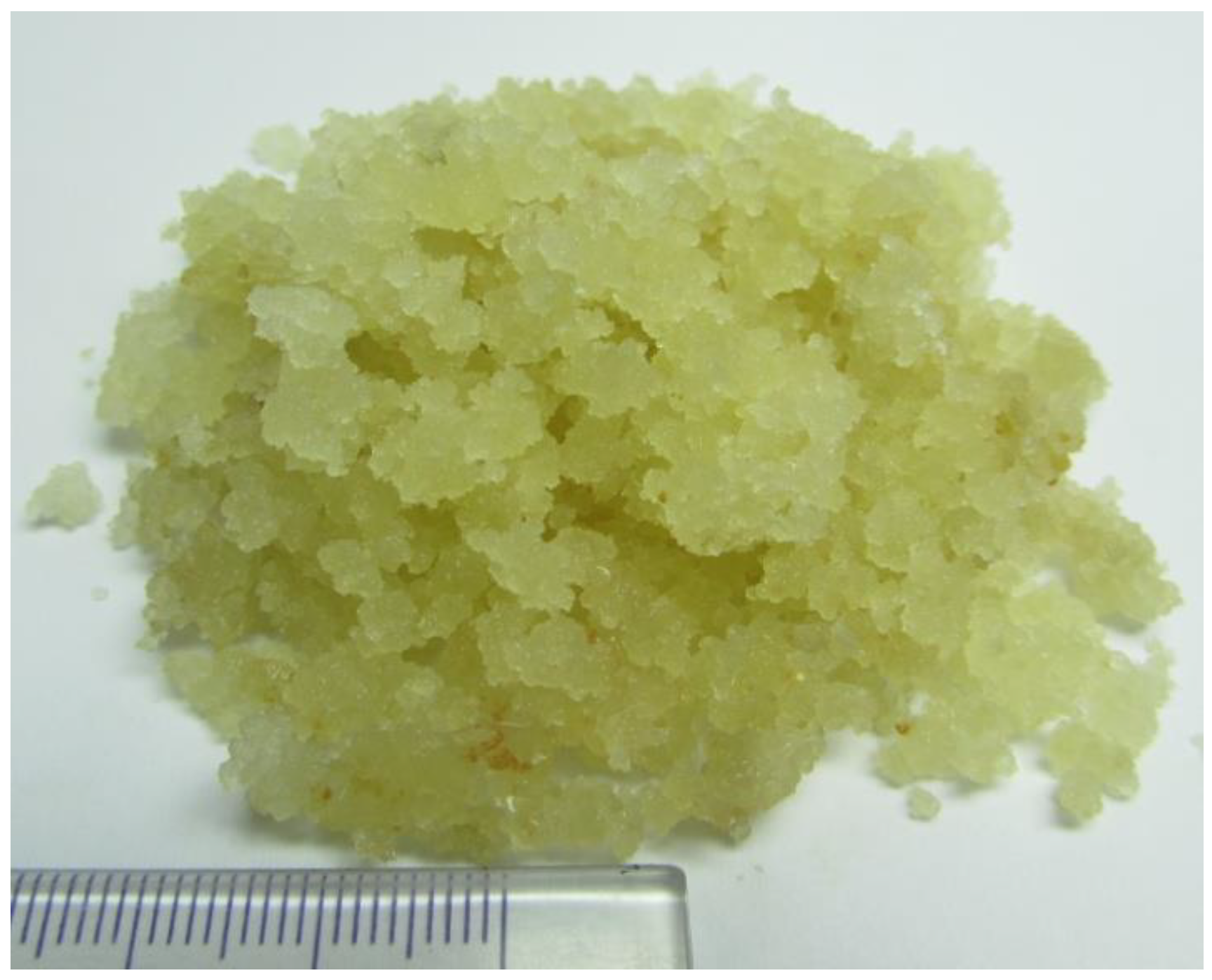
2.1. Callus Line Sr-L1

The callus line Sr-L1 of S. radiata was selected as the most actively growing culture. The selection was carried out in the usual way, that is, the selection of actively growing cell aggregates during the first years of cultivation. This line has been maintained by subculturing in the Plant Cell Culture Collection of the Federal Scientific Center for Terrestrial Biodiversity of East Asia (Vladivostok, Russia) for 20 years without any additional manipulations and has retained its excellent growth characteristics all this time. Sr-L1 produces a friable, vigorously growing callus of light green-yellow color (Figure 1).
The calli demonstrate the usual growth dynamics with an exponential phase of growth, a linear phase, and a stationary phase ending on day 34–37 by the time of collecting calli for drying. The growth index of the Sr-L1 callus line was 16.5 ± 0.06, with an inoculum mass of 0.2 g. Dry biomass obtained by drying calli at 50 °C to constant weight was 3.15% of fresh biomass.

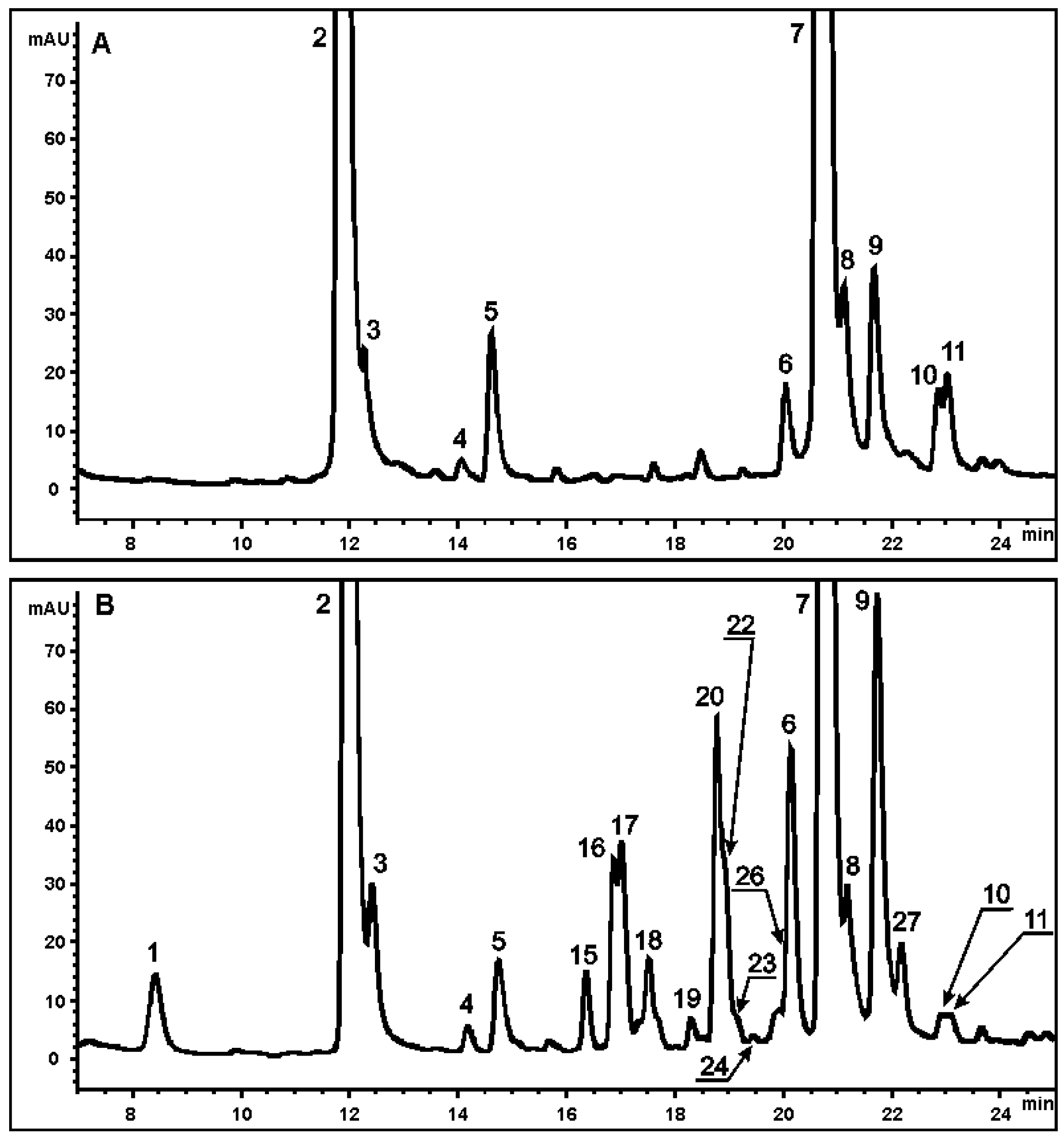
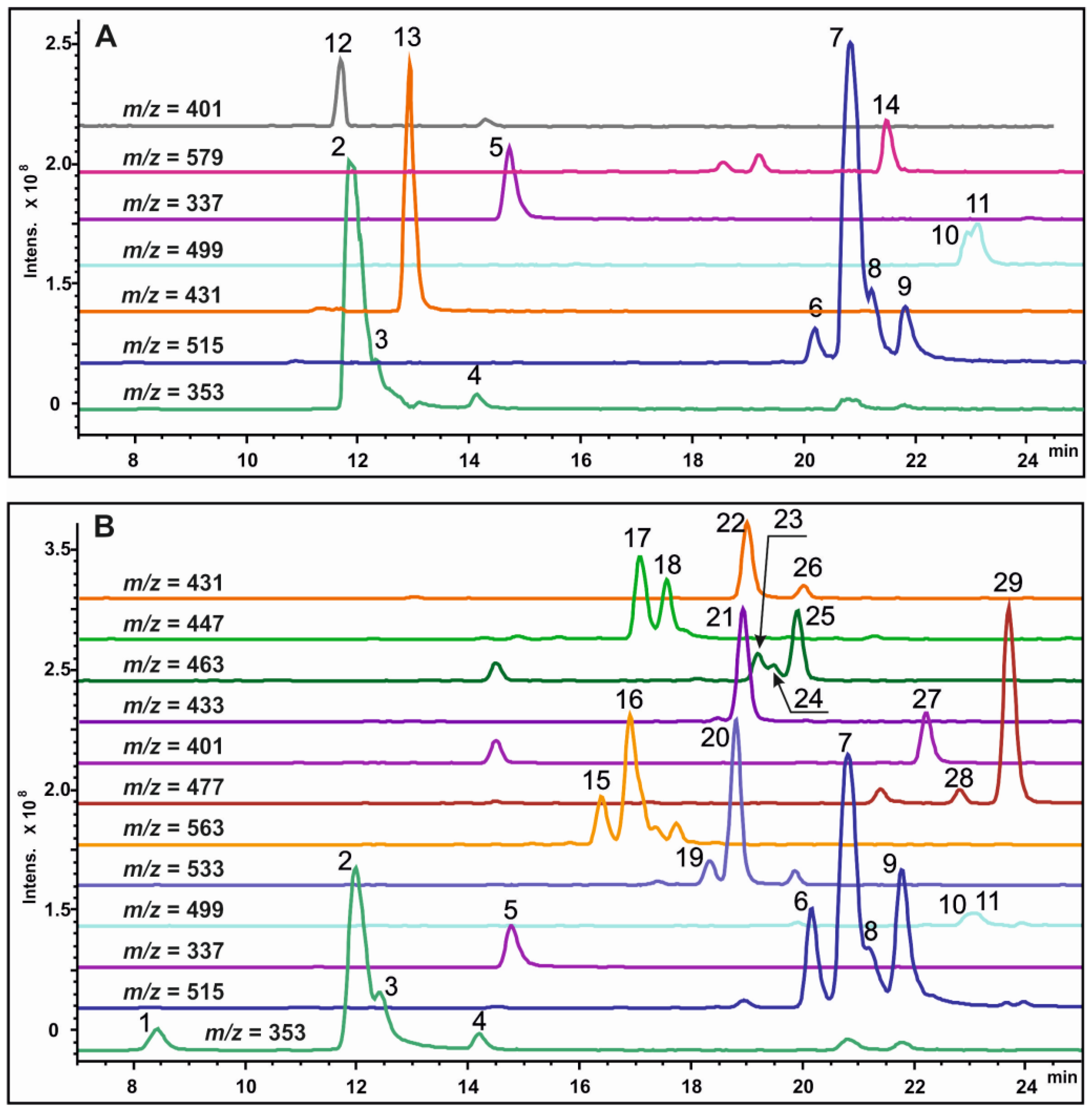
2.2. Analysis of Secondary Metabolites in the Sr-L1 Callus Line and Leaves of S. radiata Plants

HPLC-UV and ESI-MS chromatograms of crude methanol extracts of callus tissues and leaves of S. radiata are presented in Figure 2 and Figure 3.
A total of 29 substances were detected in extracts of the Sr-L1 callus line and leaves of S. radiata: 14 in the Sr-L1 line and 26 in leaves. Eleven compounds found were common to both callus and leaf extracts. All determined components of the extracts were identified on the base of UV spectra, recorded with a DAD detector, mass spectral data, and chromatographic separation using a reverse phase column. The values of the monoisotopic molecular masses were obtained using the high-resolution mass spectrometry. Based on these mass data, molecular formulas were obtained that are in good agreement with the theoretical data for each found component. A mass error was less than 5 ppm, thus confirming the elemental composition of each component. To detect the characteristic fragmentation patterns of regioisomers, multistage MS analysis was performed, and target fragmentation spectra were obtained in the negative ion mode using an ion trap mass spectrometer. For the qualitative determination of all peaks, their ESI-MS data were compared with those of chemical standards and/or literature data, and each peak was assigned on the basis of the structural diagnostic hierarchical keys previously developed [9,10,11,12,13,32]. All identified metabolites belonged to four classes: CQAs, lignans (presented as lignol derivatives), flavonoids, and stilbenoids (presented as dihydrostilbene derivatives). A total of 11 CQAs were found in the Sr-L1 line and leaves of S. radiata (Table 1), 3 lignol derivatives were found in the Sr-L1 line, and 15 flavonoids and dihydrostilbene derivatives were found in the leaves of S. radiata. Thus, calli produced CQAs and lignol derivatives, but did not produce flavonoids and dihydrostilbenes. The leaves, in turn, did not contain lignols.
Table 1. List of caffeoyl- and coumaroylquinic acid derivatives identified in methanol extracts of Sr-L1 callus culture and S. radiata leaves by their retention times and UV and MS data.
Peak No.tR (min)λmax (nm)[M-H] (m/z Detected)[M-H] (m/z Calculated)Molecular FormulaMS2 Fragmentation (Precursor Ions [M-H]) (% Base Peak) (m/z)MS3 Fragmentation (% Base Peak) (m/z) **MS4 Fragmentation (% Base Peak) (m/z) **Assignment
18.4324353.0888353.0878C16H18O9191(100) *, 179(46), 135(6)173(91), 155(41), 127(82), 111(48), 93(73), 85(100)-3-O-CQA
211.9325353.0892353.0878C16H18O9191(100), 179(2)173(100), 127(80), 111(46), 109(48), 93(48), 85(94)-5-O-CQA
312.4325353.0889353.0878C16H18O9191(38), 179(62), 173(100), 135(5)155(35), 137(18), 111(30), 93(100)-4-O-CQA
414.2320353.0887353.0878C16H18O9191(100), 179(2)173(100), 127(75), 111(26), 109(27), 93(42), 85(92)-cis-5-O-CQA
514.7315337.0939337.0929C16H18O8191(100), 163(3)173(93), 127(100), 111(40), 93(48), 85(88)-5-O-p-coumaroylquinic acid
620.1326515.1213515.1195C25H24O12353(100), 335(9), 299(1), 255(3), 203(5), 179(11), 173(30)191(50), 179(61), 173(100), 135(10)155(65), 111(23), 93(100)3,4-O-diCQA
720.7326515.1207515.1195C25H24O12353(100), 191(5)191(100), 179(38), 173(4), 135(6)173(100), 127(75), 111(63), 85(83)3,5-O-diCQA
821.1325515.1208515.1195C25H24O12353(100), 191(6)191(100), 179(32), 173(3), 135(5)173(100), 127(70), 111(55), 85(81)cis-3,5-O-diCQA
921.7326515.1206515.1195C25H24O12353(100), 335(3), 317(4), 299(5), 255(3), 203(11), 179(8), 173(10)191(29), 179(53), 173(100), 135(7)111(60), 93(100)4,5-O-diCQA
1022.9316499.1259499.1246C25H24O11353(5), 337(100), 335(3), 173(5), 163(12)173(59), 163(100), 119(6)119(100)3-O-p-coumaroyl-5-O-CQA
1123.1317499.1257499.1246C25H24O11353(100), 337(21), 191(5), 179(5)191(100), 179(16)173(100), 127(54), 111(68), 93(42), 85(64)3-O-caffeoyl-5-O-p-coumaroylquinic acid
* Precursor ions for the subsequent MSn fragmentation are shown in bold. ** The characteristic fragmentation patterns of regioisomers are shown.

2.3. Determination and Quantification of CQAs

Among detected compounds, 11 CQAs were identified (Figure 2 and Figure 3). Five monoacyl (peaks 1–5) and six diacyl (peaks 6–11) CQAs were identified according to a previously described approach [33] based on comparison of their MSn fragmentation patterns, elution/retention time, and UV absorption spectra with standards and published data [9,10,11,12,13,32]. Chromatographic and spectral characteristics of the studied metabolites are presented in Table 1.
According to the hierarchical keys previously developed [9,10,11,12,13,32] and reference standards, all eleven CQAs were designated as 3-O-CQA (1), 5-O-CQA (2), 4-O-CQA (3), cis-5-O-CQA (4), 5-O-p-coumaroylquinic acid (5), 3,4-O-diCQA (6), 3,5-O-diCQA (7), cis-3,5-O-diCQA (8), 4,5-O-diCQA (9), 3-O-p-coumaroyl-5-O-CQA (10), and 3-O-caffeoyl-5-O-p-coumaroylquinic acid (11) (Table 1). Compounds 4 and 8, presented in minor amounts and showing the MSn fragmentation pattern similar to those of compounds 2 and 7, respectively, were tentatively assigned to the cis isomers of compounds 2 and 7. Since it is known that chlorogenic acid undergoes trans–cis isomerization under the action of UV light [13], we tested this assumption in experiments with UV irradiation of all samples. After UV irradiation of extracts, the compounds 4 and 8 were detected in chromatograms as peaks with a significant increased intensity, which confirmed the presence of two cis isomers in our samples, cis-5-O-CQA (4) and cis-3,5-O-diCQA (8). Figure 4 presents the general structures of CQAs, diCQAs, and p-coumaroylquinic acids in S. radiate plants and the Sr-L1 callus line.
All identified CQAs were quantified using HPLC with UV detection at a wavelength of 325 nm on the basis of four-point regression curves built with the reference commercial standards. Chlorogenic acid was used for quantitative determination of monoacyl derivatives, and 1,3-O-diCQA (cynarin) was used for quantification of diacyl derivatives. The content of CQAs in the S. radiata Sr-L1 callus line and leaves is presented in Table 2. It should be noted that these measurements were started in November 2014 during the revision of callus cultures of various plant species from the Sakhalin collection. It was surprising to find that the Sr-L1 callus culture showed a high content of secondary metabolites, since by this time 13 years had passed after establishing the culture. Table 2 presents a summary of quantification data for the period 2014–2022. Currently, there is no trend towards a decrease in the content of CQAs or depletion of their composition. There was only one difference in the composition of CQAs: 3-CQA was not detected in Sr-L1 during 2014–2019, but was found in the samples of 2020–2022, although in small amounts. For the entire period of observation, the maximum content of the total CQAs in the calli of the Sr-L1 line was 40.61 mg/g dry weight, which was obtained in March 2022.
Table 2. Content of CQA derivatives (mg/g dry weight ± SE) in the callus line Sr-L1 and leaves of S. radiate. Results were obtained as an average of ten biological samples from different passages for callus line and three biological samples for leaves, with three analytical replicates each.
Peak NumberMetaboliteAbbreviationSr-L1Leaves
13-O-caffeoylquinic acid3-CQA0.02 ± 0.010.31 ± 0.04
25-O-caffeoylquinic acid5-CQA7.54 ± 0.807.29 ± 0.06
34-O-caffeoylquinic acid4-CQA0.04 ± 0.000.19 ± 0.03
4cis-5-O-caffeoylquinic acidcis-5-CQA0.03 ± 0.010.07 ± 0.01
55-O-p-coumaroylquinic acid 0.45 ± 0.030.26 ± 0.01
Total monoacyl derivatives 8.05 ± 0.798.11 ± 0.07
63,4-O-dicaffeoylquinic acid3,4-diCQA0.29 ± 0.030.77 ± 0.05
73,5-O-dicaffeoylquinic acid3,5-diCQA18.52 ± 1.9810.28 ± 0.37
8cis-3,5-O-dicaffeoylquinic acidcis-3,5-diCQA0.10 ± 0.030.09 ± 0.01
94,5-O-dicaffeoylquinic acid4,5-diCQA0.40 ± 0.051.34 ± 0.12
103-O-p-coumaroyl-5-O-caffeoylquinic acid 0.24 ± 0.040.06 ± 0.01
113-O-caffeoyl-5-O-p-coumaroylquinic acid 0.34 ± 0.030.06 ± 0.01
Total diacyl derivatives 19.90 ± 2.0312.60 ± 0.39
Total 27.95 ± 1.8620.71 ± 0.31

2.4. Determination of Lignol Derivatives

The HPLC-UV-MS analysis of extracts from the Sr-L1 callus line of S. radiata showed the presence of three interesting peaks (12–14) (Figure 3), which were absent in leaf extracts. These components displayed absorption maxima at nearly 266 nm and were identified as monolignol (12, 13) and dilignol (14) derivatives on the basis of their detailed MS investigation (Table 3). The UV profile and MS data of component 13 corresponded to those described for syringin (sinapyl alcohol hexoside, also known as eleutheroside B) [34,35,36]. In particular, the perfectly shown negative [M+CH3COO] ions at m/z 431.1594 and positive [M+Na]+ ions at m/z 395.1329 of peak 13 were exactly matched to the molecular formula of syringin (C17H24O9). The negative MS2 spectrum of [M+CH3COO] precursor ions demonstrated two main signals at m/z 371 and 209, corresponding to the fragments produced by consistent elimination of acetic acid and dehydrated hexose moiety, with the compositions [M-H] and [M-H-C6H10O5], respectively (Table 3). In addition, the MS3 stage of ions at m/z 209 produced the fragmentation pattern that coincided with typical ion fragmentation for sinapyl alcohol [36]. Component 12 displayed the mass spectrometric behavior similar to that of component 13, with a slight difference.
Table 3. List of lignol derivatives identified in the methanol extracts of Sr-L1 callus culture of S. radiata by their retention times, UV and MS data, and by comparison with published data.
Peak No. *tR (min)λmax (nm)Detected Ions Compositionm/z Detectedm/z CalculatedMolecular FormulaMS2 Fragmentation (% Base Peak) (m/z)MS3 Fragmentation (% Base Peak) (m/z)Assignment
1211.7258[M+CH3COO]401.1462401.1453C16H22O8341(46), 221(2), 179(100) **, 164(3), 161(3), 146(4)164(62),
161(100), 146(43)
Coniferyl alcohol O-hexoside (coniferin, coniferoside, and abietin)
[M+Na]+365.1221365.1207--
1312.9266[M+CH3COO]431.1594431.1573C17H24O9371(16), 221(8), 209(100), 194(9), 179(3), 176(4), 161(2)194(100), 191(32), 179(3), 176(19), 161(3)Sinapyl alcohol O-hexoside (syringin and eleutheroside B)
[M+Na]+395.1329395.1313--
1421.3275[M-H]579.2096579.2083C28H36O13417(100), 181(5)402(40), 387(3), 371(3), 205(3), 181(100), 175(3), 166(38), 151(15)Syringaresinol-O-hexoside (eleutheroside E1)
[M+Na]+603.2082603.2054--
* The peaks are numbered as shown in Figure 3. ** Ions subjected to subsequent MSn fragmentation are shown in bold.
The calculated molecular formula (using HRMS) of component 12 was C16H22O8, which had one less methoxy group compared to compound 13, defined as syringin. We designated the component 12 as coniferyl alcohol O-hexoside, also known as coniferin [37]. The MS2 fragmentation pattern of compound 12 was also consistent with previously published data for coniferin [37].
Similarly, component 14 was identified as syringaresinol-O-hexoside (also known as eleutheroside E1), the UV and MS characteristics of which were described previously [38,39]. In the ESI conditions, component 14 showed the formation of deprotonated ions [M-H] at m/z 579.2096 and positive sodium adduct ions [M+Na]+ at m/z 603.2082. These data are in good agreement with the molecular formula of syringaresinol-hexoside (C28H36O13). MS2 fragmentation of [M-H] showed an intensive signal at m/z 417 due to the loss of the hexoside moiety, and the MS3 stage of precursor ions at m/z 417 provided fragmentation patterns identical to syringaresinol [34,40].

2.5. Determination of Flavonoid and Dihydrostilbene Derivatives

Peaks 15–29 were detected only in leaves of S. radiate and were absent in callus extracts. A DAD profile of leaf extracts at 325 nm in conjunction with extracted ion chromatograms (Figure 2B and Figure 3B) indicated several peaks corresponding to flavonoid glycosides (flavones 15–20, 22, 26 and 27; flavonoles 23 and 24) (Table 4). The complete structural assignments of these peaks were performed by means of comparison of all collected UV and MS data with previously published information on the genus Scorzonera [26,40,41].
Table 4. List of flavonoid and dihydrostilbene derivatives identified in the methanol extracts of S. radiata leaves by their retention times, UV and MS data, and by comparison with published data.
Peak No. *tR (min)λmax (nm)Detected Ions Compositionm/z Detectedm/z CalculatedMolecular FormulaMS2 Fragmentation (% Base Peak) (m/z)Assignment
1516.4271, 338[M-H]563.1429563.1406C26H28O14503(11), 473(68), 443(98), 383(86), 353(100)Apigenin-C-hexoside-C-pentoside I
[M+H]+565.1531565.1552-
1616.9271, 238[M-H]563.1430563.1406C26H28O14503(5), 473(50), 443(95), 383(57), 353(100)Apigenin-C-hexoside-C-pentoside II
[M+H]+565.1542565.1552-
1717.0257sh., 269, 348[M-H]447.0953447.0933C21H20O11429(25), 357(82), 327(100)Luteolin-6-C-glucoside
(isoorientin)
[M+H]+449.1075449.1079-
1817.5256sh., 268, 350[M-H]447.0942447.0933C21H20O11357(50), 327(100)Luteolin-8-C-glucoside
(Orientin)
[M+H]+449.1093449.1079-
1918.3270, 339[M-H]533.1325533.1300C25H26O13515(15), 473(71), 443(100), 383(60), 353(56)Apigenin-di-C-pentoside I
[M+H]+535.1427535.1446-
2018.7271, 338[M-H]533.1323533.1300C25H26O13515(19), 473(85), 443(100), 413(16), 383(72), 353(68)Apigenin-di-C-pentoside II
[M+H]+535.1463535.1446-
2118.9285[M-H]433.1523433.1504C22H26O9271(100), 165(4)Scorzodihydrostilbene C
[M-H-C6H10O5]271.0968271.0970229(4), 165(100), 149(22)
[M+Na]+457.1458457.1475-
2219.0271, 337[M-H]431.0998431.0984C21H20O10341(6), 311(100)Apigenin-8-C-glucoside
(vitexin)
[M+H]+433.1119433.1129-
2319.2255, 265sh., 360[M-H]463.0898463.0882C21H20O12301(100), 271(2), 179(3), 151(1)Quercetin-3-O-galactoside
(hyperoside)
[M+H]+465.1011465.1028-
2419.5255, 265sh., 358[M-H]463.0901463.0882C21H20O12301(100), 271(1), 179(3), 151(2)Quercetin-3-O-glucoside (isoquercitrin)
[M+H]+465.1015465.1028-
2519.9284[M-H]463.1626463.1610C23H28O10301(100), 165(2)Scorzodihydrostilbene A
[M-H-C6H10O5]301.1072301.1081283(10), 259(4), 165(100), 149(48)
[M+Na]+487.1571487.4580-
2620.0Nd**[M-H]431.1005431.0984C21H20O10341(2), 311(100)Apigenin-6-C-glucoside
(isovitexin)
[M+H]+433.1141433.1129-
2722.2269, 340[M-H]401.0891401.0878C20H18O9341(29), 311(100)Apigenin-C-pentoside
[M+H]+403.1037403.1024-
2822.8280[M-H]477.1777477.1766C24H30O10315(100)Scorzodihydrostilbene D
[M+Na]+501.1755501.1737-
2923.7285[M-H]477.1785477.1766C24H30O10357(23), 315(100), 163(1)Scorzodihydrostilbene B
[M-H-C6H10O5]315.1229305.1238299(22), 297(44), 281(25), 163(100), 149(31)
[M+Na]+501.1751501.1737-
* The peaks are numbered as shown in Figure 2 and Figure 3. ** Not detected.
Compounds related to peaks 15 and 16 were identified as a pair of isomers by the identity of their UV and MS data (Table 4). The molecular formula of 15 and 16 was calculated as C26H28O14. The MS2 spectra of [M-H] at m/z 563 exhibited the pattern of fragmentation, which is typical for di-C-glycoside conjugates: [(M-H)-60] at m/z 503, [(M-H)-90] at m/z 473, [(M-H)-120] at m/z 443, [Agl+113] at m/z 383, and [Agl+83] at m/z 353. The presence of [(M-H)-60] fragments in the MS2 spectra corresponded to elimination of the C-pentosyl ring moiety, whereas fragment ions with the composition [(M-H)-90] and [(M-H)-120] indicated the C-hexosylated molecule.
Thus, compounds 15 and 16 were identified as isomers of apigenin-C-hexoside-C-pentoside, namely isoschaftoside and schaftoside, respectively, found earlier in the Asteraceae family [40,42]. The remaining C-glycosides were determined the same way using a well-known algorithm [43,44]. On the basis of retention times and UV and MS data (Table 4), these compounds were identified as luteolin-6-C-glucoside (isoorientin) (17), luteolin-8-C-glucoside (orientin) (18) [26,40,41], two isomers of apigenin-di-C-pentoside (19 and 20), apigenin-8-C-glucoside (vitexin) (22), apigenin-6-C-glucoside (isovitexin) (26) [41,42], and apigenin-C-pentoside (27). Components corresponding to peaks 23 and 24 were identified as two isomers of flavonol O-glycoside due to typical UV profiles and MS2 fragmentations of deprotonated ions (Table 4). As a result, quercetin-3-O-galactoside (hyperoside) (23) and quercetin-3-O-glucoside (isoquercitrin) (24) were identified, according to [40,41,42]. Flavonoid derivative content in extracts of S. radiata leaves is presented in Table 5.
A DAD profile of leaf extracts at 280 nm demonstrated four low-intensity peaks matched to dihydrostilbene glycosides. The compound corresponding to peak 21 has the molecular formula C22H26O9, as determined by accurate mass measurement of its deprotonated molecules [M-H] at m/z 433.1523 and sodium adduct ions [M+Na]+ at m/z 457.1458 (Table 3, Figure 3B). MS2 investigation of [M-H] at m/z 433 showed major fragment ions formed by the elimination of the O-hexoside residue [M-H-C6H10O5]. Based on these data, compound 21 was identified as scorzodihydrostilbene C, first discovered in S. radiata and described by Wang et al. [5]. Using the same method, components 25, 28, and 29, with similar UV and MS2 profiles, were assigned as scorzodihydrostilbene A, scorzodihydrostilbene D, and scorzodihydrostilbene B, respectively [5].

3. Discussion

Efficient production of secondary metabolites requires their stable production by plant cell cultures during long-term cultivation. In this regard, the study of the biosynthetic properties stability of cultured cells should be carried out during long-term cultivation ([45]—Section “Increasing cell productivity: what next?”). Extremely high yields of secondary metabolites are desirable, but they are difficult to attain in combination with such characteristics as long-term stability and vigorous growth [45,46]. There are few examples of long-term preservation of high productivity of secondary metabolites in plant cell cultures. The most prominent examples are isoflavones (25 years of stable production, [47]), naphthoquinones and rosmarinic acid derivatives (15 years, [45]), and anthraquinones (14 years, [48]). The callus line Sr-L1 was established 21 years ago. From 2014 to the present, it has been under intense scrutiny for the production of secondary metabolites. The composition of CQAs in the Sr-L1 culture was identical to that in the S. radiata leaves. The total content of CQA derivatives in Sr-L1 was 1.35 times higher than in leaves (27.95 vs. 20.71 mg/g dry weight). Among CQAs, the highest content was detected for 5-CQA (7.54 mg/g dry weight) and 3,5-diCQA (18.52 mg/g dry weight) (Table 2).
These values are close to the maximum yields of CQAs previously obtained in cultured cells or tissues of various plant species. For example, 5-CQA was produced in the cell suspension culture of Nicotiana plumbaginifolia at the level of 4–6.5 mg/g dry weight; the Fabiana imbricate callus culture contained 5-CQA at the level of 4.6 mg/g dry weight, and the highest content (9.5 mg/g dry weight) was found in Cichorium intybus hairy roots, transformed with the pRi-1855 Agrobacterium rhizogenes strain (reviewed by Bulgakov et al. [46]). Likewise, the content of 3,5-diCQA was 3 mg/g dry weight in Rhaponticum carthamoides hairy roots; an in vitro culture of Tanacetum vulgare roots produced 10.2 mg/g dry weight of 3,5-diCQA [49]. Most likely, the maximum yield of 3,5-diCQA was achieved in the hairy root culture of Cichorium intybus, which amounted to 55.7 mg/g dry weight [46]. Interestingly, artichoke (the cultivated cardoon or Cynara cardunculus var. altilis DC (Asteraceae)) calli transformed in our laboratory with the rolC gene (which is a known inducer of secondary metabolism) produced predominantly 1,5-diCQA, 3,4-diCQA, and chlorogenic acid, but not 3,5-diCQA [33]. The total content of CQAs was 15.4 mg/g dry weight, which is less than that of the Sr-L1 line.
Regarding biological activity of the most abundant compound, 3,5-diCQA, it should be noted that considerable attention has been paid to this compound in recent years. Raineri et al. [50] reported that 3,5-diCQA treatment increased the level of the transcription factor Nrf2 (nuclear factor erythroid 2-related factor 2) that induces the expression of the heme oxygenase 1 (HO-1) with a subsequent inhibition of both C/EBP-α (late adipogenic transcription factor C/EBP) expression and adipocyte terminal differentiation. This molecular mechanism is probably common to other compounds containing a caffeic acid moiety [50], however, 3,5-diCQA seems to be more effective that other CQA derivatives [51]. Another important action of 3,5-diCQA is its beneficial effect in neurodegenerative diseases. In particular, 3,5-diCQA improved learning and alleviated memory deficits in mice by preventing neuronal apoptosis through the protection of mitochondrial activities and the repression of apoptotic signaling molecules [52]. 3,5-DCQA attenuates inflammation-mediated hypersensitivity to pain through inhibition of MCP3-induced JAK2/STAT3 (a protein module that induces the expression of genes regulating inflammation) signaling [53]. Therefore, 3,5-DCQA could be a potential therapeutic agent for alleviating inflammatory pain. Pang et al. [54] showed that the methyl derivatives of 5-CQA (MCGA) and 3,5-diCQA are potent antiproliferation agents in hepatocellular carcinoma cells, exceeding this effect of 5-CQA. MCGA and 3,5-diCQA perturb the expression of the HIF-1α-GLUT1/3-glycolysis pathway, thus inhibiting hepatocellular carcinoma proliferation and metastasis. The authors indicated these effects as a new research hotspot in carcinoma treatment.
It is also interesting to compare the chemical composition of the plants used in our experiments with the literature data. Previously, the chemical composition of S. radiata metabolites from Mongolia was studied by Wang et al. [5,26] and Tsevegsuren et al. [27]. Totally, these authors isolated 20 metabolites from the aerial parts of the plant, including 6 flavonoids, 5 dihydrostilbenes, and 9 derivatives of CQA. Overall, we identified 26 polyphenolic compounds in leaves of S. radiata, with the most significant differences between samples observed in the composition of flavonoids. Four of the five dihydrostilbenes first discovered by Wang et al. [5] in S. radiata from Mongolia, scorzodihydrostilbene A, B, C, and D, were also detected in Sakhalin plants. At the same time, only one flavonoid, luteolin-6-C-glucoside I (isoorientin), was detected in both samples, while five other flavonoids (scorzonerin A, scorzonerin B, violanthin, kaempferol 3-O-rutinoside, and rutin), previously identified in Mongolian plants [26,27], were not detected in Sakhalin samples. Ten flavonoids were found in Sakhalin samples, which are not described for Mongolian plants, and thus, they are described for the first time for S. radiata (Table 4 and Table 5). As shown in a recent review by Lendzion et al. [8], the aerial parts of various species of the Scorzonera genus contain a wide variety of flavonoids. Apigenin, luteolin, and quercetin, as well as apigenin-C-glycosides, luteolin-C-glycosides, luteolin-O-glycosides, and quercetin-O-glycosides are the most frequently found in Scorzonera species. Luteolin 6-C-glucoside (isoorientin), found in the present work and by Wang et al. [26] in S. radiata plants, was also identified in nine other Scorzonera species [8].
As for CQAs, their composition also differs in the samples from Mongolia and Sakhalin Island. Quinic acid and eight CQA derivatives, two monoacyl and six diacyl ones, were isolated from Mongolian samples of S. radiata [26,27]. We identified 11 CQAs in S. radiata originating from Sakhalin, namely 5 monoacyl and 6 diacyl derivatives (Table 1, Figure 4). Four of them, 5-CQA, 5-O-p-coumaroylquinic acid, 3,5-diCQA, and 4,5-diCQA, were common for both Mongolian and Sakhalin samples. CQAs easily undergo structural transformation due to the spontaneous migration of caffeoyl residues among the hydroxyl groups of quinic acid under heating [16,55,56,57,58]. Taking into account that CQA derivatives are unstable under illumination and heating, the mildest methods of chemical analysis were used in the present investigation. Thus, 3,4-diCQA and monoesters of quinic acid, 3-CQA and 4-CQA, were identified for S. radiata plants for the first time (Table 1). Similarly, mixed diesters such as 3-O-p-coumaroyl-5-O-CQA and 3-O-caffeoyl-5-O-p-coumaroylquinic acid were identified in S. radiata for the first time (Table 1).

4. Materials and Methods

4.1. Plant Material and Callus Culture

Samples of Scorzonera radiata Fisch. (Asteraceae) were collected on Sakhalin Island in 2001 during a joint Russian–American–Japanese expedition (International Sakhalin Island Project, https://www.burkemuseum.org/static/okhotskia/isip/Results/reports/nsf/02report.htm: accessed on 17 October 2022). The plants were identified at the Department of Botany of the Federal Scientific Center of the East Asia Terrestrial Biodiversity (Vladivostok, Russia). Callus cultures were established from young leaves of the collected plants in the laboratory of the research vessel Akademik Oparin. Calli were established as described previously [59]. Callus cultures were cultivated in 100 mL Erlenmeyer flasks on WB/A medium at 25 °C in the dark at 30-day subculture intervals. The WB/A medium contained standard Murashige and Skoog macrosalts, microsalts, and Fe-EDTA, with the exception of NH4NO3, the concentration of which was decreased up to 400 mg/L. The following components were added to the WB/A medium (mg/L): thiamine-HCl (0.2), nicotinic acid (0.5), pyridoxine-HCl (0.5), meso-inositol (100), peptone (100), sucrose (25,000), agar (6000), 6-benzylaminopurine (0.5), and α-naphthaleneacetic acid (2.0). All reagents were obtained from Sigma-Aldrich (St. Louis, MO, USA, “Tissue Culture Grade”). The actively growing callus culture of leaf origin was selected and designated as the Sr-L1 line. The growth index of Sr-L1 was calculated as Wf-Wi/Wi, where Wf is the final biomass and Wi represents the inoculum biomass (0.2 g). The obtained callus culture is maintained by subculturing in the Collection of Plant Cell Cultures at the Federal Scientific Center of the East Asia Terrestrial Biodiversity.

4.2. Chemicals

All used solvents were of high-performance liquid chromatography (HPLC) grade. Chemical standards for the quantitative determination of CQAs (chlorogenic acid, CAS number: 327-97-9, and cynarin, 30964-13-7) and flavonoids (rutin trihydrate, CAS number: 250249-75-3) were purchased from Sigma-Aldrich (St. Louis, MO, USA).

4.3. Sample Preparation for Analytical Chromatography

Scorzonera radiata plants were brought from Sakhalin Island and transplanted into pots. Fresh leaves were collected for analysis, washed in distilled water, and subjected to convection drying at 50 °C in the darkness to a constant weight. Callus tissue was collected from the culture vessels and immediately dried under the same conditions. Leaf and callus samples were prepared for further analysis in the same way. Dried and powdered samples (about 100 mg, in duplicate) were sonicated at room temperature for 20 min in 3 mL of methanol, equilibrated for 3 h in the darkness, and then centrifuged (15,000× g, 10 min). The supernatant was filtered, and the residue was re-extracted once more in the same manner. The extracts were combined, cleared with a 0.45 μm membrane (Millipore, Bedford, MA, USA), and used for HPLC analysis. Then, 1 mL aliquots were employed for UV irradiation.

4.4. UV Irradiation

UV irradiation was performed to confirm the presence of cis isomers, since chlorogenic acid derivatives are known to undergo trans–cis isomerization under UV light [13]. If, after UV irradiation, the compound increases the peak intensity on the HPLC chromatogram, this means that this compound is indeed a cis isomer. The prepared samples of plant material (1 mL of each) were irradiated under a shortwave UV lamp at 245 nm for 40 min and used for HPLC analysis.

4.5. Analytical Chromatography and Mass Spectrometry

HPLC analysis of the samples was performed at the Instrumentation Center for Biotechnology and Genetic Engineering of the Federal Scientific Center for Terrestrial Biodiversity of East Asia using a 1260 Infinity analytical HPLC system (Agilent Technologies, Santa Clara, CA, USA) equipped with a G1315D photodiode array detector, G1311C quaternary pump, G1316A column oven, and G1329B auto sampler. The HPLC system was interfaced with an ion trap mass spectrometer (Bruker HCT ultra PTM Discovery System, Bruker Daltonik GmbH, Bremen, Germany) equipped with an electrospray ionization (ESI) source. The MS analyses were carried out with negative ions detection. The following settings were used: the range of m/z detection was 100–1000 and the drying gas (N2) flow rate was 8.0 L/min. The nebulizer gas (N2) pressure was 175 kPa, the ion source potential was −4.0 kV, and the drying gas temperature was 325 °C. Tandem mass spectra were acquired in Auto-MSn mode (smart fragmentation) using a ramping of the collision energy. The fragmentation amplitude was set to 1 V. If necessary, multistage MS experiments were performed for the parent ions of the monitoring compounds.
HPLC with high-resolution mass spectrometry (HPLC-UV-HRMS) was carried out using a LCMS-IT-TOF tandem mass spectrometer (Shimadzu, Japan) including LC-20AD Prominence liquid chromatograph (Shimadzu, Japan) and an ion trap/time-of-flight mass spectrometer. The mass spectra were recorded applying electrospray ionization (ESI) with simultaneous negative and positive ion detection with a resolution of 12,000. The following settings were used: the range of m/z detection was 100–1000, the drying gas (N2) pressure was 195 kPa, the nebulizer gas flow rate was 1.5 L/min, the ion source potential changed from −3.8 to 4.5 kV, and the interface temperature was 200 °C.
An analytical Zorbax C18 column (150 mm, 2.1 mm i.d., 3.5 μm part size, Agilent Technologies, Santa Clara, CA, USA) was applied for separation. The column temperature was maintained at 40 °C. The mobile phase consisted of a gradient elution of 0.1% aqueous acetic acid (A) and acetonitrile (B). The gradient profile with a flow rate of 0.2 mL/min was the following: 0 min 5% B; 20 min 30% B; 30 min 100% B; and then eluent B until 40 min. The injection volume was 1–5 μL. UV spectra were recorded with a DAD detector in the range between 200 and 400 nm. Chromatograms for quantification were acquired at a wavelength of 325 nm.
All identified phenolic acids and flavonoid glycosides were quantified using HPLC with UV detection at a wavelength of 325 nm on the basis of four-point regression curves built with the reference commercial standards. For quantification of CQAs, two external standards were used: chlorogenic acid for monoacyl derivatives and cynarin for diacyl derivatives. Quantitative data of all flavonoid glycosides were obtained using rutin trihydrate solution as the external standard. The amount of each individual compound was calculated to correct for the difference in molecular weights.

4.6. Statistical Analysis

All values were expressed as the mean ± SE using Statistica 10.0 (StatSoft Inc., Tulsa, OK, USA).

5. Conclusions

The Sr-L1 callus line produced CQAs and lignol derivatives but did not produce flavonoids and dihydrostilbenes. The leaves of S. radiata, in turn, did not contain lignols. The high content of CQAs and the exceptional stability of biosynthesis make the callus culture of S. radiata Sr-L1 a promising source for the development of drugs and nutraceuticals for daily and dietary nutrition. This is especially important in connection with the search for effective means to combat obesity and other metabolic disorders, as well as potential activity of 3,5-diCQA in improving cognitive functions. From a biotechnological point of view, it is interesting to note that the Sr-L1 line produces 3,5-diCQA in high amounts, 1.9% of the dry cell weight, for many years. Apparently, this observation will contribute to the increased interest of biotechnology companies in this class of secondary metabolites.

Author Contributions

Conceptualization, V.P.B.; HPLS-MS analysis, assessment of results, data interpretation, V.P.G.; cell culture, O.V.G. and G.K.T.; writing—original draft preparation, V.P.B., O.G.K., O.V.G., and V.P.G.; writing—figure preparation, V.P.G.; writing—review and editing, V.P.B., O.G.K., O.V.G., and V.P.G.; supervision, V.P.B. All authors have read and agreed to the published version of the manuscript.

Funding

Financial support was provided by the Russian Science Foundation, grant no. 20-16-00016.

Institutional Review Board Statement

Not applicable.

Informed Consent Statement

Not applicable.

Acknowledgments

The analyses described in this work were performed using equipment from the Instrumental Center for Biotechnology and Gene Engineering at the Federal Scientific Center of East Asia Terrestrial Biodiversity of the Far East Branch of the Russian Academy of Sciences. We would like to thank Barkalov V.Y. and Yakubov V.P. for their help in collecting and identifying the material.

Conflicts of Interest

The authors declare no conflict of interest.

References

  1. Kamelin, R.V.; Tagaev, I.U. A review of the Scorzonera species (Asteraceae). Bot. Zhurnal 1986, 71, 1672–1682. (In Russian) [Google Scholar]
  2. Zaika, M.A.; Kilian, N.; Jones, K.; Krinitsina, A.A.; Nilova, M.V.; Speranskaya, A.S.; Sukhorukov, A.P. Scorzonera sensu lato (Asteraceae, Cichorieae)—Taxonomic reassessment in the light of new molecular phylogenetic and carpological analyses. PhytoKeys 2020, 137, 1–85. [Google Scholar] [CrossRef] [PubMed]
  3. Zidorn, C.; Ellmerer, E.P.; Sturm, S.; Stuppner, H. Tyrolobibenzyls E and F from Scorzonera humilis and distribution of caffeic acid derivatives, lignans and tyrolobibenzyls in European taxa of the subtribe Scorzonerinae (Lactuceae, Asteraceae). Phytochemistry 2003, 63, 61–67. [Google Scholar] [CrossRef]
  4. Tsevegsuren, N.; Edrada, R.; Lin, W.; Ebel, R.; Torre, C.; Ortlepp, S.; Wray, V.; Proksch, P. Biologically active natural products from Mongolian medicinal plants Scorzonera divaricata and Scorzonera pseudodivaricata. J. Nat. Prod. 2007, 70, 962–967. [Google Scholar] [CrossRef] [PubMed]
  5. Wang, Y.; Edrada-Ebel, R.; Tsevegsuren, N.; Sendker, J.; Braun, M.; Wray, V.; Lin, W.; Proksch, P.J. Dihydrostilbene derivatives from the mongolian medicinal plant Scorzonera radiata. J. Nat. Prod. 2009, 72, 671–675. [Google Scholar] [CrossRef]
  6. Bahadır, A.Ö.; Hošek, J.; Babula, P.; Cvačka, J.; Budešínský, M.; Dračinský, M.; Saltan, İ.G.; Kadlecová, D.; Ballová, L.; Šmejkal, K. Turkish. Scorzonera species extracts attenuate cytokine secretion via inhibition of NF-κB activation, showing anti-inflammatory effect in vitro. Molecules 2016, 21, 43. [Google Scholar] [CrossRef] [Green Version]
  7. Gil, M.; Wianowska, D. Chlorogenic acids—Their properties, occurrence and analysis. Ann. Univ. Mariae Curie-Sklodowska Sect. AA–Chem. 2017, LXXII, 61–104. [Google Scholar] [CrossRef]
  8. Lendzion, K.; Gornowicz, A.; Bielawski, K.; Bielawska, A. Phytochemical composition and biological activities of Scorzonera species. Int. J. Mol. Sci. 2021, 22, 5128. [Google Scholar] [CrossRef]
  9. Clifford, M.N.; Johnston, K.L.; Knight, S.; Kuhnert, N. Hierarchical scheme for LC-MSn identification of chlorogenic acids. J. Agric. Food Chem. 2003, 51, 2900–2911. [Google Scholar] [CrossRef]
  10. Clifford, M.N.; Knight, S.; Kuhnert, N. Discriminating between the six isomers of dicaffeoylquinic acid by LC-MSn. J. Agric. Food Chem. 2005, 53, 3821–3832. [Google Scholar] [CrossRef]
  11. Clifford, M.N.; Marks, S.; Knight, S.; Kuhnert, N. Characterization by LC-MSn of four new classes of p-coumaric acid-containing diacyl chlorogenic acids in green coffee beans. J. Agric. Food Chem. 2006, 54, 4095–4101. [Google Scholar] [CrossRef] [PubMed]
  12. Clifford, M.N.; Zheng, W.; Kuhnert, N. Profiling the chlorogenic acids of aster by HPLC-MSn. Phytochem. Anal. 2006, 17, 384–393. [Google Scholar] [CrossRef] [PubMed]
  13. Clifford, M.N.; Kirkpatrick, J.; Kuhnert, N.; Roozendaal, H.; Salgado, P.R. LC-MSn analysis of the cis isomers of chlorogenic acids. Food Chem. 2008, 106, 379–385. [Google Scholar] [CrossRef]
  14. IUPAC. Nomenclature of cyclitols. Biochem. J. 1976, 153, 23–31. [Google Scholar]
  15. Kim, S.; Thiessen, P.A.; Bolton, E.E.; Chen, J.; Fu, G.; Gindulyte, A.; Han, L.; He, J.; He, S.; Shoemaker, B.A.; et al. PubChem Substance and Compound databases. Nucleic Acids Res. 2016, 44, D1202–D1213. [Google Scholar] [CrossRef] [PubMed]
  16. Magaña, A.A.; Kamimura, N.; Soumyanath, A.; Stevens, J.F.; Maier, C.S. Caffeoylquinic acids: Chemistry, biosynthesis, occurrence, analytical challenges, and bioactivity. Plant J. 2021, 107, 1299–1319. [Google Scholar] [CrossRef]
  17. Zhu, Y.; Wu, Q.; Hu, P.; Wu, W. Biguaiascorzolides A and B: Two novel dimeric guaianolides with a rare skeleton, from Scorzonera austriaca. Food Chem. 2009, 114, 1316–1320. [Google Scholar] [CrossRef]
  18. Wu, Q.X.; Su, Y.B.; Zhu, Y. Triterpenes and steroids from the roots of Scorzonera austriaca. Fitoterapia 2011, 82, 493–496. [Google Scholar] [CrossRef]
  19. Xie, Y.; Guo, Q.-S.; Wang, G.-S. Preparative separation and purification of the total flavonoids in Scorzonera austriaca with macroporous resins. Molecules 2016, 21, 768. [Google Scholar] [CrossRef]
  20. Sezer, S.F.; Bahadir, A.O.; Saltan, C.G.; Erdogan, O.L.; Dall’Acqua, S.; Ozgokce, F. Prospective neurobiological effects of the aerial and root extracts and some pure compounds of randomly selected Scorzonera species. Pharm. Biol. 2014, 52, 873–882. [Google Scholar] [CrossRef]
  21. Zidorn, C.; Ellmerer-Müller, E.P.; Stuppner, H. Sesquiterpenoids from Scorzonera hispanica. Pharmazie 2000, 55, 550–555. [Google Scholar] [PubMed]
  22. Zidorn, C.; Ellmerer-Muller, E.P.; Stuppner, H. Tyrolobibenzyls—Novel secondary metabolites from Scorzonera humilis. Helv. Chim. Acta 2000, 83, 2920–2925. [Google Scholar] [CrossRef]
  23. Zidorn, C.; Petersen, B.O.; Udovičić, V.; Larsen, T.O.; Duus, J.O.; Rollinger, J.M.; Ongania, K.H.; Ellmerer, E.P.; Stuppner, H. Podospermic acid, 1,3,5-tri-O-(7,8-dihydrocaffeoyl)quinic acid from Podospermum laciniatum (Asteraceae). Tetrahedron Lett. 2005, 46, 1291–1294. [Google Scholar] [CrossRef]
  24. Paraschos, S.; Magiatis, P.; Kalpoutzakis, E.; Harvala, C.; Skaltsounis, A.L. Three new dihydroisocoumarins from the Greek endemic species Scorzonera cretica. J. Nat. Prod. 2001, 64, 1585–1587. [Google Scholar] [CrossRef]
  25. Sari, A.; Zidorn, C.; Ellmerer, E.P.; Ozgokce, F.; Ongania, K.H.; Stuppner, H. Phenolic compounds from Scorzonera tomentosa L. HeIv. Chim. Acta 2007, 90, 311–317. [Google Scholar] [CrossRef]
  26. Wang, Y.; Wray, V.; Tsevegsuren, N.; Lin, W.; Proksch, P. Phenolic compounds from the mongolian medicinal plant Scorzonera radiata. Z. Naturforsch. 2012, 67c, 135–143. [Google Scholar] [CrossRef]
  27. Tsevegsuren, N.; Proksch, P.; Wang, Y.; Davaakhuu, G. Bioactive phenolic acids from Scorzonera radiata Fisch. Mong. J. Chem. 2014, 12, 78–84. [Google Scholar] [CrossRef]
  28. Bahadır, A.Ö.; Ergene, Ö.B.; Bakar, F.; Saltan, Ç.G.; Nebioğlu, S. Evaluation of antioxidant activities and phenolic compounds of Scorzonera latifolia (Fisch. & Mey.) DC. collected from different geographic origins in Turkey. Turk. J. Pharm. Sci. 2017, 14, 179–184. [Google Scholar]
  29. Banerjee, S.K.; Bonde, C.G. Total phenolic content and antioxidant activity of extracts of Bridelia retusa Spreng bark: Impact of dielectric constant and geographical location. J. Med. Plants Res. 2011, 5, 817–822. [Google Scholar]
  30. Bryanskii, O.V.; Tolstikhina, V.V.; Semenov, A.A. A glycoside of syringaresinol from a tissue culture of Scorzonera hispanica. Chem. Nat. Compd. 1992, 25, 519–520. [Google Scholar] [CrossRef]
  31. Bryanskii, O.V.; Tolstikhina, V.V.; Zinchenko, S.V.; Semenov, A.A. A sesquiterpene glucoside from cultivated cells of Scorzonera hispanica. Chem. Nat. Compd. 1992, 28, 556–560. [Google Scholar] [CrossRef]
  32. Jaiswal, R.; Sovdat, T.; Vivan, F.; Kuhnert, N. Profiling and characterization by LC-MSn of the chlorogenic acids and hydroxycinnamoylshikimate esters in mate (Ilex paraguariensis). J. Agric. Food Chem. 2010, 58, 5471–5484. [Google Scholar] [CrossRef] [PubMed]
  33. Vereshchagina, Y.V.; Bulgakov, V.P.; Grigorchuk, V.P.; Veremeichik, G.N.; Tchernoded, G.K.; Gorpenchenko, T.Y.; Koren, O.G.; Phan, N.H.T.; Minh, N.T.; Chau, L.T.; et al. The rolC gene increases caffeoylquinic acid production in transformed artichoke cells. Appl. Microbiol. Biotechnol. 2014, 98, 7773–7780. [Google Scholar] [CrossRef] [PubMed]
  34. Eyles, A.; Jones, W.; Riedl, K.; Cipollini, D.; Schwartz, S.; Chan, K.; Herms, D.A.; Bonello, P. Comparative phloem chemistry of Manchurian (Fraxinus mandshurica) and two north American ash species (Fraxinus Americana and Fraxinus pennsylvanica). J. Chem. Ecol. 2007, 33, 1430–1448. [Google Scholar] [CrossRef]
  35. Sundaram, C.K.S.; Subramanian, I.P.I.; Subramanian, S.P.S. Isolation, characterization of syringin, phenylpropanoid glycosidefrom Musa paradisiaca tepal extract and evaluation of its antidiabeticeffect in streptozotocin-induced diabetic rats. Biomed. Prev. Nutr. 2014, 4, 105–111. [Google Scholar] [CrossRef]
  36. Sun, H.; Liu, J.; Zhang, A.; Zhang, Y.; Meng, X.; Han, Y.; Zhang, Y.; Wang, X. Characterization of the multiple components of Acanthopanax senticosus stem by ultra high performance liquid chromatography with quadrupole time-of-flight tandem mass spectrometry. J. Sep. Sci. 2015, 39, 496–502. [Google Scholar] [CrossRef]
  37. Tchoumtchoua, J.; Mathiron, D.; Pontarin, N.; Gagneul, D.; van Bohemen, A.-I.; N’nang, E.O.; Mesnard, F.; Petit, E.; Fontaine, J.-X.; Molinié, R.; et al. Phenolic profiling of flax highlights contrasting patterns in winter and spring varieties. Molecules 2019, 24, 4303. [Google Scholar] [CrossRef] [Green Version]
  38. Matsuda, F.; Hirai, M.Y.; Sasaki, E.; Akiyama, K.; Yonekura-Sakakibara, K.; Provart, N.J.; Sakurai, T.; Shimada, Y.; Saito, K. AtMetExpress development: A phytochemical atlas of Arabidopsis development. Plant Physiol. 2010, 152, 566–578. [Google Scholar] [CrossRef] [Green Version]
  39. Sanz, M.; de Simón, B.F.; Cadahía, E.; Esteruelas, E.; Muñoz, A.M.; Hernández, T.; Estrellac, I.; Pintod, E. LC-DAD/ESI-MS/MS study of phenolic compounds in ash (Fraxinus excelsior L. and F. americana L.) heartwood. Effect of toasting intensity at cooperage. J. Mass Spectrom. 2012, 47, 905–918. [Google Scholar] [CrossRef]
  40. Granica, S.; Lohwasser, U.; Jöhrer, K.; Zidorn, C. Qualitative and quantitative analyses of secondary metabolites in aerial and subaerial of Scorzonera hispanica L. (black salsify). Food Chem. 2015, 173, 321–331. [Google Scholar] [CrossRef]
  41. Granica, S.; Zidorn, C. Phenolic compounds from aerial parts as chemosystematic markers in the Scorzonerinae (Asteraceae). Biochem. Syst. Ecol. 2015, 58, 102–113. [Google Scholar] [CrossRef]
  42. Olennikov, D.N.; Chirikova, N.K.; Kashchenko, N.I.; Nikolaev, V.M.; Kim, S.-W.; Vennos, C. Bioactive phenolics of the genus Artemisia (Asteraceae): HPLC-DAD-ESI-TQ-MS/MS profile of the Siberian species and their inhibitory potential against α-amylase and α-glucosidase. Front. Pharmacol. 2018, 9, 756. [Google Scholar] [CrossRef] [PubMed]
  43. Gattuso, G.; Caristi, C.; Gargiulli, C.; Bellocco, E.; Toscano, G.; Leuzzi, U. Flavonoid glycosides in bergamot juice (Citrus bergamia Risso). J. Agric. Food Chem. 2006, 54, 3929–3935. [Google Scholar] [CrossRef]
  44. Ferreres, F.; Andrade, P.B.; Valentão, P.; Gil-Izquierdo, A. Further knowledge on barley (Hordeum vulgare L.) leaves O-glycosyl-C-glycosyl flavones by liquid chromatography-UV diode-array detection-electrospray ionisation mass spectrometry. J. Chromatogr. A 2008, 1182, 56–64. [Google Scholar] [CrossRef] [PubMed]
  45. Bulgakov, V.P.; Inyushkina, Y.V.; Fedoreyev, S.A. Rosmarinic acid and its derivatives: Biotechnology and applications. Crit. Rev. Biotechnol. 2012, 32, 203–217. [Google Scholar] [CrossRef]
  46. Bulgakov, V.P.; Vereshchagina, Y.V.; Veremeichik, G.N. Anticancer Polyphenols from Cultured Plant Cells: Production and New Bioengineering Strategies. Curr. Med. Chem. 2018, 25, 4671–4692. [Google Scholar] [CrossRef] [PubMed]
  47. Federici, E.; Touché, A.; Choquart, S.; Avanti, O.; Fay, L.; Offord, E.; Courtois, D. High isoflavone content and estrogenic activity of 25 year-old Glycine max tissue cultures. Phytochemistry 2003, 64, 717–724. [Google Scholar] [CrossRef]
  48. Veremeichik, G.N.; Bulgakov, V.P.; Shkryl, Y.N.; Silantieva, S.A.; Makhazen, D.S.; Tchernoded, G.K.; Mischenko, N.P.; Fedoreyev, S.A.; Vasileva, E.A. Activation of anthraquinone biosynthesis in long-cultured callus culture of Rubia cordifolia transformed with the rolA plant oncogene. J. Biotechnol. 2019, 306, 38–46. [Google Scholar] [CrossRef]
  49. Devrnja, N.; Krstić-Milošević, D.; Janošević, D.; Tešević, V.; Vinterhalter, B.; Savić, J.; Ćalić, D. In vitro cultivation of tansy (Tanacetum vulgare L.): A tool for the production of potent pharmaceutical agents. Protoplasma 2021, 258, 587–599. [Google Scholar] [CrossRef] [PubMed]
  50. Raineri, A.; Campagnari, R.; Dal Toso, R.; Copetti, S.; Gomez-Lira, M.; Menegazzi, M. 3,5-Dicaffeoylquinic acid lowers 3T3-L1 mitotic clonal expansion and adipocyte differentiation by enhancing heme oxygenase-1 expression. Molecules 2021, 26, 5027. [Google Scholar] [CrossRef]
  51. Jang, Y.S.; Kim, H.-Y.; Zuo, G.; Lee, E.H.; Kang, S.K.; Lim, S.S. Constituents from Solidago virgaurea var. Gigantea and their inhibitory effect on lipid accumulation. Fitoterapia 2020, 146, 104683. [Google Scholar] [CrossRef] [PubMed]
  52. Kang, J.Y.; Park, S.K.; Guo, T.J.; Ha, J.S.; Lee, D.S.; Kim, J.M.; Lee, U.; Kim, D.O.; Heo, H.J. Reversal of Trimethyltin-Induced Learning and Memory Deficits by 3,5-dicaffeoylquinic acid. Oxidative Med. Cell. Longev. 2016, 2016, 6981595. [Google Scholar] [CrossRef] [PubMed] [Green Version]
  53. Park, J.; Kim, Y.; Lee, C.; Kim, Y.T. 3,5-Dicaffeoylquinic acid attenuates microglial activation-mediated inflammatory pain by enhancing autophagy through the suppression of MCP3/JAK2/STAT3 signaling. Biomed. Pharmacother. 2022, 153, 113549. [Google Scholar] [CrossRef] [PubMed]
  54. Pang, Y.; Lin, Y.; Wang, X.; Wang, J.; Liu, Q.; Ding, N.; Huang, L.; Xiang, Q.; Fang, J.; Tan, G.; et al. Inhibition of abnormally activated HIF-1α-GLUT1/3-glycolysis pathway enhances the sensitivity of hepatocellular carcinoma to 5-caffeoylquinic acid and its derivatives. Eur. J. Pharmacol. 2022, 920, 174844. [Google Scholar] [CrossRef]
  55. Moglia, A.; Lanteri, S.; Comino, C.; Hill, L.; Knevitt, D.; Cagliero, C.; Rubiolo, P.; Bornemann, S.; Martin, C. Dual catalytic activity of hydroxycinnamoyl-coenzyme A quinate transferase from tomato allows it to moonlight in the synthesis of both mono- and dicaffeoylquinic acids. Plant Physiol. 2014, 166, 1777–1787. [Google Scholar] [CrossRef]
  56. Deshpande, S.; Jaiswal, R.; Matei, M.F.; Kuhnert, N. Investigation of acyl migration in mono- and dicaffeoylquinic acids under aqueous basic, aqueous acidic, and dry roasting conditions. J. Agric. Food Chem. 2014, 62, 9160–9170. [Google Scholar] [CrossRef]
  57. Xue, M.; Shi, H.; Zhang, J.; Liu, Q.-Q.; Guan, J.; Zhang, J.-Y.; Ma, Q. Stability and degradation of CQAs under different storage conditions studied by high-performance liquid chromatography with photo diode array detection and high-performance liquid chromatography with electrospray ionization collision-induced dissociation tandem mass spectrometry. Molecules 2016, 21, 948. [Google Scholar]
  58. Dawidowicz, A.L.; Typek, R. Transformation of chlorogenic acids during the coffee beans roasting process. Eur. Food Res. Technol. 2017, 243, 379–390. [Google Scholar] [CrossRef]
  59. Bulgakov, V.P.; Tchernoded, G.K.; Mischenko, N.P.; Khodakovskaya, M.; Glazunov, V.P.; Radchenko, S.V.; Zvereva, E.V.; Fedoreyev, S.A.; Zhuravlev, Y.N. Effect of salicylic acid, methyl jasmonate, ethephon and cantharidin on anthraquinone production by Rubia cordifolia callus cultures transformed with the rolB and rolC gene. J. Biotechnol. 2002, 97, 213–221. [Google Scholar] [CrossRef]
Figure 1. View of the Sr-L1 callus line of Scorzonera radiata at the stationary phase of growth, 30 days.
Molecules 27 07989 g001
Figure 2. Representative HPLC-UV profiles of S. radiata extracts, monitored at 325 nm. (A) Methanolic extract of the Sr-L1 callus line; (B) methanolic extract of S. radiata leaves. Peak numbers correspond to each of the identified components listed in Table 1, Table 2, Table 3 and Table 4.
Molecules 27 07989 g002
Figure 3. The HPLC-ESI-MS analysis of extracts obtained from the Sr-L1 callus line (A) and leaves (B) of S. radiata. Extracted ion chromatograms of the identified components in negative ion detection mode are presented. The monitored ions correspond to the most abundant deprotonated molecules [M-H] for 1–11 and 14–29, and acetylated molecular ions [M+CH3COO] for 12 and 13. Peak numbers correspond to each of the identified components listed in Table 1, Table 2, Table 3 and Table 4.
Molecules 27 07989 g003
Figure 4. Structures of caffeoylquinic acids found in S. radiate plants and calluses. A scheme explaining geometric isomerization of caffeoyl moieties under UV irradiation is presented.
Molecules 27 07989 g004
Table 5. Content of flavonoid derivatives (mg/g dry weight) in leaves of S. radiata. The results are presented as mean ± SE of three biological samples.
Peak NumberMetaboliteContent
15Apigenin-C-arabinoside-C-glucoside I0.61 ± 0.05
16Apigenin-C-arabinoside-C-glucoside II0.99 ± 0.07
17Luteolin-6-C-glucoside I1.32 ± 0.05
18Luteolin-8-C-glucoside II0.57 ± 0.09
19Apigenin-di-C-arabinoside I0.31 ± 0.03
20Apigenin-di-C-arabinoside II2.02 ± 0.19
22Apigenin-C-glucoside I0.79 ± 0.10
23Quercetin-O-glucoside I0.25 ± 0.02
24Quercetin-O-glucoside II0.15 ± 0.01
26Apigenin-C-glucoside II0.31 ± 0.02
27Apigenin-C-arabinoside0.67 ± 0.04
Total7.98 ± 0.29
Publisher’s Note: MDPI stays neutral with regard to jurisdictional claims in published maps and institutional affiliations.

