
Inhibition of Phytopathogenic and Beneficial Fungi Applying Silver Nanoparticles In Vitro
 , ,
, ,
Abstract
:1. Introduction
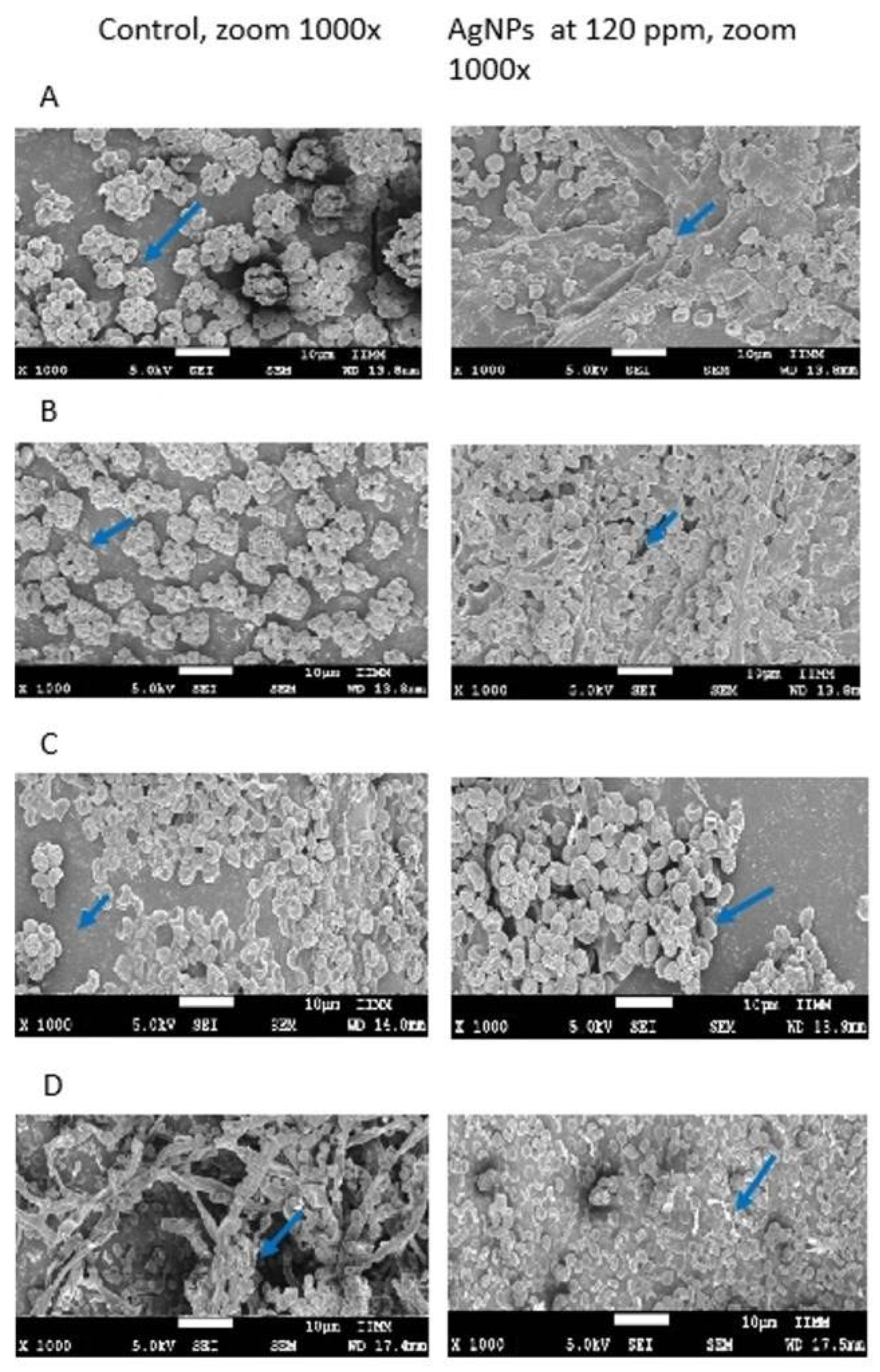
2. Results
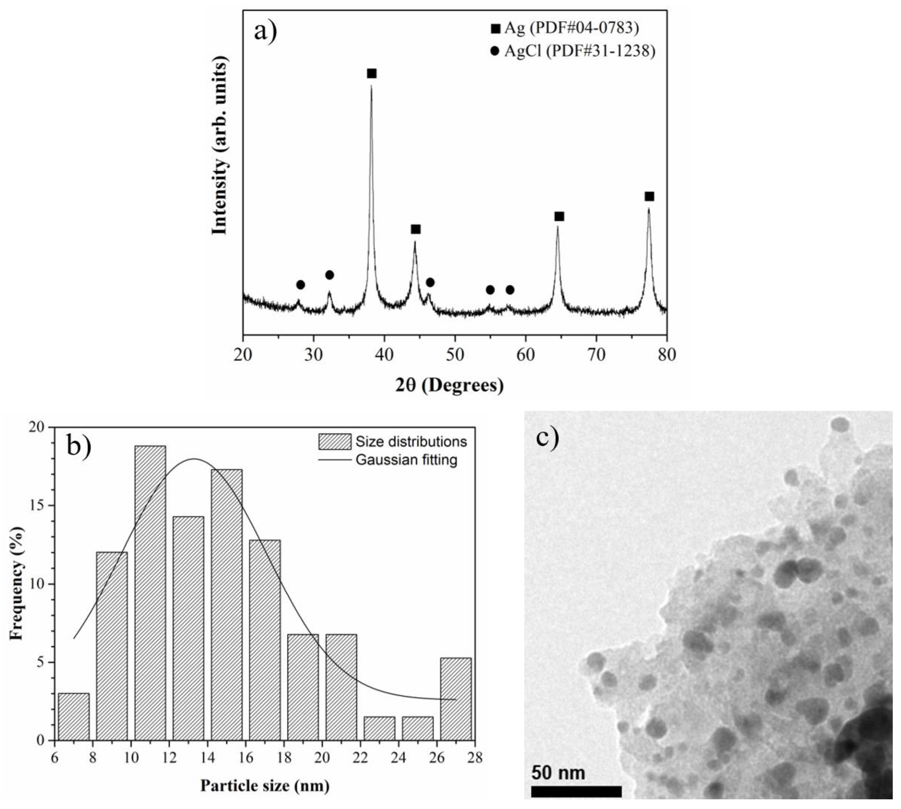
2.1. Characterization of Silver Nanoparticles (AgNP)
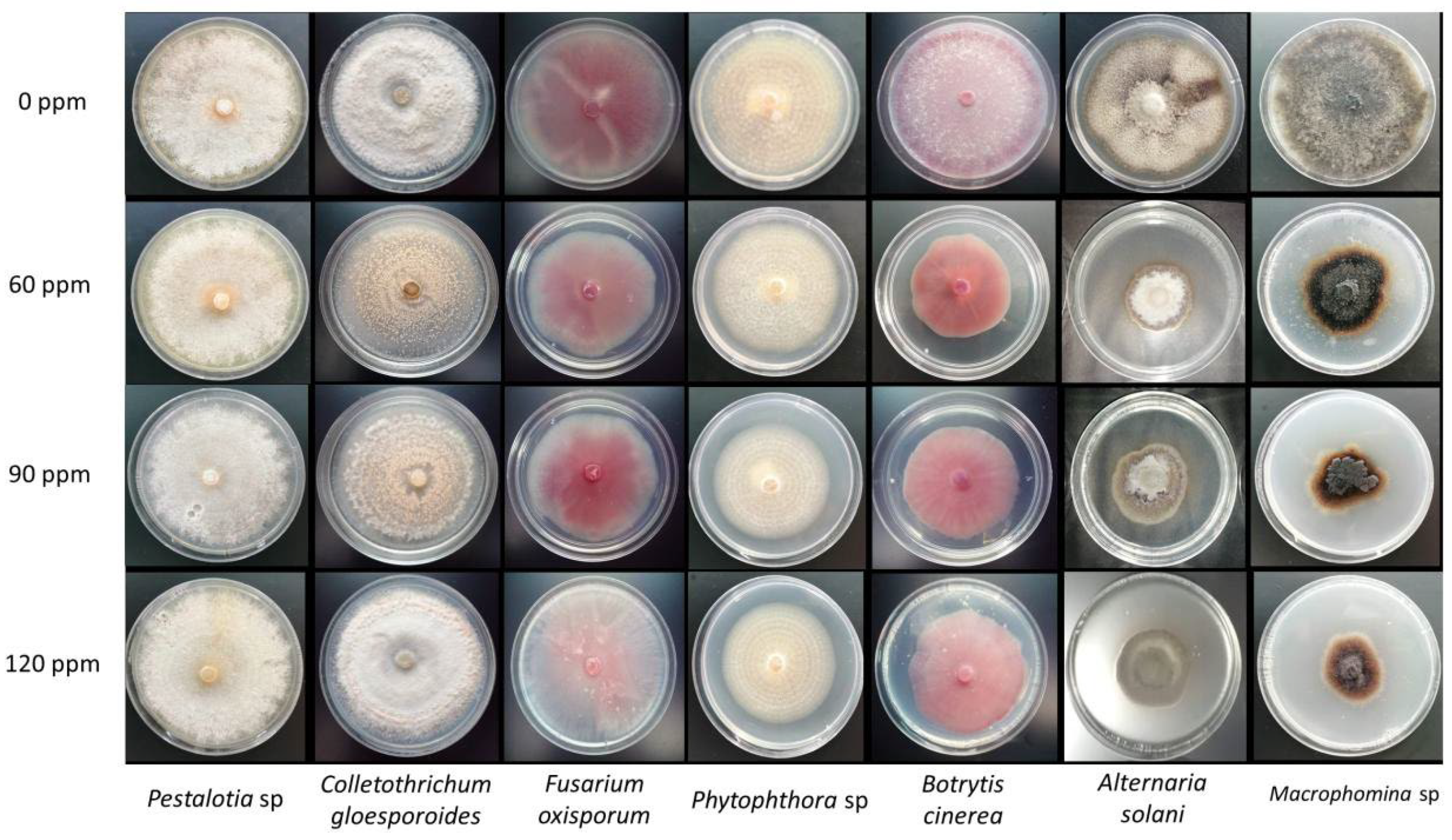
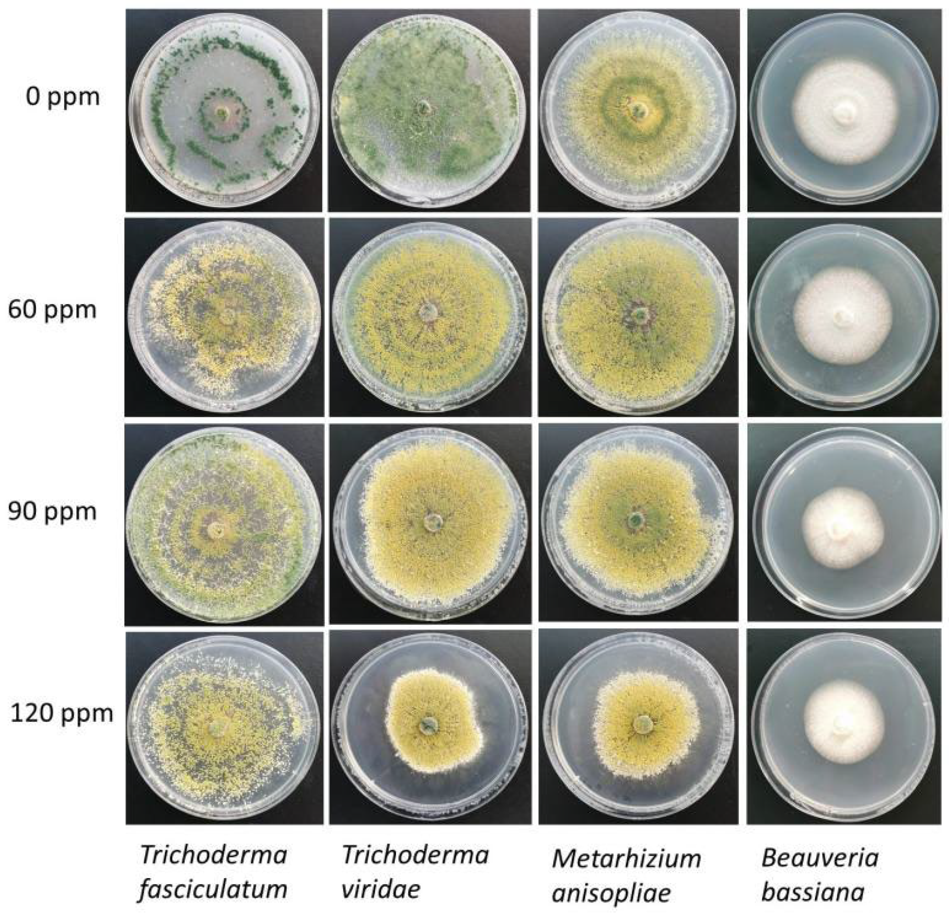
2.2. Antifungal Activity of AgNP
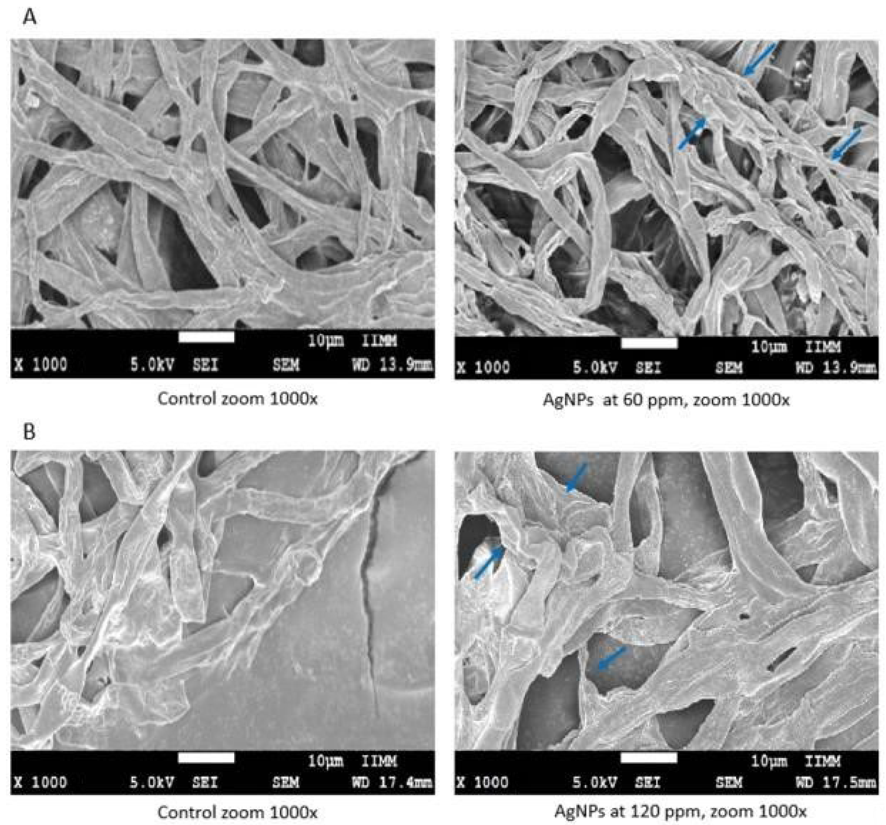
2.3. Morphometric Characterization of the Fungi
3. Discussion
3.1. Antifungal Activity of AgNP
3.2. Morphometric Characterization of Fungi
4. Material and Methods
4.1. Plant Material
4.2. Silver Nanoparticles (AgNP)
4.3. Sources of Phytopathogenic and Beneficial Fungi
4.4. Antifungal Activity of AgNP
4.5. Mycelial Growth Inhibition
4.6. Morphometric Characterization of Fungi
4.7. Data Analysis
5. Conclusions
Supplementary Materials
Author Contributions
Funding
Institutional Review Board Statement
Informed Consent Statement
Data Availability Statement
Conflicts of Interest
Sample Availability
References
- ONCN (Oficina Nacional de Coordinación de la Nanotecnología). Acerca de la Nanotecnología Iniciativa Nacional de Nanotecnología, Consultation date 9 September 2022. Available online: http://www.nano.gov/about-nanotechnology (accessed on 1 September 2022).
- Mondal, P.; Anweshan, A.; Purkait, M.K. Green synthesis and environmental application of iron-based nanomaterials and nanocomposite: A review. Chemosphere 2020, 259, 127509. [Google Scholar] [CrossRef] [PubMed]
- Garipov, I.T.; Khaydarov, R.R.; Gapurova, O.U.; Khaydarov, R.A.; Firdaus, M.L.; Efimova, I.L.; Evgrafova, S.Y. Silver nanoparticles as a new generation of antimicrobial prophylaxis. J. Sib. Fed. Univ. Biol. 2019, 12, 266–276. [Google Scholar] [CrossRef]
- Prasad, R.; Jha, A.; Prasad, K. (Eds.) Exploring the Realms of Nature for Nanosynthesis; Springer International Publishing: Berlin/Heidelberg, Germany, 2018. [Google Scholar]
- Husen, A.; Jawaid, M. (Eds.) Nanomaterials for Agriculture and Forestry Applications; Elsevier: Cambridge, UK, 2020. [Google Scholar] [CrossRef]
- Chhipa, H. Applications of nanotechnology in agriculture. In Methods in Microbiology; Academic Press: Cambridge, MA, USA, 2019; Volume 46, pp. 115–142. [Google Scholar] [CrossRef]
- Mahato, D.K.; Mishra, A.; Kumar, P. Nanoencapsulation for agri-food application and associated health and environmental concerns. Front. Nutr. 2021, 8, 146. [Google Scholar] [CrossRef] [PubMed]
- Pascoli, M.; Lopes-Oliveira, P.J.; Fraceto, L.F.; Seabra, A.B.; Oliveira, H.C. State of the art of polymeric nanoparticles as carrier systems with agricultural applications: A minireview. Energy Ecol. Environ. 2018, 3, 137–148. [Google Scholar] [CrossRef]
- Chen, X.; Schluesener, H. Nanosilver: A nanoproduct in medical application. Toxicol. Lett. 2008, 176, 1–12. [Google Scholar] [CrossRef] [PubMed]
- Necula, B.; Fratila-Apachitei, L.; Zaat, S.; Apachitei, I.; Duszczyk, J. In vitro antibacterial activity of porous TiO2–Ag composite layers against methicillin-resistant Staphylococcus aureus. Acta Biomater. 2009, 5, 3573–3580. [Google Scholar] [CrossRef]
- Bernardo-Mazariegos, E.; Valdez-Salas, B.; González-Mendoza, D.; Abdelmoteleb, A.; Camacho, O.T.; Duran, C.C.; Gutiérrez-Miceli, F. Silver nanoparticles from Justicia spicigera and their antimicrobial potentialities in the biocontrol of foodborne bacteria and phytopathogenic fungi. Rev. Argent. Microbiol. 2019, 51, 103–109. [Google Scholar] [CrossRef]
- Win, T.T.; Khan, S.; Fu, P. Fungus-(Alternaria sp.) mediated silver nanoparticles synthesis, characterization, and screening of antifungal activity against some phytopathogens. J. Nanotechnol. 2020, 2020, 8828878. [Google Scholar] [CrossRef]
- Grün, A.L.; Manz, W.; Kohl, Y.L.; Meier, F.; Straskraba, S.; Jost, C.; Emmerling, C. Impact of silver nanoparticles (AgNP) on soil microbial community depending on functionalization, concentration, exposure time, and soil texture. Environ. Sci. Eur. 2019, 31, 1–22. [Google Scholar] [CrossRef] [Green Version]
- McGee, C.F.; Clipson, N.; Doyle, E. Exploring the influence of raising soil pH on the ecotoxicological effects of silver nanoparticles and micron particles on soil microbial communities. Water Air Soil Pollut. 2020, 231, 174. [Google Scholar] [CrossRef]
- Hashemi, S.F.; Tasharrofi, N.; Saber, M.M. Green synthesis of silver nanoparticles using Teucrium polium leaf extract and assessment of their antitumor effects against MNK45 human gastric cancer cell line. J. Mol. Struct. 2020, 1208, 127889. [Google Scholar] [CrossRef]
- Ahmad, S.; Munir, S.; Zeb, N.; Ullah, A.; Khan, B.; Ali, J.; Ali, S. Green nanotechnology: A review on green synthesis of silver nanoparticles—An ecofriendly approach. Int. J. Nanomed. 2019, 14, 5087–5107. [Google Scholar] [CrossRef] [Green Version]
- Jadoun, S.; Arif, R.; Jangid, N.K.; Meena, R.K. Green synthesis of nanoparticles using plant extracts: A review. Environ. Chem. Lett. 2021, 19, 355–374. [Google Scholar] [CrossRef]
- Méndez-Andrade, R.; Vallejo-Perez, M.R.; Loera-Alvarado, E.; de los Santos-Villarreal, G.; García-Cerda, L.A.; Vera-Reyes, I. Efficacy of biosynthesized silver nanoparticles from Larrea tridentata against Clavibacter michiganensis. J. Phytopathol. 2022, 170, 91–99. [Google Scholar] [CrossRef]
- Mansoor, S.; Zahoor, I.; Baba, T.R.; Padder, S.A.; Bhat, Z.A.; Koul, A.M.; Jiang, L. Fabrication of silver nanoparticles against fungal pathogens. Front. Nanotechnol. 2021, 3, 679358. [Google Scholar] [CrossRef]
- Abdel-Hafez, S.I.; Nafady, N.A.; Abdel-Rahim, I.R.; Shaltout, A.M.; Daròs, J.A.; Mohamed, M.A. Assessment of protein silver nanoparticles toxicity against pathogenic Alternaria solani. 3 Biotech 2016, 6, 199. [Google Scholar] [CrossRef] [Green Version]
- Zaki, S.A.; Ouf, S.A.; Abd-Elsalam, K.A.; Asran, A.A.; Hassan, M.M.; Kalia, A.; Albarakaty, F.M. Trichogenic Silver-Based Nanoparticles for Suppression of Fungi Involved in Damping-Off of Cotton Seedlings. Microorganisms 2022, 10, 344. [Google Scholar] [CrossRef]
- Mohamed, Y.M.A.; Elshahawy, I.E. Antifungal activity of photo-biosynthesized silver nanoparticles (AgNPs) from organic constituents in orange peel extract against phytopathogenic Macrophomina phaseolina. Eur. J. Plant Pathol. 2022, 162, 725–738. [Google Scholar] [CrossRef]
- Ouda, S.M. Antifungal activity of silver and copper nanoparticles on two plant pathogens, Alternaria alternata and Botrytis cinerea. Res. J. Microbiol. 2014, 9, 34–42. [Google Scholar] [CrossRef] [Green Version]
- Álvarez-Carvajal, F.; Gonzalez-Soto, T.; Armenta-Calderón, A.D.; Méndez Ibarra, R.; Esquer-Miranda, E.; Juarez, J.; Encinas-Basurto, D. Silver nanoparticles coated with chitosan against Fusarium oxysporum causing the tomato wilt. Biotecnia 2020, 22, 73–80. [Google Scholar] [CrossRef]
- Vinh, T.N.; Van Khanh, Q.T.; Quyen, N.T. Effect of oligochitosan-coated silver nanoparticles (OCAgNPs) on the growth and reproduction of three species Phytophthora in vitro. Arch. Phytopathol. Plant Prot. 2018, 51, 227–244. [Google Scholar] [CrossRef]
- Ali, M.; Kim, B.S.; Belfield, K.D.; Norman, D.; Brennan, M.; Ali, G.S. Inhibition of Phytophthora parasitica and P. capsici by silver nanoparticles synthesized using aqueous extract of Artemisia absinthium. Fitopatología 2015, 105, 1183–1190. [Google Scholar] [CrossRef] [PubMed] [Green Version]
- Priyadarshini, E.; Priyadarshini, S.S.; Cousins, B.G.; Pradhan, N. Metal-Fungus interaction: Review on cellular processes underlying heavy metal detoxification and synthesis of metal nanoparticles. Chemosphere 2021, 274, 129976. [Google Scholar] [CrossRef]
- Lamsal, K.; Kim, S.W.; Jung, J.H.; Kim, Y.S.; Kim, K.S.; Lee, Y.S. Application of silver nanoparticles for the control of Colletotrichum species in vitro and pepper anthracnose disease in field. Mycobiology 2011, 39, 194–199. [Google Scholar] [CrossRef] [Green Version]
- Aguilar-Méndez, M.A.; San Martín-Martínez, E.; Ortega-Arroyo, L.; Cobián-Portillo, G.; Sánchez-Espíndola, E. Synthesis and characterization of silver nanoparticles: Effect on phytopathogen Colletotrichum gloesporioides. J. Nanopart. Res. 2011, 13, 2525–2532. [Google Scholar] [CrossRef]
- Guilger-Casagrande, M.; Germano-Costa, T.; Pasquoto-Stigliani, T.; Fraceto, L.F.; de Lima, R. Biosynthesis of silver nanoparticles employing Trichoderma harzianum with enzymatic stimulation for the control of Sclerotinia sclerotiorum. Sci. Rep. 2019, 9, 14351. [Google Scholar] [CrossRef] [Green Version]
- Mahawar, H.; Prasanna, R. Prospecting the interactions of nanoparticles with beneficial microorganisms for developing green technologies for agriculture. Environ. Nanotechnol. Monit. Manag. 2018, 10, 477–485. [Google Scholar] [CrossRef]
- Vazquez-Muñoz, R.; Bogdanchikova, N.; Huerta-Saquero, A. Beyond the nanomaterials approach: Influence of culture conditions on the stability and antimicrobial activity of silver nanoparticles. ACS Omega 2020, 5, 28441–28451. [Google Scholar] [CrossRef]
- Jian, Y.; Chen, X.; Ahmed, T.; Shang, Q.; Zhang, S.; Ma, Z.; Yin, Y. Toxicity and action mechanisms of silver nanoparticles against the mycotoxin-producing fungus Fusarium graminearum. J. Adv. Res. 2022, 38, 1–12. [Google Scholar] [CrossRef]
- Dasgupta, N.; Ramalingam, C. Silver nanoparticle antimicrobial activity explained by membrane rupture and reactive oxygen generation. Environ. Chem. Lett. 2016, 14, 477–485. [Google Scholar] [CrossRef]
- Kumari, M.; Giri, V.P.; Pandey, S.; Kumar, M.; Katiyar, R.; Nautiyal, C.S.; Mishra, A. An insight into the mechanism of antifungal activity of biogenic nanoparticles than their chemical counterparts. Pestic. Biochem. Physiol. 2019, 157, 45–52. [Google Scholar] [CrossRef]
- Raghunath, A.; Perumal, E. Metal oxide nanoparticles as antimicrobial agents: A promise for the future. Int. J. Antimicrob. Agents 2017, 49, 137–152. [Google Scholar] [CrossRef] [PubMed]
- Hirpara, D.G.; Gajera, H.P. Green synthesis and antifungal mechanism of silver nanoparticles derived from chitin- induced exometabolites of Trichoderma interfusant. Appl. Organomet. Chem. 2020, 34, e5407. [Google Scholar] [CrossRef]
- Liao, C.; Li, Y.; Tjong, S.C. Bactericidal and cytotoxic properties of silver nanoparticles. Int. J. Mol. Sci. 2019, 20, 449. [Google Scholar] [CrossRef] [PubMed] [Green Version]
- Muñoz-Silva, L.; Olivera-Gonzales, P.; Santillán Torres, M.; Tamariz-Angeles, C. Microorganismos tolerantes a metales pesados del pasivo minero Santa Rosa, Jangas (Perú). Rev. Peru. Biol. 2019, 26, 109–118. [Google Scholar] [CrossRef] [Green Version]
- Bayat, M.; Zargar, M.; Chudinova, E.; Astarkhanova, T.; Pakina, E. In vitro evaluation of antibacterial and antifungal activity of biogenic silver and copper nanoparticles: The first report of applying biogenic nanoparticles against Pilidium concavum and Pestalotia sp. fungi. Molecules 2021, 26, 5402. [Google Scholar] [CrossRef]
- Ruiz-Romero, P.; Valdez-Salas, B.; González-Mendoza, D.; Mendez-Trujillo, V. Antifungal effects of silver phytonanoparticles from Yucca shilerifera against strawberry soil-borne pathogens: Fusarium solani and Macrophomina phaseolina. Mycobiology 2018, 46, 47–51. [Google Scholar] [CrossRef] [Green Version]
- Correll, D.S.; Johnston, M.C. Manual of the vascular plants of Texas. In Contributions from Texas Research Foundation. A Series of Botanical Studies 6; Texas Research Foundation: Renner, TX, USA, 1970; p. 535. [Google Scholar]
- Dirección General de Repositorios Universitarios, Universidad Nacional Autónoma de México. Portal de Datos Abiertos UNAM, Colecciones Universitarias. Consultation date 9 September 2022. Available online: https://datosabiertos.unam.mx/ (accessed on 1 September 2022).
- Ellis, M.B. More Dematiaceous Hyphomycetes; Commonwealth Mycological Institute: Surrey, UK, 1971; p. 608. [Google Scholar] [CrossRef]
- Romero, C.S. Hongos Fitopatógenos; Primera Reimpresión en Español; Universidad Autónoma Chapingo: Texcoco, Mexico, 1993. [Google Scholar]
- Phillips, A.J.L.; Alves, A.; Abdollahzadeh, J.; Slippers, B.; Wingfield, M.J.; Groenewald, J.Z.; Crous, P.W. The Botryosphaeriaceae: Genera and species known from culture. Stud. Mycol. 2013, 76, 51–167. [Google Scholar] [CrossRef] [Green Version]
- Ajay Kumar, G. Colletotrichum gloeosporioides: Biology, pathogenicity and management in India. J. Plant Physiol. Pathol. 2014, 2, 2–11. [Google Scholar] [CrossRef]
- Badía, M.; Hernández, B.; Murrel, J.; Mahillon, J.; Peréz, M. Aislamiento y caracterización de cepas de Bacillus asociadas al cultivo de arroz (Oryza sativa L). Rev. Bras. Agroecol. 2011, 6, 90–99. Available online: https://orgprints.org/id/eprint/23097/1/Bad%C3%ADa_Aislamiento.pdf (accessed on 1 October 2022).

| Strain | Mycelial Growth Inhibition (%) | ||
|---|---|---|---|
| 60 ppm | 90 ppm | 120 ppm | |
| Alternaria solani | 70.76 ± 2.68 a | 60.82 ± 7.09 b | 60.23 ± 2.03 b |
| Macrophomina sp. | 48.94 ± 7.31 b | 64.89 ± 0.00 a | 65.43 ± 7.20 a |
| Botrytis cinerea | 51.75 ± 3.80 a | 46.05 ± 2.28 b | 43.86 ± 1.52 b |
| Fusarium oxisporum | 39.04 ± 0.76 a | 37.28 ± 11.94 a | 21.05 ± 3.95 b |
| Collethotrichum gloesporoides | 8.57 ± 2.47 a | 0.00 ± 3.78 ab | 2.86 ± 2.47 ab |
| Phytophthora cinnamomi | 8.97 ± 1.11 c | 23.08 ± 0.00 a | 21.15 ± 0.00 b |
| Pestalotia sp. | −2.70 ± 0.00 b | 12.61 ± 8.69 a | 11.71 ± 7.80 a |
| Strain | Mycelial Growth Inhibition (%) | ||
|---|---|---|---|
| 60 ppm | 90 ppm | 120 ppm | |
| Beauveria bassiana | 0.00 ± 2.17 b | 16.25 ± 2.17 a | 15.00 ± 2.17 a |
| Metarhizium anisopliae | 0.00 ± 0.00 a | 5.70 ± 5.93 a | 17.98 ± 25.53 a |
| Trichoderma viridae | 12.28 ± 0.76 b | 14.04 ± 4.98 b | 34.21 ± 0.00 a |
| Trichoderma fasciculatum | 25.44 ± 0.76 a | 1.75 ± 3.04 b | 30.26 ± 18.28 a |
Publisher’s Note: MDPI stays neutral with regard to jurisdictional claims in published maps and institutional affiliations. |
© 2022 by the authors. Licensee MDPI, Basel, Switzerland. This article is an open access article distributed under the terms and conditions of the Creative Commons Attribution (CC BY) license (https://creativecommons.org/licenses/by/4.0/).
Share and Cite
Vera-Reyes, I.; Altamirano-Hernández, J.; Reyes-de la Cruz, H.; Granados-Echegoyen, C.A.; Loera-Alvarado, G.; López-López, A.; Garcia-Cerda, L.A.; Loera-Alvarado, E. Inhibition of Phytopathogenic and Beneficial Fungi Applying Silver Nanoparticles In Vitro. Molecules 2022, 27, 8147. https://doi.org/10.3390/molecules27238147
Vera-Reyes I, Altamirano-Hernández J, Reyes-de la Cruz H, Granados-Echegoyen CA, Loera-Alvarado G, López-López A, Garcia-Cerda LA, Loera-Alvarado E. Inhibition of Phytopathogenic and Beneficial Fungi Applying Silver Nanoparticles In Vitro. Molecules. 2022; 27(23):8147. https://doi.org/10.3390/molecules27238147
Chicago/Turabian StyleVera-Reyes, Ileana, Josué Altamirano-Hernández, Homero Reyes-de la Cruz, Carlos A. Granados-Echegoyen, Gerardo Loera-Alvarado, Abimael López-López, Luis A. Garcia-Cerda, and Esperanza Loera-Alvarado. 2022. "Inhibition of Phytopathogenic and Beneficial Fungi Applying Silver Nanoparticles In Vitro" Molecules 27, no. 23: 8147. https://doi.org/10.3390/molecules27238147