Article

Advanced Photoelectrochemical Hydrogen Generation by CdO-g-C3N4 in Aqueous Medium under Visible Light

1
Department of Chemistry, College of Science, University of Hafr Al Batin, P.O. Box 1803, Hafr Al Batin 39524, Saudi Arabia
2
Department of Mechanical Engineering, College of Engineering, University of Hafr Al Batin, P.O. Box 1803, Hafr Al Batin 39524, Saudi Arabia
3
Department of Physics, College of Science, University of Hafr Al Batin, P.O. Box 1803, Hafr Al Batin 39524, Saudi Arabia
4
Interdisciplinary Research Center for Hydrogen & Energy Storage (IRC-HES), King Fahd University of Petroleum & Minerals (KFUPM), P.O. Box 5040, Dhahran 31261, Saudi Arabia
*
Author to whom correspondence should be addressed.
Molecules 2022, 27(24), 8646; https://doi.org/10.3390/molecules27248646
Submission received: 21 October 2022 / Revised: 3 December 2022 / Accepted: 4 December 2022 / Published: 7 December 2022
(This article belongs to the Special Issue Metal-Based Nanocomposite: Synthesis and Applications)

Abstract

:
Herein, hydrothermal fabrication of CdO-g-C3N4 photocatalyst for a substantially better photocatalytic recital in water splitting is presented. The XRD analysis confirms the cubic phase of CdO-g-C3N4, whereas FTIR and UV-VIS studies revealed the presence of respective groups and a median band gap energy (2.55 eV) of the photocatalyst, respectively, which further enhanced its photo-electrochemical (PEC) properties. The SEM displays the oblong structures of g-C3N4 sheets and nano rod-like morphology of CdO and CdO-g-C3N4, respectively. The HR-TEM exhibits morphology & orientation of the grains and substantiates the polycrystal-line nature of CdO-g-C3N4 nanocomposite. The photocatalytic water-splitting concert is evaluated by PEC experiments under 1 SUN visible light irradiation. Linear sweep voltammetry (LSV), chronoamperometry (CA), and electrochemical impedance spectroscopy (EIS) comprehend the CdO-g-C3N4 as a hydrogen evolution photocatalyst. A photocurrent density beyond ≥5 mA/cm2 is recorded from CdO-g-C3N4, which is 5–6 folds greater than pure CdO and g-C3N4. The efficient separation and transfer of charges allocated to CdO-g-C3N4 and fabricating heterojunctions between g-C3N4 and CdO suppresses the unfavorable electron-hole pairs recombination process. Thus, it recesses charge transfer resistance, augmenting enhanced photocatalytic performance under 1 SUN irradiation.

Graphical Abstract

1. Introduction

Semiconducting nanomaterials for photoelectrochemical water splitting earned substantial consideration in recent decades. This is imperative to fulfill the intensifying global energy challenges and to meet the renewable energy demand [1,2,3,4,5,6,7,8,9,10]. Solar energy is an important renewable energy source that can split water into molecular hydrogen and oxygen with the aid of photocatalysts [11,12]. Solar energy is the most abundant and promising source of sustainable H2 production. One of the key challenges is its proper utilization to develop innovative systems that can use this renewable energy source effectively as an energy intake to drive the reactions for producing H2 in an environmentally friendly way. Hydrogen (H2), the future fuel, is a resourceful energy carrier. It is among the promising sources of clean energy to confront several energy challenges. H2 bears remarkable properties that makes it a potential source of energy. H2 is abundantly found in combination with other elements such as oxygen, carbon, and nitrogen. 75% of the universe’s mass contains H2. It is a colorless, odorless gas that accounts for higher energy source with zero greenhouse emissions, high gravimetric energy density, low heating value of ∼120 MJ kg−1, etc. [13,14,15]. Since the last few decades, hydrogen has been used majorly in the synthesis of methanol, ammonia and in petroleum refinement. Furthermore, cosmonauts use hydrogen as fuel for their space vehicles and in fuel cells that provide heat, electricity and drinking water in space. To utilize hydrogen, it must be separated from other substances. Fuel cells are one of the stratagems by which hydrogen can be directly converted into electricity. In the near future, hydrogen could be used to fuel automobiles and aircraft and to provide power to fulfill domestic and commercial needs. Furthermore, H2 can be employed in current industrial setups that are being used for conventional fuels. Therefore, lately several countries have launched sustainable energy strategies and programs to nurture the implementation of H2 based technologies on various fronts, which will help in instituting a low carbon environment [13,15].
Photoelectrochemical water splitting consists of an oxidation/reduction of half-cell reactions—i.e., four-proton, four-electron process—which requires large over-potential in order to restrain the competing electron-hole recombination processes at the expense of solar energy [11,12,16,17,18,19]. Photoelectrochemical solar water splitting for H2 and O2 production involves an appropriate material with substantial band gap energy that could assist the electron holes’ mobility in an effective manner. Usually, semiconductors with a bandgap below 3 eV are considered efficient for solar driven water splitting [16,17,18]. For an effective PEC process, the photocatalysts and the PEC system should be efficient, stable, durable under harsh experimental or operational conditions and result in minimal or no environmental impacts. It should also be capable of absorbing a large fraction of the visible light and retaining well-aligned band energy for thermodynamically promising charge transfer reactions. Above all such systems should be cost-effective [19,20,21]. To comprehend all the aforementioned characteristics required for effectual solar water splitting with significant solar-to-chemical energy conversion and in order to make these systems commercially viable, significant research efforts are in progress [22,23,24,25,26]. Nevertheless, there are enough opportunities for finding superior, inexpensive, and effective photocatalyts.
Several transition-metal oxides have been widely reported due to their intriguing electrical, optical and catalytic properties [27,28,29,30,31,32]. Cadmium oxide (CdO) is among one of the nanomaterials which is an n-type semiconductor with a cubic (FCC) crystal structure and owes many imperative properties such as wide band-gap (2.16–2.6 eV) and refractive index (n = 2.75), high density (8150 kg/m3), low resistance and high transmittance in the visible range [33,34]. Based on these properties, CdO is used in solar cells, phototransistors, catalysts, transparent electrodes, gas sensors, photodiodes, photoelectric components [34,35,36,37], etc. However, its photoelectrochemical properties are not fully explored, especially in solar driven water splitting. It is worth mentioning here that CdS and CdSe are also known light absorption photocatalysts but severe photo corrosion due to sulfur leaching or Se degradation greatly affect its practical stability in the PEC water splitting process. Furthermore, their toxicity also limits their large-scale application in industry [38]. Thus, oxygen incorporation is more suitable with cadmium as compared to S or Se in addition to the doping or intermixing of CdO with suitable semiconducting supporting materials such as graphitic Carbon Nitride (g-C3N4) to achieve maximal output.
The g-C3N4 has attracted widespread attention in recent years [39] as an emerging carbon based semiconductor. Its excellent features, such as suitable band gap (2.71 eV), easily tunable electronic structure and absorption of visible light (460nm wavelength) and high physical and chemical stability make it an attractive entrant in semiconducting industrial applications. g-C3N4 has two basic structural units: s-triazine (C3N4) and tris-s-triazine (C6N7) rings; for g-C3N4, the regular three s-triazine units structure is considered to be among the most stable phase studies by Density Functional Theory (DFT) [40] Despite several advantages, g-C3N4 also have shortcomings, such as high recombination rate for photogenerated electron/hole pairs, poor electron conductivity, slow hole- Shifts, etc., that limit its use as a photoelectrode material. The PEC performance of g-C3N4 can be improved by constructing different nanoscale morphologies of g-C3N4 [14,16,39] doping or physical mixing of different metals to produce heterogeneous photocatalysts that could result in improved PEC efficiency and visible light absorption. So, morphologically tuned g-C3N4 nanomaterials dopped or intermixed with metal or metal oxides could develop nanoscale heterogeneous nanomaterials with the potential for PEC water splitting [14,16,39] properties.
In Table 1, we compared several recent examples of Cd based g-C3N4 nanomaterials which are used in photoelectrochemical water splitting and in photodegradation applications. It is evident that ample of g-C3N4 materials impregnated or dopped with metal/metal oxides following various synthetic strategies are explored.
In the current research, we aimed at developing more stable, inexpensive and effective metal oxide based visible light-driven photocatalysts nanocomposites as photoanode materials to address the challenges in PEC water splitting. Thus, to enhance the PEC water splitting, we used CdO, graphitic Carbon Nitride (g-C3N4) and CdO-g-C3N4 nanocomposites. These photocatalytic materials were prepared by Ultrasonication, precipitation and hydrothermal methods. The nanomaterials were characterized by physicochemical analysis methods such as XRD, FTIR, UV-VIS, PL and SEM, HR-TEM for estimation of their structure property relationships, elemental composition, and morphological properties. For solar driven water splitting CdO, g-C3N4 and CdO-g-C3N4 photoanode materials are also evaluated for enhanced PEC properties by electrochemical methods under 1 SUN solar irradiation.

2. Results and Discussion

2.1. X-ray Diffraction (XRD) Analysis

The XRD diffraction spectrum of pristine g-C3N4, CdO, and the composite CdO-g-C3N4 are shown in Figure 1. In the comparison spectrum, all the developed materials appeared in their respective characteristic peaks and confirms their crystalline nature after undergoing the complete hydrothermal process.
The analysis was performed to assess the phase formation during the processing of pristine g-C3N4, CdO, and the composite CdO-g-C3N4, the diffraction patterns of each material are shown in Figure 1. The patterns confirmed the presence of g-C3N4, CdO, and CdO-g-C3N4 in their respective materials. The diffraction peaks in the spectrum for g-C3N4 were observed at Bragg angles 13° and 27° concerning their miller indices 100 & 002, and the peaks are in agreement with the JCPDS card numbers 87–1526 [39,40]. Furthermore, the XRD pattern of CdO exhibited the formation of cubic phase crystal structure and indexed the following miller indices (111), (200), (220), (311), (222) at the respective diffraction peaks at 2θ values. The observed diffraction peaks are well aligned with the JCPDS card # 05–0640 [38]. The XRD pattern of CdO-g-C3N4 nanomaterials also showed and confirmed the presence of all characteristic peaks of CdO and g-C3N4 at their respective 2θ values. It supports that the precipitation method has converted the cadmium nitrate into cadmium oxide and the hydrothermal method supports the CdO and g-C3N4 intermixing into a crystalline CdO-g-C3N4 nanomaterial. The XRD studies further revealed that in CdO-g-C3N4 the CdO crystallizes into the cubic Fm 3 ¯ m space group and that each Cd2+ is bonded to six equivalent O2− atoms to form a mixture of edge and corner-sharing CdO on the triazine rings of g-C3N4. All Cd–O bond lengths are 2.39 Å. O2− and are bonded to six equivalent Cd2+ atoms to form a mixture of edge and corner-sharing OCd6 octahedra.

2.2. Scaning Electron Microscopy (SEM) and High Resolution Transmission Electron Microscopy (HR-TEM)

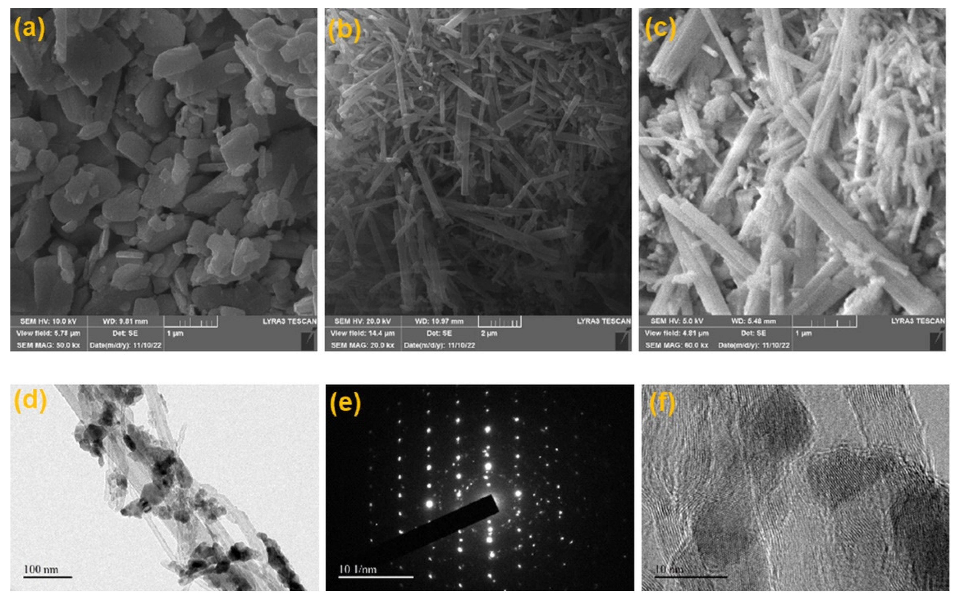
The topography and morphology of developed materials were investigated using an advanced scanning electron microscopic technique. Figure 2a shows the morphology of g-C3N4, and the oblong or long-flattened grains show the nature of g-C3N4 to be nanosheets. In addition, Figure 2b presents the micrographs of CdO, and it can be observed that the CdO consists of nanorods morphology with an average thickness of 50–150 nm and 1–3 ± μm length. Furthermore, Figure 2c displays the structural nature of the CdO-g-C3N4 composite, and it is evident that a blend of both the entities—nanosheets and nanorods (50–150 nm and 1–3± μm length)—are present in the SEM micrograph. Since both the major ingredients, g-C3N4 and CdO, exhibited their sizes in the nanoscale, and due to their lower grain sizes, the chances of agglomeration are quite bright due to large surface affinity. Nevertheless, Figure 2c attests to the validity of the process and parameters to produce uniform and homogenized CdO-g-C3N4 composite nanorods which will be further put through photoelectrochemical (PEC) characterization.
The HR-TEM micrographs of CdO-g-C3N4 are demonstrated in Figure 2d–f. Nanorods like structure of the CdO can be observed in Figure 2d with some irregular shaped g-C3N4 nanosheets. The HR-TEM Figure 2d reveals that the CdO nanorods were grown from nanoscale crystals, and the visible gaps and holes in between the nanorods were developed due to detachment and further vaporization of H2O from the Cd (NO3)2·4H2O precursor, while CdO synthesis was followed by heat treatment in autoclave for 180 °C. In addition, the diameter of nanorods ranges from 50–60 nm with an average length of 2–3 μm which makes its surface area huge, making its surface more active for electron and holes interaction. Thus, producing CdO-g-C3N4 is a promising candidate for photoelectrochemical and photocatalytic applications. Figure 2e exhibits the selected area diffraction (SAED) pattern of a CdO-g-C3N4 nanocomposite which is polycrystalline in nature and crystals are placed at different orientations which can be deduced from the numerous bright dots available on the pattern. The concentric rings in the pattern are in good agreement with the XRD planes (111), (200), (220), (311) and (222) [44,49,50]. A higher magnification micrograph, as in Figure 2f, demonstrated the size and morphology of grains and nanorod shaped like structures. The different orientation of the lattices in various grains manifests the polycrystalline nature of the CdO-g-C3N4 and shows the lattice spacing of 0.2 to 0.3 nm in Figure 2f, which is conforming to the standard lattice spacing of 0.235 nm in CdO for (200) crystal plane [51,52].

2.3. FTIR-Spectroscopic Analysis

To assess the chemical bonds and functional groups in the fabricated materials g-C3N4, CdO, and CdO-g-C3N4, FTIR spectroscopy was performed within the range 370–4000 cm−1 as described in Figure 3. The presence of triazine structure in g-C3N4 nanosheets are validated by the emergence of characteristics heterocyclic ν (C–N/C = N) stretching vibrations and ν (N–H) shearing vibrations bands from 1250 to 1650 cm−1 and 3100–3600 cm−1 respectively. The large broad bands in g-C3N4 and CdO-g-C3N4 are the distinctive vibrational ν (C–N/C = N) bands of triazine units appeared at 12,501,327–70 cm−1 are assigned to stretching vibrations of C–NH–C, i.e., partially condensed, and C–N(–C)–C, i.e., fully condensed units, respectively. A strong peak at 819 cm−1 complemented the characteristic breathing mode vibrations of triazine units, which also reinforces growth of g-C3N4 [53]. The broad vibrational bands in g-C3N4 and Cdo-g-C3N4 from 3100 to 3600 cm−1 are assigned to free uncondensed terminal amino groups (-NH2 or =NH).
As discussed earlier, the 1200–1700 cm−1 bands are arising due to skeletal C = N heterocycle stretches, consisting of trigonal (N-(C)3) and bridging C-NH-C units of the extended C-N-C network in the g-C3N4 structure [53]. The FTIR absorption bands recorded beneath ~ 1000 cm−1 are the fingerprint zone of metal oxides. Hence, the peak witnessed at 425 cm−1 in the FTIR spectrum of both the pristine CdO and CdO-g-C3N4 composite is referred to typical vibrational mode of Cd-O linkage, and it is well aligned with the reported results [54]. The other peak at 3457 cm−1 corresponds to hydroxyl group (-OH) due to moisture. In addition, the FTIR characterization of the CdO, g-C3N4 & CdO-g-C3N4 confirms its unadulteration by providing evidence in the form of spectra which attests that the secondary phase or impurity formation did not take place during the hydrothermal processing of the materials. Furthermore, Figure 3 also validates that g-C3N4 and CdO co-evolved during the processing as their peculiar vibrational modes reported at the same positions [33,38,54], and the findings from XRD and SEM and HR-TEM characterization confirm the idea. Consequently, it can be surmised that the intermixing of CdO with g-C3N4 by following the specific conditions and parameters of hydrothermal treatment is feasible to fabricate crystalline nanorods of CdO-g-C3N4 composite.

2.4. UV-Visible Analysis

The optical properties, i.e., absorption spectrum and band gap energy of pure g-C3N4, CdO, and CdO-g-C3N4 composite are evaluated through UV-VIS absorption spectroscopy [55]. The UV absorbance data is used to formulate Tauc Plots [56] as shown in Figure 4. From the given Tauc Plots, band gap energy (Eg) is determined [57,58]. Based on the calculations, it is revealed that the bandgap energies for g-C3N4, CdO & CdO-g-C3N4 are found to be 2.77 eV, 2.40 eV, and 2.55 eV, respectively. It is evident that intermixing of cadmium oxide (CdO, Eg = 2.40 eV) with graphitic carbon nitride (g-C3N4, Eg = 2.77 eV) resulted in the procurement of a mean band gap energy of 2.55 eV for CdO-g-C3N4 composite. Therefore, it can be deduced that the probe sonication, along with the hydrothermal processing, resulted in near-to-perfect impregnation of CdO-g-C3N4 composite. Moreover, the band gap energy (2.55 eV) of the CdO-g-C3N4 composite confirms its candidature to act as a promising photocatalyst.

2.5. Photoluminesence (PL) Spectroscopy

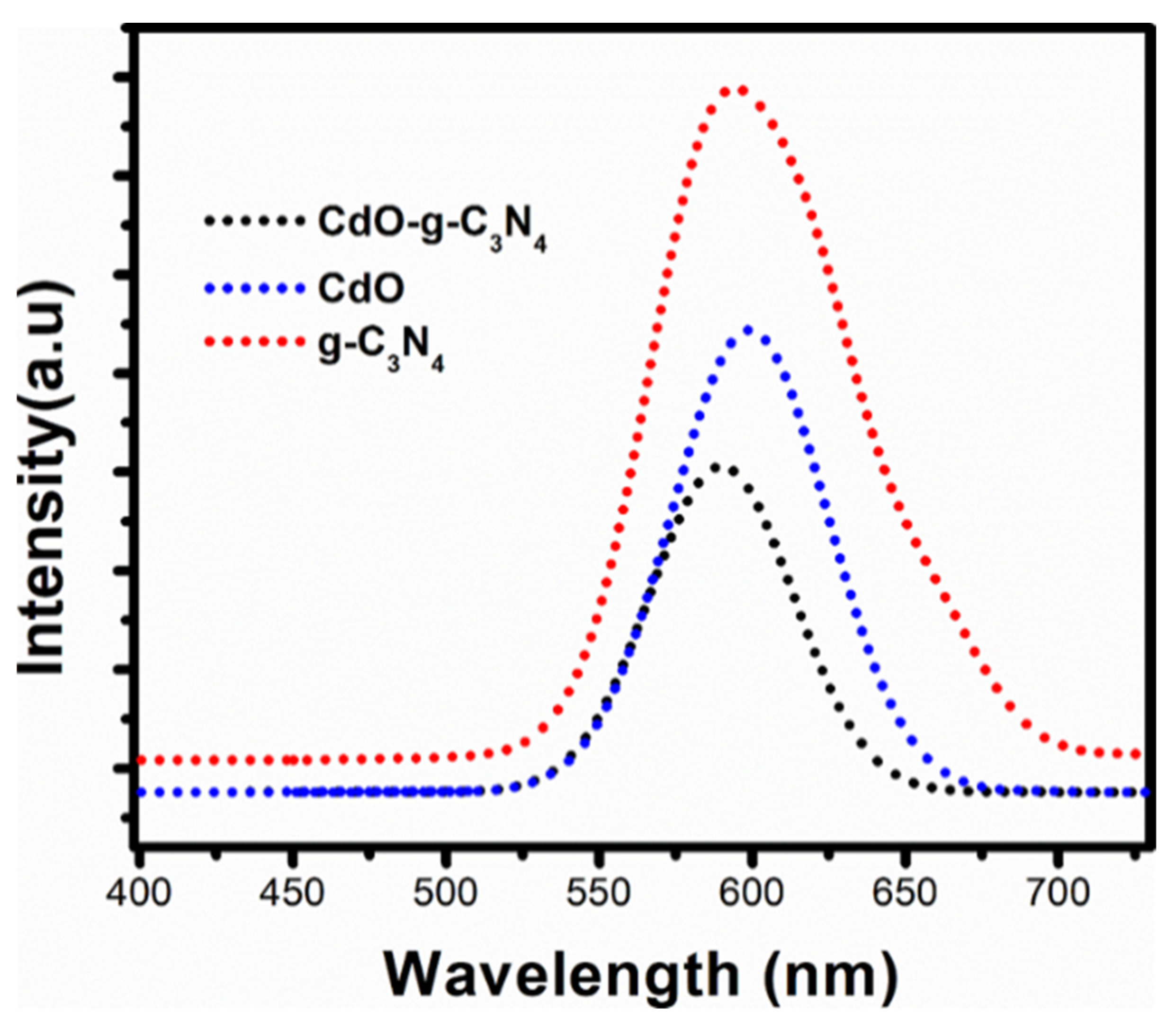
Figure 5 shows the photoluminescence spectroscopy of g-C3N4, CdO, and CdO-g-C3N4 nanomaterials to study their charge carrier separation. The PL emission spectra was recorded at an excitation of 385 nm. The PL peaks maxima for each material centered approximately in the range of 590–599 nm [38]. It is observed from PL analysis that the peak intensity of g-C3N4 is significantly higher than that of its counterparts, i.e.,CdO and CdO-g-C3N4 nanocomposite, respectively. This indicates a fast recombination rate among the charge carriers in g-C3N4 nanosheets. Nevertheless, Figure 5 shows a declining PL intensity from CdO to CdO-g-C3N4. Hence, it is a supporting slower charge recombination in electron and hole pairs in the CdO-g-C3N4 photocatalyst [38]. The PL analysis further elaborate that rate of electrons hole pair recombination among charge carriers in g-C3N4 could be tuned by impregnation of metal oxides to form heteronanostructures. This confined recombination rate is beneficial to enhance the photoelectrochemical and photocatalytic activities of the nanocomposite materials for renewable and sustainable energy applications. Such enhanced PEC and photocatalytic characteristics are sufficiently observed in our prepared photocatalysts as discussed in the sections that follow.

3. Photoelectrochemical (PEC) Measurements

3.1. Chronoamperometric Analysis (CA)

The chronoamperometric (CA) results are displayed in Figure 6a–c for the pristine g-C3N4, CdO and doped CdO-g-C3N4 nanomaterials. In the CA outcome from fabricated nanomaterials, a photocurrent profile (Jp–t) is shown, where photocurrent produced is plotted as a function of time under 1 Sun visible light exposure in a neutral electrolyte (0.5 M Na2SO4). In Figure 6a,b it is observed that the pristine g-C3N4 nanosheets and CdO nanorods generated photocurrent densities of 5 uA/cm2 and 800 µA/cm2 respectively. Conversely, in similar circumstances, CdO-g-C3N4 composite manifests a photocurrent density of 4.5 mA/cm2, and the photocurrent density is 500% more than the photocurrent density recorded for pristine g-C3N4 nanosheets and CdO nanorods. Based on the photocurrent densities of the fabricated materials, it can be inferred that the CdO-g-C3N4 composite produced better PEC outcomes than their counter parts (CdO and g-C3N4). Likewise, from Figure 6c it can be learned that the CdO-g-C3N4 composite continuously maintained the same amount of photocurrent upon exposure to 1 SUN irradiation in regular intervals under the same experimental conditions. Consistent behavior of the material under visible light shows sound adherence and stability of fabricated material layers on the FTO substrates. Moreover, the larger photocurrent density of the CdO-g-C3N4 composite also indicates that pre and post hydrothermal processing of the CdO-g-C3N4 composite played a vital role in its heterojunction formation and charge transferability among g-C3N4 & CdO has drastically amplified. Consequently, the two factors ensued enhanced photocatalytic activity of the CdO-g-C3N4 composite by inhibiting the recoupling of the electron-hole pairs.

3.2. Linear Sweep Voltammetry (LSV)

The linear sweep voltammetry was performed for g-C3N4, CdO, and composite CdO-g-C3N4. The results are shown in Figure 7a–c, respectively. In each figure, various trends were observed for bare FTO, pure g-C3N4, CdO, and composite CdO-g-C3N4, with and without the exposure to 1 SUN irradiation. It is evident from the graphs that the bare FTO substrates showed insignificant response to one SUN irradiation. In contrast, g-C3N4, CdO, registered mild responses in terms of photocurrent densities of 0.18 µA·cm−2 and 400 µA·cm−2 respectively at a scan rate of 100 mv/s in a potential range of 1 V to −1 V in 0.5 M Na2SO4 electrolyte. Under the same parameters, CdO-g-C3N4 nanorod composite performed outstandingly by displaying the highest value of photocurrent density 5 mA·cm−2. These results are in good agreement with the chronoamperometric (CA) measurements as presented in Figure 6. It is evident from Figure 7c that the photocurrent density of CdO-g-C3N4 improved drastically and thus induced a notable uncoupling of electron-hole pairs by offering a lesser amount of resistance rather than individual components g-C3N4 and CdO. Hence, the composite CdO-g-C3N4 offered significantly more hindrance in electron-hole rejoining. In addition, the artful impregnation of CdO and g-C3N4 through sonochemical treatment, followed by hydrothermal processing produced a synergistic effect by amplifying the photocatalytic properties of the CdO-g-C3N4 composite. Furthermore, the photocurrent densities of each material; g-C3N4 and CdO, and CdO-g-C3N4 are correlated with their capability to accommodate hydrogen evolution by reduction reactions and charge separation of photoexcited elections from holes.

3.3. Electrochemical Impedance Spectroscopic (EIS) Analysis

Electrochemical impedance spectroscopy (EIS) was conducted to determine the charge transfer resistance of the synthesized materials, i.e., g-C3N4, CdO, and CdO-g-C3N4. The EIS characterization was performed in 0.5 M Na2SO4 aqueous solution under visible light illumination in Constant E mode with frequency range of 15–300,000 Hz. As a result, the Nyquist plots were obtained for the synthesized materials under visible light exposure as displayed in Figure 8. It is noticed that all the synthesized materials showed a semicircle along with a straight-line slope under visible light. The semicircles are in the area of high frequency. Generally, the semicircle in the region of high frequency corresponds to the electron transfer processes. In contrast, the straight slope in the low-frequency region corresponds to the electrode’s ion diffusion. Therefore, the value of charge transfer resistance (Rt) is directly proportional to the diameter of the semicircle. Figure 8 demonstrates that the arc diameter of CdO-g-C3N4 is lower than that of CdO and g-C3N4 EIS curves. Yet again, it confirmed that the sonochemical fabrication, followed by a hydrothermal approach, has improved the electrochemical conductivity of the CdO-g-C3N4 photocatalyst. Likewise, the straight slope of the CdO-g-C3N4 photocatalyst is higher than that of CdO and g-C3N4, which supports the relatively enhanced ionic diffusion rate. Thus, EIS studies and the PEC outcomes as discussed above have revealed that enhanced photocurrent density, low resistance, improved charge transfer, formation of heterojunction, lower electrical resistance and higher ionic diffusion are the reasons for better photocatalytic efficiency of CdO-g-C3N4 photocatalyst that can generate hydrogen from an aqueous medium.

3.4. Charge Transfer Mechanism

The charge transfer diagram shown in Figure 9 also supports that the photocurrent densities correspond to oxidation (oxygen production) and reduction reactions (hydrogen generation). The increase in the electrochemical efficiency of the CdO-g-C3N4 photoanodes compared to CdO, and g-C3N4 photoanodes, is attributed to effective separation of electron-hole pairs and the low resistance offered by CdO-g-C3N4 compared to CdO and g-C3N4, photocatalysts. Moreover, the highest photocurrent density by CdO-g-C3N4 is credited to the synergistic effect achieved by the sonochemical and hydrothermal fabrication of CdO with g-C3N4. The interaction between g-C3N4 and CdO in the CdO-g-C3N4 photocatalyst effectively reduced the charge transfer distance of photoexcited electrons and inhibited their further recombination with holes. Subsequently, this charge separation predominantly facilitated photocurrent generation and H2 evolution, thus showing enhanced PEC and photocatalytic characteristics of the CdO-g-C3N4 composite.

4. Experimental Strategies

4.1. Material and Chemicals

All the chemicals/solvents used (as received) during the experiments were of analytical grade and purchased from well-known companies/suppliers, i.e., Sigma Aldrich/Alfa Aesar. A local vendor was also contacted to obtain Fluorine-doped tin oxide (FTO) substrates of the following specific features: size (l × w × t = 25 × 25 × 1.1 mm), R (resistivity= 7–15 ohms) and transmittance efficiency of more than 80%. Before using the FTO substrates, the FTO substrates underwent ultrasonication treatment for 15 min in Deionized (DI) water and ethanol.

4.2. Synthesis of Graphitic-Carbon Nitride (g-C3N4)

The graphitic carbon nitride g-C3N4 was synthesized from melamine by following the steps already reported in the literature with some alterations [59,60,61]. In the beginning, melamine powder was weighed properly, and the known amount of the substance was subjected to heat treatment (annealing) in a muffle furnace at 550 °C for 300 min to finally obtain a yellow-colored bulk g-C3N4. To convert bulk g-C3N4 to nanosized sheets, the process underwent a physical transformation procedure [62]. Initially, the powder of bulk g-C3N4 was mixed with ethanol (70% sol.) using a probe sonication technique for 180 min. Then the solution was centrifuged (6000 rpm) to collect the desired nanosheets. Eventually, the g-C3N4 was dried at 150 °C in a vacuum oven for 120 min.

4.3. Synthesis of Cadmium Oxide (CdO)

The CdO nanoparticles were synthesized by chemical precipitation method with trivial changes as described by Li et al. [63] and Sumeet et al. [64]. In a typical chemical reaction, Cd (NO3)2·4H2O salt was put into the water (DI) to obtain a 0.5 M solution. Sodium hydroxide (1 M) was added to the solution in the form of droplets by using a magnetic stirrer for mixing until the solution became slightly basic (pH = 8). Meanwhile, adding NaOH to the solution, the CdO will precipitate out. The reaction was allowed to take place while continuously stirring at 60 °C for 80 min. In the next stage, an ultrasonication of the solution was performed at 60 °C for another hour. To collect the CdO precipitates from the solution, it was subjected to centrifuge for 20 min at 10,000 rpm. The obtained precipitates were washed several times using DI water and ethanol to remove the impurities and unreacted ingredients. Later, the precipitates obtained were dried in the oven at 100 °C for 12 h [34]. Subsequently, CdO nanorods were obtained after annealing at 700 °C for 4 h.

4.4. Synthesis of Cadmium Oxide Dopped Graphitic Carbon Nitride (CdO-g-C3N4) Nanocomposite

A hydrothermal method was adopted to produce CdO-g-C3N4 nanocomposite. Initially, two separate 0.1 molar (0.642 g of CdO & 0.460 g of g-C3N4 nanosheets) aqueous suspensions in 50 mL DI water were prepared by 1 h sonication. In the next step, to obtain a homogeneous mixture, the CdO and g-C3N4 aqueous solutions were mixed using sonication for another hour. After the sonication, the homogenous solution was transferred into PPL-lined vessels (stainless steel autoclave). A day-long (24 h) heat-treatment of the homogenous solution was performed in the autoclave at 180 °C. Later, the thermally heat treated homogenous aqueous solution was subjected to centrifugation at 6000 RPMs for 5 min, and the obtained CdO-g-C3N4 nanocomposite was washed using ethanol and DI water at least three times before being placed in the vacuum oven for further drying at 150 °C for 120 min. After the drying, the powdered CdO-g-C3N4 nanocomposite underwent annealing for a further 4 h at 550 °C.

5. Characterization

The manufactured materials were characterized using advanced characterization techniques; UV-VIS, FTIR, PL, XRD, FE-SEM and HR-TEM, etc. A 750-Watt probe sonicater (Model: Cole Parmer 500) was used to perform the sonochemical treatment. The powder obtained after the sonochemical procedure was further characterized by XRD to determine phase formation in the developed materials. The characterization was performed within the 2θ range of 10–80° with a step size of 5° min−1 (Model: XRD-6100 by SHIMADZU), and the X-Rays source was Cu Kα (wavelength of 1.5409 Å) operated at 80 mA/60 kV. The XRD pattern acquired was matched by the database of the ICDD-PDF-2 library and it confirmed the formation of g-C3N4, CdO and CdO-g-C3N4. The shape and morphology of the materials under investigation were observed using a scanning electron microscope TESCAN Lyra 3 Field Emission Dual Beam (Electron/Focused Ion Beam) system combined with a high-end field-emission scanning electron microscope (FE-SEM). The morphology at higher resolution and selected area diffracted pattern (SAED) from CdO-g-C3N4 nanocomposite were recorded on JEOL (JEM2100F) high resolution transmission electron microscopy (HRTEM) at 200 KV accelerating voltage. The optical characterizations of the developed materials (g-C3N4, CdO & CdO-g-C3N4) was performed by using a special UV-VIS spectrometer (Model: Jenway 6850 double beam with variable bandwidth) to measure the absorbance. The FTIR (SHIMADZU IRAffinity-1s) was used to conduct vibrational spectroscopic studies. A fluorolog-3 imaging spectrophotometer was used to measure the photoluminescence activity of the nanomaterials at an excitation wavelength of 385 nm and a slit width of 2 nm. The photoelectrochemical studies of the materials were carried out using a potentiostat (IVIUM-n-stat). The electrochemical workstation was equipped with multichannel electrochemical analysis. Solar Simulator (Model: ABET 10500) was used to generate 1 SUN visible light for photoelectrochemical studies. In addition, the solar simulator contained the specifications: AM 1.5 G output, UV-Cut of filter & D.C. Xenon arc lamp, and it was calibrated as per the standards ASTM, IEC & JIS class A.

6. Fabrication of g-C3N4/FTO, CdO/FTO, and CdO-g-C3N4/FTO Photoanodes for Photoelectrochemical (PEC) Studies

The slurries of the developed materials g-C3N4, CdO and CdO-g-C3N4 were coated over the conducting substrates of Fluorinated Tin Oxide glass (FTO) and were used for photoelectrochemical (PEC) studies. Before the coating, the FTO glass substrates were thoroughly cleaned using acetone and DI water in order, following the ultrasonication. The slurries were prepared separately by taking 100 mg of each material and blending in a mixture containing 70% ethanol, 30% DI water and a few drops of 5% Nafion. The FTO substrates that were pretreated by drop-casting were taken and a slurry of each prepared material was coated over the conducting side. Then, the coated slurry was exposed to 100 °C for 2 h to completely evaporate the ethanol and DI water from the films. After heat treatment, uniform layers/films of g-C3N4, CdO and CdO-g-C3N4 were obtained over the FTO substrates. These FTO substrates were subjected to photoelectrochemical (PEC) characterization using Ivium multichannel Potentiostat (Ivium-n-Stat) in a 3-electrode system in which the electrolyte used was sodium sulphate (0.5 M) with a neutral pH of 7. The platinum electrode (wired) was employed as a counter electrode, the prepared material films, i.e., g-C3N4, CdO, and CdO-g-C3N4 acted as working electrodes and the standard calomel electrode worked as a reference electrode. To generate solar light of the equivalent intensity of one SUN, a solar simulator 10500 was used.

7. Conclusions

Hydrothermal and sonochemical methods are successfully applied for the synthesis of CdO-g-C3N4 photocatalyst. The sonochemical fabrication of CdO with g-C3N4 yielded a highly effective photocatalyst for solar driven water splitting to produce hydrogen. All the nanomaterials were characterized using SEM, HR-TEM, XRD, UV-VIS, PL and FTIR techniques. The XRD confirmed the formation of g-C3N4 and CdO and CdO-g-C3N4 phases with good crystallinity. FTIR spectroscopy showed characteristic peaks of corresponding functional groups in all the nanomaterials. The scanning electron microscopy revealed the nanorod like structure and morphology of Cd-Og-C3N4. The HR-TEM micrographs of CdO-g-C3N4 further confirmed nanorods like structure with a diameter range of 50–60 nm with an average length of 2–3 μm, which makes its surface area huge, making its surface more active for electron and holes interaction. SAED patterns also revealed its polycrystalline in nature and crystals are placed at different orientations which can be deduced from the numerous bright dots.
Photoelectrochemical studies showed excellent water degradation efficiency of the CdO-g-C3N4 photocatalyst for Hydrogen evolution under 1 SUN visible light illumination. The Linear sweep voltammetry measurements revealed that the enhanced current density of 5 mA·cm−2 by CdO-g-C3N4 is due to the effective separation of photogenerated electron-hole pairs and low resistance offered by the photocatalyst. Similarly, the chronoamperometric analysis revealed the significant increment in photocurrent attributed to synergism between CdO and g-C3N4 that lowered the electron-hole recombination while moving from VB to CB.
In comparison, the electrochemical impedance spectroscopy exposed the lower electrical resistance and higher ionic diffusion for CdO-g-C3N4 photocatalyst. Thus, the EIS study is also augmenting PL analysis, where low charge recombination in electron and hole pairs for CdO-g-C3N4 are observed. In summary, the CdO-g-C3N4 photocatalyst is a potential photocatalyst for hydrogen generation.

Author Contributions

Conceptualization, N.I.; Methodology, N.I., M.Z., S.A.K. and A.A. (Abdullatif Alrashidi); Formal analysis, M.S.K., M.Z., S.A.K., N.A., S.A., M.A., A.E., T.A. and A.A. (Asghar Ali); Investigation, N.I, M.Z., N.A., S.A., M.A., A.E., T.A. and A.A. (Asghar Ali); Resources, N.I.; Data curation, N.I., M.S.K. and A.A. (Abdullatif Alrashidi); Writing—original draft, N.I. and A.A. (Abdullatif Alrashidi); Writing—review & editing, M.S.K.; Supervision, N.I.; Funding acquisition, N.I. All authors have read and agreed to the published version of the manuscript.

Funding

This research work was funded by institutional fund projects under no. (IFP-A-2022-2-4-15). Therefore, authors gratefully acknowledge the technical and financial support from the Ministry of Education and University of Hafr Al Batin, Hafr Al Batin, Saudi Arabia.

Institutional Review Board Statement

Not applicable.

Informed Consent Statement

Not applicable.

Data Availability Statement

Not applicable.

Acknowledgments

The authors appreciate support of Deanship of Scientific Research (DSR), University of Hafr Al Batin, Saudi Arabia. This research work was funded by institutional fund projects under no. (IFP-A-2022-2-4-15). Therefore, authors gratefully acknowledge the technical and financial support from the Ministry of Education and University of Hafr Al Batin, Hafr Al Batin, Saudi Arabia.

Conflicts of Interest

The authors declare no conflict of interest.

Sample Availability

Not applicable.

References

  1. Sivula, K.; van de Krol, R. Semiconducting materials for photoelectrochemical energy conversion. Nat. Rev. Mater. 2016, 1, 15010. [Google Scholar] [CrossRef]
  2. He, F.; Chen, G.; Zhou, Y.; Yu, Y.; Li, L.; Hao, S.; Liu, B. ZIF-8 derived carbon (C-ZIF) as a bifunctional electron acceptor and HER cocatalyst for g-C3N4: Construction of a metal-free, all carbon-based photocatalytic system for efficient hydrogen evolution. J. Mater. Chem. A 2016, 4, 3822–3827. [Google Scholar] [CrossRef]
  3. Kuang, Y.; Jia, Q.; Nishiyama, H.; Yamada, T.; Kudo, A.; Domen, K. A Front-Illuminated Nanostructured Transparent BiVO4 Photoanode for >2% Efficient Water Splitting. Adv. Energy Mater. 2016, 6. [Google Scholar] [CrossRef]
  4. Lai, C.W. Surface Morphology and Growth of Anodic Titania Nanotubes Films: Photoelectrochemical Water Splitting Studies. J. Nanomater. 2015, 2015, 7. [Google Scholar] [CrossRef] [Green Version]
  5. Appavoo, K.; Liu, M.; Black, C.T.; Sfeir, M.Y. Quantifying Bulk and Surface Recombination Processes in Nanostructured Water Splitting Photocatalysts via In Situ Ultrafast Spectroscopy. Nano Lett. 2015, 15, 1076–1082. [Google Scholar] [CrossRef]
  6. Pihosh, Y.; Turkevych, I.; Mawatari, K.; Uemura, J.; Kazoe, Y.; Kosar, S.; Makita, K.; Sugaya, T.; Matsui, T.; Fujita, D.; et al. Photocatalytic generation of hydrogen by core-shell WO3/BiVO4 nanorods with ultimate water splitting efficiency. Sci. Rep. 2015, 5, 11141. [Google Scholar] [CrossRef] [PubMed] [Green Version]
  7. Zhao, Y.; Kuai, L.; Liu, Y.; Wang, P.; Arandiyan, H.; Cao, S.; Zhang, J.; Li, F.; Wang, Q.; Geng, B.; et al. Well-Constructed Single-Layer Molybdenum Disulfide Nanorose Cross-Linked by Three Dimensional-Reduced Graphene Oxide Network for Superior Water Splitting and Lithium Storage Property. Sci. Rep. 2015, 5, 8722. [Google Scholar] [CrossRef]
  8. Ai, G.; Mo, R.; Li, H.; Zhong, J. Cobalt phosphate modified TiO2 nanowire arrays as co-catalysts for solar water splitting. Nanoscale 2015, 7, 6722–6728. [Google Scholar] [CrossRef]
  9. Qurashi, A.; Zhang, Z.; Asif, M.; Yamazaki, T. Template-less surfactant-free hydrothermal synthesis NiO nanoflowers and their photoelectrochemical hydrogen production. Int. J. Hydrogen Energy 2015, 40, 15801–15805. [Google Scholar] [CrossRef]
  10. Avasare, V.; Zhang, Z.; Avasare, D.; Khan, I.; Qurashi, A. Room-temperature synthesis of TiO2 nanospheres and their solar driven photoelectrochemical hydrogen production. Int. J. Energy Res. 2015, 39, 1714–1719. [Google Scholar] [CrossRef]
  11. Kanan, M.W.; Surendranath, Y.; Nocera, D.G. Cobalt-phosphate oxygen-evolving compound. Chem. Soc. Rev. 2009, 38, 109–114. [Google Scholar] [CrossRef] [PubMed]
  12. Lewis, N.S.; Nocera, D.G. Powering the planet: Chemical challenges in solar energy utilization. Proc. Natl. Acad. Sci. USA 2006, 103, 15729–15735. [Google Scholar] [CrossRef] [PubMed] [Green Version]
  13. Momirlan, M.; Veziroglu, T.N. The properties of hydrogen as fuel tomorrow in sustainable energy system for a cleaner planet. Int. J. Hydrogen Energy 2005, 30, 795–802. [Google Scholar] [CrossRef]
  14. Iqbal, N. Ultrasonically anchored MoO3-g-C3N4 photocatalyst for enhanced solar driven hydrogen generation and environmental remediation. J. Photochem. Photobiol. A Chem. 2022, 427, 113813. [Google Scholar] [CrossRef]
  15. Song, H.; Luo, S.; Huang, H.; Deng, B.; Ye, J. Solar-Driven Hydrogen Production: Recent Advances, Challenges, and Future Perspectives. ACS Energy Lett. 2022, 7, 1043–1065. [Google Scholar] [CrossRef]
  16. Iqbal, N.; Afzal, A.; Khan, I.; Khan, M.S.; Qurashi, A. Molybdenum impregnated g-C3N4 nanotubes as potentially active photocatalyst for renewable energy applications. Sci. Rep. 2021, 11, 16886. [Google Scholar] [CrossRef]
  17. Sher, M.; Javed, M.; Shahid, S.; Iqbal, S.; Qamar, M.A.; Bahadur, A.; Qayyum, M.A. The controlled synthesis of g-C3N4/Cd-doped ZnO nanocomposites as potential photocatalysts for the disinfection and degradation of organic pollutants under visible light irradiation. RSC Adv. 2021, 11, 2025–2039. [Google Scholar] [CrossRef]
  18. Iqbal, N.; Khan, I.; Yamani, Z.H.; Qurashi, A. Sonochemical Assisted Solvothermal Synthesis of Gallium Oxynitride Nanosheets and their Solar-Driven Photoelectrochemical Water-Splitting Applications. Sci. Rep. 2016, 6, 32319. [Google Scholar] [CrossRef] [Green Version]
  19. Fujishima, A.; Honda, K. Electrochemical Photolysis of Water at a Semiconductor Electrode. Nature 1972, 238, 37–38. [Google Scholar] [CrossRef]
  20. Grätzel, M. Photoelectrochemical cells. Nature 2001, 414, 338–344. [Google Scholar] [CrossRef]
  21. Khaselev, O.; Turner, J.A. A monolithic photovoltaic-photoelectrochemical device for hydrogen production via water splitting. Science 1998, 280, 425–427. [Google Scholar] [CrossRef] [PubMed]
  22. Barroso, M.; Cowan, A.J.; Pendlebury, S.R.; Grätzel, M.; Klug, D.R.; Durrant, J.R. The Role of Cobalt Phosphate in Enhancing the Photocatalytic Activity of α-Fe2O3 toward Water Oxidation. J. Am. Chem. Soc. 2011, 133, 14868–14871. [Google Scholar] [CrossRef]
  23. Wang, J.; Osterloh, F.E. Limiting factors for photochemical charge separation in BiVO4/Co3O4, a highly active photocatalyst for water oxidation in sunlight. J. Mater. Chem. A 2014, 2, 9405–9411. [Google Scholar] [CrossRef]
  24. Osterloh, F.E.; Parkinson, B.A. Recent developments in solar water-splitting photocatalysis. MRS Bull. 2011, 36, 17–22. [Google Scholar] [CrossRef] [Green Version]
  25. Osterloh, F.E. Inorganic materials as catalysts for photochemical splitting of water. Chem. Mater. 2007, 20, 35–54. [Google Scholar] [CrossRef]
  26. Maeda, K.; Domen, K. Photocatalytic Water Splitting: Recent Progress and Future Challenges. J. Phys. Chem. Lett. 2010, 1, 2655–2661. [Google Scholar] [CrossRef]
  27. Liao, J.; Sa, B.; Zhou, J.; Ahuja, R.; Sun, Z. Design of High-Efficiency Visible-Light Photocatalysts for Water Splitting: MoS2/AlN(GaN) Heterostructures. J. Phys. Chem. C 2014, 118, 17594–17599. [Google Scholar] [CrossRef]
  28. Lu, Y.; Yao, X.; Yin, J.; Peng, G.; Cui, P.; Xu, X. MoS2 nanoflowers consisting of nanosheets with a controllable interlayer distance as high-performance lithium ion battery anodes. RSC Adv. 2015, 5, 7938–7943. [Google Scholar] [CrossRef]
  29. Mak, K.F.; He, K.; Shan, J.; Heinz, T.F. Control of valley polarization in monolayer MoS2 by optical helicity. Nat. Nano. 2012, 7, 494–498. [Google Scholar] [CrossRef]
  30. Xiang, Q.; Yu, J.; Jaroniec, M. Synergetic Effect of MoS2 and Graphene as Cocatalysts for Enhanced Photocatalytic H2 Production Activity of TiO2 Nanoparticles. J. Am. Chem. Soc. 2012, 134, 6575–6578. [Google Scholar] [CrossRef]
  31. Yang, M.-Q.; Han, C.; Xu, Y.-J. Insight into the Effect of Highly Dispersed MoS2 versus Layer-Structured MoS2 on the Photocorrosion and Photoactivity of CdS in Graphene–CdS–MoS2 Composites. J. Phys. Chem. C 2015, 119, 27234–27246. [Google Scholar] [CrossRef]
  32. Zeng, Y.-X.; Zhong, X.-W.; Liu, Z.-Q.; Chen, S.; Li, N. Preparation and enhancement of thermal conductivity of heat transfer oil-based MoS 2 nanofluids. J. Nanomater. 2013, 2013, 3. [Google Scholar] [CrossRef] [Green Version]
  33. Gulino, A.; Tabbì, G. CdO thin films: A study of their electronic structure by electron spin resonance spectroscopy. Appl. Surf. Sci. 2005, 245, 322–327. [Google Scholar] [CrossRef]
  34. Mahdi, R.R.; Makki, S.A. Synthesis and Properties of Cadmium Oxide Thin Films Prepared By Simple Chemical Method. Energy Procedia 2019, 157, 261–269. [Google Scholar] [CrossRef]
  35. Chu, T.L.; Chu, S.S. Degenerate cadmium oxide films for electronic devices. J. Electron. Mater. 1990, 19, 1003–1005. [Google Scholar] [CrossRef]
  36. Ishihara, K.; Kubota, M.; Kurihara, H.; Yamamoto, H. Scandium Trifluoromethanesulfonate as an Extremely Active Acylation Catalyst. J. Am. Chem. Soc. 1995, 117, 4413–4414. [Google Scholar] [CrossRef]
  37. Saravanan, P.; Singh, V.K. An efficient method for acylation reactions. Tetrahedron Lett. 1999, 40, 2611–2614. [Google Scholar] [CrossRef]
  38. Munusamy, T.D.; Yee, C.S.; Khan, M.M.R. Construction of hybrid g-C3N4/CdO nanocomposite with improved photodegradation activity of RhB dye under visible light irradiation. Adv. Powder Technol. 2020, 31, 2921–2931. [Google Scholar] [CrossRef]
  39. Iqbal, N. Tailoring g-C3N4 with Lanthanum and Cobalt Oxides for Enhanced Photoelectrochemical and Photocatalytic Activity. Catalysts 2022, 12, 15. [Google Scholar] [CrossRef]
  40. Wang, L.; Si, W.; Tong, Y.; Hou, F.; Pergolesi, D.; Hou, J.; Lippert, T.; Dou, S.X.; Liang, J. Graphitic carbon nitride (g-C3N4)-based nanosized heteroarrays: Promising materials for photoelectrochemical water splitting. Carbon Energy 2020, 2, 223–250. [Google Scholar] [CrossRef]
  41. Tadjarodi, A.; Imani, M.; Kerdari, H. Experimental design to optimize the synthesis of CdO cauliflower-like nanostructure and high performance in photodegradation of toxic azo dyes. Mater. Res. Bull. 2013, 48, 935–942. [Google Scholar] [CrossRef]
  42. Venkata Reddy, C.; Bandaru, N.; Shim, J.; Vattikuti, S.V.P. Synthesis of CdO/ZnS heterojunction for photodegradation of organic dye molecules. Appl. Phys. A 2017, 123, 396. [Google Scholar] [CrossRef]
  43. Dhanalekshmi, K.I.; Magesan, P.; Umapathy, M.J.; Zhang, X.; Srinivasan, N.; Jayamoorthy, K. Enhanced photocatalytic and photodynamic activity of chitosan and garlic loaded CdO–TiO2 hybrid bionanomaterials. Sci. Rep. 2021, 11, 20790. [Google Scholar] [CrossRef] [PubMed]
  44. Li, W.; Li, M.; Xie, S.; Zhai, T.; Yu, M.; Liang, C.; Ouyang, X.; Lu, X.; Li, H.; Tong, Y. Improving the photoelectrochemical and photocatalytic performance of CdO nanorods with CdS decoration. CrystEngComm 2013, 15, 4212–4216. [Google Scholar] [CrossRef]
  45. Saravanan, R.; Shankar, H.; Prakash, T.; Narayanan, V.; Stephen, A. ZnO/CdO composite nanorods for photocatalytic degradation of methylene blue under visible light. Mater. Chem. Phys. 2011, 125, 277–280. [Google Scholar] [CrossRef]
  46. Zhao, Y.; Qin, H.; Wang, Z.; Wang, H.; He, Y.; Tian, Q.; Luo, Q.; Xu, P.J.E.S.; Research, P. Facile synthesis of cadmium-doped graphite carbon nitride for photocatalytic degradation of tetracycline under visible light irradiation. Environ. Sci. Pollut. Res. 2022, 29, 74062–74080. [Google Scholar] [CrossRef]
  47. Abu Hanif, M.; Akter, J.; Akherul Islam, M.; Sapkota, K.P.; Hahn, J.R. Visible-light-driven enhanced photocatalytic performance using cadmium-doping of tungsten (VI) oxide and nanocomposite formation with graphitic carbon nitride disks. Appl. Surf. Sci. 2021, 565, 150541. [Google Scholar] [CrossRef]
  48. Karimi, M.A.; Atashkadi, M.; Ranjbar, M.; Habibi-Yangjeh, A. Novel visible-light-driven photocatalyst of NiO/Cd/g-C3N4 for enhanced degradation of methylene blue. Arab. J. Chem. 2020, 13, 5810–5820. [Google Scholar] [CrossRef]
  49. Lu, H.B.; Liao, L.; Li, H.; Tian, Y.; Wang, D.F.; Li, J.C.; Fu, Q.; Zhu, B.P.; Wu, Y. Fabrication of CdO nanotubes via simple thermal evaporation. Mater. Lett. 2008, 62, 3928–3930. [Google Scholar] [CrossRef]
  50. Yang, Z.-x.; Zhong, W.; Yin, Y.-x.; Du, X.; Deng, Y.; Au, C.; Du, Y.-w. Controllable Synthesis of Single-Crystalline CdO and Cd(OH)2Nanowires by a Simple Hydrothermal Approach. Nanoscale Res. Lett. 2010, 5, 961. [Google Scholar] [CrossRef]
  51. Yin, L.; Ding, X.; Wei, W.; Wang, Y.; Zhu, Z.; Xu, K.; Zhao, Z.; Zhao, H.; Yu, T.; Yang, T. Improving catalysis for electrochemical water splitting using a phosphosulphide surface. Inorg. Chem. Front. 2020, 7, 2388–2395. [Google Scholar] [CrossRef]
  52. Przezdziecka, E.; Wierzbicka, A.; Dluzewski, P.; Sankowska, I.; Morawiec, K.; Pietrzyk, M.; Kozanecki, A.J. CdO/MgO superlattices grown by MBE as cubic CdMgO quasi-alloys. arXiv 2020, arXiv:2003.04704. [Google Scholar]
  53. Elshafie, M.; Younis, S.A.; Serp, P.; Gad, E.A.M. Preparation characterization and non-isothermal decomposition kinetics of different carbon nitride sheets. Egypt. J. Pet. 2020, 29, 21–29. [Google Scholar] [CrossRef]
  54. Prakash, T.; Thirugnanasambantham, A.; Raj, D.; Rajan, J. Surfactant-liaised Variation in CdO Nanocomposites Morphology. Phys. Procedia 2013, 49, 36–43. [Google Scholar] [CrossRef] [Green Version]
  55. Dharma, J.; Pisal, A.; Shelton, C.J.A.N.S. CT: PerkinElmer, Simple Method of Measuring the Band Gap Energy Value of TiO2 in the Powder Form Using a UV/Vis/NIR Spectrometer. Appl. Note Shelton CT: PerkinElmer. 2009, pp. 1–4. Available online: https://www.perkinelmer.com/libraries/APP_UVVISNIRMeasureBandGapEnergyValue (accessed on 20 October 2022).
  56. Makuła, P.; Pacia, M.; Macyk, W. How To Correctly Determine the Band Gap Energy of Modified Semiconductor Photocatalysts Based on UV–Vis Spectra. J. Phys. Chem. Lett. 2018, 9, 6814–6817. [Google Scholar] [CrossRef] [Green Version]
  57. Tauc, J.; Grigorovici, R.; Vancu, A. Optical Properties and Electronic Structure of Amorphous Germanium. Phys. Status Solidi B 1966, 15, 627–637. [Google Scholar] [CrossRef]
  58. Davis, E.A.; Mott, N.F. Conduction in non-crystalline systems V. Conductivity, optical absorption and photoconductivity in amorphous semiconductors. Philos. Mag. A J. Theor. Exp. Appl. Phys. 1970, 22, 0903–0922. [Google Scholar] [CrossRef]
  59. Shi, J.; Zheng, B.; Mao, L.; Cheng, C.; Hu, Y.; Wang, H.; Li, G.; Jing, D.; Liang, X. MoO3/g-C3N4 Z-scheme (S-scheme) system derived from MoS2/melamine dual precursors for enhanced photocatalytic H2 evolution driven by visible light. Int. J. Hydrogen Energy 2021, 46, 2927–2935. [Google Scholar] [CrossRef]
  60. Pawade, V.; Salame, P.H.; Bhanvase, B.A. Multifunctional Nanostructured Metal Oxides for Energy Harvesting and Storage Devices; CRC Press: Boca Raton, FL, USA, 2020. [Google Scholar]
  61. Wrede, S.; Tian, H. Towards sustainable and efficient p-type metal oxide semiconductor materials in dye-sensitised photocathodes for solar energy conversion. Phys. Chem. Chem. Phys. 2020, 22, 13850–13861. [Google Scholar] [CrossRef]
  62. Rehman, A.; Ehsan, M.A.; Afzal, A.; Ali, A.; Iqbal, N. Aerosol-assisted nanostructuring of nickel/cobalt oxide thin films for viable electrochemical hydrazine sensing. Analyst 2021, 146, 3317–3327. [Google Scholar] [CrossRef] [PubMed]
  63. Li, G.; Wang, B.; Zhang, J.; Wang, R.; Liu, H. Rational construction of a direct Z-scheme g-C3N4/CdS photocatalyst with enhanced visible light photocatalytic activity and degradation of erythromycin and tetracycline. Appl. Surf. Sci. 2019, 478, 1056–1064. [Google Scholar] [CrossRef]
  64. Kumar, S.; Ahmed, B.; Ojha, A.K.; Das, J.; Kumar, A. Facile synthesis of CdO nanorods and exploiting its properties towards supercapacitor electrode materials and low power UV irradiation driven photocatalysis against methylene blue dye. Mater. Res. Bull. 2017, 90, 224–231. [Google Scholar] [CrossRef]
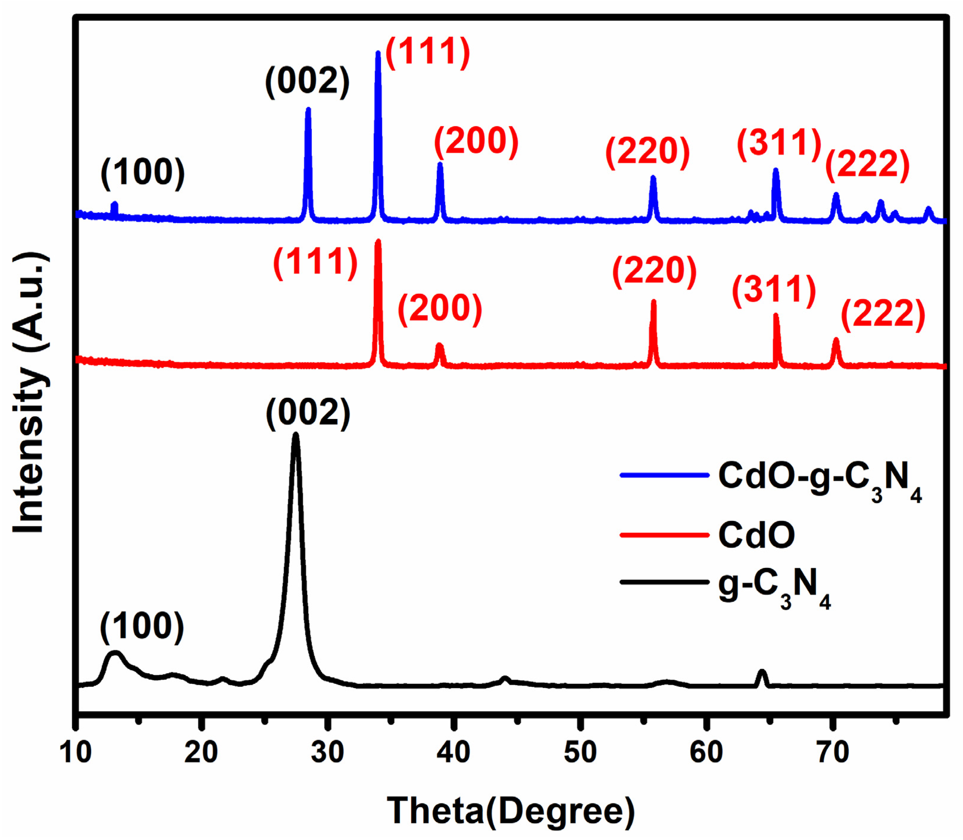
Figure 1. The XRD patterns of g-C3N4, CdO, and CdO-g-C3N4 nanomaterials.
Molecules 27 08646 g001
Figure 2. SEM and HR-TEM micrographs of (a) oblong shape g-C3N4 nanosheets (b) CdO nanorods indicating homogenous distribution and uniform thickness of 50−150 nm and 1–3 ±μm length (c) Nanorod-like structures of CdO-g-C3N4 photocatalyst. (d) H-RTEM micrograph of CdO-g-C3N4 nanocomposite. (e) SAED pattern of CdO-g-C3N4 composite nanorods. (f) Fringe spacing of polycrystalline CdO-g-C3N4 nanocomposite.
Molecules 27 08646 g002
Figure 3. FTIR spectra of pure g-C3N4, CdO nanorods, and CdO-g-C3N4 photocatalysts.
Molecules 27 08646 g003
Figure 4. Tauc Plots of all the prepared materials, (a) pure g-C3N4, (b) CdO nanorods, and (c) CdO-g-C3N4 photocatalysts showing band gap energies (Eg). The band gap of the composite lies in between both of pure g-C3N4, CdO nanorods.
Molecules 27 08646 g004
Figure 5. Photoluminescence spectroscopy of g-C3N4 nanosheets, CdO nanorods, and CdO-g-C3N4 nanocomposite.
Molecules 27 08646 g005
Figure 6. Chronoamperometric responses recorded at Open Circuit Potential (0 V applied potential) for (a) g-C3N4 nanosheets (b) CdO -nanorods and (c) CdO-g-C3N4 nanocomposite in 0.5 M NaSO4 electrolyte by a three-electrode system under chopped on/off light from a solar simulator of AM 1.5 G filter and 100 mW cm−2 (1 Sun) light intensity of Xe lamp.
Molecules 27 08646 g006
Figure 7. Linear Sweep Voltammetry profile in applied potential range of −1 V to 1 V, at a scan rate of 100 mV/s in 0.5 M Na2SO4 aqueous solution (a) g-C3N4 nanosheets (b) CdO -nanorods and (c) CdO-g-C3N4 nanocomposite in a three-electrode system under 1 Sun light irradiation in an OFF and ON light mode.
Molecules 27 08646 g007
Figure 8. EIS analysis and Nyquist plot of g-C3N4, CdO and CdO-g-C3N4.
Molecules 27 08646 g008
Figure 9. The proposed charge transfer mechanism by g-C3N4, CdO and CdO-g-C3N4 Photocatalyst.
Molecules 27 08646 g009
Table 1. A comparison of various recent Cd/CdO nanomaterials or their composites with g-C3N4 and others for Photoelectrochemical/Photodegradation applications.
#PhotocatalystSynthetic
Strategy
Photoelectrochemical/
Photodegradation
Study
Band Gap Energy (eV)Current Density/Hydrogen/Oxygen GenerationRef.
1CdO cauliflowerMechanochemical process followed by heating treatment95%, 91.5%, and 98% photodegradation Congo red, Malachite green and Crystal violet2.22_[41]
2CdS/CdOCo-precipitation92% Photodegradation of the RhB2.9_[42]
3CdO–TiO2sol–gel method>70% Methylene blue (MB), Methyl orange (MO) and Rhodamine B (Rh-B) degradation efficiency3.12–3.19_[43]
4CdO–CdSElectrochemical deposition71.1% of photodegradation efficiency of MB.2.25–2.292.6 mA/cm2[44]
5ZnO/CdOThermal decomposition97.8% degradation efficiency for MB2.99_[45]
6g-C3N4/CdOChemical precipitation & self-assembly96% rhodamine B(RhB) dye removal efficiency.2.35_[38]
7Cd-g-C3N4Thermal polymerization98.1% tetracycline (TC) degradation efficiency.__[46]
8Cd-WO3 (CWC) and Cd-doped WO3@g-C3N4 (CWCC)Ultrasonication, stirring & thermolysis95.98% Photodegradation Efficiency2.06 & 1.85_[47]
9NiO/Cd/g-C3N4Microwave-assisted81.8% MB degradation efficiency3.6 [48]
10Cd-ZnO/g-C3N4Co-precipitation & hydrothermal>90% MB degradation efficiency2.50 [17]
11CdO-g-C3N4Precipitation and Hydrothermal method92% efficiency of MB degradation2.55>5 mA/cm2/
H2 generation
Current work
Publisher’s Note: MDPI stays neutral with regard to jurisdictional claims in published maps and institutional affiliations.