Review

Ursolic Acid Analogs as Potential Therapeutics for Cancer

by
Siva S. Panda
1,*,
Muthusamy Thangaraju
2,3 and
Bal L. Lokeshwar
2,3,4,*
1
Department of Chemistry and Physics, Augusta University, Augusta, GA 30912, USA
2
Department of Biochemistry and Molecular Biology, Augusta University, Augusta, GA 30912, USA
3
Georgia Cancer Center, Augusta University, Augusta, GA 30912, USA
4
Research Service, Charlie Norwood Veterans Administration Medical Center, Augusta, GA 30912, USA
*
Authors to whom correspondence should be addressed.
Molecules 2022, 27(24), 8981; https://doi.org/10.3390/molecules27248981
Submission received: 1 December 2022 / Revised: 13 December 2022 / Accepted: 14 December 2022 / Published: 16 December 2022
(This article belongs to the Special Issue Natural Product-Inspired Molecules: From Weed to Remedy)

Abstract

:
Ursolic acid (UA) is a pentacyclic triterpene isolated from a large variety of vegetables, fruits and many traditional medicinal plants. It is a structural isomer of Oleanolic Acid. The medicinal application of UA has been explored extensively over the last two decades. The diverse pharmacological properties of UA include anti-inflammatory, antimicrobial, antiviral, antioxidant, anti-proliferative, etc. Especially, UA holds a promising position, potentially, as a cancer preventive and therapeutic agent due to its relatively non-toxic properties against normal cells but its antioxidant and antiproliferative activities against cancer cells. Cell culture studies have shown interference of UA with multiple pharmacological and molecular targets that play a critical role in many cells signaling pathways. Although UA is considered a privileged natural product, its clinical applications are limited due to its low absorption through the gastro-intestinal track and rapid elimination. The low bioavailability of UA limits its use as a therapeutic drug. To overcome these drawbacks and utilize the importance of the scaffold, many researchers have been engaged in designing and developing synthetic analogs of UA via structural modifications. This present review summarizes the synthetic UA analogs and their cytotoxic antiproliferative properties reported in the last two decades.

1. Introduction

Cancer is the first or the second leading cause of death worldwide, accounting for 19.2 million new cases and 9.6 million deaths, in 2020 [1]. The most common causes of cancer death in 2020 were lung, colorectal, liver, stomach, and breast cancer. In addition, each year more than 40,000 children develop cancer [1]. In the United States, there will be an estimated 1.9 million new cancer cases and 609,360 cancer deaths in 2022 [2].
The cancer burden continues to grow globally, exerting tremendous physical, emotional, and financial strain on individuals, families, communities, and even the national health systems. Compared to developed counties, low- and middle-income countries have low survival rates because of the lack of accessible early detection, quality diagnosis, treatment, and survivorship care. The global economic burden of cancer is unknown, as accurate economic data are not available from many countries [1]. In the United States, the national patient economic burden associated with cancer care was USD 21.09 billion in 2019 [3].
Current treatments for malignant cancers include surgery, radiation, chemotherapy, adjuvant therapy, hormone therapy, and immunotherapy. Despite all these therapies and the tremendous progress, refinement, and entry of novel drugs, procedures, and treatment options, cancer continues to be a leading cause of death. The challenges to reducing cancer burden, and specifically for treatment failures, are associated with the development of drug resistance, disease progression, and dose-limiting systemic toxicities of potent drugs [4]. The much-heralded immunotherapy has low efficacy, with less than 20% of patients responding to the treatment. Novel effective and efficient pharmacological agents that act through unconventional mechanisms to enhance existing therapies or kill tumor cells resistant to other existing therapies are urgently needed.
Over four decades, bioactive compounds isolated from natural sources, such as plant and marine organisms, the natural products, have dominated the cancer prevention, treatment, and drug development areas. Natural products (NP) are one of the main sources of diverse pharmacologically active compounds [5,6,7,8,9,10,11]. NPs and NP-scaffolds comprise a large portion of current-day pharmaceutical agents (over 70% of FDA-approved drugs) [12,13]. At present, several native or modified NPs have attained the status of cancer therapeutics. These include irinotecan, vincristine, etoposide, and paclitaxel from plants, actinomycin D, and mitomycin C from bacteria, as well as marine-derived bleomycin.

2. Triterpenes as Bioactive NPs

Triterpenoids are compounds with a carbon skeleton based on six isoprene units, metabolites of isopentenyl pyrophosphate oligomers, representing the largest group of phytochemicals. These are a large and structurally diverse group of NPs having about 200 distinct skeletons, and more than 20,000 triterpenoids are isolated and identified from nature [14,15].
In the 1920s, for the first time, Ursolic acid (UA) was isolated and identified from epicuticular waxes of apples. UA is one of the most abundant and well-studied triterpenoids found in various cuticular waxes of edible fruits (apple, blueberry, cherry, cranberry, Japanese loquat, lemon, olive, orange, peach, pear, prune, quince, tangerine, and tembusu), leaves (coffee, elder, glossy privet, hawthorn, lavender, nerium, marjoram, olive, organum, rosemary, thyme, and whorled rosinweed), flowers (loquat, marigold), and bark (elder, olive, and silver birch) of medicinal plants [16,17,18,19,20]. UA is present in most edible plant products.
UA holds an important place among various triterpenoids because of its wide range of biological activities. UA is abundantly present in medicinal plants and shows many pharmacological activities including antiproliferative [21,22,23], antimicrobial [24,25], antiviral [26,27], antioxidant [28,29,30], and anti-inflammatory activities [30,31].
UA has been extensively applied in cancer treatment in Traditional Chinese Medicine (TCM) for several years. Over the last two decades, UA has been tested on several malignant cancers for both the prevention of cancer progression and as a treatment. Most studies are limited to its activities against energy metabolism, cell proliferation, and antioxidant activities [32,33,34,35,36,37,38,39,40,41,42,43]. These studies have established at least in vitro, that UA inhibits many activities of cancer cell growth, energy utilization, and tumor cell-induced inflammation as summarized in Figure 1 [44,45,46,47,48,49,50,51,52,53].
Despite its versatile functions, UA has limitations. Most studies on UA are limited to cell cultures in vitro where UA has access to cells and it is not a concern. UA diluted in biocompatible solvents are added to cell cultures, and its activities on cells are determined by many analytical techniques. However, the availability of UA at the site of the tumors in an intact body is a significant concern. Like many natural, medicinal compounds, administration of UA parenterally is seldom used due to its poor solubility and potential toxicity of the solvents, such as Dimethyl Sulfoxide or Dimethyl Formamide. The oral route, via its addition to diets or drinking water, is of little use. The bioavailability of UA, in plasma, is limited to ≤500 nM [54]. Most in vitro studies have shown the inhibition of tumor cell proliferation in low micromolar concentrations. Studies using transgenic prostate cancer models have reported good tumor control within 12 weeks of treatment in well-established transgenic tumors via a solid diet at 1% w/w. The authors reported a ~60% reduction in prostate tumor volume following a 12-week observation, indicating the high efficacy of UA in the prevention of tumor progression in this model [55]. Studies in human volunteers were less promising, concerning UA as a potential therapeutic for cancer [18]. The mode of delivery of the compound by nano-formulations such as nanoliposomes has not shown much improvement in increased plasma levels [56]. In addition, the Phase I study of nanoliposome formulation showed no cumulative accumulation of UA in the body as performed by the multi-phase pharmacokinetics study. These studies suggest that while UA itself has promise as an anticancer therapeutic, structural modification for the UA molecule to enhance its solubility, absorption, low-protein binding, and longer tissue accumulation should significantly enhance its application in cancer patients.
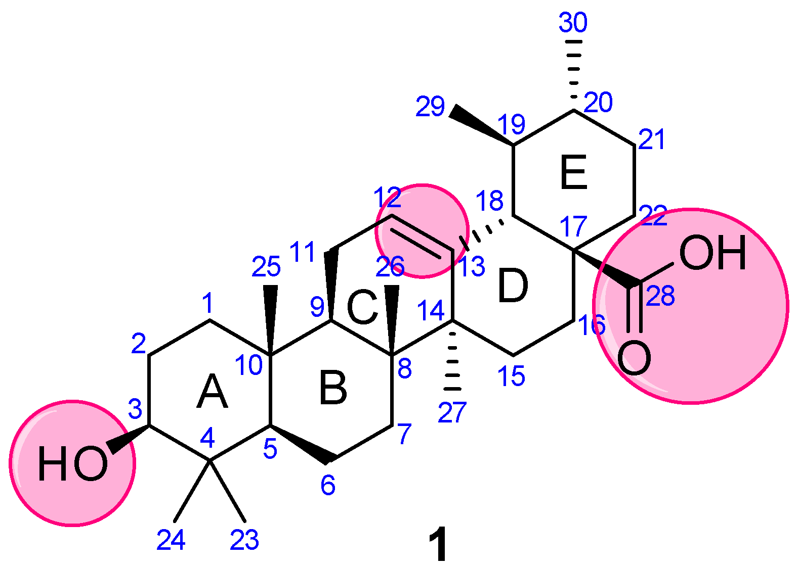
Concerning the versatile properties of UA, we focused on compiling the synthetic analogs of UA, which show potential anticancer properties, and analyzed their structure-activity relationship (SAR). UA (3-(β-hydroxy-urs-12-en-28-oic acid), 1) is a ubiquitous pentacyclic compound that possesses functional groups such as a carboxylic group at C28, β-hydroxy function at C3, and an alkene at C12–C13 (Figure 2). Considering UA as a lead molecule, researchers extensively modified and hybridized UA at these sites with the aim of enhancing its anticancer properties and overcoming the associated poor absorption and low bioavailability drawbacks. We have categorized our analysis based on modifications and/or alterations at different pharmacophoric sites.

3. Materials and Methods

According to the databases of PubMed, Science Detect, Web of Science, American Chemical Society, Springer, and Scopus, a comprehensive and systematic review was performed. The keywords “ursolic acid” AND “anticancer” were used and further filtered by “analogs”, “derivatives” and “synthesis”. The systematic search of electronic databases identified 183 articles after excluding patents, clinical trials, and conference abstracts. We carefully reviewed all the articles and chose the articles that discuss and/or report on modified ursolic acids and their anticancer properties.

4. Synthetic Analogs of UA

Molecular hybridization (the conjugation of two or more bioactive scaffolds via a covalent bond, MH) is a powerful strategy in drug discovery and can play an effective and efficient tool for the development of new drug candidates. MH is a strategy of rational design of new ligands or prototypes based on the combination of pharmacophoric moieties and has emerged as an important strategy for the development of new hybrid architectures that maintain pre-selected characteristics of the original templates and can act as multitarget ligands. This approach could contribute to the design of multifunctional cancer drug candidates and might be useful in overcoming the problems associated with parent drugs [57,58,59,60,61,62,63,64,65]. In the last two decades, the MH approach has been extensively incorporated in UA research and has reported several modified UA products as anticancer agents [66]. We believe the compiled structure analysis and rationale could help the medicinal chemist to develop potential anticancer drug candidates.

4.1. Modification of UA at C3

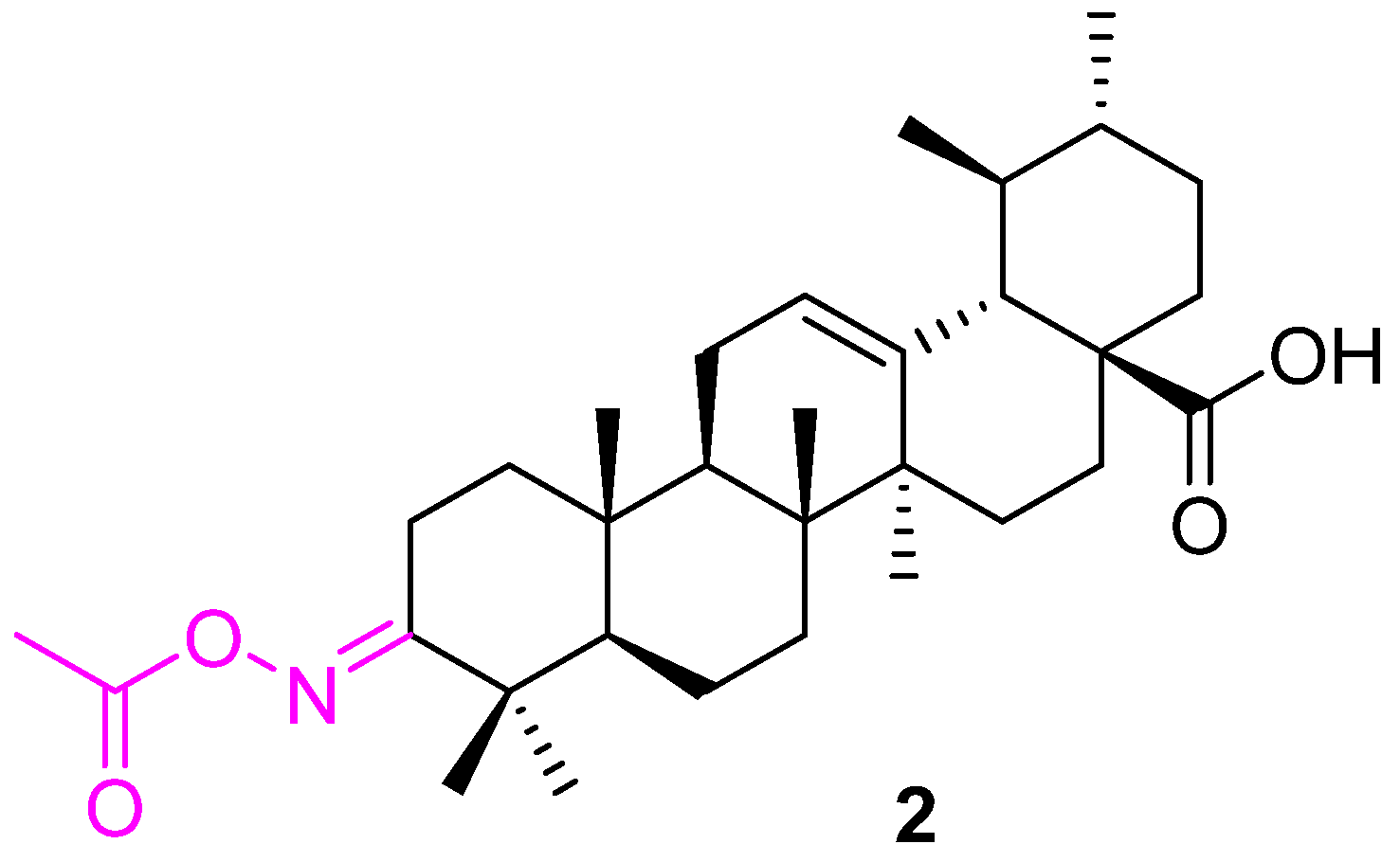
Several attempts were made to investigate the anticancer effect of the alcohol group at the C3 position of ursolic acid. Thien et al. synthesized compound 2 starting from UA. Compound 2 (Figure 3) was found to be 2–3 times more active than the parent UA against human mouth epidermal carcinoma (KB), human hepatocellular carcinoma (HepG2), human breast carcinoma (MCF7), and human lung carcinoma (Lu) cell lines [67].
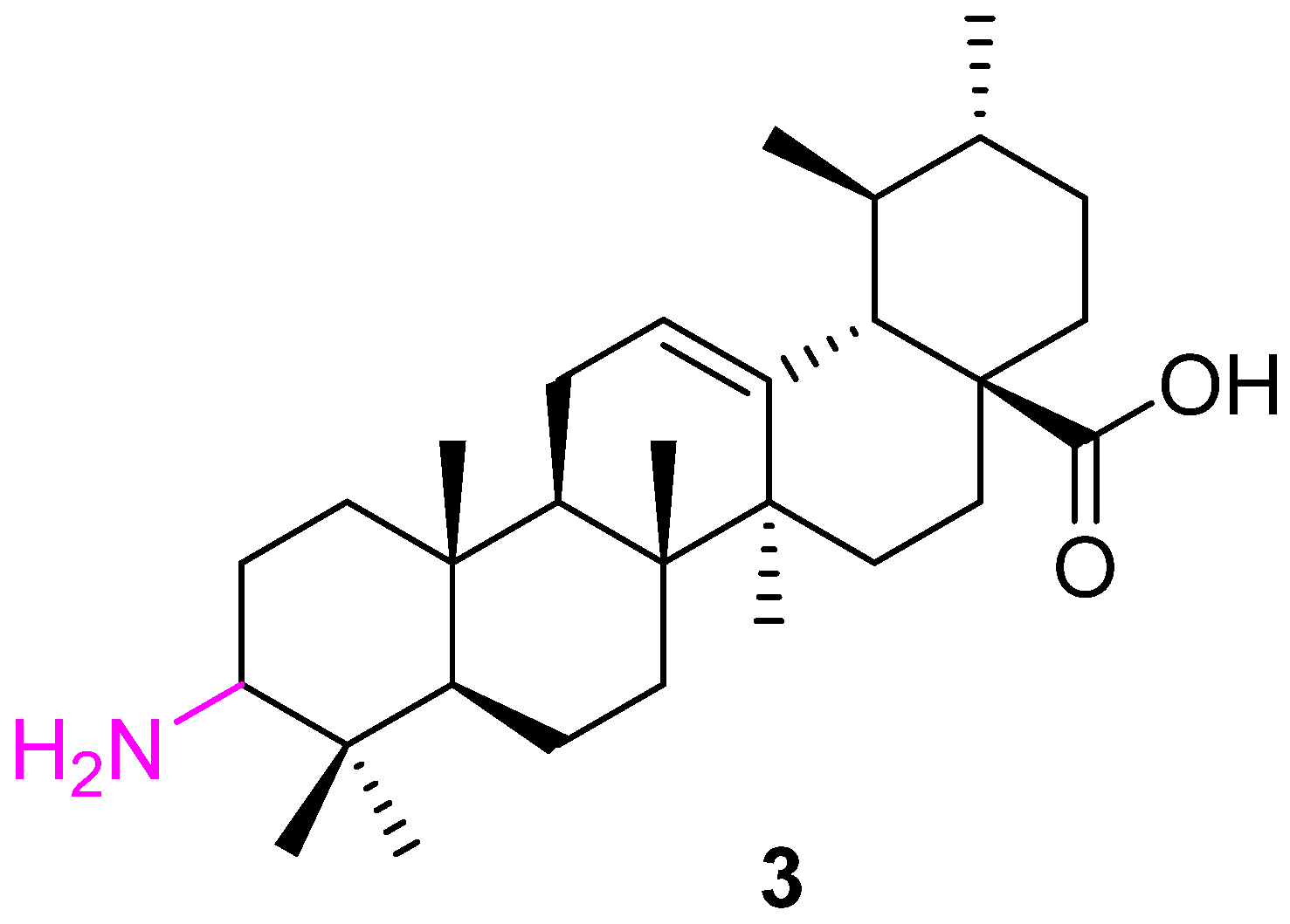
Synergistic or combination therapy is one of the promising approaches in cancer treatment. da Silva et al. converted the C3 alcohol group to an amino group (3) and used it in combination with imatinib to create the impact of a co-drug on leukemia cells (K562). Compound 3 (Figure 4), itself, shows potential inhibition against K562 (IC50 value of 5.2 μM). Further investigation suggests that it induced cell death (apoptosis) via activating caspases-3 and -8 and caused cell cycle arrest. K562 cells treated with Compound 3 were arrested in the G1 phase of the cell cycle and decreased the cell population in the G2 phase. A synergistic effect was observed when 3 was used along with imatinib to treat leukemia [68].
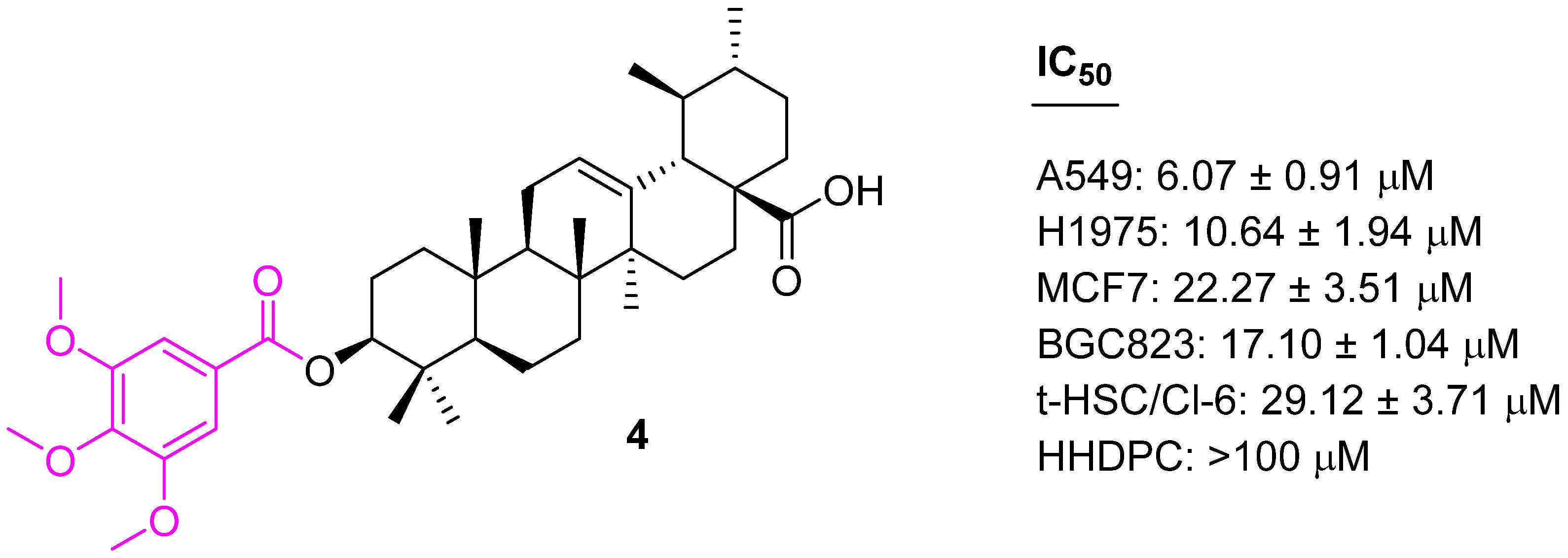
Xu et al. introduced the 3,4,5-methoxy benzoic acid moiety at the C3 position of UA and evaluated their cytotoxicity properties against A549, MCF7, H1975, and BGC823. The UA derivative containing 3,4,5-methoxy-phenacyl at the C3 position (4) (Figure 5) shows a significant selective antiproliferative effect with IC50 values in the range of 6.07 to 22.27 µM [69].
Another attempt was made to modify the C3 alcohol group of UA at a carbonyl group (5) (Figure 6) and also introduce three carbons using the Barbier–Grignard method. The synthesized stereoisomeric analogs of UA (6 and 7) were found to be effective against three cancer cell lines (HepG2, Hep3B, and HA22T/VGH). Further studies confirm the inhibition of NF-κB activation. However, analog 5 was ineffective against all three cell lines [70].
UA analogs (modification at C3) listed in Table 1 reduced cell cycle arrest and induced apoptosis by activating Bax and caspases-8, -9, and -3 and also by reducing Bcl2 and MDM2. Apoptosis is involved in maintaining homeostasis of cell numbers by removing damaged or unwanted cells; therefore, UA derivatives could be important chemotherapeutic compounds to reduce tumor growth. UA-derivatives also inhibit NF−κB, which in turn, reduces cell growth arrest. NF−κB signaling plays an important role in cell growth and inhibition. NF−κB signaling will reduce cell growth by inducing cell cycle arrest (Table 1).

4.2. Modification of UA at C28

The methyl ester derivative of UA (8) (Figure 7) did not affect the anticancer properties of UA, as revealed by several reports [71]. However, the benzyl ester of UA (9) showed a decrease in anticancer properties against the K562 leukemia cell line [72]. Nevertheless, the isopropyl ester moiety at C17-COOH (C28) showed a potent inhibitory effect on the growth of the human bladder cancer (NTUB1) cell line. Flow cytometric analysis demonstrated that treatment of NTUB1 with 10 led to cell cycle arrest in addition to an increase in apoptotic cell death. These data suggest that the possibility of G1 phase arrest and apoptosis in 10-treated NTUB1 for 24 h was mediated through an increased amount of ROS in cells exposed with 10, while the presence of G2/M arrest before the accumulation of cells in the sub-G1 phase in 7-treated cells for 48 h was also due to an increased amount of ROS in cells exposed to 10 [73].
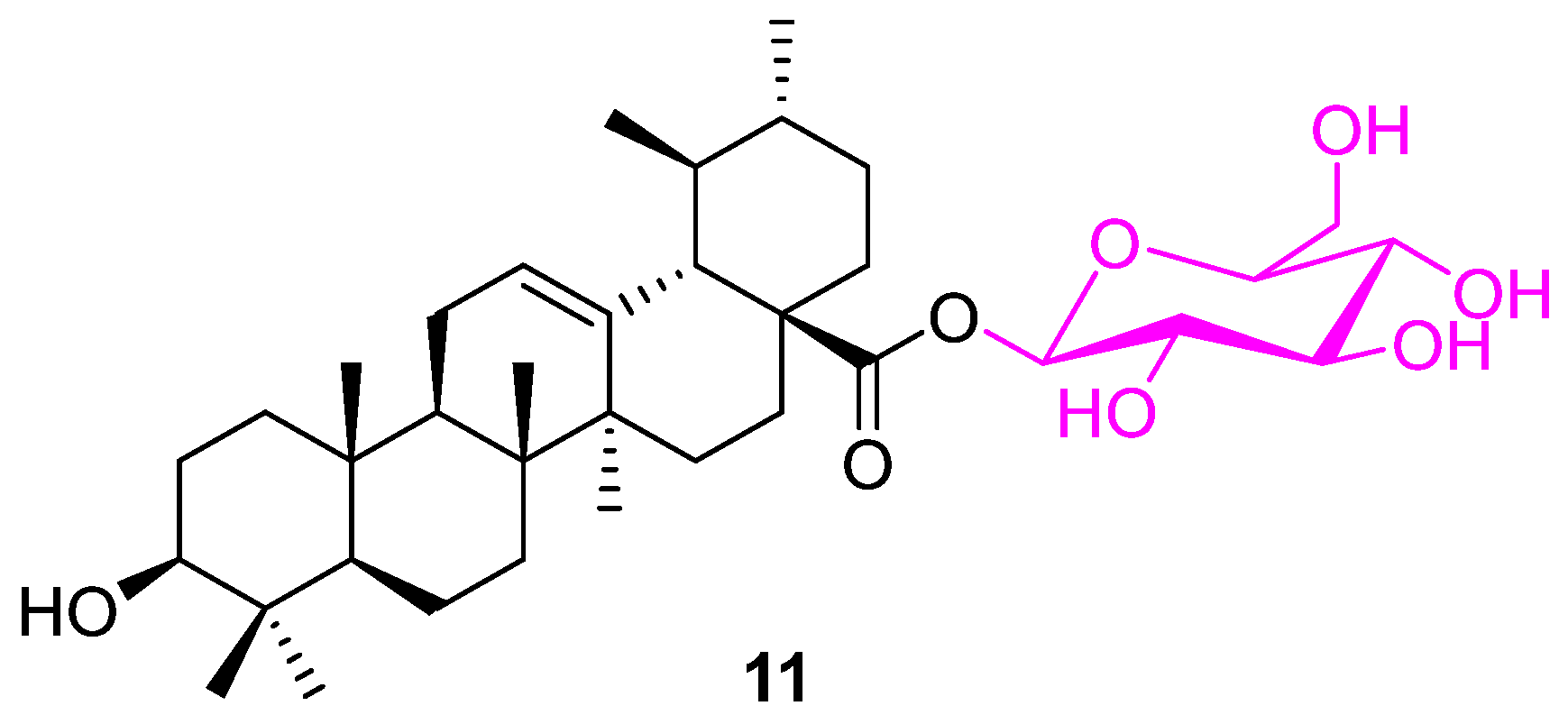
Several modifications were made to UA by incorporating a glycosyl or arylpropenoyl scaffold at the C28 and/or C3 positions. The observed experimental data suggest that only compound 11 (Figure 8) was found to have enhanced anticancer properties against human colon adenocarcinoma (HT29) cell lines with an IC50 value of 8 µM in comparison to UA (IC50: 30 µM) [74].
Bai et al. synthesized a set of UA conjugates with structural modifications at the C3 and C28 positions. Compound 12 (Figure 9) stood out from the pool compound by showing cytotoxicity with hepatoma (HepG2), gastric carcinoma (AGS), colorectal carcinoma (HT29), and prostatic carcinoma (PC3) cell lines. However, compound 12 is more effective against AGS cell lines with an IC50 value of 11.4 µM. The cell cycle distribution and sub-G0/G1 ratio data indicate that compound 12 is effective with 92.64 ± 3.13% inhibition. The presence of -OH and -NH2 groups contributes to improving the log P. Theoretical (arithmetic) and computer-assistant calculations were then used to predict the logP value, such as ACD\logP and XlogP3 and Molinspiration\logP [75]. The similar analog, 13, was prepared and tested against MCF7, Hela, and A549 cell lines. Analog 13 show improved IC50 values (9.19 ± 0.82, 8.56 ± 0.53, and 12.72 ± 0.79 µM for MCF7, Hela, and A549 cell lines, respectively). In addition, it is also observed to increase its carbon chain length between NH and NH2, which decreases the potency [76].
Liu et al. investigated the application of C28 modified UA and found that keeping the C3 alcohol group unchanged as a polar substituent at the C3 position is essential for pharmacological activities. From the series of synthesized conjugates, compound 14 was identified as the most potent against human gastric cancer (MGC803) and human breast cancer (Bcap37) cell lines with an IC50 of 2.50 ± 0.25 µM and 9.24 ± 0.53 µM, respectively. Further mechanistic studies, such as acridine orange/ethidium bromide staining, Hoechst 33,258 staining, terminal deoxynucleotidyl transferase biotin-dUTP nick end-labeling (TUNEL) assay, and flow cytometry, indicate 14 (Figure 10) can induce apoptosis in MGC803 cells [77].
The same research group synthesized another series of C28-modified UA based on the above results. Compound 15 was found to be the most active from the new series among all the synthesized analogs. Several variations have been explored with different chain lengths and secondary heterocycles. The combination of our carbon chain linker and piperidine gave the best outcome against MGC803 and Bcap37 human cancer cell lines with 4.53 and >20 µM IC50 values, respectively. Further study of the mechanism via acridine orange/ethidium bromide staining, Hoechst 33,258 staining, and TUNEL assay and flow cytometry suggest 15 can induce apoptosis in MGC803 cells at 10 μM [78].
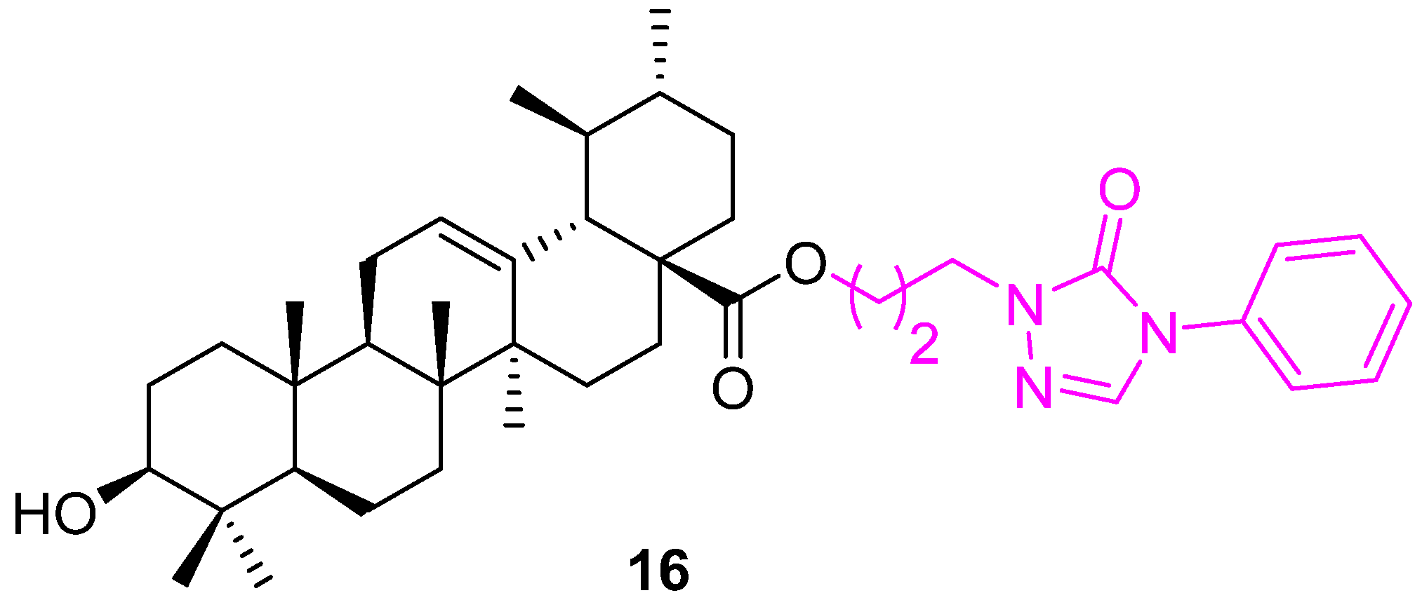
Hypoxia-inducible factor-1α (HIF-1α) is one of the key mediators of angiogenesis and survival in tumor metastasis that has been established as an important cancer drug target. Several UA derivatives incorporating oxadiazole, triazolone, and piperazine moieties at the C28 position were designed and synthesized as HIF-1α inhibitors. Most of the derivatives demonstrated the ability to inhibit the expression of HIF-1α, but 16 (Figure 11) exceptionally inhibited HIF-1α transcriptional activity under hypoxic conditions with IC50 = 36.9 µM. None of the UA derivatives showed cytotoxic activity (IC50 > 100 µM). These compounds require further investigation [79].
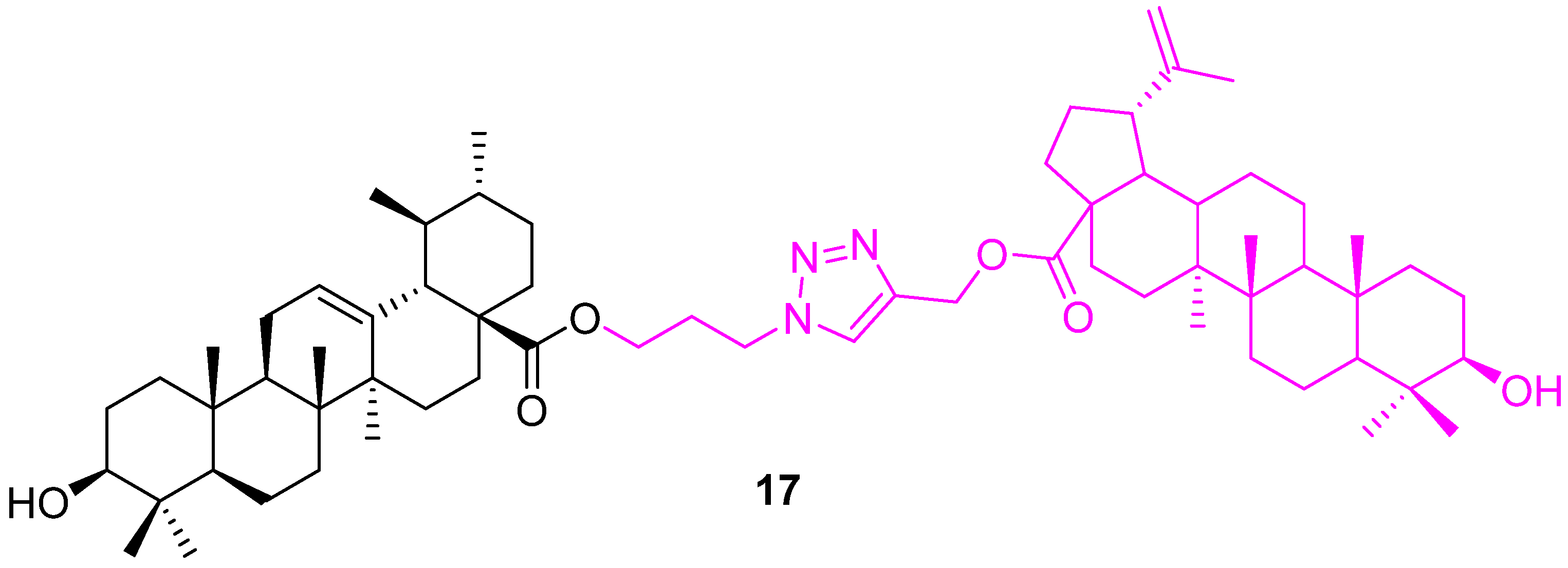
Patnaik et al. synthesized UA hybrids by modification at C28 and the incorporation of zidopropyl-3β-hydroxy-urs-12-en-28-oate via a cycloaddition reaction. The synthesized hybrid compounds were tested for their anticancer potential against two human breast cancer cell lines (MCF7 & MDA-MB231) using a sulfarhodamine B cell proliferation assay. These hybrids showed more selectivity towards the MDA-MB231 cell line than MCF7. Compound 17 (Figure 12) is the highly effective one, with a GI50 value of 1.4 ± 0.1 μM, which is 2.9 times more active than the standard doxorubicin against MDA-MB231. In addition, a mechanistic study suggested that compound 17 arrests cells in the mitotic phase of the cell cycle [80].
Semenova et al. synthesized a set of 1,2,4 triazole-incorporated UA derivatives. The conjugates were evaluated for their antiproliferative activity on MCF7, U-87 MG (glioblastoma multiform cells), A549 (lung carcinoma), and HepG2 (hepatocarcinoma) cell lines. The compounds were moderately active and, interestingly, they showed selectivity towards A549. Compounds 18 and 19 (Figure 13) are azole-based thiones that show selective inhibition (IC50 values 11.25 ± 0.77 and 13.45 ± 1.14 µM) [81].
UA conjugated with a lipophilic triphenylphosphonium cation was screened with three human cancer cell lines (MCF7, HCT116, and TET21N). The triphenylphosphonium UA derivative (20) (Figure 14) not only showed higher cytotoxicity as compared to UA but was also markedly superior in triggering mitochondria-dependent apoptosis, as assessed using a range of apoptosis markers such as cytochrome c release, stimulation of caspase-3 activity, and cleavage of poly(ADP-ribose) polymerase [82].
The published studies indicate that these C28 modified UA derivatives induce apoptotic cell death by activating death-caspases, caspase-3 or/and caspase-8, unlike the C3 derivatives, which seem to have caused cell cycle arrest and mitotic catastrophe-induced cell death. However, a lack of data on non-proliferating cells or cells with intact p53 makes these studies hard to compare for their tumor cell specific toxicity. Further, a lack of testing in vivo is another weakness when assessing their potential as future antitumor drugs (Table 2).

4.3. Modification of UA at Both C3 and C28

Esterification of 3-OH and 17-COOH significantly reduces the anticancer properties [71,72]. Maintaining a polar group at either position is essential for the activity [83]. Popov and co-workers explored the variation on UA at C3 and C28 by introducing 1,3.4-oxadiazole, 1,3.4-oxadiazole, and/or 1,2,3-triazole moieties. All the possible analogs were synthesized and screened against different cancer cell lines along with immortalized human fibroblasts. Hybrid conjugates of 1,3,4-oxadiazoles attached at the C3 and C28 positions of the UA frame via a triazole spacer, and combinations of 1,2,5-oxadiazole or 1,3,4-oxadiazole, tethered with a succinate linker, and 1,2,3-triazole at C3 of the UA backbone, were found inactive. From the series of conjugates, 22a and 22b (Figure 15) show the best cytotoxic activity and selectivity on HepG2 and MCF7 cells. The introduction of an additional ester-type linker between triazole (24) and the UA scaffold or UA frame and furoxan, either at C3 (23) or at C28 (25), led to the loss of cytotoxicity (Table 3) [84].
Shao et al. developed several UA analogs, retaining the polar properties at the C3 and C28 positions. Among the several synthesized analogs, compound 26 (Figure 16) showed potential anticancer properties against human hepatoma (HepG2), human gastric cancer (BGC823), human neuroblastoma (SH-SY5Y), human cervical carcinoma (Hela), and human embryonic lung fibroblast (HELF) cell lines. Further, a flow cytometric analysis and morphologic changes study suggested compound 26 arrests cell cycle progression at the S phase in HepG2 cells [85]. The same compound 26 was prepared by another research group, who evaluated its EC50 values against several cancer cell lines; they validated the potency of 26, however, it also had a lower ED50 against the non-malignant fibroblasts NIH 3T3 [86].
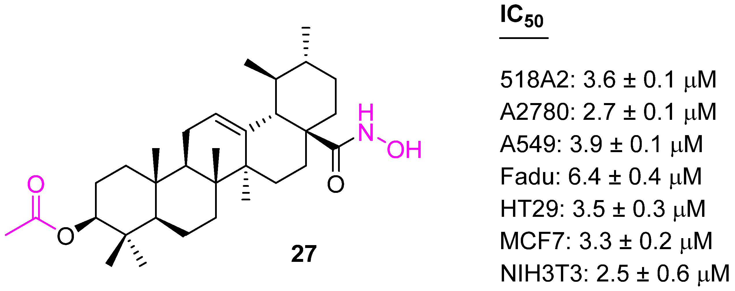
UA-derived hydroxamates were synthesized by adding a C3 acetyl group; they were screened for their cytotoxicity utilizing SRB assays against several human tumor cell lines. Among the various hydroxamates, compound 27 (Figure 17) was found to be the most potent against seven cancer cell lines. However, unfortunately, these hydroxamates were nonselective and showed toxicity to nonmalignant mouse fibroblasts (NIH 3T3) [87].
The structure–activity relationship confirms the acetyl group at C3 is more effective than the butyl group. Keeping the acetyl group at the C3 position, another set of conjugates was developed with variations at the C28 position. Compound 28 (Figure 18) was found to be more potent than the other synthesized ones against Hela, SKOV3, and BGC823 cell lines [88].
Another similar modification was attempted to prepare compound 29 (Figure 19), which targeted gastric cancer. Compound 29 was tested against BGC823 cells and a human normal gastric (GES1) cell line to demonstrate its selectivity. The studies uncovered that 29 could induce apoptotic effects in the treated BGC823 cells, such as a comet-like DNA bend, sub-G0/G1 phase accumulation, and phosphatidylserine externalization (apoptosis). In addition, the activity of caspase-3 was found to be up-regulated, while the expression of Bcl2 and survivin were down-regulated in 29 treated cells. Data from studies in mice indicate compound 29 is safer than Taxol [89].
Compound 30 was synthesized to have an acetyl group at the C3 position. The modified UA 30 was 2–3 times more active than the parent UA against human mouth epidermal carcinoma (KB), human hepatocellular carcinoma (HepG2), human breast carcinoma (MCF7), and human lung carcinoma (Lu) cell lines [67].
It is important to understand the impact of harsh environments on cancer cells as several discoveries on the metabolic pathways of cancer cells have led to a better overall understanding of their ability to proliferate and adapt to their microenvironment [90]. To uncover the pathways involved in cancer cells, or highly proliferative tumors, recently “cancer metabolism” and “metabolic reprogramming” have been investigated [91,92,93,94,95]. A new analog (31) (Figure 20) was designed by using a computer-aided drug design (CADD) approach to have an acetyl group at C3. The molecular docking studies suggest 31 could bind to the active sites of glucokinase (GK), glucose transporter 1 (GLUT1), and ATPase, which are the key enzymes involved in cancer glucose metabolism. Further, experimental observations confirm the synergistic effect of 31 and glycolysis inhibitor 2-deoxy-D-glucose (2-DG) in inhibiting the glucose metabolism of cancer cells. The depletion of intracellular ATP and decrease in lactate production triggers the cancer cell’s arrest in the S and G2/M cycle phases. In addition, the combination selectively down-regulated the expression of the Bcl2 and HKII proteins and up-regulated the expression of Bax and p53, which results in enhanced apoptosis. The Western blotting assay illustrates the molecular targets of 31, which includes Bax, Bcl2, p53, and HK proteins. The LC-MS analysis of animal serum proved the bioavailability of 31 [96].
Yang et al. synthesized a similar set of UA analogs containing the C3 acetyl group. The synthesized UA analogs were inhibiting cell growth when assayed against various tumor cell lines and a non-pathogenic cell line of normal human embryonic lung fibroblasts (HELF). A theoretical toxicity risk analysis was carried out using OSIRIS and the results indicated that most compounds showed moderate to low risks. However, compound 32 stands alone with significant IC50 values ranging from 4.09 ± 0.27 to 7.78 ± 0.43 µM against 12 different tumor cell lines. Flow cytometry analysis data indicated that 32 induced G0/G1 arrest in three of these cell lines (HT29, HepG2, and RL95-2). Observed experimental data were validated by structural docking studies, which confirmed that UA or its derivatives, could bind to cyclins D1 (CycD1) and cyclin-dependent kinases (CDK6), the key regulators of G0/G1 transition in the cell cycle, while the piperazine moiety of 32 could bind with glucokinase (GK), glucose transporter 1 (GLUT1), and ATPase, which are the key proteins involved in cancer cell metabolism. Further, the inducing apoptosis capability of 32 was confirmed by acridine orange/ethidium bromide staining and decreased cell viability in a dose-dependent mode [97].
Keeping the acetyl group at the C3 position intact, the C28 position was further explored, and several analogs were synthesized and screened against five cancer cell lines (MGC803, HCT116, T24, HepG2, and A549 cell lines) and a normal cell (HL7702) using an MTT assay. Compound 33 was identified as the most potent analog and investigated via cell distribution by acridine orange/ethidium bromide staining, Hoechst 33,258 staining, JC-1 mitochondrial membrane potential staining, and flow cytometry, which confirmed that the potency of 33 (Figure 21) was probably achieved via the induction of cell apoptosis by G1 cell-cycle arrest. Further, Western blot and qRT-PCR (quantitative real-time PCR) experiments confirmed that 33 induces apoptosis via both intrinsic and extrinsic mechanisms [98].
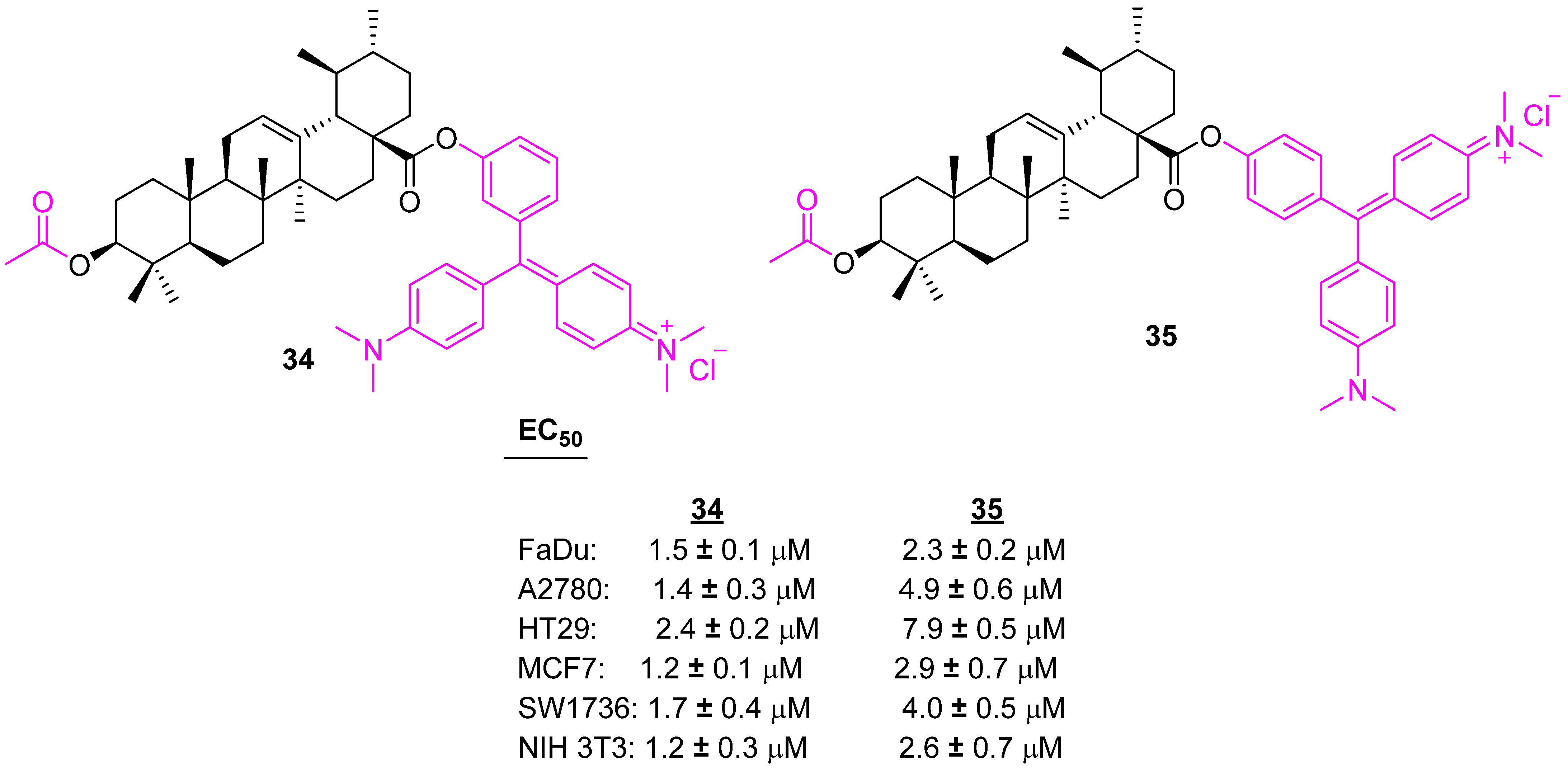
Another set of piperazine-like UA conjugates were developed and tested with several cancer cell lines. Among the conjugates, 34 and 35 (Figure 22)were shown to be potent against several cancer cells, however, these showed high toxicity on nonmalignant fibroblasts [99].
Transcription factor nuclear factor-kappa B (NF-κB), known to be a regulator of a wide variety of anti-apoptotic proteins, is commonly over-expressed and constitutively activated in different types of hematologic cancers and solid tumors. NF-κB is one of the prime targets for cancer therapy. A series of UA analogs containing long-chain diamine moieties were designed as NF-κB inhibitors. Compound 36 shows anticancer potential against the test tumor cell lines including multidrug-resistant human cancer lines, with the IC50 values ranging from 5.22 to 8.95 μM. A further mechanistic study revealed compound 36 (Figure 23) arrests the cell cycle at the G1 phase and triggered apoptosis in A549 cells through blockage of the NF-κB signaling pathway [100].
A set of ethylenediamine-spacer carboxamides of UA (37 and 38) (Figure 24) were prepared and evaluated for their cytotoxicity against several human tumor cell lines using SRB assays. All the synthesized carboxamides showed increased cytotoxicity for the cancer cells than UA. However, unfortunately, the toxicity was not selective and exerted enhanced toxicity against non-malignant mouse fibroblasts (NIH 3T3). The presence of the acetyl group in these carboxamides helps in increasing the cytotoxicity. These carboxamides require further attention to improve their selectivity [101].
Polyamines contain positively charged nitrogen atoms at physiological pH and can serve as electrostatic bridges between negatively charged phosphates of DNA. Polyamines are precursors of aminoalkylguanidines, which could be considered as a scaffold for the development of chemotherapeutic agents [102,103]. Spivak et al. synthesized a C28 Guanidine-Functionalized UA analog (39) (Figure 25). The impact of the guanidine group on the antitumor properties of UA was examined and 39 showed enhanced selective cytotoxicity with five human cancer cell lines (Jurkat, K562, U937, HEK, and Hela). Mechanistic study indicated the cell cycle was arrested at the S-phase of Jurkat cells and led to apoptosis [104].
A library of UA derivatives was prepared by incorporating 2-mercapto-1,3,4-oxadiazoles, 2-amino-1,3,4-oxadiazoles, and 3-mercapto-1,2,4-triazoles at the C28 position. From the library, the compound having 2-amino-1,3,4-oxadiazole scaffold (40) (Figure 26) was found to be selective towards MCF7 cell lines and showed an anticancer potential comparable to doxorubicin. In addition, the acetyl group at the C3 position enhanced activity in comparison to the deacetylated analog (41) [105].
Ma et al. synthesized several analogs of UA via structural modification at C3, C28, and C11 and investigated their anticancer properties. Among all the modifications, compound 42 (Figure 27) was identified as the most potent against human leukemia cancer (HL-60), human gastric cancer (BGC), human hepatocellular carcinoma (Bel7402), and human cervical cancer (Hela) cell lines. The chirality also played an important role as the β-form (42a) is 20-fold more active than the α-form (42b) as well as UA [71].
The incorporation of a triazole moiety is one of the most efficient and most adopted approaches in developing drug candidates [106,107]. Rashid et al. synthesized several ursolic acid-triazolyl derivatives and screened them against A549 (lung), MCF7 (breast), HCT116 (colon), THP1 (leukemia), and a normal human epithelial cell line (FR2) using the sulforhodamine-B assay. Four compounds (43ad) (Figure 28) out of eighteen compounds were found to be most active. Compounds 43ac with o-bromo, o-methoxy and o-chloro substitution at the aromatic ring were selective towards cancer cells, however, 43a showed cytotoxicity for normal cell line (FR-2) cells (Table 4) [108].
UA derivatives were prepared with a modification at the positions C3 and C28 of UA. Compounds 44a (Figure 29) and 44b were found to be most active against Hela, HepG2, and BGC823 cell lines in comparison to the reference drug, gefitinib (IC50 value 17.1, 20.7, and 19.3 µM for Hela, HepG2, and BGC-823, respectively) [109].
Meng et al. synthesized eighteen UA derivatives with C3 and C28 modifications. Among them, compound 45 (Figure 30) was identified as the most active against BEL7402 and SGC7901 cell lines. The carbon chain length impacts the potency and from the analogs, it indicates a six-carbon chain is optimal [110].
Zhao et al. synthesized UA analog (46) (Figure 31) by modifying at the C3 and C28 positions and incorporating an O-[4-(1-piperazinyl)-4-oxo-butyryl moiety. Compound 46 showed 8–10-fold enhanced anticancer properties (against MCF7, Hela, and A549 cell lines) in comparison to the parent UA [111].
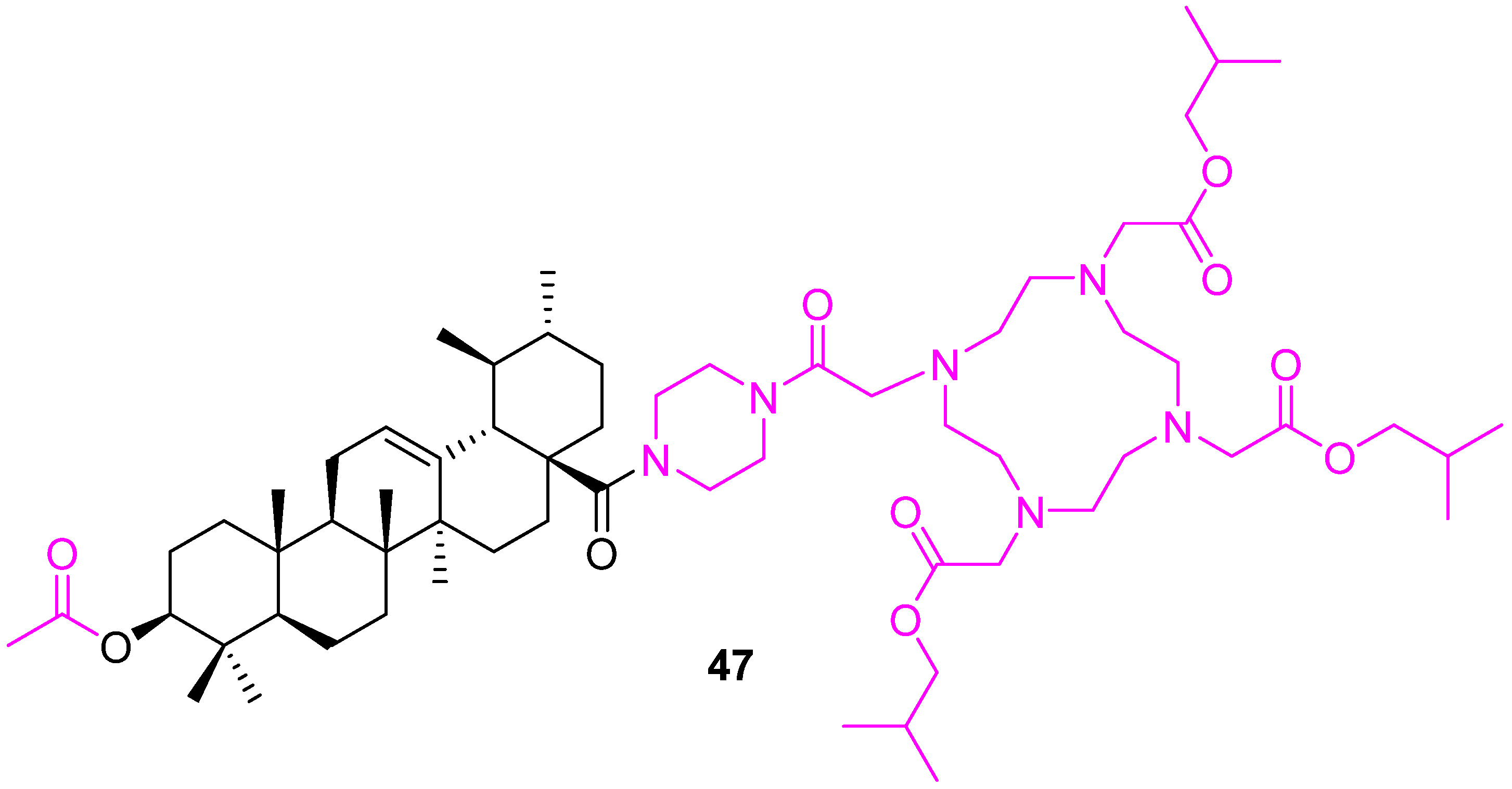
Kahnt et al. reported several UA-1,4,7,10-tetraazacyclododecane-1,4,7,10-tetraacetic acid (DOTA) conjugates with various oligo-methylene diamines as linkers. From them, compound 47 (Figure 32) was found to be the most promising and showed EC50 values of 1.5 µM (for A375 melanoma) and 1.7 µM (for A2780 ovarian carcinoma) [112].
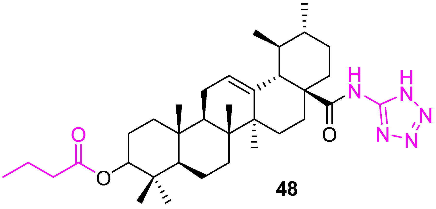
The hypoxia-inducible factor-1α (HIF-1α) pathway has been implicated in tumor angiogenesis, growth, and metastasis. In the continuous effort to develop potential chemotherapeutics, another potent HIF-1α inhibitor (48) (Figure 33) was reported by Zhang et al. with an IC50 value 0.8 ± 0.2 µM. The tetrazole located at C28 is the critical component for the potency [113].
UA analogs generated upon modification of both the C3 and C28 positions, reduced cell cycle arrest and induced apoptosis by activating p53, Bax and caspases-8, -9, and -3, and also by reducing Bcl2 and survivin. Apoptosis is involved in maintaining homeostasis of cell numbers by removing damaged or unwanted cells; therefore, UA derivatives are important chemotherapeutic compounds to reduce tumor growth. UA derivatives also inhibit NF−κB, which in turn, reduces cell growth arrest. NF−κB signaling plays an important role in cell growth and the inhibition of NF−κB signaling will reduce cell growth by inducing cell cycle arrest (Table 5).

4.4. Modification of UA at Other Positions

In addition to C3 and C28 modifications, several structural changes are reported that help enhance anticancer properties. Leal et al. synthesized a series of heterocyclic derivatives of UA as therapeutics for pancreatic cancer. Compound 49 (Figure 34), which is an α,β-unsaturated ketone at C3 in conjugation with a heterocyclic ring at C28, shows 7-fold more antiproliferative activities than UA with an IC50 of 1.9 µM against the pancreatic cancer (AsPC1) cell line. Further investigation confirms 49 arrests the cell cycle in the G1 phase and induces apoptosis in AsPC1 cells with the up-regulation of p53, p21waf1 and NOXA protein levels [114]. The same research group developed another potent compound, 50, which is showing anticancer properties against various cancer cell lines at significantly low concentrations. Compound 50 also arrests the cell cycle at the G1 phase with the up-regulation of p21waf1. Apoptosis was induced at an inhibitor concentration of 8 µm with up-regulation of NOXA and down-regulation of c-FLIP [115].
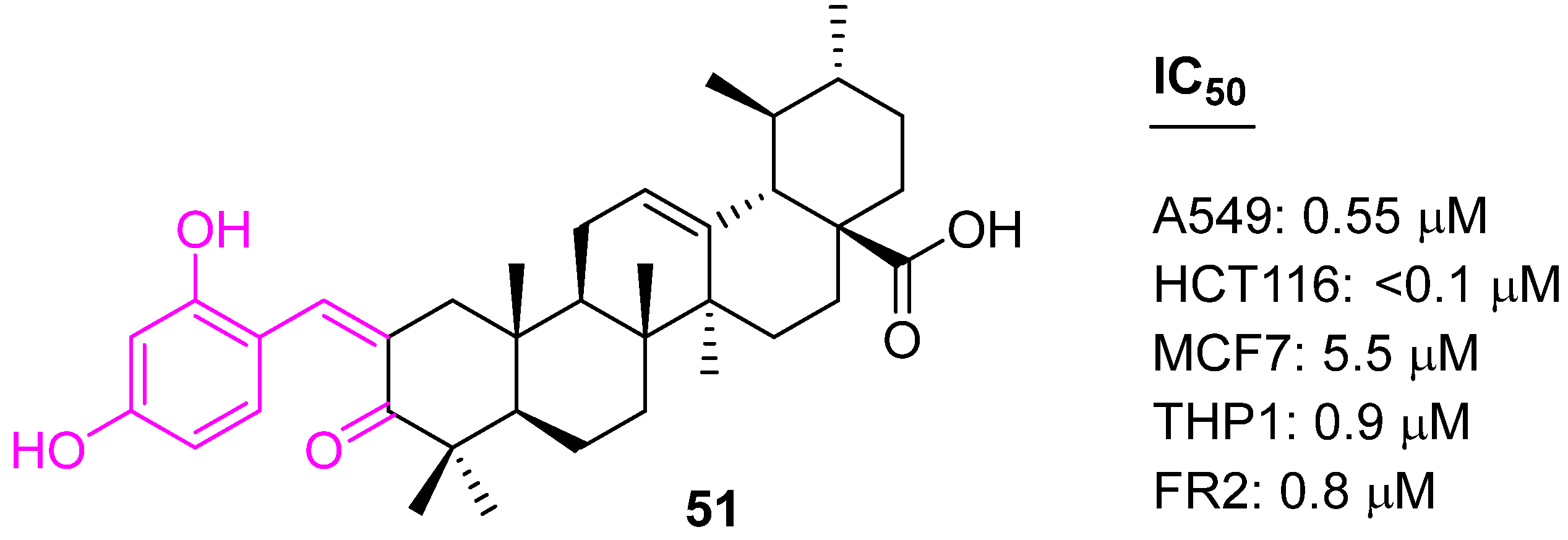
UA analogs were prepared using a Claisen Schmidt condensation reaction with aromatic aldehydes with the aim of developing potential anticancer drug candidates. Compound 51 (Figure 35) was found to be selective and has shown potency against four human carcinoma cell lines, including A549 (lung), MCF7 (breast), HCT116 (colon), and THP1 (leukemia), at significantly low concentrations. Further mechanistic studies concluded that 51 induced apoptosis in HCT-116 cell lines through the mitochondrial pathway, arrested the cell cycle in the G1 phase, caused accumulation of cytochrome c in the cytosol, and increased the expression levels of the caspase-9 and caspase-3 proteins [116].
In an attempt to develop potent anticancer agents, a set of carbazole derivatives of UA were prepared. Among the various carbazoles, compound 52 (Figure 36) showed promising IC50 values of 1.08 ± 0.22 and 1.26 ± 0.17 µM against hepatocarcinoma cell lines (SMMC7721 and HepG2), respectively, which are comparable to doxorubicin. The reduced cytotoxicity against noncancerous LO2 cells (IC50 value of 5.75 ± 0.48 lM) makes 52 a potential lead molecule [117].
Similarly, Wang et al. synthesized a set of 2-amino-4-aryl-pyrimidine derivatives of UA and tested their anticancer potential against MCF7 and Hela cells. Compound 53 showed IC50 values of 0.48 ± 0.11 and 0.74 ± 0.13 mM for MCF7 and Hela cells, respectively, and significantly low cytotoxicity to LO2 cells. A further molecular mechanistic study revealed that compound 53 inhibits cell migration, induces cell cycle arrest at the S phase, and triggers mitochondrial-related apoptosis by increasing the generation of intracellular ROS and decreasing the mitochondrial membrane potential (MMP). In addition, it participates in the up-regulation of the protein expression level of Bax and the downregulation of the level of Bcl2. Furthermore, molecular docking indicates that MEK1 kinase could be one of the possible targets for 53 [118].
Gu et al. synthesized quinoline derivatives of UA and evaluated their in vitro cytotoxicity against three human cancer cell lines (MDA-MB231, Hela, and SMMC7721). Compound 54 (Figure 37) was found to be the most active one, with IC50 values of 0.61 ± 0.07, 0.36 ± 0.05, 12.49 ± 0.08 µM against MDA-MB231, Hela, and SMMC7721 cells, respectively, stronger than the positive control, etoposide. The mechanistic study by the Annexin V-FITC/PI dual staining assay confirmed that compound 54 induces the apoptosis of MDA-MB231 cells in a dose-dependent manner and arrests the cell cycle MDA-MB231 cells at the G0/G1 phase [119].
As we discussed earlier, an aminoguanidine moiety scaffold plays an important role in the development of potential drug candidates. Here, Wu et al. introduced the scaffold at the C3 position of the UA and synthesized compound 55 (Figure 38). The compound was tested for the inhibition of HIF-1α transcriptional activity under hypoxia conditions using a Hep3B cell-based luciferase reporter assay (IC50 = 4.0 µM). Further investigation indicated the down-regulation of HIF-1α protein expression, possibly by suppressing its synthesis, reducing the production of vascular endothelial growth factor, and inhibiting the proliferation of the cancer cells [120].
Nitric oxide (NO) is an important messenger molecule in the human body that plays a key role in many physiological processes. High levels of NO can inhibit the proliferation of cancer cells, but unfortunately, the in vivo half-life of NO is very short. Recently, NO-donating derivatives, which can transport and release NO within the body, were used to explore the development of chemotherapeutic agents [121,122]. The approach was adopted, and a set of NO-donor UA derivatives were prepared and evaluated for their in vitro cytotoxicity against four human cancer cell lines (HepG2, MCF7, HT29, and A549). Compound 56 was found to be the most active with IC50 4.28 μM against HT29. An additional bioassay explained that this compound induced cell cycle arrest at the G1 phase and apoptosis in a dose-dependent manner. In addition, compound 56 (Figure 39) was found to up-regulate pro-apoptotic Bax, p53, and down-regulate antiapoptotic Bcl2 [123]. Another potential NO-donating UA–benzylidene derivative (57) was developed, which showed a 3- to 9-fold higher cytotoxicity as compared with the parent drug in A549, MCF7, HepG2, HT29, and HeLa cells. Further analysis concluded that 57 arrested the MCF7 cell cycle in the G1 phase, which was associated with caspase activation and a decrease in the Bcl2/Bax ratio. A molecular docking study unveiled that the nitroxyethyl moiety of 57 possibly establishes hydrogen bonds with caspase-8 amino acid residues (SER256 and HIS255) [124].
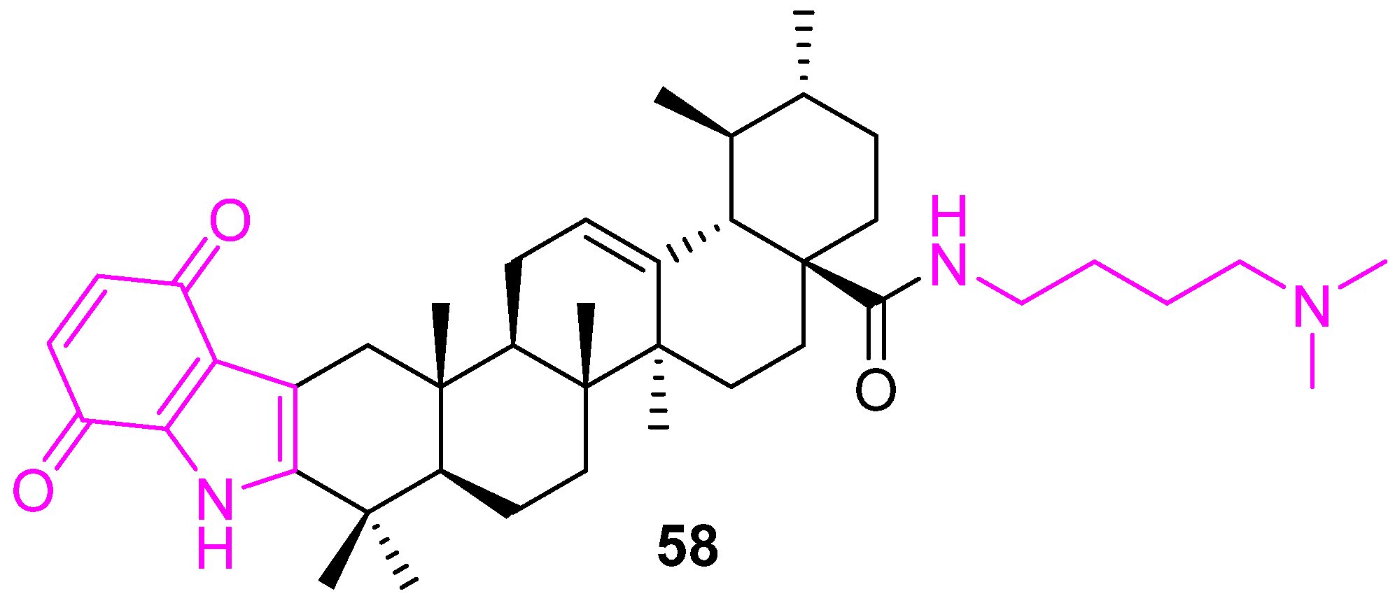
A series of indolequinone derivatives of UA having ester, hydrazide, or amide moieties were prepared and tested for their in vitro antiproliferative properties against MCF-7, Hela, and HepG2 cell lines and a normal gastric mucosal cell line (Ges1). From the series, compound 58 (Figure 40) was identified as the most potent, with IC50 values of 1.66 ± 0.21, 3.16 ± 0.24, and 10.35 ± 1.63 μM, respectively, against the cancer cell lines. Detailed molecular mechanism studies disclosed that compound 58 could arrest the cell cycle at the S phase, suppress the migration of MCF-7 cells, elevate the intracellular reactive oxygen species (ROS) level, and decrease mitochondrial membrane potential. In addition, the Western blot analysis confirmed that compound 58 up-regulated Bax, cleaved caspases-3 and -9, cleaved PARP levels, and down-regulated the Bcl2 level of MCF7 cells. Meanwhile, compound 58 markedly decreased p-AKT and p-mTOR expression, which revealed that compound 58 probably exerts its cytotoxicity by targeting the PI3K/AKT/mTOR signaling pathway [125].
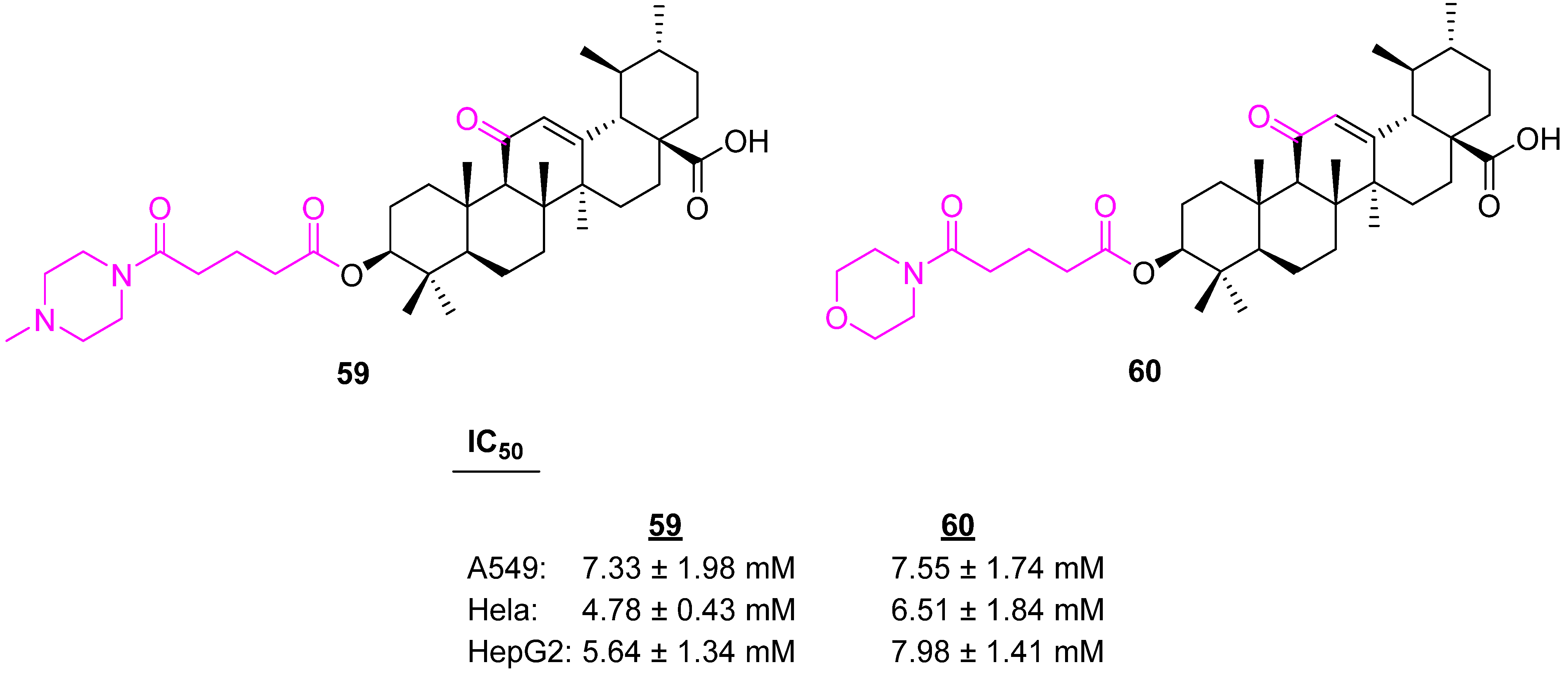
Secondary amines are the key building blocks of many pharmaceutically active compounds, such as gefitinib and imatinib [126,127]. Wang et al. introduced the piperazine and morpholine rings at the C3 position of a modified UA via a linker. The piperazine-containing analog 59 (Figure 41) was identified as being more potent against three different cancer cell lines than the morpholine-containing one, 60 [128].
Other analogs of UA listed in Table 6 reduced cell cycle arrest by inducing p21 and induced apoptosis by activating Bax, Noxo, and caspases-8, -9, and -3 and also by reducing Bcl2 and cFLIP. Apoptosis is involved in maintaining homeostasis of cell numbers by removing damaged or unwanted cells and, therefore, UA derivatives are important chemotherapeutic compounds for reducing tumor growth. UA derivatives also inhibit RAS/RAF/ERK/MAPK signaling pathways, which are involved in cell growth promotion.

5. Conclusions and Future

Structural modification of some complex natural products, such as UA, offer an unrivaled opportunity to develop novel anticancer compounds with diverse biological activities and limited systemic toxicity. As the description of the various classes of UA modified to enhance their anti-proliferative activities show, tremendous potential for modifications exists both structurally and functionally. The structural modifications of UA showed increased potency and diverse targets, able to inhibit processes responsible for cancer pathology, such as altered energy metabolism, mitochondrial functions, cancer cell migration, and an invasive potential, which are critical for sustaining malignancy and progression.
There are still plenty of opportunities to explore UA and design potential therapeutics for cancer. Fewer reports were found on the C3 modification because there is the assumption that the presence of a polar group at the C3 position is essential for the anticancer properties. Several reports confirm that the acetyl group at the C3 position also increases anticancer properties. In addition, the incorporation of triazole, piperazine, and guanidine scaffolds enhances activity and selectivity. In addition, including an NO donor is one of the interesting approaches.
A recognized deficiency in many of these studies is the lack of data concerning the efficacy of these altered UA analogs in vivo. Although in vitro studies are indicative of the expected functions of the compounds on tumor cells, favorable pharmacological properties, such as pharmacokinetics, and more importantly, pharmacodynamics, in either model animals (usually rodents) or in human volunteers, will move these compounds from the laboratory bench to the clinic. These studies are economically challenging for most laboratories engaged in creating analogs. However, select compounds, such as compounds 33ad, 40, and 41 offer significant potential to be successful anticancer compounds in vivo. Perhaps future pharmacological studies for bioavailability, systemic toxicity, and antitumor efficacy may prove some if not all of these compounds have clinical use.

Funding

We thank the Pilot Study Research Program (PSRP), Augusta University, for its financial support. The authors’ work in their laboratories is supported by a Veterans Health Administration Office of Research and Development Merit Award I01BX-003862 (BLL), J.H. Harrison Endowment funds, and Augusta University New Investigator Startup funds.

Institutional Review Board Statement

Not applicable.

Informed Consent Statement

Not applicable.

Data Availability Statement

Not applicable.

Acknowledgments

We thank the Department of Chemistry and Physics, Department of Biochemistry and Molecular Biology, and the Georgia Cancer Center at Augusta University for their support.

Conflicts of Interest

The authors declare no conflict of interest

References

  1. Sung, H.; Ferlay, J.; Siegel, R.L.; Laversanne, M.; Soerjomataram, I.; Jemal, A.; Bray, F. Global Cancer Statistics 2020: GLOBOCAN Estimates of Incidence and Mortality Worldwide for 36 Cancers in 185 Countries. CA A Cancer J. Clin. 2021, 71, 209–249. [Google Scholar] [CrossRef] [PubMed]
  2. Siegel, R.L.; Miller, K.D.; Jemal, A. Cancer statistics, 2022. CA A Cancer J. Clin. 2022, 72, 7–33. [Google Scholar] [CrossRef] [PubMed]
  3. Yabroff, K.R.; Mariotto, A.M.; Tangka, F.; Zhao, J.; Islami, F.; Sung, H.; Sherman, R.L.; Henley, S.J.; Jemal, A.; Ward, E.M. Annual Report to the Nation on the Status of Cancer, Part II: Patient Economic Burden Associated with Cancer Care. J. Natl. Cancer Inst. 2021, 113, 1670–1682. [Google Scholar] [CrossRef] [PubMed]
  4. Schirrmacher, V. From chemotherapy to biological therapy: A review of novel concepts to reduce the side effects of systemic cancer treatment (review). Int. J. Oncol. 2019, 54, 407–419. [Google Scholar]
  5. Pejin, B.; Jovanovi, K.K.; Mojovi, M.; Savi, A.G. New and Highly Potent Antitumor Natural Products from Marine-Derived Fungi: Covering the Period from 2003 to 2012. Curr. Top. Med. Chem. 2013, 13, 2745–2766. [Google Scholar] [CrossRef]
  6. Sisodiya, P.S. Plant Derived Anticancer Agents: A Review. Int. J. Res. Dev. Pharm. Life Sci. 2013, 2, 293–308. [Google Scholar]
  7. Pejin, B.; Glumac, M.A. brief review of potent anti-CNS tumourics from marine sponges: Covering the period from 1994 to 2014. Nat. Prod. Res. 2018, 32, 375–384. [Google Scholar] [CrossRef]
  8. Calcabrini, C.; Catanzaro, E.; Bishayee, A.; Turrini, E.; Fimognari, C. Marine Sponge Natural Products with Anticancer Potential: An Updated Review. Mar. Drugs 2017, 15, 310. [Google Scholar] [CrossRef] [Green Version]
  9. Cragg, G.M.; Pezzuto, J.M. Natural Products as a Vital Source for the Discovery of Cancer Chemotherapeutic and Chemopreventive Agents. Med. Princ. Pract. 2016, 25, 41–59. [Google Scholar] [CrossRef]
  10. Demain, A.L.; Vaishnav, P. Natural products for cancer chemotherapy. Microb. Biotechnol. 2011, 4, 687–699. [Google Scholar] [CrossRef] [Green Version]
  11. Rajesh, E.; Sankari, L.S.; Malathi, L.; Krupaa, J.R. Naturally occurring products in cancer therapy. J. Pharm. Bioallied Sci. 2015, 7, S181–S183. [Google Scholar] [CrossRef] [PubMed]
  12. Newman, D.J.; Cragg, G.M. Natural products as sources of new drugs over the 30 years from 1981 to 2010. J. Nat. Prod. 2012, 75, 311–335. [Google Scholar] [CrossRef] [PubMed]
  13. Katz, L.; Baltz, R.H. Natural product discovery: Past, present, and future. J. Ind. Microbiol. Biotechnol. 2016, 43, 155–176. [Google Scholar] [CrossRef]
  14. Liby, K.T.; Yore, M.M.; Sporn, M.B. Triterpenoids and rexinoids as multifunctional agents for the prevention and treatment of cancer. Nat. Rev. Cancer 2007, 7, 357–369. [Google Scholar] [CrossRef] [PubMed]
  15. Bishayee, A.; Ahmed, S.; Brankov, N.; Perloff, M. Triterpenoids as potential agents for the chemoprevention and therapy of breast cancer. Front. Biosci. 2011, 16, 980–996. [Google Scholar] [CrossRef] [PubMed] [Green Version]
  16. Aksit, H.; Gozcu, S.; Altay, A. Isolation and cytotoxic activities of undescribed iridoid and xanthone glycosides from Centaurium erythraea Rafn. (Gentianaceae). Phytochemistry 2023, 205, 113484. [Google Scholar] [CrossRef]
  17. Mlala, S.; Oyedeji, A.O.; Gondwe, M.; Oyedeji, O.O. Ursolic Acid and Its Derivatives as Bioactive Agents. Molecules 2019, 24, 2751. [Google Scholar] [CrossRef] [PubMed] [Green Version]
  18. Seo, D.Y.; Lee, S.R.; Heo, J.-W.; No, M.-H.; Rhee, B.D.; Ko, K.S.; Kwak, H.-B.; Han, J. Ursolic acid in health and disease. Korean J. Physiol. Pharmacol. 2018, 22, 235–248. [Google Scholar] [CrossRef] [Green Version]
  19. Lopez-Hortas, L.; Perez-Larran, P.; Gonzalez-Munoz, M.J.; Falque, E.; Dominguez, H. Recent developments on the extraction and application of ursolic acid. A review. Food Res. Int. 2018, 103, 130–149. [Google Scholar] [CrossRef]
  20. Li, C.; Chen, W.; Zheng, L.; Zhang, B.; Yang, X.; Zhang, Q.; Wang, N.; Wang, Y.; Yang, J.; Sha, J. Ameliorative effect of ursolic acid on ochratoxin A-induced renal cytotoxicity mediated by Lonp1/Aco2/Hsp75. Toxicon 2019, 168, 141–146. [Google Scholar] [CrossRef]
  21. Ozdemir, Z.; Wimmer, Z. Selected plant triterpenoids and their amide derivatives in cancer treatment: A review. Phytochemistry 2022, 203, 113340. [Google Scholar] [CrossRef] [PubMed]
  22. Kornel, A.; Nadile, M.; Tsiani, E. Evidence of the Beneficial Effects of Ursolic Acid against Lung Cancer. Molecules 2022, 27, 7466. [Google Scholar] [CrossRef] [PubMed]
  23. Wang, L.; Yin, Q.; Liu, C.; Tang, Y.; Sun, C.; Zhuang, J. Nanoformulations of ursolic acid: A modern natural anticancer molecule. Front. Pharmacol. 2021, 12, 706121. [Google Scholar] [CrossRef] [PubMed]
  24. Sycz, Z.; Tichaczek-Goska, D.; Wojnicz, D. Anti-Planktonic and Anti-Biofilm Properties of Pentacyclic Triterpenes-Asiatic Acid and Ursolic Acid as Promising Antibacterial Future Pharmaceuticals. Biomolecules 2022, 12, 98. [Google Scholar] [CrossRef] [PubMed]
  25. Jesus, J.A.; Lago, J.H.G.; Laurenti, M.D.; Yamamoto, E.S.; Passero, L.F.D. Antimicrobial activity of oleanolic and ursolic acids: An update. Evid. Based Complement. Altern. Med. 2015, 2015, 620472. [Google Scholar] [CrossRef] [PubMed] [Green Version]
  26. Al-kuraishy, H.M.; Al-Gareeb, A.I.; Negm, W.A.; Alexiou, A.; Batiha, G.E.S. Ursolic acid and SARS-CoV-2 infection: A new horizon and perspective. Inflammopharmacology 2022, 30, 1493–1501. [Google Scholar] [CrossRef]
  27. Bachar, S.C.; Mazumder, K.; Bachar, R.; Aktar, A.; Al Mahtab, M. A review of medicinal plants with antiviral activity available in Bangladesh and mechanistic insight into their bioactive metabolites on SARS-CoV-2, HIV and HBV. Front. Pharmacol. 2021, 12, 732891. [Google Scholar] [CrossRef]
  28. Gudoityte, E.; Arandarcikaite, O.; Mazeikiene, I.; Bendokas, V.; Liobikas, J. Ursolic and oleanolic acids: Plant metabolites with neuroprotective potential. Int. J. Mol. Sci. 2021, 22, 4599. [Google Scholar] [CrossRef]
  29. Li, W.; Sun, K.; Hu, F.; Chen, L.; Zhang, X.; Wang, F.; Yan, B. Protective effects of natural compounds against oxidative stress in ischemic diseases and cancers via activating the Nrf2 signaling pathway: A mini review. J. Biochem. Mol. Toxicol. 2021, 35, e22658. [Google Scholar] [CrossRef]
  30. Habtemariam, S. Antioxidant and anti-inflammatory mechanisms of neuroprotection by ursolic acid: Addressing brain injury, cerebral ischemia, cognition deficit, anxiety, and depression. Oxid. Med. Cell. Longev. 2019, 2019, 8512048. [Google Scholar] [CrossRef] [Green Version]
  31. Kashyap, D.; Sharma, A.; Tuli, H.S.; Punia, S.; Sharma, A.K. Ursolic Acid and Oleanolic Acid: Pentacyclic Terpenoids with Promising Anti-Inflammatory Activities. Recent Pat. Inflamm. Allergy Drug Discov. 2016, 10, 21–33. [Google Scholar] [CrossRef] [PubMed]
  32. Luan, M.; Wang, H.; Wang, J.; Zhang, X.; Zhao, F.; Liu, Z.; Meng, Q. Advances in anti-inflammatory activity, mechanism and therapeutic application of ursolic acid. Mini-Revi. Med. Chem. 2022, 22, 423–437. [Google Scholar]
  33. Achiwa, Y.; Hasegawa, K.; Udagawa, Y.; Achiwa, Y.; Hasegawa, K.; Udagawa, Y. Effect of ursolic acid on MAPK in cyclin D1 signaling and RING-type E3 ligase (SCF E3s) in two endometrial cancer cell lines. Nutr. Cancer 2013, 65, 1026–1033. [Google Scholar] [CrossRef] [PubMed]
  34. Prasad, S.; Yadav, V.R.; Sung, B.; Gupta, S.C.; Tyagi, A.K. Ursolic acid inhibits the growth of human pancreatic cancer and enhances the antitumor potential of gemcitabine in an orthotopic mouse model through suppression of the inflammatory microenvironment. Oncotarget 2016, 7, 13182. [Google Scholar] [CrossRef] [PubMed]
  35. Huang, C.Y.; Lin, C.Y.; Tsai, C.W.; Yin, M.C. Inhibition of cell proliferation, invasion and migration by ursolic acid in human lung cancer cell lines. Toxicol. Vitr. 2011, 25, 1274–1280. [Google Scholar] [CrossRef] [PubMed]
  36. Kim, S.; Ryu, H.G.; Lee, J.; Shin, J.; Harikishore, A.; Jung, H.Y.; Kim, Y.S.; Lyu, H.N.; Oh, E.; Baek, N.I.; et al. Ursolic acid exerts anti-cancer activity by suppressing vaccinia-related kinase 1-mediated damage repair in lung cancer cells. Sci. Rep. 2015, 5, 14570. [Google Scholar] [CrossRef] [PubMed] [Green Version]
  37. Kassi, E.; Papoutsi, Z.; Pratsinis, H.; Aligiannis, N.; Manoussakis, M.; Moutsatsou, P. Ursolic acid, a naturally occurring triterpenoid, demonstrates anticancer activity on human prostate cancer cells. J. Cancer Res. Clin. Oncol. 2007, 133, 493–500. [Google Scholar] [CrossRef]
  38. Shanmugam, M.K.; Rajendran, P.; Li, F.; Nema, T.; Vali, S.; Abbasi, T.; Kapoor, S.; Sharma, A.; Kumar, A.P.; Ho, P.C.; et al. Ursolic acid inhibits multiple cell survival pathways leading to suppression of growth of prostate cancer xenograft in nude mice. J. Mol. Med. 2011, 89, 713–727. [Google Scholar] [CrossRef]
  39. Gai, W.T.; Yu, D.P.; Wang, X.S.; Wang, P.T. Anti-cancer e_ect of ursolic acid activates apoptosis through ROCK/PTEN mediated mitochondrial translocation of cofilin-1 in prostate cancer. Oncol. Lett. 2016, 12, 2880–2885. [Google Scholar] [CrossRef] [Green Version]
  40. Yang, L.; Liu, X.; Lu, Z.; Yuet, W.J.; Zhou, L.L.; Fung, K.P.; Wu, P.; Wu, S. Ursolic acid induces doxorubicin-resistant HepG2 cell death via the release of apoptosis-inducing factor. Cancer Lett. 2010, 298, 128–138. [Google Scholar] [CrossRef]
  41. Zheng, Q.; Li, P.; Jin, F.; Yao, C.; Zhang, G.; Zang, T.; Ai, X. Ursolic acid induces ER stress response to activate ASK1-JNK signaling and induce apoptosis in human bladder cancer T24 cells. Cell. Signal. 2013, 25, 206–213. [Google Scholar] [CrossRef] [PubMed]
  42. Xu, X.; Zhu, G.; Zhang, K.; Zhou, Y.; Li, X.; Xu, W.; Zhang, H.; Shao, Y.; Zhang, Z.; Sun, W. Cyclooxygenase-2 mediated synergistic effect of ursolic acid in combination with paclitaxel against human gastric carcinoma. Oncotarget 2017, 8, 92770–92777. [Google Scholar] [CrossRef] [Green Version]
  43. Kim, E.S.; Moon, A. Ursolic acid inhibits the invasive phenotype of SNU-484 human gastric cancer cells. Oncol. Lett. 2015, 9, 897–902. [Google Scholar] [CrossRef] [PubMed]
  44. Prasad, S.; Yadav, V.R.; Kannappan, R.; Aggarwal, B.B. Ursolic acid, a pentacyclin triterpene, potentiates TRAIL-induced apoptosis through p53-independent up-regulation of death receptors. J. Biol. Chem. 2016, 291, 16924. [Google Scholar] [CrossRef] [PubMed] [Green Version]
  45. Wang, C.; Lin, C.; Hua, C.; Jou, Y.; Liao, C.; Chang, Y.; Wan, L.; Huang, S.; Hour, M.; Lin, C. Cis-3-O-p-hydroxycinnamoyl ursolic acid induced ROS-dependent p53-mediated mitochondrial apoptosis in oral cancer cells. Biomol. Ther. 2019, 27, 54–62. [Google Scholar] [CrossRef]
  46. Jiang, K.; Chi, T.; Li, T.; Zheng, G.; Fan, L.; Liu, Y.; Chen, X.; Chen, S.; Jia, L.; Shao, J. A smart pH-responsive nano-carrier as a drug delivery system for the targeted delivery of ursolic acid: Suppresses cancer growth and metastasis by modulating P53/MMP-9/PTEN/CD44 mediated multiple signaling pathways. Nanoscale 2017, 9, 9428–9439. [Google Scholar] [CrossRef]
  47. Kim, J.; Kim, Y.H.; Song, G.; Kim, D.; Jeong, Y.; Liu, K.; Chung, Y.; Oh, S. Ursolic acid and its natural derivative corosolic acid suppress the proliferation of APC-mutated colon cancer cells through promotion of β-catenin degradation. Food Chem. Toxicol. 2014, 67, 87–95. [Google Scholar] [CrossRef]
  48. Zhang, R.; Li, Y.; Tian, D.; Liu, Y.; Nian, W.; Zou, X.; Chen, Q.; Zhou, L.; Deng, Z.; He, B. Ursolic acid inhibits proliferation and induces apoptosis by inactivating Wnt/β-catenin signaling in human osteosarcoma cells. Int. J. Oncol. 2016, 49, 1973–1982. [Google Scholar] [CrossRef] [Green Version]
  49. Kim, S.; Jin, H.; Meng, R.Y.; Kim, D.Y.; Liu, Y.C.; Chai, O.H.; Park, B.H.; Kim, S. Activating hippo pathway via Rassf1 by ursolic acid suppresses the tumorigenesis of gastric cancer. Int. J. Mol. Sci. 2019, 20, 4709. [Google Scholar] [CrossRef] [Green Version]
  50. Choi, W.; Hyung; Lee, I. A. The mechanism of action of ursolic acid as a potential anti-toxoplasmosis agent, and its immunomodulatory effects. Pathogens 2019, 8, 61. [Google Scholar] [CrossRef] [Green Version]
  51. Pathak, A.K.; Bhutani, M.; Nair, A.S.; Kwang, S.A.; Chakraborty, A.; Kadara, H.; Guha, S.; Sethi, G.; Aggarwal, B.B. Ursolic acid inhibits STAT3 activation pathway leading to suppression of proliferation and chemosensitization of human multiple myeloma cells. Mol. Cancer Res. 2007, 5, 943–955. [Google Scholar] [CrossRef] [PubMed] [Green Version]
  52. Wang, W.; Zhao, C.; Jou, D.; Lü, J.; Zhang, C.; Lin, L.; Lin, J. Ursolic acid inhibits the growth of colon cancer-initiating cells by targeting STAT3. Anticancer Res. 2013, 33, 4279–4284. [Google Scholar] [PubMed]
  53. Liu, T.; Ma, H.; Shi, W.; Duan, J.; Wang, Y.; Zhang, C.; Li, C.; Lin, J.; Li, S.; Lv, J.; et al. Inhibition of STAT3 signaling pathway by ursolic acid suppresses growth of hepatocellular carcinoma. Int. J. Oncol. 2017, 51, 555–562. [Google Scholar] [CrossRef] [PubMed]
  54. Xia, Y.; Wei, G.; Si, D.; Liu, C. Quantitation of ursolic acid in human plasma by ultra performance liquid chromatography tandem mass spectrometry and its pharmacokinetic study. J. Chromatogr. B Analyt. Technol. Biomed. Life Sci. 2011, 879, 219–224. [Google Scholar] [CrossRef] [PubMed]
  55. Shanmugam, N.K.N.; Ong, T.H.; Kumar, A.P.; Lun, C.K.; Ho, P.C.; Wong, P.T.H.; Hui, K.M.; Sethi, G. Ursolic Acid Inhibits the Initiation, Progression of Prostate Cancer and Prolongs the Survival of TRAMP Mice by Modulating Pro-Inflammatory Pathways. PLoS ONE 2012, 7, e32476. [Google Scholar] [CrossRef] [Green Version]
  56. Zhu, Z.; Qian, Z.; Yan, Z.; Zhao, C.; Wang, H.; Ying, G. A phase I pharmacokinetic study of ursolic acid nanoliposomes in healthy volunteers and patients with advanced solid tumors. Int. J. Nanomed. 2012, 8, 129–136. [Google Scholar]
  57. Bahia, S.B.B.; Reis, W.J.; Jardim, G.A.; Souto, F.T.; de Simone, C.A.; Gatto, C.C.; Menna-Barreto, R.F.; de Castro, S.L.; Cavalcanti, B.C.; Pessoa, C.; et al. Molecular hybridization as a powerful tool towards multitarget quinoidal systems: Synthesis, trypanocidal and antitumor activities of naphthoquinone-based 5-iodo-1,4-disubstituted-, 1,4- and 1,5-disubstituted-1,2,3-triazoles. MedChemComm 2016, 7, 1555–1563. [Google Scholar] [CrossRef]
  58. Bosquesi, P.L.; Melo, T.R.F.; Vizioli, E.O.; dos Santos, J.L.; Chung, M.C. Anti-inflammatory drug design using a molecular hybridization approach. Pharmaceuticals 2011, 4, 1450–1474. [Google Scholar] [CrossRef]
  59. Fershtat, L.L.; Makhova, N.N. Molecular Hybridization Tools in the Development of Furoxan-Based NO-Donor Prodrugs. ChemMedChem 2017, 12, 622–638. [Google Scholar] [CrossRef]
  60. Meunier, B. Hybrid molecules with a dual mode of action: Dream or reality? Acc. Chem. Res. 2008, 41, 69–77. [Google Scholar] [CrossRef]
  61. Panda, S.S.; Tran, Q.L.; Rajpurohit, P.; Pillai, G.G.; Thomas, S.J.; Bridges, A.E.; Capito, J.E.; Thangaraju, M.; Lokeshwar, B.L. Design, Synthesis, and Molecular Docking Studies of Curcumin Hybrid Conjugates as Potential Therapeutics for Breast Cancer. Pharmaceuticals 2022, 15, 451. [Google Scholar] [CrossRef] [PubMed]
  62. Panda, S.S.; Girgis, A.S.; Thomas, S.J.; Capito, J.E.; George, R.F.; Salman, A.; El-Manawaty, M.A.; Samir, A. Synthesis, pharmacological profile and 2D-QSAR studies of curcumin-amino acid conjugates as potential drug candidates. Eur. J. Med. Chem. 2020, 196, 112293. [Google Scholar] [CrossRef] [PubMed]
  63. Panda, S.S.; Asiri, A.M.; Elagawany, M.; Buchanan, D.D.; Torkian, B.; Bathala, K.; Thomas, S.J.; Capito, J.E.; Arshad, M.N.; Al-Romaizan, A.N. Efficient synthesis of pyrazinoic acid hybrid conjugates. SynOpen 2017, 1, 50–58. [Google Scholar] [CrossRef] [Green Version]
  64. Tangutur, A.D.; Kumar, D.; Krishna, V.; Kantevari, S. Microtubule targeting agents as cancer chemotherapeutics: An overview of molecular hybrids as stabilizing and destabilizing agents. Curr. Top. Med. Chem. 2017, 17, 2523–2537. [Google Scholar] [CrossRef]
  65. Viegas-Junior, C.; Danuello, A.; da Silva Bolzani, V.; Barreiro, E.J.; Fraga, C.A.M. Molecular Hybridization: A Useful Tool in the Design of New Drug Prototypes. Curr. Med. Chem. 2007, 14, 1829–1852. [Google Scholar] [CrossRef]
  66. Khwaza, V.; Oyedeji, O.O.; Aderibigbe, B.A. Ursolic Acid-Based Derivatives as Potential Anti-Cancer Agents: An Update. Int. J. Mol. Sci. 2020, 21, 5920. [Google Scholar] [CrossRef]
  67. Thien, D.D.; Nguyen, T.T.; Thien, D.G.; Nguyen, T.H.A.; Van Sung, T. Synthesis and Cytotoxic Activity of Ursolic Acid Derivatives. Z. Naturforsch. B 2013, 68, 201–206. [Google Scholar] [CrossRef]
  68. da Silva, E.F.; de Vargas, A.S.; Willig, J.B.; de Oliveira, C.B.; Zimmer, A.R.; Pilger, D.A.; Buffon, A.; Gnoatto, S.C.B. Synthesis and antileukemic activity of an ursolic acid derivative: A potential co-drug in combination with imatinib. Chem. Biol. Interact. 2021, 344, 109535. [Google Scholar] [CrossRef]
  69. Xu, J.; Wang, X.; Zhang, H.; Yue, J.; Sun, Y.; Zhang, X.; Zhao, Y. Synthesis of triterpenoid derivatives and their anti-tumor and anti-hepatic fibrosis activities. Nat. Prod. Res. 2020, 34, 766–772. [Google Scholar] [CrossRef]
  70. Fontana, G.; Bruno, M.; Notarbartolo, M.; Labbozzetta, M.; Poma, P.; Spinella, A.; Rosselli, S. Cytotoxicity of oleanolic and ursolic acid derivatives toward hepatocellular carcinoma and evaluation of NF-κB involvement. Bioorg. Chem. 2019, 90, 103054. [Google Scholar] [CrossRef]
  71. Ma, C.-M.; Cai, S.-Q.; Cui, J.-R.; Wang, R.-Q.; Tu, P.-F.; Hattori, M.; Daneshtalab, M. The cytotoxic activity of ursolic acid derivatives. Eur. J. Med. Chem. 2005, 40, 582–589. [Google Scholar] [CrossRef] [PubMed]
  72. Ciftci, H.I.; Radwan, M.O.; Ozturk, S.E.; Ulusoy, N.G.; Sozer, E.; Ellakwa, D.E.; Ocak, Z.; Can, M.; Ali, T.F.S.; Abd-Alla, H.; et al. Design, synthesis and biological evaluation of pentacyclic triterpene derivatives: Optimization of anti-ABL kinase activity. Molecules 2019, 24, 3535. [Google Scholar] [CrossRef] [PubMed] [Green Version]
  73. Tu, H.-Y.; Huang, A.-M.; Wei, B.-L.; Gan, K.-H.; Hour, T.-C.; Yang, S.-C.; Pu, Y.-S.; Lin, C.-N. Ursolic acid derivatives induce cell cycle arrest and apoptosis in NTUB1 cells associated with reactive oxygen species. Bioorg. Med. Chem. 2009, 17, 7265–7274. [Google Scholar] [CrossRef] [PubMed]
  74. Baglin, I.; Poumaroux, A.; Nour, M.; Tan, K.; Mitaine-Offer, A.C.; Lacaille-Dubois, M.A.; Chauffert, B.; Cave, C. New Ursolic and Betulinic Derivatives as Potential Cytotoxic Agents. J. Enzym. Inhib. Med. Chem. 2003, 18, 111–117. [Google Scholar] [CrossRef] [PubMed]
  75. Bai, K.-K.; Yu, Z.; Chen, F.-L.; Li, F.; Li, W.-Y.; Guo, Y.-H. Synthesis and evaluation of ursolic acid derivatives as potent cytotoxic agents. Bioorg. Med. Chem. Lett. 2012, 22, 2488–2493. [Google Scholar] [CrossRef]
  76. Tian, T.; Liu, X.; Lee, E.-S.; Sun, J.; Feng, Z.; Zhao, L.; Zhao, C. Synthesis of novel oleanolic acid and ursolic acid in C-28 position derivatives as potential anticancer agents. Arch. Pharm. Res. 2017, 40, 458–468. [Google Scholar] [CrossRef]
  77. Liu, M.-C.; Yang, S.-J.; Jin, L.-H.; Hu, D.-Y.; Xue, W.; Song, B.-A.; Yang, S. Synthesis and cytotoxicity of novel ursolic acid derivatives containing an acyl piperazine moiety. Eur. J. Med. Chem. 2012, 58, 128–135. [Google Scholar] [CrossRef]
  78. Liu, M.-C.; Yang, S.-J.; Jin, L.-H.; Hu, D.-Y.; Xue, W.; Yang, S. Synthesis and evaluation as potential antitumor agents of novel ursolic acid derivatives. Med. Chem. Res. 2016, 25, 2267–2279. [Google Scholar] [CrossRef]
  79. Chi, K.-Q.; Wei, Z.-Y.; Wang, K.-S.; Wu, J.; Chen, W.-Q.; Jin, X.-J.; Piao, H.-R. Design, synthesis, and evaluation of novel ursolic acid derivatives as HIF-1α inhibitors with anticancer potential. Bioorg. Chem. 2017, 75, 157–169. [Google Scholar] [CrossRef]
  80. Pattnaik, B.; Lakshmi, J.K.; Kavitha, R.; Jagadeesh, B.; Bhattacharjee, D.; Jain, N.; Mallavadhani, U.V. Synthesis, structural studies, and cytotoxic evaluation of novel ursolic acid hybrids with capabilities to arrest breast cancer cells in mitosis. J. Asian Nat. Prod. Res. 2017, 19, 260–271. [Google Scholar] [CrossRef]
  81. Semenova, M.D.; Popov, S.A.; Golubeva, T.S.; Baev, D.S.; Shults, E.E.; Turks, M. Synthesis and Cytotoxicity of Sulfanyl, Sulfinyl and Sulfonyl Group Containing Ursane Conjugates with 1,3,4-Oxadiazoles and 1,2,4-Triazoles. ChemistrySelect 2021, 6, 6472–6477. [Google Scholar] [CrossRef]
  82. Khazanov, V.A.; Manuylova, A.V.; Gogvadze, V.; Spivak, A.Y. Mitochondria-targeted betulinic and ursolic acid derivatives: Synthesis and anticancer activity. MedChemComm 2017, 8, 1934–1945. [Google Scholar]
  83. Saraswat, B.P.; Visen, K.; Agarwal, D.P. Ursolic acid isolated from Eucaliptus tereticornis protects against ethanol toxicity in isolated rat hepatocytes. Phytother. Res. 2000, 14, 163–166. [Google Scholar]
  84. Popov, S.A.; Semenova, M.D.; Baev, D.S.; Frolova, T.S.; Shestopalov, M.A.; Wang, C.; Qi, Z.; Shults, E.E.; Turks, M. Synthesis and cytotoxicity of hybrids of 1,3,4- or 1,2,5-oxadiazoles tethered from ursane and lupane core with 1,2,3-triazole. Steroids 2020, 162, 108698. [Google Scholar] [CrossRef]
  85. Shao, J.-W.; Dai, Y.-C.; Xue, J.-P.; Wang, J.-C.; Lin, F.-P.; Guo, Y.-H. In vitro and in vivo anticancer activity evaluation of ursolic acid derivatives. Eur. J. Med. Chem. 2011, 46, 2652–2661. [Google Scholar] [CrossRef]
  86. Toni, C.; Denner, T.C.; Hoenke, S.; Kraft, O.; Deigner, H.-P.; Al-Harrasi, A.; Csuk, R. Hydroxyethylamide substituted triterpenoic acids hold good cytotoxicity for human tumor cells. Results Chem. 2022, 4, 100371. [Google Scholar]
  87. Wiemann, J.; Heller, L.; Csuk, R. Targeting cancer cells with oleanolic and ursolic acid derived hydroxamates. Bioorg. Med. Chem. Lett. 2016, 26, 907–909. [Google Scholar] [CrossRef]
  88. Meng, Y.; Song, Y.; Yan, Z.; Xia, Y. Synthesis and in vitro Cytotoxicity of Novel Ursolic Acid Derivatives. Molecules 2010, 15, 4033–4040. [Google Scholar] [CrossRef] [Green Version]
  89. Bai, K.-K.; Chen, F.-L.; Yu, Z.; Zheng, Y.-Q.; Li, Y.-N.; Guo, Y.-H. Synthesis of [3β-acetoxy-urs-12-en-28-oyl]-1-monoglyceride and investigation on its anti tumor effects against BGC-823. Bioorg. Med. Chem. 2011, 19, 4043–4050. [Google Scholar] [CrossRef]
  90. Quail, D.; Joyce, J.A. Microenvironmental regulation of tumor progression and metastasis. Nat. Med. 2013, 19, 1423–1437. [Google Scholar] [CrossRef]
  91. Kroemer, G.; Pouyssegur, J. Tumor cell metabolism: Cancer’s Achilles’ heel. Cancer Cell 2008, 13, 472–482. [Google Scholar] [CrossRef] [PubMed]
  92. Faubert, B.; Solmonson, A.; DeBerardinis, R.J. Metabolic reprogramming and cancer progression. Science 2020, 368, 5473. [Google Scholar] [CrossRef] [PubMed]
  93. Pavlova, N.N.; Zhu, J.; Thompson, C.B. The hallmarks of cancer metabolism: Still emerging. Cell Metab. 2022, 34, 355–377. [Google Scholar] [CrossRef] [PubMed]
  94. Cassim, S.; Vučetić, M.; Ždralević, M.; Pouyssegur, J. Warburg and Beyond: The Power of Mitochondrial Metabolism to Collaborate or Replace Fermentative Glycolysis in Cancer. Cancers 2020, 12, 1119. [Google Scholar] [CrossRef] [PubMed]
  95. Cassim, S.; Raymond, V.A.; Lacoste, B.; Lapierre, P.; Bilodeau, M. Metabolite profiling identifies a signature of tumorigenicity in hepatocellular carcinoma. Oncotarget 2018, 9, 26868–26883. [Google Scholar] [CrossRef] [Green Version]
  96. Dong, H.; Yang, X.; Xie, J.; Xiang, L.; Li, Y.; Ou, M.; Chi, T.; Liu, Z.; Yu, S.; Gao, Y.; et al. UP12, a novel ursolic acid derivative with potential for targeting multiple signaling pathways in hepatocellular carcinoma. Biochem. Pharmacol. 2015, 93, 151–162. [Google Scholar] [CrossRef]
  97. Yang, X.; Li, Y.; Jiang, W.; Ou, M.; Chen, Y.; Xu, Y.; Wu, Q.; Zheng, Q.; Wu, F.; Wang, L.; et al. Synthesis and Biological Evaluation of Novel Ursolic acid Derivatives as Potential Anticancer Prodrugs. Chem. Biol. Drug Des. 2015, 86, 1397–1404. [Google Scholar] [CrossRef]
  98. Hua, S.-X.; Huang, R.-Z.; Ye, M.-Y.; Pan, Y.-M.; Yao, G.-Y.; Zhang, Y.; Wang, H.-S. Design, synthesis and in vitro evaluation of novel ursolic acid derivatives as potential anticancer agents. Eur. J. Med. Chem. 2015, 95, 435–452. [Google Scholar] [CrossRef]
  99. Friedrich, S.; Serbian, I.; Hoenke, S.; Wolfram, R.K.; Csuk, R. Synthesis and cytotoxic evaluation of malachite green derived oleanolic and ursolic acid piperazineamides. Med. Chem. Res. 2020, 29, 926–933. [Google Scholar] [CrossRef] [Green Version]
  100. Jiang, W.; Huang, R.-Z.; Zhang, J.; Guo, T.; Zhang, M.-T.; Huang, X.-C.; Zhang, B.; Liao, Z.-X.; Sun, J.; Wang, H.-S. Discovery of antitumor ursolic acid long-chain diamine derivatives as potent inhibitors of NF-κB. Bioorg. Chem. 2018, 79, 265–276. [Google Scholar] [CrossRef]
  101. Kahnt, M.; Fischer, L.; Al-Harrasi, A.; Csuk, R. Ethylenediamine Derived Carboxamides of Betulinic and Ursolic Acid as Potential Cytotoxic Agents. Molecules 2018, 23, 2558. [Google Scholar] [CrossRef] [Green Version]
  102. Blanchet, M.; Borselli, D.; Brunel, J.M. Polyamine derivatives: A revival of an old neglected scaffold to fight resistant Gram-negative bacteria? Future Med. Chem. 2016, 8, 963–973. [Google Scholar] [CrossRef] [PubMed]
  103. Nowotarski, S.L.; Woster, P.M.; Casero, R.A. Polyamines and cancer: Implications for chemotherapy and chemoprevention. Expert Rev. Mol. Med. 2013, 15, e3. [Google Scholar] [CrossRef] [PubMed] [Green Version]
  104. Spivak, A.; Khalitova, R.; Nedopekina, D.; Dzhemileva, L.; Yunusbaeva, M.; Odinokov, V.; D’yakonov, V.; Dzhemilev, U. Synthesis and Evaluation of Anticancer Activities of Novel C-28 Guanidine-Functionalized Triterpene Acid Derivatives. Molecules 2018, 23, 3000. [Google Scholar] [CrossRef] [PubMed] [Green Version]
  105. Popov, S.A.; Semenova, M.D.; Baev, D.S.; Frolova, T.S.; Shults, E.E.; Wang, C.; Turks, M. Synthesis of cytotoxic urs-12-ene- and 28-norurs-12-ene- type conjugates with amino- and mercapto-1,3,4-oxadiazoles and mercapto-1,2,4-triazoles. Steroids 2020, 153, 108524. [Google Scholar] [CrossRef]
  106. Dheer, D.; Singh, V.; Shankar, R. Medicinal attributes of 1,2,3-triazoles: Current developments. Bioorg. Chem. 2017, 71, 30–54. [Google Scholar] [CrossRef]
  107. Bozorov, K.; Zhao, J.; Aisa, H.A. 1,2,3-Triazole-containing hybrids as leads in medicinal chemistry: A recent overview. Bioorg. Med. Chem. 2019, 27, 3511–3531. [Google Scholar] [CrossRef] [PubMed]
  108. Rashid, S.; Dar, B.A.; Majeed, R.; Hamid, A.; Bhat, B.A. Synthesis and biological evaluation of ursolic acid-triazolyl derivatives as potential anti-cancer agents. Eur. J. Med. Chem. 2013, 66, 238–245. [Google Scholar] [CrossRef]
  109. Meng, Y.-Q.; Zhang, L.-F.; Liu, D.Y.; Liu, L.-W.; Zhang, Y.; Zhao, Y. Synthesis and antitumor activity evaluation of novel ursolic acid derivatives. J. Asian Nat. Prod. Res. 2016, 18, 280–288. [Google Scholar] [CrossRef]
  110. Meng, Y.-Q.; Xu, C.-D.; Yu, T.-T.; Li, W.; Li, Q.-W.; Li, X.-X. Synthesis and antitumor activity evaluation of ursolic acid derivatives. J. Asian Nat. Prod. Res. 2020, 22, 359–369. [Google Scholar] [CrossRef]
  111. Zhao, C.-H.; Zhang, C.-L.; Shi, J.-J.; Hou, X.-Y.; Feng, B.; Zhao, L.-X. Design, synthesis, and biofunctional evaluation of novel pentacyclic triterpenes bearing O-[4-(1-piperazinyl)-4-oxo-butyryl moiety as antiproliferative agents. Bioorg. Med. Chem. Lett. 2015, 25, 4500–4504. [Google Scholar] [CrossRef] [PubMed]
  112. Kahnt, M.; Hoenke, S.; Fischer, L.; Al-Harrasi, A.; Csuk, R. Synthesis and Cytotoxicity Evaluation of DOTA-Conjugates of Ursolic Acid. Molecules 2019, 24, 2254. [Google Scholar] [CrossRef] [PubMed] [Green Version]
  113. Zhang, L.-H.; Zhang, Z.-H.; Li, M.-Y.; Wei, Z.-Y.; Jin, X.-J.; Piao, H.-R. Synthesis and evaluation of the HIF-1α inhibitory activities of novel ursolic acid tetrazole derivatives. Bioorg. Med. Chem. Lett. 2019, 29, 1440–1445. [Google Scholar] [CrossRef] [PubMed]
  114. Leal, A.S.; Wang, R.; Salvador, J.A.R.; Jing, Y. Synthesis of novel ursolic acid heterocyclic derivatives with improved abilities of antiproliferation and induction of p53, p21waf1 and NOXA in pancreatic cancer cells. Bioorg. Med. Chem. 2012, 20, 5774–5786. [Google Scholar] [CrossRef]
  115. Leal, A.S.; Wang, R.; Salvador, J.A.R.; Jing, Y. Semisynthetic Ursolic Acid Fluorolactone Derivatives Inhibit Growth with Induction of p21waf1 and Induce Apoptosis with Upregulation of NOXA and Downregulation of c-FLIP in Cancer Cells. ChemMedChem 2012, 7, 1635–1646. [Google Scholar] [CrossRef] [PubMed]
  116. Dar, B.A.; Lone, A.M.; Shah, W.A.; Qurishi, M.A. Synthesis and screening of ursolic acid-benzylidine derivatives as potential anti-cancer agents. Eur. J. Med. Chem. 2016, 111, 26–32. [Google Scholar] [CrossRef]
  117. Gu, W.; Hao, Y.; Zhang, G.; Wang, S.-F.; Miao, T.-T.; Zhang, K.-P. Synthesis, in vitro antimicrobial and cytotoxic activities of new carbazole derivatives of ursolic acid. Bioorg. Med. Chem. Lett. 2015, 25, 554–557. [Google Scholar] [CrossRef]
  118. Wang, W.-Y.; Yang, Z.-H.; Li, A.-L.; Liu, Q.-S.; Sun, Y.; Gu, W. Design, synthesis, anticancer activity and mechanism studies of novel 2-amino-4-arylpyrimidine derivatives of ursolic acid. New J. Chem. 2022, 46, 2335–2350. [Google Scholar] [CrossRef]
  119. Gu, W.; Jin, X.-Y.; Li, D.-D.; Wang, S.-F.; Tao, X.-B.; Chen, H. Design, synthesis and in vitro anticancer activity of novel quinoline and oxadiazole derivatives of ursolic acid. Bioorg. Med. Chem. Lett. 2017, 27, 4128–4132. [Google Scholar] [CrossRef]
  120. Wu, J.; Zhang, Z.-H.; Zhang, L.-H.; Jin, X.-J.; Ma, J.; Piao, H.-R. Design, synthesis, and screening of novel ursolic acid derivatives as potential anti-cancer agents that target the HIF-1α pathway. Bioorg. Med. Chem. Lett. 2019, 29, 853–858. [Google Scholar] [CrossRef] [PubMed]
  121. Monteiro, H.P.; Costa, P.E.; Reis, A.; Stern, A. Nitric Oxide: Protein tyrosine phosphorylation and protein S-nitrosylation in cancer. Biomed. J. 2015, 38, 380–388. [Google Scholar] [PubMed]
  122. Ishima, Y.; Kragh-Hansen, U.; Maruyama, T.; Otaqiri, M. Poly-S-nitrosated albumin as a safe and effective multifunctional antitumor agent: Characterization, biochemistry and possible future therapeutic applications. BioMed Res. Int. 2013, 2013, 353892. [Google Scholar] [CrossRef] [PubMed] [Green Version]
  123. Zhang, T.; He, B.; Yuan, H.; Feng, G.; Chen, F.; Wu, A.; Zhang, L.; Lin, H.; Zhuo, Z.; Wang, T. Synthesis and antitumor evaluation in vitro of NO-donating ursolic acid-benzylidene derivatives. Chem. Biodivers. 2019, 16, e1900111. [Google Scholar] [CrossRef]
  124. He, B.; Zhu, Z.; Chen, F.; Zhang, R.; Chen, W.; Zhang, T.; Wang, T.; Lei, J. Synthesis and antitumor potential of new arylidene ursolic acid derivatives via caspase-8 activation. Arch. Pharm. 2021, 354, 2000448. [Google Scholar] [CrossRef] [PubMed]
  125. Wang, W.-Y.; Wu, W.-Y.; Liu, Q.-S.; Sun, T.; Gu, W. Synthesis, anticancer evaluation and mechanism studies of novel indolequinone derivatives of ursolic acid. Bioorg. Chem. 2021, 109, 104705. [Google Scholar] [CrossRef]
  126. Cui, Z.L.; Liu, Z.; Zeng, J.X.; Zhang, S.L.; Chen, L.; Zhang, G.; Xu, W.; Song, L.; Guo, X. TRIM59 promotes gefitinib resistance in EGFR mutant lung adenocarcinoma cells. Life Sci. 2019, 224, 23–32. [Google Scholar] [CrossRef]
  127. Onmaz, D.E.; Abusoglu, S.; Unlu, A.; Dagli, B.M.; Bagci, M.; Tok, O.; Abusoglu, G. Determination of serum imatinib and its’ metabolite in patients chronic myeloid leukemia. Clin. Chim. Acta 2019, 497, 120–124. [Google Scholar] [CrossRef]
  128. Wang, Q.; Jin, M.; Liu, Y.; Sun, L.; Lu, B.; Zhao, L. Synthesis, characterization and in vitro anti-proliferative effects of pentacyclic triterpenoids. Med. Chem. Res. 2021, 30, 2055–2068. [Google Scholar] [CrossRef]
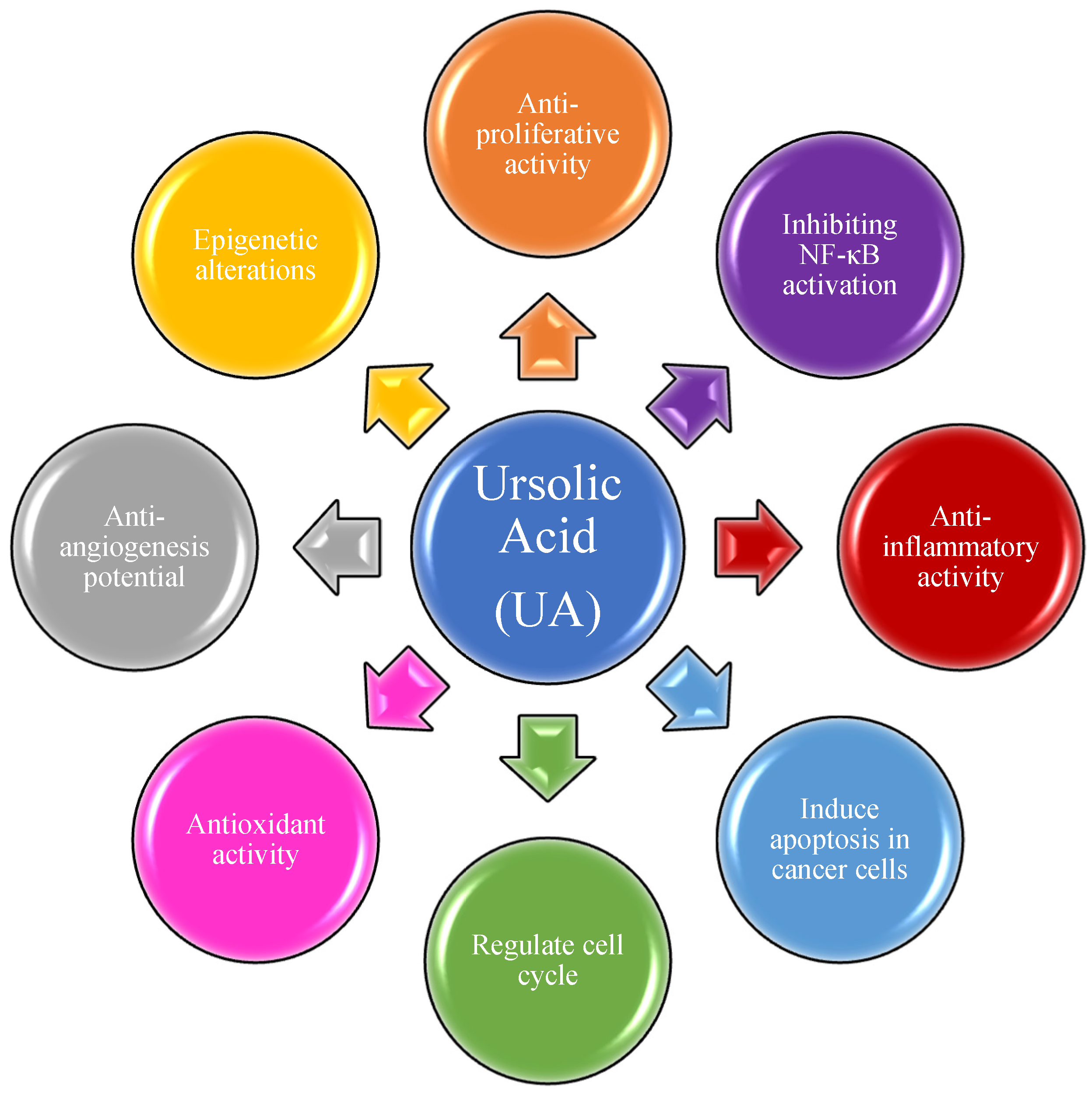
Figure 1. Ursolic acid and its molecular mechanism of action in cancer treatment.
Molecules 27 08981 g001
Figure 2. Structure of UA with highlighted pharmacophoric sites.
Molecules 27 08981 g002
Figure 3. Structure of 2.
Molecules 27 08981 g003
Figure 4. Structure of 3.
Molecules 27 08981 g004
Figure 5. Structure of 4 which shows antiproliferative effect.
Molecules 27 08981 g005
Figure 6. Structure of 5, 6, 7.
Molecules 27 08981 g006
Figure 7. Structure of 8, 9, 10.
Molecules 27 08981 g007
Figure 8. Structure of 11.
Molecules 27 08981 g008
Figure 9. Structure of 12, 13.
Molecules 27 08981 g009
Figure 10. Structure of 14, 15.
Molecules 27 08981 g010
Figure 11. Structure of 16.
Molecules 27 08981 g011
Figure 12. Structure of 17.
Molecules 27 08981 g012
Figure 13. Structure of 18, 19.
Molecules 27 08981 g013
Figure 14. Structure of 20.
Molecules 27 08981 g014
Figure 15. Structure of 21ac, 22a, 22b, 23a, 23b, 24ad, 25.
Molecules 27 08981 g015
Figure 16. Structure of 26.
Molecules 27 08981 g016
Figure 17. Structure of 27.
Molecules 27 08981 g017
Figure 18. Structure of 28.
Molecules 27 08981 g018
Figure 19. Structure of 29, 30.
Molecules 27 08981 g019
Figure 20. Structure of 31, 32.
Molecules 27 08981 g020
Figure 21. Structure of 33.
Molecules 27 08981 g021
Figure 22. Structure of 34, 35.
Molecules 27 08981 g022
Figure 23. Structure of 36.
Molecules 27 08981 g023
Figure 24. Structure of 37, 38.
Molecules 27 08981 g024
Figure 25. Structure of 39.
Molecules 27 08981 g025
Figure 26. Structure of 40, 41.
Molecules 27 08981 g026
Figure 27. Structure of 42a, 42b.
Molecules 27 08981 g027
Figure 28. Structure of 43ad.
Molecules 27 08981 g028
Figure 29. Structure of 44a, 44b.
Molecules 27 08981 g029
Figure 30. Structure of 45.
Molecules 27 08981 g030
Figure 31. Structure of 46.
Molecules 27 08981 g031
Figure 32. Structure of 47.
Molecules 27 08981 g032
Figure 33. Structure of 48.
Molecules 27 08981 g033
Figure 34. Structure of 49, 50.
Molecules 27 08981 g034
Figure 35. Structure of 51.
Molecules 27 08981 g035
Figure 36. Structure of 52, 53.
Molecules 27 08981 g036
Figure 37. Structure of 54.
Molecules 27 08981 g037
Figure 38. Structure of 55.
Molecules 27 08981 g038
Figure 39. Structure of 56, 57.
Molecules 27 08981 g039
Figure 40. Structure of 58.
Molecules 27 08981 g040
Figure 41. Structure of 59, 60.
Molecules 27 08981 g041
Table 1. Summarized information on UA analogs (C3 modifications).
CompoundPathway/MechanismRegulationCell Cycle Arrest (Phase)Animal Study[Ref]
3Activation of caspases-8, -9, and -3Apoptosis NASub G1 & G2/Mno[68]
4Activation of p53Bcl2↓
MDM2↓
Bax↑
Sub G1 & G1no[69]
6NF−κBNANAno[70]
Table 2. Summarized information on UA analogs (C28 modifications).
CompoundPathway/MechanismRegulationCell Cycle Arrest (Phase)Animal Study[Ref]
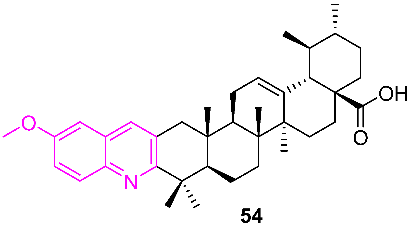
9Inhibition of extracellular signal-regulated kinase: ERKNANAno[72]
10Increased production of reactive oxygen species (ROS)apoptosisSub G1no[73]
12NANAG2/Mno[76]
14DNA cleavageNANAno[77]
16Inhibition of HIF-1α transcriptional activityHIF-1α↓ mRNA↓G1no[79]
17Mitotic catastropheNAG2/Mno[80]
Table 3. Concentrations of the half-maximal inhibition (IC50 ± SEM, μM) of compounds 2125 on immortalized human fibroblasts, MCF7, U-87 MG, A549, HepG2 cells.
Compd.Immortalized Human FibroblastsMCF7U-87 MGA549HepG2
Doxorubicin3.33 ± 0.674.51 ± 1.122.05 ± 0.226.17 ± 1.1710.02 ± 1.67
UA74.9 ± 4.5874.9 ± 4.5874.9 ± 4.5874.9 ± 4.5874.9 ± 4.58
21a>100>100>100>100>100
21b>100>100>100>100>100
21c>100>100>100>100>100
22a25.85 ± 3.0422.9 ± 10.0229.71 ± 6.52>10012.89 ± 2.63
22b10.41 ± 1.251.55 ± 0.08>100>100>100
23a82.59 ± 16.8726.23 ± 8.76>100>10035.58 ± 8.83
23b>100>100>100>100>100
24a74.9 ± 4.58>100>10040.26 ± 7.55>100
24b>100>100>100>100>100
24c>100>100>100>100>100
24d>100>100>100>100>100
2548.35 ± 11.47>10081.67 ± 2.89>100>100
Table 4. IC50 values (µM) of compounds 43ad against various cell lines.
CompoundA549MCF7HTC116THP1FR2
UA33 ± 0.0337 ± 0.0742 ± 0.089.1 ± 0.0731 ± 0.08
43a0.5 ± 0.055.5 ± 0.08<0.1 ± 0.090.9 ± 0.0210 ± 0.04
43b2.9 ± 0.05<0.1 ± 0.0515 ± 0.06<0.1 ± 0.0369 ± 0.05
43c<0.1 ± 0.001<0.1 ± 0.090.3 ± 0.001<0.1 ± 0.001>50 ± 4.1
43d0.15 ± 0.01<0.1 ± 0.0019.1 ± 0.1<0.1 ± 0.001>50 ± 3.9
Table 5. Summarized information on UA analogs (C3 and C28 modifications).
CompoundPathway/MechanismRegulationCell Cycle Arrest (Phase)Animal Study[Ref]
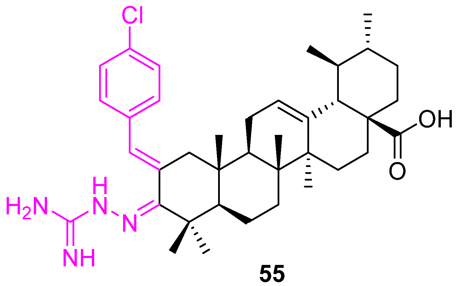
26Activation of caspase-3ApoptosisSyes[85]
29Activation of the caspase-3
mitochondria pathway
Bcl2↓
Survivin↓
NAyes[89]
31Activation of caspases-8, -9, and -3Bcl2↓
HKII↓
Bax↑
p53↑
S
G2/M
yes[96]
32NANAG0/G1no[97]
33Activation of caspases-8, -9, and -3
Increased production of reactive oxygen species (ROS)
Bcl2↓G1no[98]
36NF-κBNAG1no[100]
Table 6. Summarized information on UA analogs (modifications at multiple positions).
CompoundPathway/MechanismRegulationCell Cycle Arrest (Phase)Animal Study[Ref]
49NAc-FLIP↓
NOXA↑
p21waf1
G1no[115]
50NAc-FLIP↓
NOXA↑
p21waf1
G1no[115]
51Activation of caspases-8, -9, and -3 G1no[116]
53Activation of caspase cascade
RAS/Raf/MEK/ERK
PI3K/AKT/mTOR
Increased production of reactive oxygen species (ROS)
Bcl-2↓
Bax↑
Sno[118]
54NANAG0/G1no
55VEGFHIF-1α↓NAno
56Caspase activation
Increased production of reactive oxygen species (ROS)
Bcl2↓
Bax↑
G1no[124]
57Caspase activation
Increased production of reactive oxygen species (ROS)
Bcl2↓
Bax↑
G1no[124]
58PI3K/AKT/mTOR
Increased production of reactive oxygen species (ROS)
Bcl2↓
PARP↑
Bax↑
caspase-3/9↑
Sno[125]
Publisher’s Note: MDPI stays neutral with regard to jurisdictional claims in published maps and institutional affiliations.