
Anti-Diabetes, Anti-Gout, and Anti-Leukemia Properties of Essential Oils from Natural Spices Clausena indica, Zanthoxylum rhetsa, and Michelia tonkinensis

 ,
,  ,
,  ,
,  , ,
, ,  and
and 
Abstract
:1. Introduction
2. Results
2.1. Phytochemical Composition of EOs
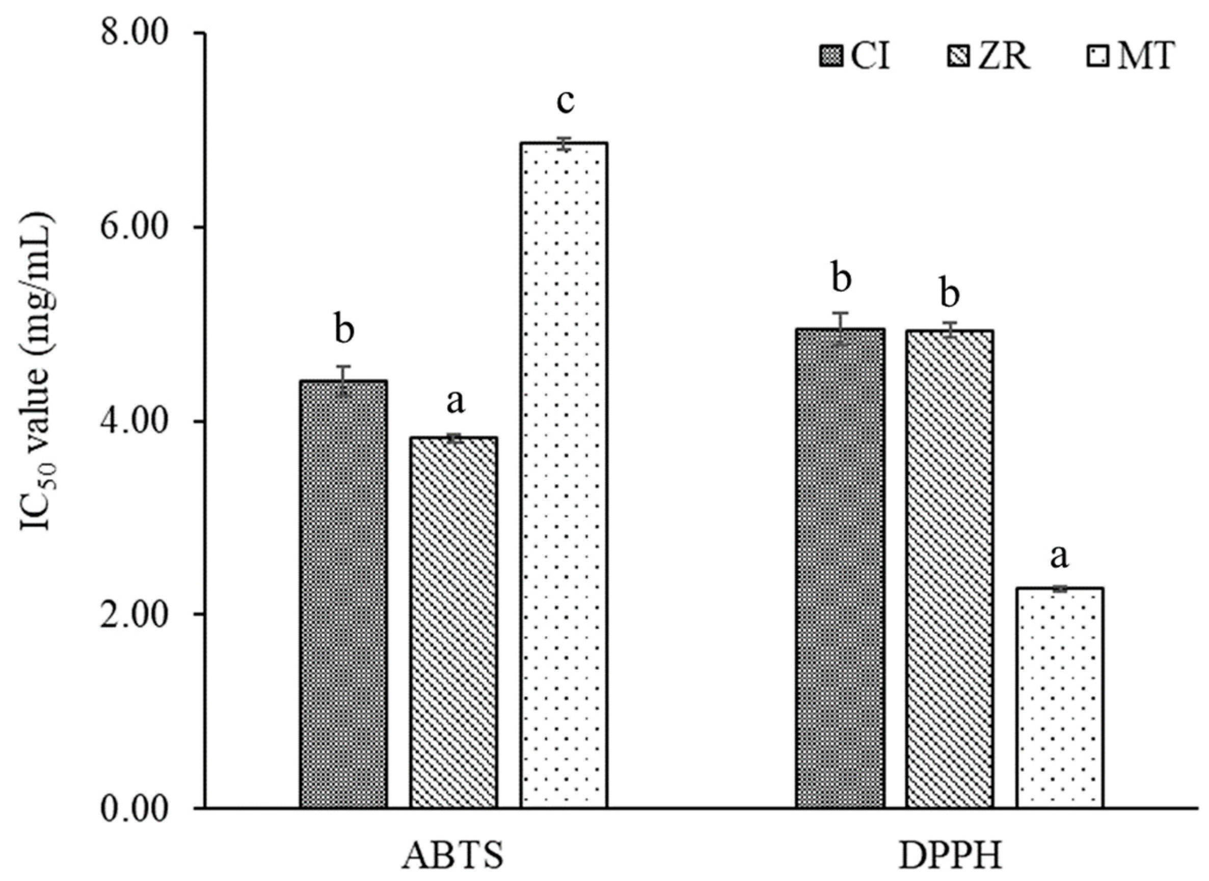
2.2. Antioxidant Activity
2.3. Enzymatic Inhibitory Activity
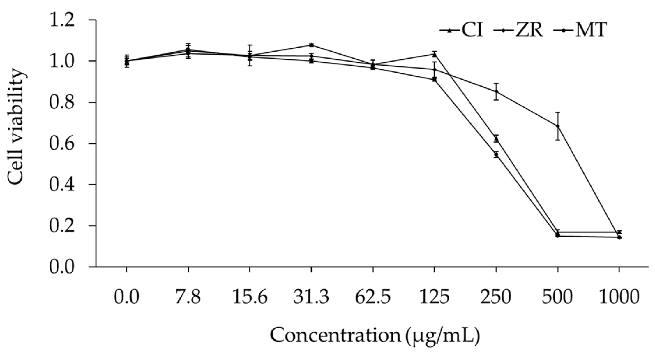
2.4. Cytotoxic Activity
3. Discussion
4. Materials and Methods
4.1. Instrumentations and Reagents
4.2. Plant Materials
4.3. Essential Oil Extraction
4.4. Identification of Phytochemical Composition by GC–MS
4.5. Evaluation of Biological Activities
4.5.1. Antioxidant Activity
4.5.2. Enzymatic Assays
4.5.3. Cytotoxic Assay on Meg-01 Cell Line
4.6. Statistical Analysis
5. Conclusions
Supplementary Materials
Author Contributions
Funding
Institutional Review Board Statement
Informed Consent Statement
Data Availability Statement
Acknowledgments
Conflicts of Interest
References
- Teschke, R.; Xuan, T.D. Active nature based ingredients for drug discovery with pivotal role of clinical efficacy: Review and prospective. J. Mod. Med. Chem. 2020, 8, 4–18. [Google Scholar] [CrossRef]
- Skrinjar, M.; Nemet, N. Antimicrobial effects of spices and herbs essential oils. Acta Period. Technol. 2009, 2009, 195–209. [Google Scholar] [CrossRef]
- Bozin, B.; Mimica-Dukic, N.; Simin, N.; Anackov, G. Characterization of the volatile composition of essential oils of some Lamiaceae spices and the antimicrobial and antioxidant activities of the entire oils. J. Agric. Food Chem. 2006, 54, 1822–1828. [Google Scholar] [CrossRef] [PubMed]
- Quan, N.V.; Xuan, T.D.; Anh, L.H.; Tran, H.-D. Bio-guided isolation of prospective bioactive constituents from roots of Clausena indica (Dalzell) Oliv. Molecules 2019, 24, 4442. [Google Scholar] [CrossRef] [PubMed] [Green Version]
- Diep, P.T.M.; Pawlowska, A.M.; Cioni, P.L.; Van Minh, C.; Huong, L.M.; Braca, A. Chemical composition and antimicrobial activity of Clausena indica (Dalz) Oliv. (Rutaceae) essential oil from Vietnam. Nat. Prod. Commun. 2009, 4, 869–872. [Google Scholar] [CrossRef] [Green Version]
- John, J.A.; Kurup, S.R.R.; Pradeep, N.S.; Sabulal, B. Chemical composition and Antibacterial activity of the leaf oil of Clausena indica from South India. J. Essent. Oil Bear. Plants 2011, 14, 776–781. [Google Scholar] [CrossRef]
- Ahsan, M.; Haque, M.R.; Hossain, M.B.; Islam, S.N.; Gray, A.I.; Hasan, C.M. Cytotoxic dimeric quinolone-terpene alkaloids from the root bark of Zanthoxylum rhetsa. Phytochemistry 2014, 103, 8–12. [Google Scholar] [CrossRef]
- Rahman, M.; Alimuzzaman, M.; Ahmad, S.; Chowdhury, A.A. Antinociceptive and antidiarrhoeal activity of Zanthoxylum rhetsa. Fitoterapia 2002, 73, 340–342. [Google Scholar] [CrossRef]
- Alphonso, P.; Saraf, A. Chemical profile studies on the secondary metabolites of medicinally important plant Zanthoxylum rhetsa (Roxb.) DC using HPTLC. Asian Pac. J. Trop. Biomed. 2012, 2, S1293–S1298. [Google Scholar] [CrossRef]
- Tantapakul, C.; Phakhodee, W.; Ritthiwigrom, T.; Yossathera, K.; Deachathai, S.; Laphookhieo, S. Antibacterial compounds from Zanthoxylum rhetsa. Arch. Pharm. Res. 2012, 35, 1139–1142. [Google Scholar] [CrossRef]
- Wongkatiya, N.; Akekawatchai, C.; Sanguansermsri, P.; Fraser, L.H.; Pratoomsoot, C.; Sanguansermsri, D. Chemical compositions and biological properties of essential oils from Zanthoxylum rhetsa (Roxb.) DC and Zanthoxylum limonella Alston. Afr. J. Tradit. Complement. Altern. Med. 2018, 15, 12–18. [Google Scholar] [CrossRef] [Green Version]
- Theeramunkong, S.; Utsintong, M. Comparison between volatile oil from fresh and dried fruits of Zanthoxylum rhetsa (Roxb.) DC. and cytotoxicity activity evaluation. Pharmacogn. J. 2018, 10, 827–832. [Google Scholar] [CrossRef] [Green Version]
- Jiwajinda, S.; Santisopasri, V.; Murakami, A.; Kim, O.-K.; Kim, H.W.; Ohigashi, H. Suppressive effects of edible Thai plants on superoxide and nitric oxide generation. Asian Pac. J. Cancer Prev. 2002, 3, 215–223. [Google Scholar] [PubMed]
- Trung, H.D.; Thang, T.D.; Ban, P.H.; Hoi, T.M.; Dai, D.N.; Ogunwande, I.A. Terpene constituents of the leaves of five Vietnamese species of Clausena (Rutaceae). Nat. Prod. Res. 2014, 28, 622–630. [Google Scholar] [CrossRef] [PubMed]
- Dai, D.N.; Thang, T.D.; Ogunwande, I.A. Essential oil composition of four Magnoliaceae species cultivated in Vietnam. J. Herbs Spices Med. Plants 2016, 22, 279–287. [Google Scholar] [CrossRef]
- Bastos, D.H.M.; Ishimoto, E.Y.; Marques, M.O.M.; Ferri, A.F.; Torres, E.A.F.S. Essential oil and antioxidant activity of green mate and mate tea (Ilex paraguariensis) infusions. J. Food Compos. Anal. 2006, 19, 538–543. [Google Scholar] [CrossRef]
- Adams, R.P.; Elizondo, M.S.G.; Elizondo, M.G.; Slinkman, E. DNA fingerprinting and terpenoid analysis of Juniperus blancoi var. huehuentensis (Cupressaceae), a new subalpine variety from Durango, Mexico. Biochem. Syst. Ecol. 2006, 34, 205–211. [Google Scholar] [CrossRef]
- Thai, T.H.; Bazzali, O.; Hoi, T.M.; Hien, N.T.; Hung, N.V.; Félix, T.; Casanova, J.; Bighelli, A. Chemical composition of the essential oils from Vietnamese Clausena indica and C. anisum-olens. Nat. Prod. Commun. 2014, 9, 1531–1534. [Google Scholar] [CrossRef] [Green Version]
- Adams, R.P. Identification of Essential Oil Components by Gas Chromatography/Mass Spectrometry, 4th ed.; Allured Publishing Corp: Carol Stream, IL, USA, 2007; ISBN 1932633219. [Google Scholar]
- Stephane, F.F.Y.; Jules, B.K.J. Terpenoids as Important Bioactive Constituents of Essential Oils. In Essential Oils—Bioactive Compounds, New Perspectives and Applications; IntechOpen: London, UK, 2020. [Google Scholar]
- Santos, M.R.V.; Moreira, F.V.; Fraga, B.P.; Souza, D.P.D.; Bonjardim, L.R.; Quintans-Junior, L.J. Cardiovascular effects of monoterpenes: A review. Rev. Bras. Farmacogn. 2011, 21, 764–771. [Google Scholar] [CrossRef] [Green Version]
- Ciftci, O.; Oztanir, M.N.; Cetin, A. Neuroprotective effects of β-myrcene following global cerebral ischemia/reperfusion-mediated oxidative and neuronal damage in a C57BL/J6 mouse. Neurochem. Res. 2014, 39, 1717–1723. [Google Scholar] [CrossRef]
- Rufino, A.T.; Ribeiro, M.; Sousa, C.; Judas, F.; Salgueiro, L.; Cavaleiro, C.; Mendes, A.F. Evaluation of the anti-inflammatory, anti-catabolic and pro-anabolic effects of E-caryophyllene, myrcene and limonene in a cell model of osteoarthritis. Eur. J. Pharmacol. 2015, 750, 141–150. [Google Scholar] [CrossRef] [PubMed]
- Burcu, G.B.; Osman, C.; Asli, C.; Namik, O.M.; Neşe, B.T. The protective cardiac effects of Β-myrcene after global cerebral ischemia/reperfusion in C57BL/J6 mouse. Acta Cir. Bras. 2016, 31, 456–462. [Google Scholar] [CrossRef] [PubMed]
- Vieira, A.J.; Beserra, F.P.; Souza, M.C.; Totti, B.M.; Rozza, A.L. Limonene: Aroma of innovation in health and disease. Chem. Biol. Interact. 2018, 283, 97–106. [Google Scholar] [CrossRef] [PubMed] [Green Version]
- Sharma, S.; Gupta, J.; Prabhakar, P.K.; Gupta, P.; Solanki, P.; Rajput, A. Phytochemical repurposing of natural molecule: Sabinene for identification of novel therapeutic benefits using in silico and in vitro approaches. Assay Drug Dev. Technol. 2019, 17, 339–351. [Google Scholar] [CrossRef] [PubMed]
- Li, W.-R.; Li, H.-L.; Shi, Q.-S.; Sun, T.-L.; Xie, X.-B.; Song, B.; Huang, X.-M. The dynamics and mechanism of the antimicrobial activity of tea tree oil against bacteria and fungi. Appl. Microbiol. Biotechnol. 2016, 100, 8865–8875. [Google Scholar] [CrossRef] [PubMed]
- Pereira, I.; Severino, P.; Santos, A.C.; Silva, A.M.; Souto, E.B. Linalool bioactive properties and potential applicability in drug delivery systems. Colloids Surf. B Biointerfaces 2018, 171, 566–578. [Google Scholar] [CrossRef] [PubMed]
- Martins, H.B.; Selis, N.D.N.; Souza, C.L.S.E.; Nascimento, F.S.; Carvalho, S.P.d.; Gusmão, L.D.; Nascimento, J.D.S.; Brito, A.K.P.; Souza, S.I.D.; Oliveira, M.V.D.; et al. Anti-inflammatory activity of the essential oil citral in experimental infection with Staphylococcus aureus in a model air pouch. Evid.-Based Complement. Altern. Med. 2017, 2017, 1–10. [Google Scholar] [CrossRef] [Green Version]
- Gogoi, R.; Loying, R.; Sarma, N.; Begum, T.; Pandey, S.K.; Lal, M. Comparative analysis of in-vitro biological activities of methyl eugenol rich Cymbopogon khasianus Hack., leaf essential oil with pure methyl eugenol compound. Curr. Pharm. Biotechnol. 2020, 21, 927–938. [Google Scholar] [CrossRef]
- Barceloux, D.G. Nutmeg (Myristica fragrans Houtt.). In Medical Toxicology of Natural Substances: Foods, Fungi, Medicinal Herbs, Toxic Plants, and Venomous Animals, 1st ed.; Barceloux, D.G., Ed.; John Wiley & Sons: Hoboken, NJ, USA, 2008; pp. 67–70. [Google Scholar]
- Morita, T.; Jinno, K.; Kawagishi, H.; Arimoto, Y.; Suganuma, H.; Inakuma, T.; Sugiyama, K. Hepatoprotective effect of myristicin from nutmeg (Myristica fragrans) on lipopolysaccharide/D-galactosamine-induced liver injury. J. Agric. Food Chem. 2003, 51, 1560–1565. [Google Scholar] [CrossRef]
- Ansory, H.M.; Sari, E.N.; Nilawati, A.; Handayani, S.; Aznam, N. Sunscreen and antioxidant potential of myristicin in nutmeg essential oils (Myristica fragrans). In Proceedings of the 2nd Bakti Tunas Husada-Health Science International Conference (BTH-HSIC 2019), Tasikmalaya, Indonesia, 5–6 October 2019; Atlantis Press: Paris, France, 2020. [Google Scholar]
- Seneme, E.F.; Dos Santos, D.C.; Silva, E.M.R.; Franco, Y.E.M.; Longato, G.B. Pharmacological and therapeutic potential of myristicin: A literature review. Molecules 2021, 26, 5914. [Google Scholar] [CrossRef]
- Narasimhan, B.; Dhake, A.S. Antibacterial principles from Myristica fragrans seeds. J. Med. Food 2006, 9, 395–399. [Google Scholar] [CrossRef] [PubMed]
- Martins, C.; Doran, C.; Laires, A.; Rueff, J.; Rodrigues, A.S. Genotoxic and apoptotic activities of the food flavourings myristicin and eugenol in AA8 and XRCC1 deficient EM9 cells. Food Chem. Toxicol. 2011, 49, 385–392. [Google Scholar] [CrossRef] [PubMed]
- Shafi, P.M.; Saidutty, A.; Clery, R.A. Volatile constituents of Zanthoxylum rhetsa leaves and seeds. J. Essent. Oil Res. 2000, 12, 179–182. [Google Scholar] [CrossRef]
- Naik, R.R.; Shakya, A.K.; Khalaf, N.A. GC-MS Analysis and biological evaluation of essential oil of Zanthoxylum rhetsa (Roxb.) DC pericarp. Jordan J. Pharm. Sci. 2015, 8, 181–193. [Google Scholar] [CrossRef]
- Roberto, D.; Micucci, P.; Sebastian, T.; Graciela, F.; Anesini, C. Antioxidant activity of limonene on normal murine lymphocytes: Relation to H2O2 modulation and cell proliferation. Basic Clin. Pharmacol. Toxicol. 2009, 106, 38–44. [Google Scholar] [CrossRef]
- Souza, C.F.; Baldissera, M.D.; Silva, L.d.L.; Geihs, M.A.; Baldisserotto, B. Is monoterpene terpinen-4-ol the compound responsible for the anesthetic and antioxidant activity of Melaleuca alternifolia essential oil (tea tree oil) in silver catfish? Aquaculture 2018, 486, 217–223. [Google Scholar] [CrossRef]
- Seol, G.-H.; Kang, P.; Lee, H.S.; Seol, G.H. Antioxidant activity of linalool in patients with carpal tunnel syndrome. BMC Neurol. 2016, 16, 17. [Google Scholar] [CrossRef] [Green Version]
- Holdgate, G.A.; Meek, T.D.; Grimley, R.L. Mechanistic enzymology in drug discovery: A fresh perspective. Nat. Rev. Drug Discov. 2018, 17, 115–132. [Google Scholar] [CrossRef]
- Anh, L.H.; Xuan, T.D.; Thuy, N.T.D.; Quan, N.V.; Trang, L.T. Antioxidant and α-amylase inhibitory activities and phytocompounds of Clausena indica fruits. Medicines 2020, 7, 10. [Google Scholar]
- More, T.A.; Kulkarni, B.R.; Nalawade, M.L.; Arvindekar, A.U. Antidiabetic activity of linalool and limonene in streptozotocin-induced diabetic rat: A combinatorial therapy approach. Int. J. Pharm. Pharm. Sci. 2014, 6, 159–163. [Google Scholar]
- Dawson, J.; Walters, M. Uric acid and xanthine oxidase: Future therapeutic targets in the prevention of cardiovascular disease? Br. J. Clin. Pharmacol. 2006, 62, 633–644. [Google Scholar] [CrossRef] [PubMed] [Green Version]
- Bray, F.; Ferlay, J.; Soerjomataram, I.; Siegel, R.L.; Torre, L.A.; Jemal, A. Global cancer statistics 2018: GLOBOCAN estimates of incidence and mortality worldwide for 36 cancers in 185 countries. CA Cancer J. Clin. 2018, 68, 394–424. [Google Scholar] [CrossRef] [PubMed] [Green Version]
- Poma, P.; Labbozzetta, M.; Ramarosandratana, A.V.; Rosselli, S.; Tutone, M.; Sajeva, M.; Notarbartolo, M. In vitro modulation of p-glycoprotein activity by Euphorbia intisy essential oil on acute myeloid leukemia cell line HL-60R. Pharmaceuticals 2021, 14, 111. [Google Scholar] [CrossRef] [PubMed]
- Martins, C.; Doran, C.; Silva, I.C.; Miranda, C.; Rueff, J.; Rodrigues, A.S. Myristicin from nutmeg induces apoptosis via the mitochondrial pathway and down regulates genes of the DNA damage response pathways in human leukaemia K562 cells. Chem. Biol. Interact. 2014, 218, 1–9. [Google Scholar] [CrossRef]
- Shah, B.B.; Baksi, R.; Chaudagar, K.K.; Nivsarkar, M.; Mehta, A.A. Anti-leukemic and anti-angiogenic effects of D-limonene on K562-implanted C57BL/6 mice and the chick chorioallantoic membrane model. Anim. Models Exp. Med. 2018, 1, 328–333. [Google Scholar] [CrossRef] [Green Version]
- Chang, M.-Y.; Shieh, D.-E.; Chen, C.-C.; Yeh, C.-S.; Dong, H.-P. Linalool induces cell cycle arrest and apoptosis in leukemia cells and cervical cancer cells through CDKIs. Int. J. Mol. Sci. 2015, 16, 28169–28179. [Google Scholar] [CrossRef] [Green Version]
- Quan, N.V.; Xuan, T.D.; Teschke, R. Potential hepatotoxins found in herbal medicinal products: A systematic review. Int. J. Mol. Sci. 2020, 21, 5011. [Google Scholar] [CrossRef]
- Teschke, R.; Zhu, Y.; Jing, J. Herb induced liver injury (HILI) in the Asian region and current role of RUCAM for causality assessment in 11,160 published cases: Analysis and outlook. J. Clin. Transl. Hepatol. 2020, 8, 200–214. [Google Scholar] [CrossRef]
- Teschke, R.; Eickhoff, A.; Schulze, J.; Danan, G. Herb-induced liver injury (HILI) with 12,068 worldwide cases published with causality assessments by Roussel Uclaf Causality Assessment Method (RUCAM): An overview. Transl. Gastroenterol. Hepatol. 2021, 6, 51. [Google Scholar] [CrossRef]
- Moylan, J.S.; Reid, M.B. Oxidative stress, chronic disease, and muscle wasting. Muscle Nerve 2007, 35, 411–429. [Google Scholar] [CrossRef]
- Ambade, A.; Mandrekar, P. Oxidative stress and inflammation: Essential partners in alcoholic liver disease. Int. J. Hepatol. 2012, 2012, 1–9. [Google Scholar] [CrossRef] [PubMed]
- Wardhana, W.; Rudijanto, A. Effect of uric acid on blood glucose levels. Acta Med. Indones. 2018, 50, 253–256. [Google Scholar] [PubMed]
- Victor, V.M. Mitochondrial oxidative stress in diabetes. In Diabetes: Oxidative Stress and Dietary Antioxidants; Elsevier: Amsterdam, The Netherlands, 2014; pp. 41–49. [Google Scholar]
- Quan, N.V.; Xuan, T.D.; Tran, H.-D.; Ahmad, A.; Khanh, T.D.; Dat, T.D. Contribution of momilactones A and B to diabetes inhibitory potential of rice bran: Evidence from in vitro assays. Saudi Pharm. J. 2019, 27, 643–649. [Google Scholar] [CrossRef] [PubMed]
- Bui, T.H.; Nguyen, N.T.; Dang, P.H.; Nguyen, H.X.; Nguyen, M.T.T. Design and synthesis of chalcone derivatives as potential non-purine xanthine oxidase inhibitors. Springerplus 2016, 5, 1789. [Google Scholar] [CrossRef] [Green Version]
- Anh, L.H.; Quan, N.V.; Lam, V.Q.; Iuchi, Y.; Takami, A.; Teschke, R.; Xuan, T.D. Antioxidant, anti-tyrosinase, anti-α-amylase, and cytotoxic potentials of the invasive weed Andropogon virginicus. Plants 2020, 10, 69. [Google Scholar] [CrossRef]


| Peak No. | Identified Compound | Composition (%) | LRI | KI | Identification | ||
|---|---|---|---|---|---|---|---|
| CI | ZR | MT | |||||
| 1 | β-Thujene | - | 0.3 | - | 923 | 926 | MS, ref |
| 2 | α-Pinene | 0.1 | 2.2 | - | 933 | 936 | MS, ref |
| 3 | 5-Methylfurfural | 0.1 | - | - | 955 | 959 | MS, ref |
| 4 | Sabinene | - | 4.8 | 0.1 | 973 | 976 | MS, ref |
| 5 | 6-Methyl-5-heptene-2-one | - | - | 1.0 | 981 | 983 | MS |
| 6 | β-Myrcene | 6.5 | 1.2 | - | 985 | 987 | MS, ref |
| 7 | α-Phellandrene | - | 2.2 | - | 1009 | 1010 | MS, ref |
| 8 | 3-Carene | 0.4 | - | - | 1008 | 1009 | MS, ref |
| 9 | 3,6-Dimethylene-1,7-octadiene | 0.1 | - | - | 1013 | 1015 | ref |
| 10 | α-Terpinene | - | 1.4 | - | 1019 | 1021 | MS, ref |
| 11 | p-Cymene | 0.2 | 4.2 | - | 1023 | 1026 | MS, ref |
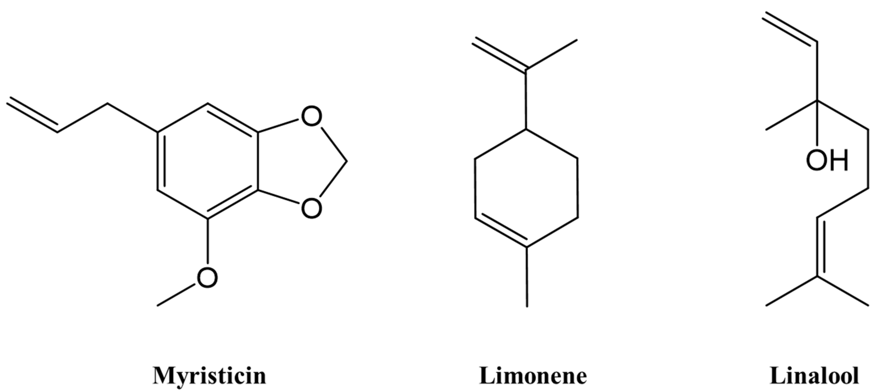
| 12 | Limonene | 5.5 | 44.2 | - | 1029 | 1031 | MS, ref |
| 13 | β-Phellandrene | - | 4.0 | - | 1034 | 1037 | MS, ref |
| 14 | Eucalyptol | - | - | 0.9 | 1036 | 1039 | MS |
| 15 | 1-Octanol | - | 0.1 | - | 1068 | 1070 | MS, ref |
| 16 | p-Cresol | 0.1 | - | - | 1066 | 1069 | MS, ref |
| 17 | cis-Linalool oxide | - | - | 0.8 | 1072 | 1074 | MS |
| 18 | cis-Linaloloxide | - | - | 0.8 | 1088 | 1089 | MS |
| 19 | α-Terpinolene | 1.6 | 1.1 | - | 1084 | 1086 | MS, ref |
| 20 | p-Cymenene | 1.2 | 0.1 | - | 1088 | 1090 | MS, ref |
| 21 | Linalool | - | 1.3 | 49.3 | 1099 | 1101 | MS |
| 22 | 6-Camphenone | 0.3 | - | - | 1095 | 1095 | ref |
| 23 | 1,5,7-Octatrien-3-ol, 3,7-dimethyl- | - | - | 0.3 | 1103 | 1104 | ref |
| 24 | 1,3,8-p-Menthatriene | 0.1 | - | - | 1111 | 1112 | MS, ref |
| 25 | cis-p-Mentha-2,8-dienol | - | 0.2 | - | 1124 | 1126 | ref |
| 26 | 2-Cyclohexen-1-ol, 1-methyl-4-(1-methylethyl)-, trans- | - | 0.5 | - | 1127 | 1129 | ref |
| 27 | trans-p-Menth-2-en-1-ol | 0.2 | 0.3 | - | 1138 | 1141 | MS, ref |
| 28 | Pinocarveol | - | 0.3 | - | 1145 | 1148 | ref |
| 29 | 1,5,7-Octatrien-3-ol, 2,6-dimethyl- | 0.4 | - | - | 1151 | 1154 | MS, ref |
| 30 | E-β-Terpineol | - | 0.2 | - | 1160 | 1163 | ref |
| 31 | Octanoic acid | - | 0.3 | - | 1164 | 1166 | ref |
| 32 | 2-Isopropenyl-5-methylhex-4-enal | 0.8 | - | - | 1178 | 1180 | MS, ref |
| 33 | Terpinen-4-ol | - | 11.5 | 0.9 | 1184 | 1186 | MS, ref |
| 34 | p-Cymen-8-ol | 1.6 | 2.8 | - | 1188 | 1189 | MS, ref |
| 35 | α-Terpineol | 0.5 | 2.3 | 0.4 | 1197 | 1198 | MS, ref |
| 36 | cis-Carveol | 0.1 | 1.8 | - | 1221 | 1222 | MS, ref |
| 37 | Citronellol | - | - | 1.7 | 1225 | 1226 | MS |
| 38 | cis-p-mentha-1(7),8-dien-2-ol | 0.1 | - | - | 1228 | 1229 | MS, ref |
| 39 | Nerol | - | 0.3 | - | 1234 | 1236 | ref |
| 40 | β-Citral | - | - | 5.6 | 1238 | 1240 | MS |
| 41 | Phenol, 2-ethyl-4,5-dimethyl- | 0.1 | - | - | 1240 | 1242 | MS, ref |
| 42 | Carvone | - | 3.3 | - | 1247 | 1249 | ref |
| 43 | trans-Geraniol | - | - | 3.8 | 1249 | 1251 | MS |
| 44 | Nonanoic acid | - | 0.2 | - | 1260 | 1262 | MS |
| 45 | α-Citral | - | - | 8.3 | 1267 | 1269 | MS |
| 46 | Phellandral | - | 0.4 | - | 1282 | 1283 | MS |
| 47 | p-Cymen-7-ol | - | 0.3 | - | 1292 | 1293 | MS, ref |
| 48 | Safrole | - | - | 4.6 | 1292 | 1293 | MS |
| 49 | p-Cymen-2-ol | - | 0.3 | - | 1297 | 1297 | MS, ref |
| 50 | Geranic acid methyl ester | - | - | 0.1 | 1319 | 1320 | MS |
| 51 | p-Mentha-1,4-dien-7-ol | - | 0.2 | - | 1329 | 1331 | MS, ref |
| 52 | Eugenol | 0.1 | - | - | 1349 | 1350 | MS, ref |
| 53 | 2,6-Octadien-1-ol, 3,7-dimethyl-, acetate | - | 1.4 | - | 1375 | 1377 | MS, ref |
| 54 | Methyleugenol | 0.4 | - | 16.9 | 1399 | 1399 | MS, ref |
| 55 | Caryophyllene | 0.6 | - | 0.2 | 1428 | 1429 | MS, ref |
| 56 | trans-α-Bergamotene | 0.1 | - | - | 1433 | 1434 | MS, ref |
| 57 | cis-β-Famesene | 0.1 | - | - | 1448 | 1449 | MS, ref |
| 58 | β-Eudesmene | - | - | 0.4 | 1497 | 1497 | MS |
| 59 | β-Bisabolene | 2.6 | - | 0.1 | 1510 | 1510 | MS, ref |
| 60 | δ-Cadinene | - | - | 0.2 | 1522 | 1523 | MS |
| 61 | Myristicin | 68.3 | - | - | 1524 | 1525 | MS, ref |
| 62 | Elemicin | 1.9 | - | - | 1540 | 1542 | MS, ref |
| 63 | Spathulenol | 0.2 | 0.1 | - | 1581 | 1582 | MS, ref |
| 64 | Isoelemicin | 0.1 | - | - | 1639 | 1640 | MS, ref |
| 65 | α-Cadinol | - | - | 0.2 | 1661 | 1662 | MS |
| 66 | α-Springene | 2.9 | - | - | 1964 | 1965 | MS, ref |
| Monoterpene hydrocarbons | 14.4 | 65.5 | 0.1 | ||||
| Oxygenated monoterpenes | 2.5 | 26.0 | 70.1 | ||||
| Sesquiterpene hydrocarbons | 3.4 | 0 | 0.9 | ||||
| Oxygenated sesquiterpenes | 0.2 | 0.1 | 0 | ||||
| Others | 76.6 | 1.9 | 25.5 | ||||
| Total identified | 96.9 | 93.5 | 96.6 | ||||
| Yield % (w/w) | 0.36 | 1.69 | 5.49 | ||||
| Sample | IC50 (mg/mL) | |||
|---|---|---|---|---|
| α-Amylase Assay | α-Glucosidase Assay | Xanthine Oxidase Assay | Meg-01 Assay | |
| CI | 7.73 ± 0.10 | 0.84 ± 0.03 b | 0.88 ± 0.05 a | 0.32 ± 0.01 a |
| ZR | - | 0.73 ± 0.01 a | 2.80 ± 0.14 c | 0.64 ± 0.04 b |
| MT | - | 1.46 ± 0.01 c | 1.73 ± 0.16 b | 0.31 ± 0.01 a |
| Acarbose | 0.01 ± 0.00 | 2.69 ± 0.07 | nd | nd |
| Palmitic acid | 1.57 ± 0.04 | 0.72 ± 0.01 | nd | nd |
| Allopurinol | nd | nd | 0.01 ± 0.00 | nd |
Publisher’s Note: MDPI stays neutral with regard to jurisdictional claims in published maps and institutional affiliations. |
© 2022 by the authors. Licensee MDPI, Basel, Switzerland. This article is an open access article distributed under the terms and conditions of the Creative Commons Attribution (CC BY) license (https://creativecommons.org/licenses/by/4.0/).
Share and Cite
Quan, N.V.; Anh, L.H.; Lam, V.Q.; Takami, A.; Teschke, R.; Khanh, T.D.; Xuan, T.D. Anti-Diabetes, Anti-Gout, and Anti-Leukemia Properties of Essential Oils from Natural Spices Clausena indica, Zanthoxylum rhetsa, and Michelia tonkinensis. Molecules 2022, 27, 774. https://doi.org/10.3390/molecules27030774
Quan NV, Anh LH, Lam VQ, Takami A, Teschke R, Khanh TD, Xuan TD. Anti-Diabetes, Anti-Gout, and Anti-Leukemia Properties of Essential Oils from Natural Spices Clausena indica, Zanthoxylum rhetsa, and Michelia tonkinensis. Molecules. 2022; 27(3):774. https://doi.org/10.3390/molecules27030774
Chicago/Turabian StyleQuan, Nguyen Van, La Hoang Anh, Vu Quang Lam, Akiyoshi Takami, Rolf Teschke, Tran Dang Khanh, and Tran Dang Xuan. 2022. "Anti-Diabetes, Anti-Gout, and Anti-Leukemia Properties of Essential Oils from Natural Spices Clausena indica, Zanthoxylum rhetsa, and Michelia tonkinensis" Molecules 27, no. 3: 774. https://doi.org/10.3390/molecules27030774