
Prediction of Antibacterial Peptides against Propionibacterium acnes from the Peptidomes of Achatina fulica Mucus Fractions


Abstract
:1. Introduction
2. Results and Discussion
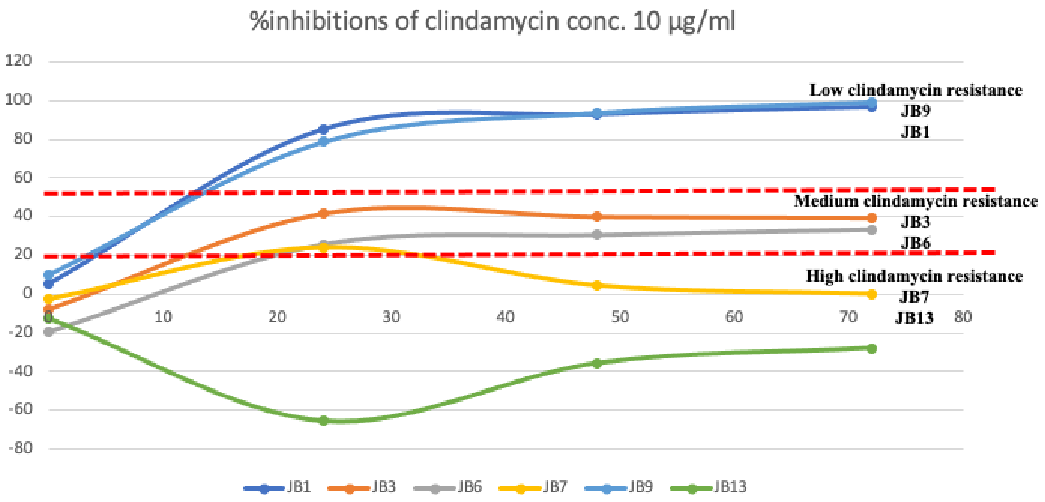
2.1. Characterization of Clindamycin Resistance P. acnes Isolates
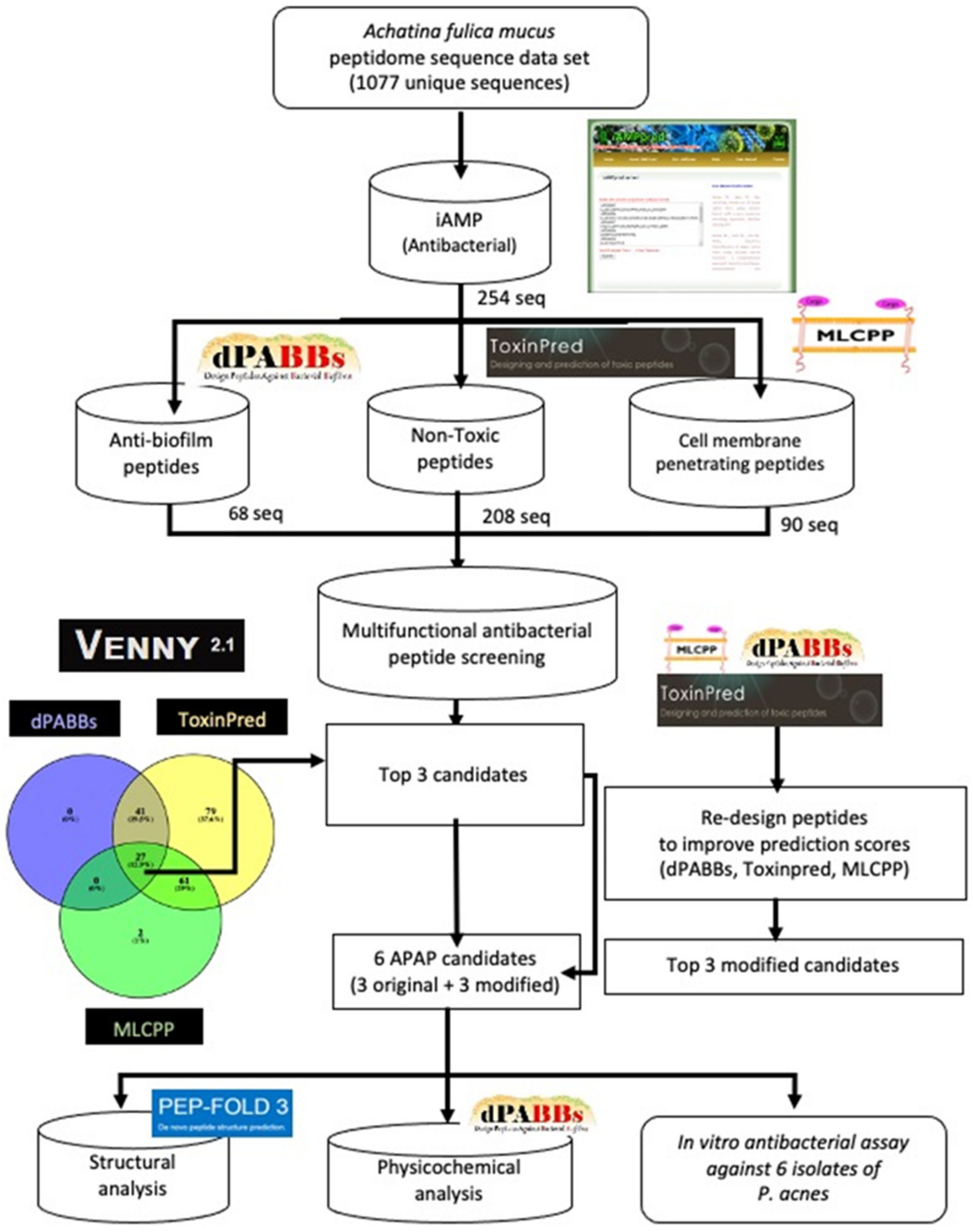
2.2. Putative Antibacterial Peptide Screening Using Bioinformatics Prediction
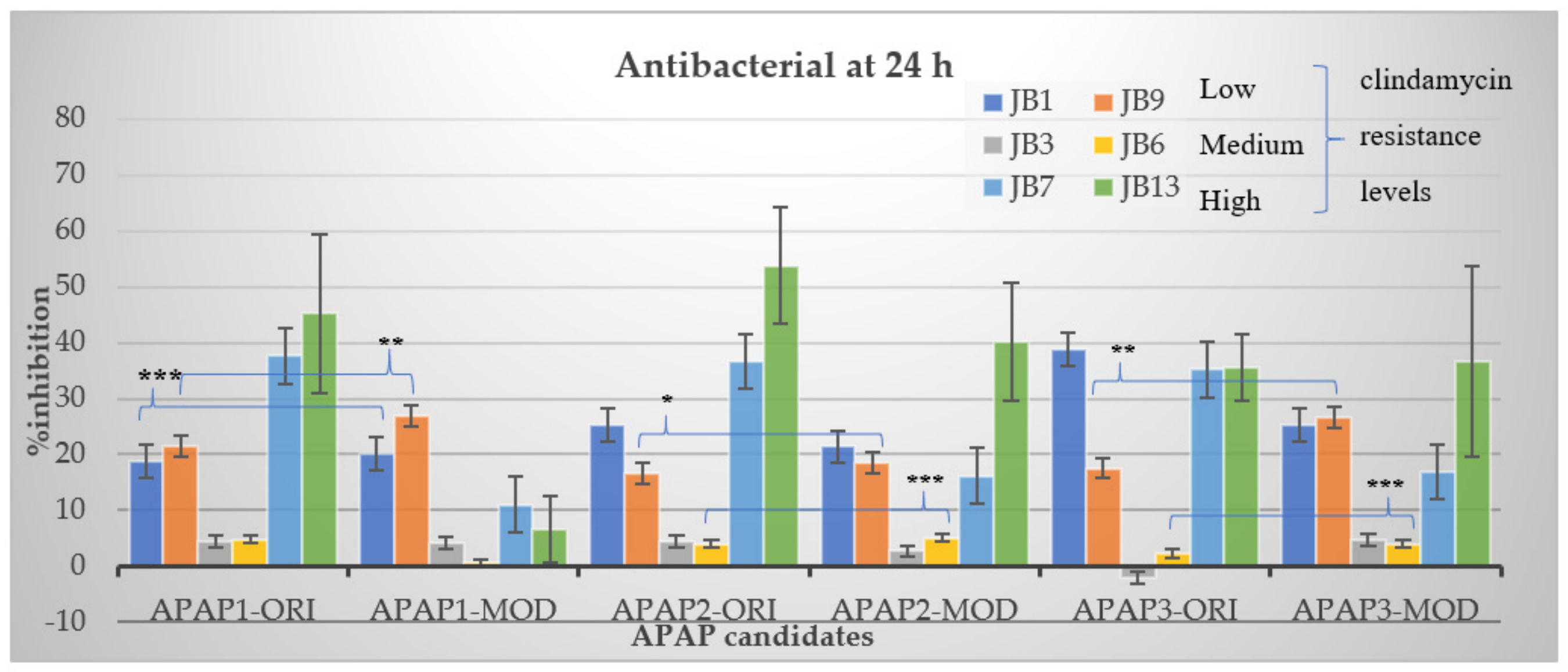
2.3. The Inhibitory Effect of Selected Putative Antibacterial Peptides against P. acnes Isolates with Different Clindamycin Resistance Levels
2.4. The Side Effects of the Original and Modified APAP2-Biomimetic Peptides on Human Fibroblast Cell Line
3. Materials and Methods
3.1. Propionibacterium acnes Isolate Preparation
3.2. Bioinformatic Prediction of Multifunctional Antibacterial Peptides
3.3. Antibacterial Assay
3.4. Side Effect Measurement on Human Skin Fibroblast Cell Line
4. Conclusions
Author Contributions
Funding
Institutional Review Board Statement
Informed Consent Statement
Data Availability Statement
Acknowledgments
Conflicts of Interest
References
- Friedlander, S.F.; Baldwin, H.E.; Mancini, A.J.; Yan, A.C.; Eichenfield, L.F. The Acne Continuum: An Age-Based Approach to Therapy. Semin. Cutan. Med. Surg. 2011, 30, S6–S11. [Google Scholar] [CrossRef]
- Liu, P.-F.; Nakatsuji, T.; Zhu, W.; Gallo, R.L.; Huang, C.-M. Passive immunoprotection targeting a secreted CAMP factor of Propionibacterium acnes as a novel immunotherapeutic for acne vulgaris. Vaccine 2011, 29, 3230–3238. [Google Scholar] [CrossRef] [Green Version]
- Huang, W.-C.; Tsai, T.-H.; Chuang, L.-T.; Li, Y.-Y.; Zouboulis, C.C.; Tsai, P.-J. Anti-bacterial and anti-inflammatory properties of capric acid against Propionibacterium acnes: A comparative study with lauric acid. J. Dermatol. Sci. 2014, 73, 232–240. [Google Scholar] [CrossRef]
- Kumar, B.; Pathak, R.; Mary, P.B.; Jha, D.; Sardana, K.; Gautam, H.K. New insights into acne pathogenesis: Exploring the role of acne-associated microbial populations. Dermatol. Sin. 2016, 34, 67–73. [Google Scholar] [CrossRef] [Green Version]
- Bojar, R.A.; Holland, K.T. Acne and propionibacterium acnes. Clin. Dermatol. 2004, 22, 375–379. [Google Scholar] [CrossRef]
- Luk, N.-M.T.; Hui, M.; Lee, H.-C.S.; Fu, L.H.; Liu, Z.H.; Lam, L.Y.; Ho, K.-M. Antibiotic-resistant Propionibacterium acnes among acne patients in a regional skin centre in Hong Kong. J. Eur. Acad. Dermatol. Venereol. 2011, 27, 31–36. [Google Scholar] [CrossRef]
- Nair, S.S.; Zolotarskaya, O.Y.; Beckwith, M.J.; Ohman, D.E.; Wynne, K.J. A Polycation Antimicrobial Peptide Mimic without Resistance Buildup against Propionibacterium Acnes. Macromol. Biosci. 2017, 17, 1700090. [Google Scholar] [CrossRef]
- Raheem, N.; Straus, S.K. Mechanisms of Action for Antimicrobial Peptides with Antibacterial and Antibiofilm Functions. Front. Microbiol. 2019, 10, 2866. [Google Scholar] [CrossRef] [Green Version]
- Qiao, X.; Wang, Y.; Yu, H. Progress in the mechanisms of anticancer peptides. Chin. J. Biotechnol. 2019, 35, 1391–1400. [Google Scholar] [CrossRef]
- Lima, M.G.; Augusto, R.D.C.; Pinheiro, J.; Thiengo, S.C.; Da Silva, J.P. Physiology and immunity of the invasive giant African snail, Achatina (Lissachatina) fulica, intermediate host of Angiostrongylus cantonensis. Dev. Comp. Immunol. 2019, 105, 103579. [Google Scholar] [CrossRef]
- E-Kobon, T.; Thongararm, P.; Roytrakul, S.; Meesuk, L.; Chumnanpuen, P. Prediction of anticancer peptides against MCF-7 breast cancer cells from the peptidomes of Achatina fulica mucus fractions. Comput. Struct. Biotechnol. J. 2016, 14, 49–57. [Google Scholar] [CrossRef] [PubMed] [Green Version]
- Suárez, L.; Pereira, A.; Hidalgo, W.; Uribe, N. Antibacterial, Antibiofilm and Anti-Virulence Activity of Biactive Fractions from Mucus Secretion of Giant African Snail Achatina fulica against Staphylococcus aureus Strains. Antibiotics 2021, 10, 1548. [Google Scholar] [CrossRef]
- Kubota, Y.; Watanabe, Y.; Otsuka, H.; Tamiya, T.; Tsuchiya, T.; Matsumoto, J.J. Purification and characterization of an antibacterial factor from snail mucus. Comp. Biochem. Physiol. Part C Comp. Pharmacol. 1985, 82, 345–348. [Google Scholar] [CrossRef]
- Tew, G.N.; Liu, D.; Chen, B.; Doerksen, R.J.; Kaplan, J.; Carroll, P.J.; Klein, M.L.; DeGrado, W.F. De novo design of biomimetic antimicrobial polymers. Proc. Natl. Acad. Sci. USA 2002, 99, 5110–5114. [Google Scholar] [CrossRef] [Green Version]
- Rotem, S.; Mor, A. Antimicrobial peptide mimics for improved therapeutic properties. Biochim. Biophys Acta 2009, 1788, 1582–1592. [Google Scholar] [CrossRef] [PubMed] [Green Version]
- Adikwu, M.; Nnamani, P.; Attama, A. Evaluation of snail mucin as bioadhesive agent for the delivery of chlorpropamide. Bio-Research 2005, 3, 75–85. [Google Scholar] [CrossRef]
- Harti, A.S.; Murharyati, A.S.D.S.; Oktariani, M. The effectiveness of snail mucus (Achatina fulica) and chitosan toward limfosit proliferation in vitro. Asian J. Pharm. Clin. Res. 2018, 11, 85–88. [Google Scholar] [CrossRef]
- Ajadi, A.R.; Oladele, S.G.; Ebenezer, B.O.; Olajide, B.K.; Gazal, O.; Otesile, E.; Kasali, O. Evaluation of Glucosamine and Snail Mucin on the Progression of Experimental Knee Osteoarthritis in Dogs. Int. J. Morphol. 2013, 31, 280–286. [Google Scholar] [CrossRef] [Green Version]
- Tachapuripunya, V.; Roytrakul, S.; Chumnanpuen, P.; E-Kobon, T. Unveiling Putative Functions of Mucus Proteins and Their Tryptic Peptides in Seven Gastropod Species Using Comparative Proteomics and Machine Learning-Based Bioinformatics Predictions. Molecules 2021, 26, 3475. [Google Scholar] [CrossRef]
- Tornesello, A.L.; Borrelli, A.; Buonaguro, L.; Buonaguro, F.M.; Tornesello, M.L. Antimicrobial Peptides as Anticancer Agents: Functional Properties and Biological Activities. Molecules 2020, 25, 2850. [Google Scholar] [CrossRef]
- Chu, H.-L.; Yip, B.-S.; Chen, K.-H.; Yu, H.-Y.; Chih, Y.-H.; Cheng, H.-T.; Chou, Y.-T.; Cheng, J.-W. Novel Antimicrobial Peptides with High Anticancer Activity and Selectivity. PLoS ONE 2015, 10, e0126390. [Google Scholar] [CrossRef] [PubMed] [Green Version]
- Chantawannakul, J.; Chatpattanasiri, P.; Wattayagorn, V.; Kongsema, M.; Noikaew, T.; Chumnanpuen, P. Virtual Screening for Biomimetic Anti-Cancer Peptides from Cordyceps militaris Putative Pepsinized Peptidome and Validation on Colon Cancer Cell Line. Molecules 2021, 26, 5767. [Google Scholar] [CrossRef]
- Sly, W.S.; Grubb, J. Isolation of fibroblasts from patients. Methods Enzymol. 1979, 58, 444–450. [Google Scholar] [CrossRef] [PubMed]
- Wunnoo, S.; Saising, J.; Voravuthikunchai, S.P. Rhodomyrtone inhibits lipase production, biofilm formation, and disorganizes established biofilm in Propionibacterium acnes. Anaerobe 2017, 43, 61–68. [Google Scholar] [CrossRef] [PubMed]
- Grievink, H.W.; Jirka, S.M.G.; Woutman, T.D.; Schoonakker, M.; Rissmann, R.; Malone, K.E.; Feiss, G.; Moerland, M. Antimicrobial Peptide Omiganan Enhances Interferon Responses to Endosomal Toll-Like Receptor Ligands in Human Peripheral Blood Mononuclear Cells. Clin. Transl. Sci. 2020, 13, 891–895. [Google Scholar] [CrossRef] [Green Version]
- Gordon, Y.J.; Romanowski, E.G.; McDermott, A.M. A Review of Antimicrobial Peptides and Their Therapeutic Potential as Anti-Infective Drugs. Curr. Eye Res. 2005, 30, 505–515. [Google Scholar] [CrossRef] [PubMed]
- Schito, A.; Schito, G.; Alfei, S. Synthesis and Antibacterial Activity of Cationic Amino Acid-Conjugated Dendrimers Loaded with a Mixture of Two Triterpenoid Acids. Polymers 2021, 13, 521. [Google Scholar] [CrossRef]
- Cutrona, K.; Kaufman, B.A.; Figueroa, D.; Elmore, D.E. Role of arginine and lysine in the antimicrobial mechanism of histone-derived antimicrobial peptides. FEBS Lett. 2015, 589, 3915–3920. [Google Scholar] [CrossRef] [Green Version]
- Mojsoska, B.; Jenssen, H. Peptides and Peptidomimetics for Antimicrobial Drug Design. Pharmaceuticals 2015, 8, 366–415. [Google Scholar] [CrossRef]
- Nakamura, M.; Kametani, I.; Higaki, S.; Yamagishi, T. Identification of Propionibacterium acnes by polymerase chain reaction for amplification of 16S ribosomal RNA and lipase genes. Anaerobe 2003, 9, 5–10. [Google Scholar] [CrossRef]
- Oprica, C.; Emtestam, L.; Lapins, J.; Borglund, E.; Nyberg, F.; Stenlund, K.; Lundeberg, L.; Sillerström, E.; Nord, C.E. Antibiotic-resistant Propionibacterium acnes on the skin of patients with moderate to severe acne in Stockholm. Anaerobe 2004, 10, 155–164. [Google Scholar] [CrossRef] [PubMed]
- Cunliffe, W.J.; Holland, K.T.; Bojar, R.; Levy, S.F. A randomized, double-blind comparison of a clindamycin phosphate/benzoyl peroxide gel formulation and a matching clindamycin gel with respect to microbiologic activity and clinical efficacy in the topical treatment of acne vulgaris. Clin. Ther. 2002, 24, 1117–1133. [Google Scholar] [CrossRef]

| APAP-ID. | Number of Amino Acid Residues | Prediction Scores | |||
|---|---|---|---|---|---|
| iAMP | dPABBs | ToxinPred | MLCPP | ||
| APAP1-original | 7 | 0.73 | 1.23 | −0.91 | 0.73 |
| APAP1-modified | 7 | 0.97 | 2.65 | −0.81 | 0.97 |
| APAP2-original | 8 | 0.61 | 0.11 | −1.17 | 0.61 |
| APAP2-modified | 8 | 0.97 | 2.77 | −1.38 | 0.97 |
| APAP3-original | 5 | 0.78 | 0.06 | −0.73 | 0.78 |
| APAP3-modified | 5 | 0.97 | 1.88 | −0.83 | 0.97 |
| ID | Sequence | Peptide Structure | Physicochemical Properties | |||||
|---|---|---|---|---|---|---|---|---|
| Hydrophobicity | Hydropathicity | Amphipathicity | Hydrophilicity | Charge | MW | |||
| APAP1-original | K RATV YR |  | −0.57 | −1.27 | 1.22 | 0.61 | 3 | 893.14 |
| APAP1-modified | K RLHV I G | 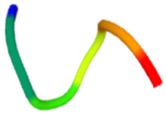 | −0.19 | 0.07 | 1.08 | 0.06 | 2.5 | 822.13 |
| APAP2-original | L ATVTV PR | 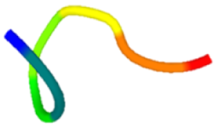 | −0.04 | 0.81 | 0.31 | −0.39 | 1 | 856.14 |
| APAP2-modified | L A I V GH K R | 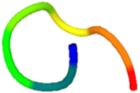 | −0.13 | 0.29 | 0.95 | −0.01 | 2.5 | 893.22 |
| APAP3-original | V I I AH | 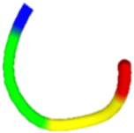 | 0.37 | 2.36 | 0.29 | −1.22 | 0.5 | 551.76 |
| APAP3-modified | VRIKL |  | −0.21 | 0.82 | 1.22 | 0.18 | 2 | 627.9 |
| P. acnes Isolates | JB1 | JB9 | JB3 | JB6 | JB7 | JB13 | Mean ± SD | |
|---|---|---|---|---|---|---|---|---|
| Peptide ID | ||||||||
| APAP2-original | 124.346 | 242.24 | 156.29 | 290.82 | 86.6996 | 143.451 | 105.5228 ± 31.41 | |
| APAP2-modified | 155.301 | 96.1367 | 55.2911 | 31.7074 | 29.7781 | 24.9624 | 27.37025 ± 20.95 | |
| Replicates | 1 | 2 | 3 | 4 | Mean | SD | SE | |
|---|---|---|---|---|---|---|---|---|
| Peptide ID | ||||||||
| APAP2-original | 73.89788 | 77.06946 | 90.70726 | 90.3901 | 83.01618 | 8.794591 | 4.397296 | |
| APAP2-modified | 101.1735 | 92.29305 | 94.196 | 81.50967 | 92.29305 | 8.139709 | 4.069854 | |
Publisher’s Note: MDPI stays neutral with regard to jurisdictional claims in published maps and institutional affiliations. |
© 2022 by the authors. Licensee MDPI, Basel, Switzerland. This article is an open access article distributed under the terms and conditions of the Creative Commons Attribution (CC BY) license (https://creativecommons.org/licenses/by/4.0/).
Share and Cite
Chalongkulasak, S.; E-kobon, T.; Chumnanpuen, P. Prediction of Antibacterial Peptides against Propionibacterium acnes from the Peptidomes of Achatina fulica Mucus Fractions. Molecules 2022, 27, 2290. https://doi.org/10.3390/molecules27072290
Chalongkulasak S, E-kobon T, Chumnanpuen P. Prediction of Antibacterial Peptides against Propionibacterium acnes from the Peptidomes of Achatina fulica Mucus Fractions. Molecules. 2022; 27(7):2290. https://doi.org/10.3390/molecules27072290
Chicago/Turabian StyleChalongkulasak, Suwapitch, Teerasak E-kobon, and Pramote Chumnanpuen. 2022. "Prediction of Antibacterial Peptides against Propionibacterium acnes from the Peptidomes of Achatina fulica Mucus Fractions" Molecules 27, no. 7: 2290. https://doi.org/10.3390/molecules27072290
APA StyleChalongkulasak, S., E-kobon, T., & Chumnanpuen, P. (2022). Prediction of Antibacterial Peptides against Propionibacterium acnes from the Peptidomes of Achatina fulica Mucus Fractions. Molecules, 27(7), 2290. https://doi.org/10.3390/molecules27072290