Communication

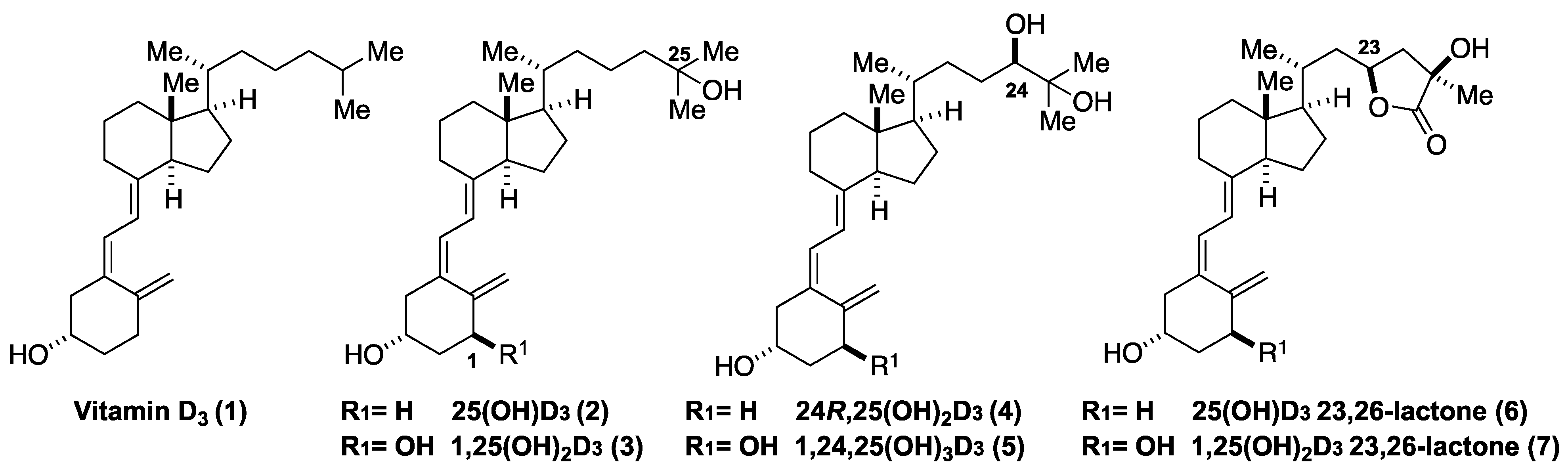
Synthesis of Deuterium-Labeled Vitamin D Metabolites as Internal Standards for LC-MS Analysis

1
Department of Biotechnology and Life Science, Tokyo University of Agriculture and Technology, Koganei, Tokyo 184-8588, Japan
2
Medical Equipment Business Operations, Management Strategy Planning Division, JEOL Ltd., Akishima, Tokyo 196-8558, Japan
*
Author to whom correspondence should be addressed.
Molecules 2022, 27(8), 2427; https://doi.org/10.3390/molecules27082427
Submission received: 19 March 2022 / Revised: 5 April 2022 / Accepted: 6 April 2022 / Published: 9 April 2022

Abstract

:
Blood levels of the vitamin D3 (D3) metabolites 25-hydroxyvitamin D3 (25(OH)D3), 24R,25-dihydroxyvitamin D3, and 1α,25-dihydroxyvitamin D3 (1,25(OH)2D3) are recognized indicators for the diagnosis of bone metabolism-related diseases, D3 deficiency-related diseases, and hypercalcemia, and are generally measured by liquid-chromatography tandem mass spectrometry (LC-MS/MS) using an isotope dilution method. However, other D3 metabolites, such as 20-hydroxyvitamin D3 and lactone D3, also show interesting biological activities and stable isotope-labeled derivatives are required for LC-MS/MS analysis of their concentrations in serum. Here, we describe a versatile synthesis of deuterium-labeled D3 metabolites using A-ring synthons containing three deuterium atoms. Deuterium-labeled 25(OH)D3 (2), 25(OH)D3-23,26-lactone (6), and 1,25(OH)2D3-23,26-lactone (7) were synthesized, and successfully applied as internal standards for the measurement of these compounds in pooled human serum. This is the first quantification of 1,25(OH)2D3-23,26-lactone (7) in human serum.

1. Introduction

Vitamin D3 (D3) (1) is metabolized by members of the cytochrome P450 (CYP) family to generate more than 50 compounds in vivo. Among them, 25-hydroxyvitamin D3 (25(OH)D3) (2) is generated from D3 (1) by CYP2R1 and/or CYP27A1-mediated hydroxylation at C25 in the liver, and the resulting 25(OH)D3 (2) is further metabolized to the active form of D3, 1α,25-dihydroxyvitamin D3 (1,25(OH)2D3) (3), by CYP27B1-mediated oxidation at C1α in the kidneys (Figure 1). 1,25(OH)2D3 (3) plays a key role in the regulation of bone metabolism in vivo [1]. In normal conditions, production of 3 from 2 is strictly controlled by the concentrations of calcium and parathyroid hormone (PTH) in the blood, and thus the concentration of 3 in the blood is a useful indicator of functional status [2] and is helpful in the diagnosis of diseases such as hypercalcemia, hyperphosphatemia, rickets, and bone metabolism-related diseases [3,4]. The concentration of 2 in the blood is also useful as an indicator for the diagnosis of various vitamin D deficiency-related diseases [3,4]. Recently, various D3 metabolites, mostly oxidized at the D-ring side chain, have also been found to show biological activities. For example, 20S,25(OH)2D3, which is generated by CYP11A1, inhibits the growth of keratinocytes, leukemia cells, and melanoma cells [5,6,7,8,9], while 24R,25(OH)2D3 (4), produced by CYP24A1, shows inhibitory activity against various cancer cell lines [10,11]. Further, 25(OH)D3-23,26 lactone (6) and 1,25(OH)2D3-23,26 lactone (7), which are thought to be final metabolites of D3, show antagonistic activity towards 1,25(OH)2D3 [12,13,14], thereby, inhibiting bone formation and resorption. Recently, compound 7 was reported to inhibit fatty acid oxidation [15]. Thus, there is a need to measure the blood levels of these metabolites.
The concentrations of metabolites 2 and 3 in blood have been measured for clinical purposes by radioimmunoassay (RIA) or chemiluminescent enzyme immunoassay (CLEIA) [16]. They, however, have disadvantages such as the need to handle radioactive materials, and insufficient discrimination of vitamin D metabolites by antibodies [17]. More recently, a liquid-chromatography tandem mass spectrometry (LC-MS/MS) method has been developed to determine the concentration of multiple vitamin D metabolites simultaneously in blood [18]. However, LC-MS/MS-based measurement also has some problems, such as the low ionization efficiency of vitamin D derivatives and interference by contaminants including multiple D3 metabolites in the blood. To address these issues, several approaches have been investigated. Cookson-type reagents have been developed to improve the ionization efficiency of D3 metabolites, affording high sensitivity even at low abundance [19,20]. The isotope dilution method has also been applied to avoid interference from contaminants in the blood. This method requires a stable isotope-labeled compound as an internal standard, and so far, deuterium-labeled 25(OH)2D3 (2), 1,25(OH)2D3 (3), and 24R,25(OH)2D3 (4), in which deuterium is introduced at C26, C27, C6, and C19, have been synthesized (Figure 2) [21,22,23,24,25].
In the synthesis of the deuterium-labeled metabolites 24-d6, deuterium was introduced into the side chain at C26 and C27 by reacting esters 8 with deuterated Grignard reagent, CD3MgBr (Figure 2B) [21,22]. On the other hand, 23-d3 were synthesized by reacting SO2 adducts of cyclic compounds 9 derived from D3 with deuterium oxide (D2O) [23,24,25]. In both strategies, the range of metabolites that can be synthesized is limited due to the restrictions imposed by the use of steroid precursors. Therefore, a more versatile approach is required. Convergent strategies, with coupling between CD-ring and A-ring moieties, have been widely applied for the synthesis of D3 derivatives [26,27]. Since the CD-ring structures of the metabolites are diverse, whereas the A-ring structures are relatively constant, we considered that deuterium-labeled A-ring synthons would be suitable for the preparation of a variety of deuterium-labeled D3 metabolites (Figure 2D). In addition, labeling in the A-ring has an advantage in metabolism studies because the side chains of the D3 are well known to be enzymatically metabolized easily. In this study, we have developed a synthesis of deuterium-labeled A-ring precursors 13-d3 and 16-d3 incorporating three deuterium atoms. These precursors were coupled with CD-ring moieties 17 and 18 to afford deuterium-labeled 25(OH)D3-d3 (2-d3) and vitamin D lactones 25(OH)D3-23,26-lactone-d3 (6-d3) and 1,25(OH)2D3-23,26-lactone-d3 (7-d3). We also confirmed that the concentrations of 2, 6, and 7 in human serum could be measured by LC-MS/MS using the corresponding deuterium-labeled compounds as the internal standards (IS) (see Supplementary Materials).

2. Results

We employed a convergent strategy using the palladium-catalyzed coupling reaction of enyne-type deuterium-labeled A-ring precursors 13-d3 and 16-d3 with bromoolefins 17 and 18 as the CD-ring moieties. The deuterium atoms in 13-d3 and 16-d3 were introduced by the H/D exchange at the a-position of the alcohol, as reported by Sajiki et al. [28]. Our synthesis of deuterium-labeled enyne 13 commenced with the H/D exchange reaction of alcohol 10, which was obtained from L-(-)-malic acid in 4 steps (Scheme 1) [29]. The alcohol 10 was subjected to the H/D exchange reaction with a catalytic amount of Ru/C in D2O at 80 °C under an H2 atmosphere to afford 10-d3 deuterium-labeled at C3 and C4 in a 96% yield with over 93% deuteride content [28]. In this reaction, the stereochemistry at C3 was isomerized (4:1 ratio of α-10a and β-10b). The deuterium-labeled alcohol 10 (enantiomeric mixture) was converted into alkyne 11 by tosylation of the primary alcohol followed by epoxidation with NaH and reaction with TMS-acetylene (22% yield from 10-d3). The hydroxyl group in alkyne 11 was protected with TBS ether, followed by deprotection of the TMS and pivaloyl groups with NaOMe in MeOH to give the alcohol 12 in an 86% yield from 11. Enyne 13 was obtained in a 51% yield from 12 via 4 steps, (i) tosylation of the primary alcohol; (ii) cyanation with NaCN; (iii) reduction of the nitrile group with DIBAL-H to aldehyde; and (iv) a Wittig reaction with Ph3PCH3I and NaHMDS. It was confirmed by 1H-NMR that the deuteration rate did not decrease in these reaction steps [30].
Next, the deuterium-labeled enyne 16 bearing a hydroxyl group at C1α was synthesized (Scheme 2). The alcohol moiety in 12 was oxidized with an IBX and the resulting aldehyde was reacted with a HWE Wittig reagent to give an unsaturated ester, whose ester group was reduced with a DIBAL-H to give allyl alcohol 14 in a 70% yield from 12 [31,32]. The allyl alcohol 14 was subjected to a Sharpless asymmetric epoxidation with a TBHP in the presence of Ti(OiPr)4 and L-(+)-DET [33], and the resulting epoxy alcohol was subjected to iodination with iodine and triphenylphosphine followed by treatment with zinc to give a secondary alcohol 15 in a 79% yield (3 steps) [31,32]. The diastereomer ratio at C1 in 15 was 10:1, and the undesired C1β diastereomer was removed by kinetic resolution, using acylation with isopropyl acid anhydride in the presence of (2S,3R)-HyperBTM [34], to give (-)-15 in an 82% yield as a single diastereomer. The undesired diastereomer at C3 was also removed via silica gel column purification. The deuterium-labeled enyne 16, in which the secondary alcohol was protected as the TBS ether, was obtained in an 82% yield.
The palladium-catalyzed coupling reaction of 13-d3 and bromoolefin 17 followed by deprotection of the silyl groups provided 25(OH)D3-d3 (2-d3) in a 36% yield [35]. Next, 25(OH)D3-23,26-lactone-d3 (6-d3) and 1,25(OH)2D3-23,26-lactone-d3 (7-d3) were similarly synthesized by reacting bromoolefin 18 and enynes 13-d3 and 16-d3, respectively [36]. In the synthesis of 2-d3 and 6-d3, the undesired diastereomers at C3α were separated by an HPLC (Scheme 3).

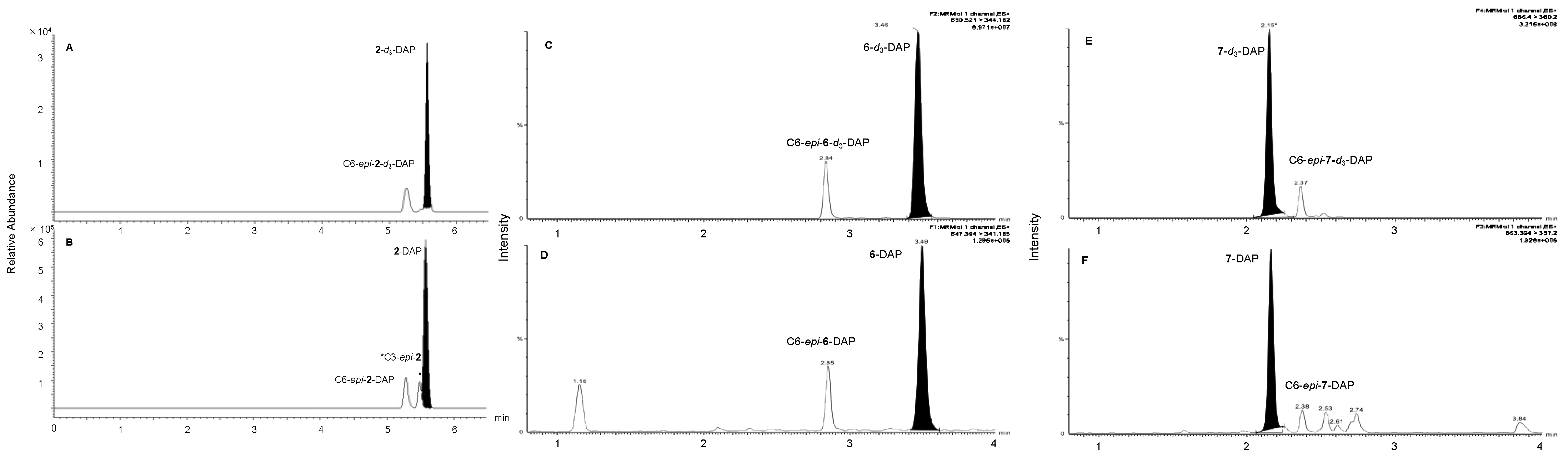
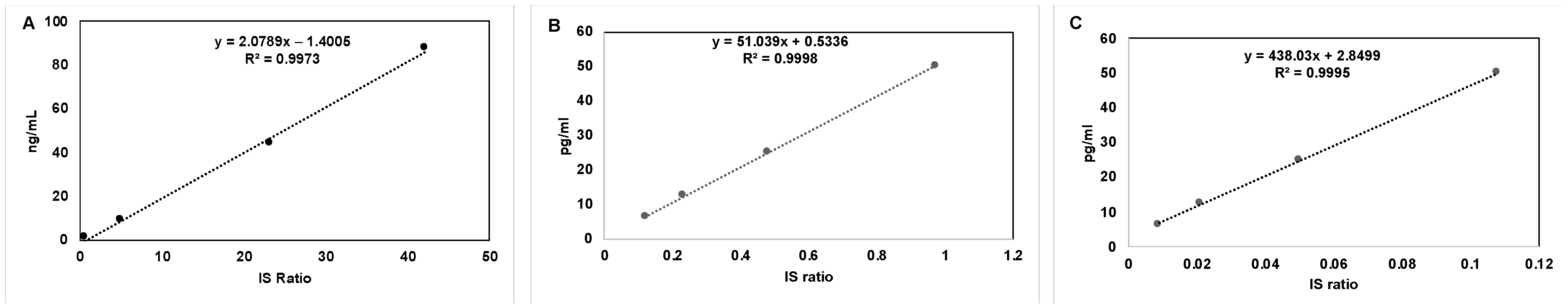
2.1. Derivatization of 2, 6, 7 for LC-MS/MS, and Preparation of Calibration Curves

With the deuterium-labeled D3 metabolites of 2-d3, 6-d3, and 7-d3 in hand, we next examined the quantitative analysis of the three D3 metabolites in pooled human serum by LC-MS/MS. First, we confirmed that our deuterium-labeled D3 metabolites were suitable as the internal standards for the isotope dilution method in an LC-MS/MS analysis. As described above, D3 and its metabolites have low ionization efficiency in an LC-MS/MS, and derivatization is necessary to improve the ionization efficiency. Thus, the D3 metabolites 2, 6, and 7, as well as 2-d3, 6-d3, and 7-d3, were derivatized with a recently developed reagent DAP-PA (4-(4′-dimethylaminophenyl)-1,2,4-triazoline-3,5-dione-phenyl anthracene) [20], and the ion peaks of the DAP adducts were detected by selective reaction monitoring (SRM) under the LC-MS/MS conditions shown in Table 1 (Figure 3).
In the case of the DAP-adducts of 2 and 2-d3 (Figure 3A,B), we observed identical ion peaks at the retention time of 5.60 min (abbreviated as tR: 5.60 min). Similarly, 6 and 6-d3 showed the same tR of 3.50 min, and 7 and 7-d3 showed the same tR of 2.15 min, indicating that the deuterium-labeled compounds are suitable as internal standards for the isotope dilution method. We also observed small peaks at the retention times of 5.20 min (Figure 3A,B), 2.65 min (Figure 3C,D), and 2.37 min (Figure 3E,F) for 2/2-d3, 6/6-d3, and 7/7-d3, respectively. These peaks are due to the epimers at C6 of the DAP adducts, because DAP-PA reacts from both the α- and β-faces.
Next, the calibration curves were prepared as follows (Figure 4). A total of 100 μL of each one of the calibrator solutions was mixed with 200 μL of the internal standard solution and evaporated to dryness. After derivatization with DAP-PA, an LC-MS/MS analysis of the unlabeled and labeled DAP-adducts was performed, and the calibration curves were prepared by plotting the concentration of unlabeled DAP-adduct against the ion peak area ratio of unlabeled versus labeled DAP-adduct. All of the calibration curves showed good linearity.

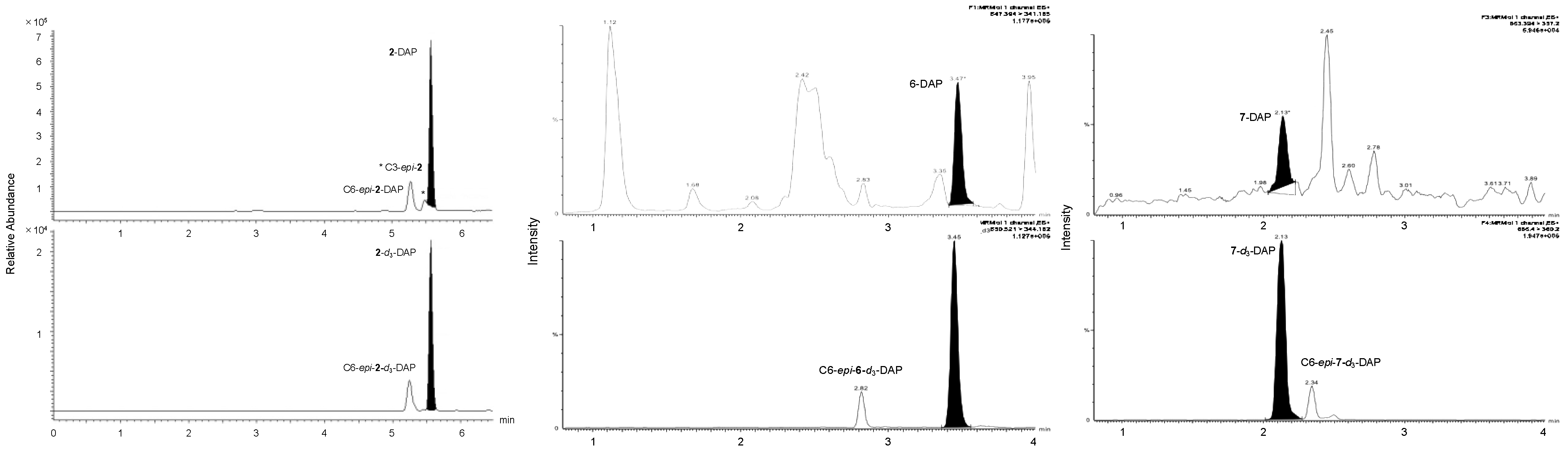
2.2. Quantification of the D3 Derivatives in Human Serum

The levels of 2, 6, and 7 in pooled human serum were quantified by the LC-MS/MS using the isotope dilution method with the constructed calibration curves. The serum was pretreated as follows. An aliquot of serum (100 μL) was mixed with the internal standards solution (200 μL). Each sample was loaded onto a supported liquid extraction column (ISOLUTE SLE+ 300 μL sample Volume, Biotage, Uppsala, Sweden) and eluted three times with 600 mL hexane/ethyl acetate (1/1, v/v) using a PRESSURE+48 positive pressure manifold (Biotage, Uppsala, Sweden). The combined eluates were evaporated to dryness in a centrifugal evaporator. The ion peaks of the metabolites matched well with those of the corresponding internal standards in the pretreated samples (Figure 5).
The concentrations of 2, 6, and 7 in human serum were calculated to be 5.1 ng/mL, 38.3 pg/mL, and 8.9 pg/mL, respectively, based on the area ratios of the detected peaks. The concentrations of 2 and 6 were in agreement with previously reported values [37], while this is the first quantification of 1α-lactone 7 in human serum.

3. Conclusions

We synthesized deuterium-labeled A-ring-d3 synthons 13 and 16 and utilized them for the convergent synthesis of deuterium-labeled D3 derivatives 25(OH)D3 (2), 25(OH)D3-23, 26-lactone (6), and 1,25(OH)2D3-23, 26-lactone (7). These deuterium-labeled D3 metabolites were successfully applied as internal standards for the quantification of the metabolites in pooled human serum by LC-MS/MS using the isotope dilution method. This is the first quantification of 1,25(OH)2D3-23, 26-lactone (7) in human serum.

Supplementary Materials

The following supporting information can be downloaded at: https://www.mdpi.com/article/10.3390/molecules27082427/s1, Experimental procedures for synthesis and characterization of compounds, Experimental procedure for LC-MS/MS analysis using the isotope dilution method, 1H and 13C NMR spectra.

Author Contributions

Conceptualization, A.N., S.F. and K.N.; investigation, A.N., K.I., R.S., Y.M. and M.I.; analysis, M.T. (Masaki Takiwakiand), Y.K. and S.F.; writing—original draft preparation, A.N. and K.N.; resources, M.O., M.T. (Masayuki Tera) and K.N.; funding acquisition, K.N. All authors have read and agreed to the published version of the manuscript.

Funding

This research received no external funding.

Acknowledgments

This work was supported by the Japan Agency for Medical Research and Development (AMED- CREST).

Conflicts of Interest

The authors declare no conflict of interest.

References and Notes

  1. Fleet, J.C. Regulation of intestinal calcium and phosphate absorption. In Vitamin D; Elsevier: Amsterdam, The Netherlands, 2018; pp. 329–342. [Google Scholar]
  2. David, V.; Quarles, L.D. FGF23/Klotho new regulators of vitamin D metabolism. In Vitamin D; Elsevier: Amsterdam, The Netherlands, 2011; pp. 747–761. [Google Scholar]
  3. Lips, P. Relative Value of 25(OH)D and 1,25(OH)2D measurements. J. Bone Miner. Res. 2007, 22, 1668–1671. [Google Scholar] [CrossRef] [PubMed]
  4. Holick, M.F. Vitamin D status: Measurement, interpretation, and clinical application. Ann. Epidemiol. 2009, 19, 73–78. [Google Scholar] [CrossRef] [PubMed] [Green Version]
  5. Janjetovic, Z.; Tuckey, R.C.; Nguyen, M.N.; Thorpe, E.M.; Slominski, A.T. 20,23-dihydroxyvitamin D3, novel P450scc product, stimulates differentiation and inhibits proliferation and NF-kB activity in human keratinocytes. J. Cell. Physiol. 2010, 223, 36–48. [Google Scholar] [PubMed] [Green Version]
  6. Janjetovic, Z.; Zmijewski, M.A.; Tuckey, R.C.; DeLeon, D.A.; Nguyen, M.N.; Pfeffer, L.M.; Slominski, A.T. 20-Hydroxycholecalciferol, product of vitamin D3 hydroxylation by P450scc, decreases NF-κB activity by increasing IκBα levels in human keratinocytes. PLoS ONE 2009, 4, e5988. [Google Scholar] [CrossRef] [Green Version]
  7. Wang, J.; Slominski, A.T.; Tuckey, R.C.; Janjetovic, Z.; Kulkarni, A.; Chen, J.; Postlethwaite, A.E.; Miller, D.; Li, W. 20-Hydroxyvitamin D3 inhibits proliferation of cancer cells with high efficacy while being non-toxic. Anticancer Res. 2012, 32, 739–746. [Google Scholar]
  8. Li, W.; Chen, J.; Janjetovic, Z.; Kim, T.K.; Sweatman, T.; Lu, Y.; Zjawiony, J.; Tuckey, R.C.; Miller, D.; Slominski, A.T. Chemical synthesis of 20S-hydroxyvitamin D3, which shows antiproliferative activity. Steroids 2010, 75, 926–935. [Google Scholar] [CrossRef]
  9. Slominski, A.T.; Janjetovic, Z.; Fuller, B.E.; Zmijewski, M.A.; Tuckey, R.C.; Nguyen, M.N.; Sweatman, T.; Li, W.; Zjawiony, J.; Miller, D.; et al. Products of vitamin D3 or 7-dehydrocholesterol metabolism by cytochrome P450scc show anti-leukemia effects, having low or absent calcemic activity. PLoS ONE 2010, 5, e9907. [Google Scholar] [CrossRef]
  10. Verma, A.; Cohen, D.J.; Schwartz, N.; Muktipaty, C.; Koblinski, J.E.; Boyan, B.D.; Schwartz, Z. 24R,25-Dihydroxyvitamin D3 regulates breast cancer cells in vitro and in vivo. Biochim. Biophys. Acta BBA—Gen. Subj. 2019, 1863, 1498–1512. [Google Scholar] [CrossRef]
  11. Verma, A.; Cohen, D.J.; Jacobs, T.W.; Boyan, B.D.; Schwartz, Z. The relative expression of ERα Isoforms ERα66 and ERα36 controls the cellular response to 24R,25-Dihydroxyvitamin D3 in breast cancer. Mol. Cancer Res. 2021, 19, 99–111. [Google Scholar] [CrossRef]
  12. Kiyoki, M.; Kurihara, N.; Ishizuka, S.; Ishii, S.; Hakea, Y.; Kumegawa, M.; Norman, A.W. The unique action for bone metabolism of 1α,25-(OH)2D3-26,23-lactone. Biochem. Biophys. Res. Commun. 1985, 127, 693–698. [Google Scholar] [CrossRef]
  13. Ishizuka, S.; Miura, D.; Eguchi, H.; Ozono, K.; Chokki, M.; Kamimura, T.; Norman, A.W. Antagonistic action of novel 1α,25-Dihydroxyvitamin D3-26,23-lactone analogs on 25-hydroxyvitamin-D3-24-hydroxylase gene expression Induced by 1α,25-dihydroxy-vitamin D3 in human promyelocytic leukemia (HL-60) cells. Arch. Biochem. Biophys. 2000, 380, 92–102. [Google Scholar] [CrossRef] [PubMed]
  14. Ishizuka, S.; Norman, A.W. The differenece of biological activity amang four diastereoisomers of 1α,25dihydoroxycholecalciferol-26,23-lactone. J. Steroid Biochem. 1986, 25, 505–510. [Google Scholar] [PubMed]
  15. Mendoza, A.; Takemoto, Y.; Cruzado, K.T.; Masoud, S.S.; Nagata, A.; Tantipanjaporn, A.; Okuda, S.; Kawagoe, F.; Sakamoto, R.; Odagi, M.; et al. Controlled lipid β-oxidation and carnitine biosynthesis by a vitamin D metabolite. Cell Chem. Biol. 2021. [Google Scholar] [CrossRef] [PubMed]
  16. Shah, I.; Akhtar, M.K.; Hisaindee, S.; Rauf, M.A.; Sadig, M.; Ashraf, S.S. Clinical diagnostic tools for vitamin D assessment. J. Steroid Biochem. Mol. Biol. 2018, 180, 105–117. [Google Scholar] [CrossRef]
  17. It is difficult to develop an antibody that specifically recognizes the structure of the side chain of vitamin D metabolites, see; Kobayashi, N.; Higashi, T.; Saito, K.; Nlurayama, T.; Douya, R.; Shimada, K. Specificity of polyclonal antibodies raised against a novel 24,25-dihydroxyvitamin D3- bovine serum albumin conjugant linked through the C-11α or C-3 position. J. Steroid Biochem. Mol. Biol. 1997, 62, 79–87. [Google Scholar]
  18. Müller, M.J.; Volmer, D.A. Mass spectrometric profiling of vitamin D metabolites beyond 25-hydroxyvitamin D. Clin. Chem. 2015, 61, 1033–1048. [Google Scholar] [CrossRef] [Green Version]
  19. Higashi, T.; Shimada, K. Application of Cookson-type reagents for biomedical HPLC and LC/MS analyses: A brief overview: Cookson-type reagents for biomedical HPLC and LC/MS analyses. Biomed. Chromatogr. 2017, 31, e3808. [Google Scholar] [CrossRef] [Green Version]
  20. Seki, M.; Sato, M.; Takiwaki, M.; Takahashi, K.; Kikutani, Y.; Satoh, M.; Nomura, F.; Kuroda, Y.; Fukuzawa, S. A novel caged Cookson-type reagent toward a practical vitamin D derivatization method for mass spectrometric analyses. Rapid Commun. Mass Spectrom. 2020, 34, e8648. [Google Scholar] [CrossRef]
  21. Nicoletti, D.; Mouriño, A.; Torneiro, M.S. Synthesis of 25-hydroxyvitamin D3 and 26,26,26,27,27,27-hexadeutero-25-hydroxyvitamin D3 on solid support. J. Org. Chem. 2009, 74, 4782–4786. [Google Scholar] [CrossRef]
  22. Perlman, K.; Schnoes, H.K.; Tanaka, Y.; DeLuca, H.F.; Kabayashi, Y.; Taguchi, T. Chemical synthesis of (24R)-24,25-dihydroxy[26,27-3H]vitamin D3 of high specific activity. Biochemistry 1984, 23, 5041–5048. [Google Scholar] [CrossRef]
  23. Ray, R.; Vicchio, D.; Yergey, A.; Holick, M.F. Synthesis of 25-hydroxy-[6,19,19′-2H3vitamin D3 and 1α,25-dihydroxy-[6,19′-2H3]vitamin D3. Steroids 1992, 57, 142–146. [Google Scholar] [CrossRef]
  24. Yamada, S.; Shimizu, M.; Fukushima, K.; Niimura, K.; Maeda, Y. Syntheses of 24R,25-dihydroxy-[6,19,19-3H]vitamin D3 and 24R,25-dihydroxy-[6,19,19-2H]vitamin D3. Steroids 1989, 54, 145–157. [Google Scholar] [CrossRef]
  25. Yamada, S.; Suzuki, T.; Takayama, H. Novel regioselective C-6 and C-19 alkylation of vitamin D3 via its sulfur-dioxide adducts. Tetrahedron Lett. 1981, 22, 3085–3088. [Google Scholar] [CrossRef]
  26. Zhu, G.D.; Okamura, W.H. Synthesis of vitamin D (Calciferol). Chem. Rev. 1995, 95, 1877–1952. [Google Scholar] [CrossRef]
  27. Gu, J.; Rodriguez, K.; Kanda, Y.; Yang, S.; Ociepa, M.; Wilke, H.; Abrishami, A.; Joergensen, L.; Skak-Nielsen, T.; Chen, J.; et al. Convergent total synthesis of (+)-Calcipotriol: A scalable, modular approach to vitamin D analogs. ChemRxiv 2021. [Google Scholar] [CrossRef]
  28. Maegawa, T.; Fujiwara, Y.; Inagaki, Y.; Monguchi, Y.; Sajiki, H. A convenient and effective method for the regioselective deuteration of alcohols. Adv. Synth. Catal. 2008, 350, 2215–2218. [Google Scholar] [CrossRef]
  29. Pattenden, G.; González, M.A.; Little, P.B.; Millan, D.S.; Plowright, A.T.; Tornos, J.A.; Ye, T. Total synthesis of (±)-phorboxazole A, a potent cytostatic agent from the sponge Phorbas sp. Org. Biomol. Chem. 2003, 1, 4173–4208. [Google Scholar] [CrossRef]
  30. The enantiomers at C3 were separated after coupling the CD rings by HPLC purification.
  31. Akagi, Y.; Usuda, K.; Tanami, T.; Yasui, K.; Asano, L.; Uesugi, M.; Nagasawa, K. Synthesis of 1α- and 1β-amino-25-hydroxyvitamin D3. Asian J. Org. Chem. 2016, 5, 1247–1252. [Google Scholar] [CrossRef]
  32. Venkanna, A.; Sreedhar, E.; Siva, B.; Babu, K.S.; Prasad, K.R.; Rao, J.M. Studies directed towards the total synthesis of koshikalide: Stereoselective synthesis of the macrocyclic core. Tetrahedron Asymmetry 2013, 24, 1010–1022. [Google Scholar] [CrossRef]
  33. Gao, Y.; Klunder, J.M.; Hanson, R.M.; Masamune, H.; Ko, S.Y.; Sharpless, K.B. Catalytic asymmetric epoxidation and kinetic resolution: Modified procedures including in situ derivatization. J. Am. Chem. Soc. 1987, 109, 5765–5780. [Google Scholar] [CrossRef]
  34. Joannesse, C.; Johnston, C.P.; Concellón, C.; Simal, C.; Philp, D.; Smith, A.D. Isothiourea-catalyzed enantioselective carboxy group transfer. Angew. Chem. Int. Ed. 2009, 48, 8914–8918. [Google Scholar] [CrossRef] [PubMed]
  35. Trost, B.M.; Dumas, J.; Villa, M. New strategies for the synthesis of vitamin D metabolites via palladium-catalyzed reactions. J. Am. Chem. Soc. 1992, 114, 9836–9845. [Google Scholar] [CrossRef]
  36. Nagata, A.; Akagi, Y.; Masoud, S.S.; Yamanaka, M.; Kittaka, A.; Uesugi, M.; Odagi, M.; Nagasawa, K. Stereoselective synthesis of four calcitriol lactone diastereomers at C23 and C25. J. Org. Chem. 2019, 84, 7630–7641. [Google Scholar] [CrossRef] [PubMed]
  37. Kaufmann, M.; Schlingmann, K.; Berezin, L.; Molin, A.; Sheftel, J.; Vig, M.; Gallagher, J.C.; Nagata, A.; Masoud, S.S.; Sakamoto, R.; et al. Differential diagnosis of vitamin D–related hypercalcemia using serum vitamin D metabolite profiling. J. Bone Miner. Res. 2021, 36, 1340–1350. [Google Scholar] [CrossRef]
Figure 1. Structures of vitamin D3 (1) and its metabolites (2)–(7).
Molecules 27 02427 g001
Figure 2. Synthetic strategy of deuterium-labeled D3 metabolites. (A) Structures of reported deuterium-labeled D3 metabolites. (B) Previous work on the synthesis of 24-d6. (C) Previous work on the synthesis of 24-d3 (D) This work: general synthesis of deuterium-labeled D3 metabolites.
Molecules 27 02427 g002
Scheme 1. Synthesis of deuterium-labeled enyne 13-d3.
Molecules 27 02427 sch001
Scheme 2. Synthesis of deuterium-labeled enyne 16-d3.
Molecules 27 02427 sch002
Scheme 3. Synthesis of vitamin D3 metabolites-d3 (2-d3, 6-d3, 7-d3).
Molecules 27 02427 sch003
Figure 3. SRM chromatograms for the DAP adducts of D3 metabolites 2, 6, and 7, as well as 2-d3, 6-d3, and 7-d3.
Molecules 27 02427 g003
Figure 4. Calibration curve of D3 metabolites; (A) 25(OH)D3 (2); (B) 25(OH)D3-23,26-lactone (6); and (C) 1,25(OH)2D3-23,26-lactone (7).
Molecules 27 02427 g004
Figure 5. SRM chromatograms for D3 derivatives 2, 6, and 7 in pooled human serum.
Molecules 27 02427 g005
Table 1. Parameters for the LC/MS/MS analysis.
CompoundSRM Transition (m/z)Cone Voltage (kv)CE (eV)
25(OH)D3-DAP (2-DAP)619.4 > 341.2
[M + H]+ [A]+
4828
25(OH)D3-23,26-lactone-DAP (6-DAP)647.4 > 341.24828
1,25(OH)2D3-23,26-lactone-DAP (7-DAP)663.4 > 357.24828
25(OH)D3-d3-DAP (2-d3-DAP)622.4 > 344.24828
25(OH)D3-23,26-lactone-d3-DAP (6-d3-DAP)650.4 > 344.24828
1,25(OH)2D3-23,26-lactone-d3-DAP (7-d3-DAP)666.4 > 360.24828
Publisher’s Note: MDPI stays neutral with regard to jurisdictional claims in published maps and institutional affiliations.

